Giant Hysteromyoma After Vaginoplasty in a Woman With Mayer-Rokitansky-Küster-Hauser (MRKH) Syndrome: Case Report and Review of the Literature
←
→
Page content transcription
If your browser does not render page correctly, please read the page content below
Giant Hysteromyoma After Vaginoplasty in a Woman With
Mayer-Rokitansky-Küster-Hauser (MRKH) Syndrome: Case
Report and Review of the Literature
Shikang Qiu
Shandong University
Yunkai Xie
Shandong Provincial Hospital
Yonghui Zou
Shandong Provincial Hospital
Fei Wang ( feiwangde@163.com )
Shandong Provincial Hospital https://orcid.org/0000-0001-6334-4829
Case report
Keywords: Case report, Leiomyomas of the uterus, Mullerian aplasia, Pelvic pain, MRKH syndrome
Posted Date: September 28th, 2021
DOI: https://doi.org/10.21203/rs.3.rs-910518/v1
License: This work is licensed under a Creative Commons Attribution 4.0 International License. Read Full License
Version of Record: A version of this preprint was published at Journal of International Medical Research on December 1st,
2021. See the published version at https://doi.org/10.1177/03000605211066394.
Page 1/11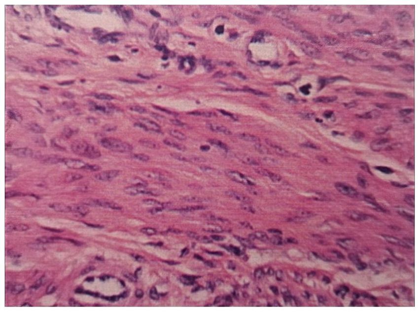
Abstract
Background: Mayer-Rokitansky-Küster-Hauser (MRKH) syndrome is a congenital disorder characterized by congenital absence
of both the uterus and vagina. Some patients may need an operation to create a neovagina. However, the preservation of
nonfunctional rudimentary uteri after surgery usually leads to some long-term complications.

Case presentation: We report a rare case of a giant hysteromyoma after vaginoplasty in a woman with MRKH syndrome. A 31-
year-old Chinese woman who was diagnosed with MRKH syndrome and received vaginal reconstruction 4 years ago presented
with abdominal distension for half a month. Transabdominal ultrasonography showed a firm mass of approximately 10 x 10
cm in the lower abdomen. She then received an exploratory laparotomy, and a leiomyoma from her rudimentary uterus was
removed.

Conclusions: Gynecologists should pay attention to the risks of pelvic complications in women with MRKH syndrome who
have undergone previous surgery and then choose suitable therapeutic methods.

Introduction
Congenital absence of both the uterus and vagina is termed Müllerian aplasia, or Mayer-Rokitansky-Küster-Hauser (MRKH)
syndrome, and affects at least 1 in 4000–5000 females(1, 2). Although rare, it is the second most common cause of primary
amenorrhea, after gonadal dysgenesis (3). MRKH syndrome is a class 1 Müllerian duct anomaly and can be divided into 2
types. Type A (44%) is characterized by symmetric muscular buds and normal fallopian tubes, while type B (56%) has
asymmetric muscular buds, abnormal fallopian tubes, and other congenital anomalies(4). Currently, there are several
approaches used to create a functional vagina, including surgical vaginoplasty and non-surgical dilation therapy. Although the
best treatment remains controversial, surgical vaginoplasty is considered a more immediate solution(5). However,
complications after surgical or invasive procedures may be inevitable, as Dabaghi has reported(6). The main complications
include dyspareunia, urinary duct obstruction, vaginal duct stenosis and infection. In other cases, the uterine remnants of
MRKH syndrome patients will also lead to some unexpected complications, including fibroids, adenomyosis, pelvic pain, and
unintended pregnancies. Here, we describe a rare clinical case of leiomyoma in a woman with MRKH syndrome after receiving
vaginoplasty. Additionally, we emphasize the necessity of intraoperative exploration and evaluation of the nonfunctional
rudimentary uterus during vaginoplasty in women with MRKH syndrome.

Case Report
A 31-year-old married woman was admitted to the gynecology ward of Shandong Provincial Hospital because of abdominal
distension for half a month. There was no significant history of cyclical vaginal bleeding or urinary or bowel complaints. This
patient had sought medical advice at our hospital 4 years ago because of amenorrhea and infertility. Further examination
showed an aplastic vagina and uterus. She was phenotypically female and had normal secondary sexual characteristics: well-
developed breasts (Tanner stage 5) and female external genitalia. The clitoris and labia majora appeared normal. However, her
labia minora showed local depigmentation. Her pubic hair showed a female type of distribution. The chromosome karyotype
analysis showed a normal female karyotype (46, XX). The endocrine evaluation showed an intact hypothalamic-pituitary-
ovarian axis. She had no known allergies. She was monogamous with her husband and did not smoke, drink alcohol, or use
illicit substances. Her family medical history was unknown.

Her abdominal ultrasonography showed a complete urinary system with normal morphology. She was then diagnosed with
MRKH syndrome (type A) and received a bowel vaginoplasty in our hospital 4 years ago. Our recent speculum examination
revealed a blind vaginal pouch 7 cm deep and the absence of the cervix. On bimanual examination, an irregular, firm, 10-cm
mobile mass arising from the pelvis was palpable. USG showed a well-defined hypoechoic mass in the pelvis with a
heterogeneous echo inside, measuring 10.8 × 9.6 cm in size. The inferior border of the mass reached a cervix-like structure
(Fig. 1). After consultation, she was primarily diagnosed with hysteromyoma. The woman then underwent a laparotomy. During
the operation, a large pelvic mass with an intact capsule was seen arising from the right rudimentary uterus (Fig. 2). The mass

                                                          Page 2/11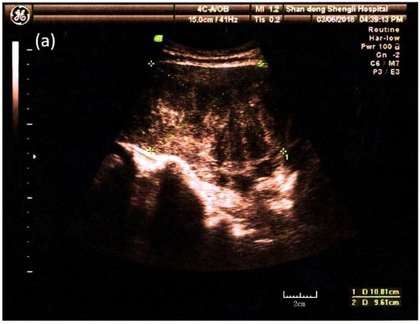
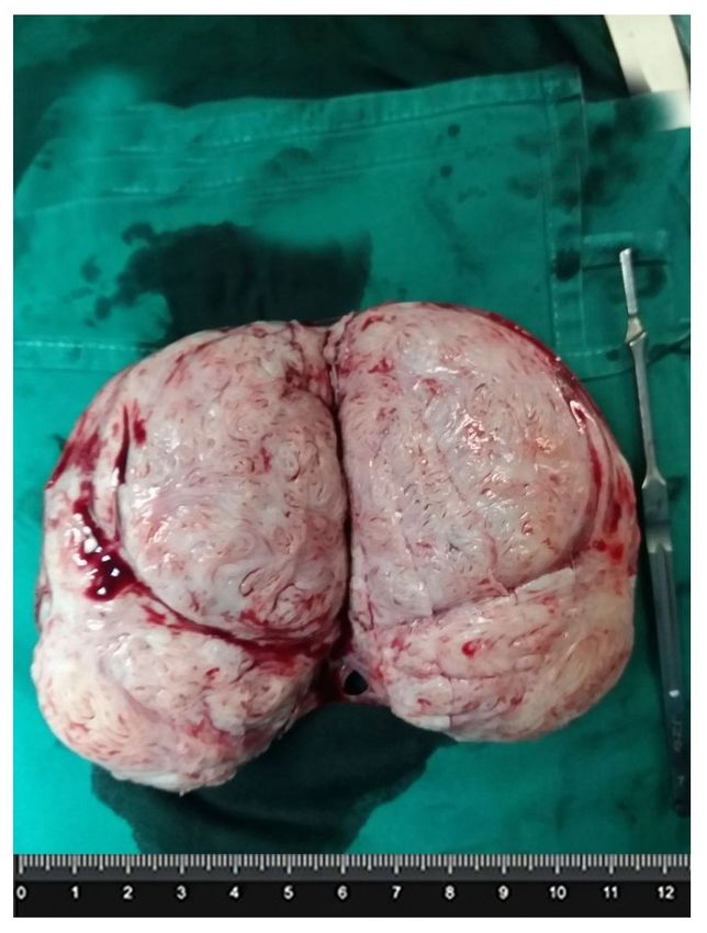
was enucleated after opening the thin cyst wall. The cut section of the mass revealed an appearance which likes a whorled
white-colored muscle-like tissue (Fig. 3), and the pathological diagnosis was leiomyoma (Fig. 4). During the surgical
exploration, we found an asymmetric fusiform uterus in the pelvic peritoneum. The rudimentary uterus was normal in size, and
structured ovaries connected with 5 cm-long fallopian tubes. The operation was ultimately successfully. Her pelvic mass,
bilateral rudimentary uterus and fallopian tube were removed.

The patient recovered and was discharged smoothly. After surgery, no sign of recurrence was found in the 1-year postoperative
check (Fig. 5), and the patient was free from lower abdominal symptoms.

Discussion
We report a case of a large leiomyoma raised from the rudimentary uterus in a woman with MRKH syndrome. The special
feature of this patient is that she had previously undergone bowel vaginoplasty, but retained the rudimentary uterus where the
leiomyoma formed. The remaining rudimentary uterus led to some complications several years later and finally resulted in a
second operation of this patient. We find few reports about leiomyomas in women with MRKH syndrome who had received
vaginoplasty. A review of the literature shows that the incidence of leiomyomas in women with MRKH syndrome was higher
than expected(7–10), while there are few reports of hysteromyoma growing so fast in such a short time after surgery. We have
to suspect that we failed to detect the growing hysteromyoma in our initial examination.

Currently, the best treatment for Müllerian agenesis remains controversial, while it has been reported that non-surgical methods,
mainly vaginal dilatation techniques, should be considered a first-line option before any surgical intervention(4). Once the
dilation fails or is inappropriate due to previous scarring or an absent vaginal dimple, surgical vaginoplasty is required. The
surgical reconstruction methods include surgical creation of a neovaginal space between the bladder and rectum, bowel
vaginoplasty, vulvaginoplasty and surgical traction(11, 12). However, there are no guidelines on whether the rudimentary uteri
should be saved during surgery. Table 1 highlights the reports about the course of patients with MRKH syndrome who saved
rudimentary uteri when receiving vaginoplasty.

                                                           Page 3/11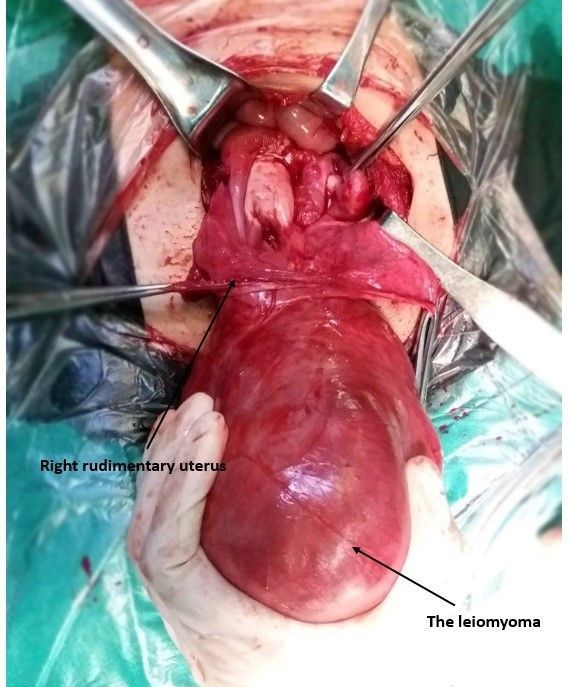
Table 1
  Course of the published cases of patients with MRKH syndrome who saved rudimentary uteri when receiving vaginoplasty
 Authors       Age     vaginoplasty    hormone     cyclic         Dimensions of      Symptoms      urinary      Surgery
                                                   endometrial    hysteromyoma                     system
                                                   changes

 Pascale       42      Abbe-           Normal      +              9.8×7.6×8.0cm      NA            UN           laparoscopy
                       McIndoe
 et al(13)             operation                                  (MR)

 Sungwook      55      Abbe-           Low         -              5.4×4.8×4.7cm      NA            UN           laparotomy
 et al(14)             McIndoe
                       operation                                  (MR)

               42      Williams’       UN          UN             5.9×5.5            Lower         unilateral   laparoscopy
                       vaginoplasty                                                  abdominal
                                                                  cm, (USG)          pain          right
                                                                                                   kidney

 Efthimios     38      Williams’       Normal      UN             4.8×3.6 cm         NA            Normal       laparoscopy
 et al(15)             vaginoplasty
                                                                  (USG)

 Nikolaos      44      Williams’       Normal      UN             9.2×7.9 cm         pelvic        Normal       laparotomy
 et al(16)             vaginoplasty
                                                                  (MR)               pain

 Kuhali        40      Vecchietti’s    Normal      +              9.1×6.7×8.6cm      Acute         UN           laparotomy
                       vaginoplasty                                                  abdominal
 et al(17)                                                        (CT)               pain

                                                                                     (torsion)

 Varpu         47      Davidov’s       UN          UN             6.0cm              NA            Normal       NA
                       vaginoplasty
 et al(18)                                                        (MR)

 Present       31      bowel           Normal      +              10.8×9.6 cm        abdominal     Normal       laparotomy
 case                  vaginoplasty                                                  distension
                                                                  (USG)

In our literature review, Abbe-McIndoe operation is the most commonly used method of vaginal reconstruction and it was
performed in two cases(13, 14). Williams' vaginoplasty was performed in 3 cases (15, 16), Vecchietti’s vaginoplasty was in one
case(17)and Davidov’s vaginoplasty was in the other one(18). Estrogen levels were normal in almost all cases(13, 15–
17)except one peri-menopausal woman, and cyclic endometrial changes were found in some ones’ rudimentary uteri by
ultrasound(13, 17). Some inconspicuous symptoms like mild abdominal pain and abdominal distension were reported in most
cases, however it can also cause emergency abdominal surgery due to acute torsion of uterine remnant leiomyoma(17).
Laparotomy is the most commonly used in our literature review, especially in the cases of Giant uterine leiomyoma and
complicated nature of its feeding arteries(14, 16), as well as emergency abdominal surgery(17). All patients underwent
hysterectomies.

Some patients with MRKH syndrome have small rudimentary Müllerian bulbs that result from abnormal development of the
Müllerian duct during embryogenesis. Oppelt has reported that 84% (239/290) of patients have uterine remnants, including
bilateral rudimentary remnants and a plastic uterine horn(19). Most of the uterine remnants or small rudimentary Müllerian
bulbs lack endometrial activity, and the rudimentary uteri are usually composed of smooth muscle cells, which may lead to
leiomyomas. 2%-7% of patients with MRKH syndrome have a functional endometrium in the rudimentary uteri(5). The presence
of endometrium in rudimentary uteri may lead to pelvic pain. Marsh reported that 48% (23/48) of females with MRKH
syndrome had uterine remnants and that 46% (22/48) had pelvic pain. He also found that the presence of endometrium was
associated with pelvic pain (RR = 2.3; 95% CI = 1.2–4.7) in females with MRKH syndrome (20). Moreover, in some patients with
a functional rudimentary uterus, as has been reported in some cases, pregnancy can be achieved by uterine and cervical
                                                          Page 4/11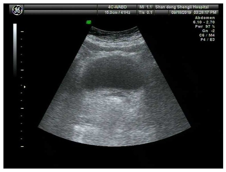
reconstruction, creation of a neovagina and placement of a uterovaginal conduit or zygote intrafallopian transfer(21, 22).
However, our review of the literature failed to find a case report of successful delivery from women with rudimentary uteri and
there are certain risks of uterine rupture during pregnancy among these women that can lead to catastrophic results, with a
death rate of 47.6% reported at the beginning of the 20th century(23). Considering the risk of pregnancy and complications of
surgical therapy, preserving the rudimentary uteri in patients with MRKH syndrome during vaginoplasty makes little sense.

Conclusion
In conclusion, because of the high incidence of uterine remnants in women with MRKH syndrome, the risk of complications
after vaginoplasty in these patients may be higher than preconceived. Females with MRKH syndrome should receive an
anatomic evaluation with transabdominal ultrasonography and MRI before receiving surgery like vaginoplasty or laparoscopic
diagnosis. Once a diagnosis of MRKH syndrome with a rudimentary uterus is clarified, gynecologists should perform a detailed
examination to detect weather the hysteromyoma has occurred or inform patients of the risk of leiomyomas and cyclic pelvic
pain. Total resection of the uterine remnants should be considered if endometrial activity or hysteromyoma in the rudimentary
uterus is found .

Abbreviations
MRKH syndrome: Mayer-Rokitansky-Küster-Hauser syndrome; USG: Ultrasonography

Declarations
Consent

Written informed consent was obtained from the patient for publication of this case report and accompanying images. A copy
of the written consent is available for review by the Editor-in-Chief of this journal.

Consent for publication

All presentations of the case reports have consent for publication.

Availability of data and materials

Not applicable.

Acknowledgement

Written informed consent was obtained from the patient for publication of this report. The research was supported by grants
from the National Natural Science Foundation of China (No. 81671434).

Statement of Ethics

The authors are accountable for all aspects of the work in ensuring that questions related to the accuracy or integrity of any
part of the work are appropriately investigated and resolved. All participants signed written informed consent forms. Ethical
approval was obtained from the Ethics Committee of the Shandong Provincial Hospital, China, in accordance with the ethical
guidelines of the 1975 Declaration of Helsinki (as revised in 2013). Written informed consent was obtained from the patient for
publication of this manuscript and any accompanying images.

Conflict of Interest Statement

The authors report no conflicts of interest in this work.

Funding Sources

                                                            Page 5/11
The authors declare that no funding was received for this study.
Author Contributions
Shikang Qiu designed, performed the study, and wrote the manuscript. Feiwang and Yonghui Zou performed the surgery
together. Shikang Qiu, Yunkai Xie performed the pathologic analysis and searched all the cases and made the analysis. All
authors read and approved the manuscript for publication.
References
1. Herlin M, Højland AT, Petersen MB. Familial occurrence of Mayer-Rokitansky-Küster-Hauser syndrome: a case report and
review of the literature. Am J Med Genet A. 2014;164A(9):2276–86.
2. Morcel K, Camborieux L, Guerrier D. Mayer-Rokitansky-Küster-Hauser (MRKH) syndrome. Orphanet J Rare Dis. 2007;2:13.
3. Rosenberg HK, Sherman NH, Tarry WF, Duckett JW, Snyder HM. Mayer-Rokitansky-Kuster-Hauser syndrome: US aid to
diagnosis. Radiology. 1986;161(3):815–9.
4. Bombard DS, Mousa SA. Mayer-Rokitansky-Kuster-Hauser syndrome: complications, diagnosis and possible treatment
options: a review. Gynecol Endocrinol. 2014;30(9):618–23.
5. Barbara LHJ, Karen D, et al. Williams Gynecology. 2016;Third Edition 3rd Edition.
6. Dabaghi S, Zandi M, Ilkhani M. Sexual satisfaction in patients with Mayer-Rokitansky-Küster-Hauser syndrome after
surgical and non-surgical techniques: a systematic review. Int Urogynecol J. 2019;30(3):353–62.
7. Kulkarni MM, Deshmukh SD, Hol K, Nene N. A rare case of Mayer-Rokitansky-Kuster-Hauser syndrome with multiple
leiomyomas in hypoplastic uterus. J Hum Reprod Sci. 2015;8(4):242–4.
8. Papa G, Andreotti M, Giannubilo SR, Cesari R, Ceré I, Tranquilli AL. Case report and surgical solution for a voluminous
uterine leiomyoma in a woman with complicated Mayer-Rokitansky-Küster-Hauser syndrome. Fertil Steril. 2008;90(5):2014.
.e5-.e6.
9. Yan CM, Mok KM. Uterine fibroids and adenomyosis in a woman with Rokitansky-Kuster-Hauser syndrome. J Obstet
Gynaecol. 2002;22(5):561–2.
10. Hoo PS, Norhaslinda AR, Reza JNS. Rare Case of Leiomyoma and Adenomyosis in Mayer-Rokitansky-Kuster-Hauser
Syndrome. Case Rep Obstet Gynecol. 2016;2016:3725043.
11. Özkan Ö, Özkan Ö, Çinpolat A, Doğan NU, Bektaş G, Dolay K, et al. Vaginal reconstruction with the modified rectosigmoid
colon: surgical technique, long-term results and sexual outcomes. J Plast Surg Hand Surg. 2018;52(4):210–6.
12. Callens N, De Cuypere G, De Sutter P, Monstrey S, Weyers S, Hoebeke P, et al. An update on surgical and non-surgical
treatments for vaginal hypoplasia. Hum Reprod Update. 2014;20(5):775–801.
13. Jadoul P, Pirard C, Squifflet J, Smets M, Donnez J. Pelvic mass in a woman with Mayer-Rokitansky-Kuster-Hauser
syndrome. Fertil Steril. 2004;81(1):203–4.
14. Chun S, Kim YM, Ji Y-I. Uterine adenomyosis which developed from hypoplastic uterus in postmenopausal woman with
mayer-rokitansky-kuster-hauser syndrome: a case report. J Menopausal Med. 2013;19(3):135–8.
15. Deligeoroglou E, Kontoravdis A, Makrakis E, Christopoulos P, Kountouris A, Creatsas G. Development of leiomyomas on the
uterine remnants of two women with Mayer-Rokitansky-Küster-Hauser syndrome. Fertil Steril. 2004;81(5):1385–7.
16. Blontzos N, Iavazzo C, Vorgias G, Kalinoglou N. Leiomyoma development in Mayer-Rokitansky-Küster-Hauser syndrome: a
case report and a narrative review of the literature. Obstet Gynecol Sci. 2019;62(4):294–7.
17. Kundu K, Cohen AW, Goldberg J. Acute torsion of uterine remnant leiomyoma with Mayer-Rokitansky-Küster-Hauser
syndrome. Fertil Steril. 2014;102(2):607–9.
18. Jokimaa V, Virtanen J, Kujari H, Ala-Nissilä S, Rantanen V. A Mayer-Rokitansky-Kuster-Hauser patient with leiomyoma and
dysplasia of neovagina: a case report. BMC Womens Health. 2020;20(1):157.
Page 6/1119. Oppelt PG, Lermann J, Strick R, Dittrich R, Strissel P, Rettig I, et al. Malformations in a cohort of 284 women with Mayer-
Rokitansky-Küster-Hauser syndrome (MRKH). Reprod Biol Endocrinol. 2012;10:57.
20. Marsh CA, Will MA, Smorgick N, Quint EH, Hussain H, Smith YR. Uterine remnants and pelvic pain in females with Mayer-
Rokitansky-Küster-Hauser syndrome. J Pediatr Adolesc Gynecol. 2013;26(3):199–202.
21. Deffarges JV, Haddad B, Musset R, Paniel BJ. Utero-vaginal anastomosis in women with uterine cervix atresia: long-term
follow-up and reproductive performance. A study of 18 cases. Hum Reprod. 2001;16(8):1722–5.
22. Thijssen RF, Hollanders JM, Willemsen WN, van der Heyden PM, van Dongen PW, Rolland R. Successful pregnancy after
ZIFT in a patient with congenital cervical atresia. Obstet Gynecol. 1990;76(5 Pt 2):902–4.
23. Kadan Y, Romano S. Rudimentary horn pregnancy diagnosed by ultrasound and treated by laparoscopy–a case report and
review of the literature. J Minim Invasive Gynecol. 2008;15(5):527–30.
Figures
Figure 1
The transabdominal ultrasonography image reveals a hypoechoic mass in the pelvis, measuring 10.8×9.6 cm in size.
Page 7/11Figure 2
Operative exploration shows a large pelvic mass with an intact capsule arising from the right rudimentary uterus.
Page 8/11Figure 3
Cut sections of the mass show white muscle-like tissue with a whorled appearance.
Page 9/11Figure 4
The pathologic diagnosis.
Page 10/11Figure 5
USG at the 1-year postoperative follow-up after surgery.
Page 11/11You can also read