Fungal infection mimicking COVID-19 infection - A case report
←
→
Page content transcription
If your browser does not render page correctly, please read the page content below
Open Medicine 2022; 17: 841–846
Case Report
Aleksandra Niemiec, Michał Kosowski*, Marcin Hachuła, Marcin Basiak, Bogusław Okopień
Fungal infection mimicking COVID-19
infection – A case report
https://doi.org/10.1515/med-2022-0443
received December 19, 2021; accepted January 31, 2022
1 Introduction
Abstract: For the last 2 years, one of the most frequent Acute respiratory distress syndrome is one of the most
causes of respiratory failure is coronavirus disease 2019 common reasons for internal ward admissions. Respiratory
(COVID-19). The symptoms are not specific. Imaging diag- failure may be due to pulmonary or extra-pulmonary causes;
nostics, especially high-resolution computed tomography, it could be caused due to pneumonia, exacerbation of obstruc-
is a diagnostic method widely used in the diagnosis of this tive lung diseases, pulmonary edema, pleural effusion, and
disease. It is important to emphasize that not only SARS- overdose of opioids or sedatives. It is necessary to take medical
CoV-2 infection may manifest as interstitial pneumonia. history at first, and perform an arterial blood gas (ABG) test
Other diseases such as other viral, fungal, atypical bac- and then chest imaging. These steps are crucial to make the
terial pneumonia, autoimmune process, and even cancer preliminary diagnosis and to implement treatment [1].
can also manifest as ground-glass opacities or consolida- For the last 2 years, one of the most frequent reasons
tions in the imaging of the lungs. In this case report, we for respiratory failure is coronavirus disease 2019 (COVID-19),
described a patient who manifested many symptoms that a viral disease caused by a coronavirus (SARS-CoV-2). It is
seemed to be COVID-19. However, all performed antigen believed that the virus is acquired from a zoonotic source
and polymerase chain reaction tests were negative. The and is transmitted via airborne respiratory droplets [2]. The
diagnostics must have been extended. Microbiological symptomatic phase, except for respiratory problems, mani-
and mycological blood cultures and sputum cultures were fests with fever, myalgia, and smell or taste disorders. The
performed. Blood cultures were negative but in sputum, infection may be detected by a rapid antigen test; however, a
Candida albicans and Candida glabrata were identified. reverse transcriptase-polymerase chain reaction (RT-PCR)
Targeted therapy with fluconazole was implemented with test is needed to confirm the diagnosis [3,4]. The specific
a satisfactory result. The patient was discharged from the image of the lungs in high resolution computed tomography
hospital in a good general condition with no complaints. (HRCT) could be a useful diagnostic tool in the differential
Keywords: COVID-19 pneumonia, fungal pneumonia, dif- diagnosis. Many studies have also investigated the rela-
ferential diagnosis, high-resolution computed tomography tionship between lung computerized tomography (CT)
scans and patients’ prognosis [5,6]. Typical radiological
manifestations presented in HRCT include ground-glass
opacities (GGO), consolidations, crazy-paving, and reti-
cular patterns [7].
However, in the time of numerous COVID-19 cases, it
is very important that we cannot forget about the non-viral
causes of pneumonia and the accompanying respiratory
disorders. In our article, we would like to present the case
* Corresponding author: Michał Kosowski, Department of Internal of a patient treated in the internal ward due to pneumonia
Medicine and Clinical Pharmacology, Medical University of Silesia, of initially unclear etiology.
Medyków 18, 40-752 Katowice, Poland,
e-mail: mkosowski@sum.edu.pl
Aleksandra Niemiec: Department of Internal Diseases, Allergology
and Clinical Immunology, Medical University of Silesia,
40-752 Katowice, Poland
2 Case report
Marcin Hachuła, Marcin Basiak, Bogusław Okopień: Department of
Internal Medicine and Clinical Pharmacology, Medical University of A 73-year-old woman was urgently admitted to the Department
Silesia, 40-752 Katowice, Poland of Internal Medicine and Clinical Pharmacology due to
Open Access. © 2022 Aleksandra Niemiec et al., published by De Gruyter. This work is licensed under the Creative Commons Attribution 4.0
International License.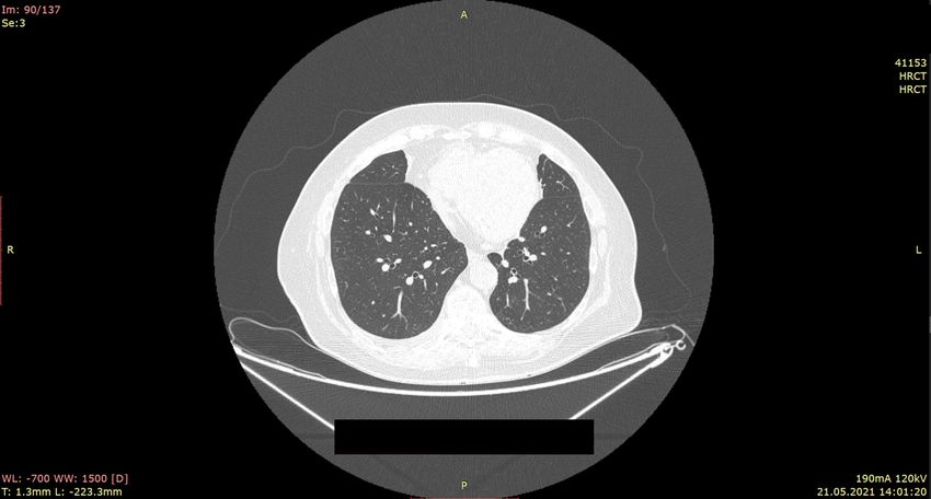
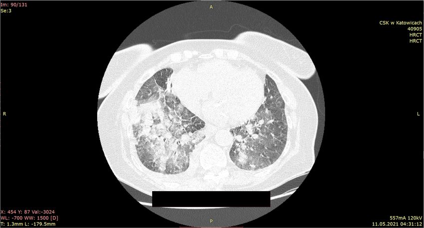
842 Aleksandra Niemiec et al. shortness of breath, fever, cough, and exercise intolerance. interleukin-6 concentration 24.3 pg/mL (reference range: The patient denied direct contact with SARS-CoV-2-infected 60 mL/min), D-dimer concentration of hypertension, diabetes type 2, hyperuricemia, chronic 1,040 ng/dL (reference range: 96%), lactate 3.15 mmol/L (reference range:
Fungal infection mimicking COVID-19 infection 843
and sputum cultures were performed. Additionally, there sensitive to fluconazole, amphotericin B, caspofungin, and
were taken blood tests for the presence of IgM and IgG micafungin. The antifungal drug Fluconazole 200 mg daily
antibodies for Mycoplasma pneumoniae, Chlamydophila per os was added to therapy.
pneumoniae, SARS-CoV-2, Influenza A, B, and Respiratory During further hospitalization, improvement in the
Syncytial Virus, but they were all negative. Also, abdominal general state of the patient was observed. Oxygen therapy
ultrasound examination and echocardiography were per- was gradually reduced. Atrial fibrillation was converted
formed. No other outbreaks of potential infection were to regular sinus rhythm with ventricular action of 65
detected. beats per minute. On the 10th day, the shortness of breath
Before the results of the laboratory tests were obtained, was not observed, and the patient did not require oxygen
due to the severe clinical condition, empirical therapy was therapy (SpO2, 94%). Auscultation of the lung revealed a
introduced using ceftriaxone, levofloxacin, low molecular reduction of crackles and wheezing; the symmetrical
weight heparin, dexamethasone, budesonide, and ipratro- respiratory sound was the dominant one. Laboratory
pium bromide. determinants of inflammation were normalized. The con-
Due to increasing respiratory failure despite oxygen trol chest HRCT showed full withdrawal of inflammatory
therapy given and deteriorating results of ABG (pO2: changes and regression of pleural effusion (Figure 2). At
45.4 mmHg, SpO2: 79%) anesthesiologist’s consultation the base of the lungs, fibrous-atelectasis changes were
was performed. Prone position and switch type of oxygen reported.
therapy to high flow oxygen delivery 60 L/min FiO2: 0.9 The patient has been discharged from the hospital in
was recommended. Moreover, it was recommended to a good general condition without any complaints and
take a nasopharyngeal swab in the SARS-CoV-2 infection one continued outpatient treatment with fluconazole.
more time, and until the result is obtained, the patient should
be treated like a COVID-19-positive person. Recommended pro- Institutional review board statement: After consulting
cedures improved saturation of patient to 98%; however, this with the Bioethics Committee of the Medical University
next swab was also negative. of Silesia in Katowice, ethical review and approval were
Despite the suggestions of anesthesiologists, remde- waived because a case report does not require the approval
sivir and tocilizumab were not included in the treatment, of the bioethics committee.
suspecting other than viral etiology of pneumonia.
In the cultures following results were obtained: blood Ethical approval: This is a description of a clinical case
and urine were negative, whereas in sputum Candida with a brief literature review. There was no formal research
albicans and Candida glabrata were identified. They were ethics approval required or no experimental intervention
Figure 2: HRCT scans after antifungal treatment: regression of changes visible on admission is described.844 Aleksandra Niemiec et al.
Table 1: Radiological findings in COVID-19 pneumonia
Stage Phase Time (days) Main radiological findings Additional radiological findings (in every phase)
1 Early 0–4 GGO Peripheral vessel widening
2 Progressive 5–8 GGO, CPP, and small Halo sign
consolidations
Atoll sign or reversed halo sign
3 Peak 9–13 Consolidative foci Overlapping of radiological findings in different phases
4 Absorption ≥14 GGO and linear consolidation Rarity of: lymphadenopathies, pleuric effusions, pulmonary
nodules
GGO, ground-glass opacities; CPP, crazy paving pattern.
in routine care. Fully informed consent from the patient described phases and the observed changes in the CT
was obtained. image are summarized in Table 1.
However, other types of pneumonia may resemble
that caused by SARS-CoV-2 in HRCT. That is the reason
why other disease entities should be taken into consid-
3 Discussion eration in the diagnostic process [11]. The symptoms and
even imaging-study findings are not peculiar for viral
During the COVID-19 pandemic, SARS-CoV-2 infection is infections [12]. Nevertheless, other viral, fungal, atypical
mainly suspected as the reason for interstitial pneu- bacterial pneumonia, autoimmune process, or cancer can
monia. Due to the lack of specific symptoms, scientists also manifest as GGO in the imaging of the lungs [13,14].
from the beginning of the pandemic were looking for a There was even a case of a patient diagnosed with amio-
test that would allow a clear diagnosis of the patient [8]. darone-induced interstitial pneumonia described [15].
Over time, however, it turned out that the identification Acute respiratory distress syndromes with a cause
of infected people is not sufficient for their proper treat- other than COVID-19 have their own specific radiolog-
ment. Tools that would be able to assess the severity of ical features that are important in the differential
the disease, and thus the prognosis, have become neces- diagnosis.
sary. These tools were the scales prepared by the researchers In the case of pneumonia caused by typical bacteria,
for the assessment of radiological examinations, both CT a characteristic feature is the air bronchogram and the
and chest X-ray [9]. fact that the lesions do not exceed pleural cleavages [16].
Pan et al. divided lung involvement on chest CT into Moreover, we can find in CT: centrilobular nodules, cavi-
four stages. Stage 1 is dominated by GGO changes. In tations, pneumatoceles, mediastinal lymphadenomega-
stage 2, additionally appear crazy paving pattern (CPP) lies, or pleural effusions [17,18]. Other types of lesions
and small consolidations. In the third phase, we observe can be found in fungal pneumonia such as Pneumocystis
the presence of consolidative foci sometimes with a halo jiroveci infection. In this type of pneumonia we see symme-
sign. And in the fourth phase, called the absorption trical, centroparenchymal and peripheral, confluent GGO,
phase, GGO and linear consolidation are again described generally with subpleural sparing [19] and a predilection
and interpreted as a sign of repair processes [10]. All the for the upper lobes [20], but we almost never observed CPP.
Table 2: Radiological features of pathologies in differential diagnosis with COVID-19 pneumonia
Pathologies GGO CPP Consolidations References
Infective pneumonia Bacterial R A C [16–18]
Viral C A R [17,23]
Fungal C R R [19,20]
Cardiovascular Acute pulmonary edema C C C [21,22]
Acute pulmonary embolism C C C [24]
Vasculities C C C [25]
GGO: ground-glass opacities, CPP: crazy paving pattern, C: common, R: rare, A: absent.Fungal infection mimicking COVID-19 infection 845
Cardiovascular disease is another group of diseases References
that can cause radiological features similar to COVID-19.
For example, in pulmonary edema, we also can find in CT [1] Singh LT, Sharara RS, Leap J, Singh AC. Management of
scans GGO, CPP, and consolidations but with different respiratory failure. Crit Care Nurs Q. 2016;39(2):94–109.
[2] Umakanthan S, Sahu P, Ranade AV, Bukelo MM, Rao JS,
timing of occurrence with respect to COVID-19 pneumonia
Abrahao-Machado LF, et al. Origin, transmission, diagnosis
and also with accompanying cardiomegaly [21,22]. How- and management of coronavirus disease 2019 (COVID-19).
ever, it should be remembered that due to the high simi- Postgrad Med J. 2020;96(1142):753–8.
larity of CT images in this group of diseases to COVID-19, [3] Sreepadmanabh M, Sahu AK, Chande A. COVID-19: advances
an anamnesis in the differential diagnosis plays a key role. in diagnostic tools, treatment strategies, and vaccine devel-
Table 2 summarizes the most common disease entities opment. J Biosci. 2020;45(1):148.
[4] Vandenberg O, Martiny D, Rochas O, van Belkum A,
requiring differentiation from COVID-19.
Kozlakidis Z. Considerations for diagnostic COVID-19 tests.
Because of this difficulty in the differential diagnosis, Nat Rev Microbiol. 2021;19(3):171–83.
CT of the chest is not recommended for routine screening [5] Francone M, Iafrate F, Masci GM, Coco S, Cilia F, Manganaro L,
in patients under investigation for COVID-19 [26]. et al. Chest CT score in COVID-19 patients: correlation with
disease severity and short-term prognosis. Eur Radiol.
2020;30(12):6808–17.
[6] Pan F, Zheng C, Ye T, Li L, Liu D, Li L, et al. Different computed
tomography patterns of Coronavirus Disease 2019 (COVID-19)
4 Conclusion between survivors and non-survivors. Sci Rep.
2020;10(1):11336.
To sum up, it is necessary to remember that not only [7] Guarnera A, Podda P, Santini E, Paolantonio P, Laghi A.
Differential diagnoses of COVID-19 pneumonia: the current
SARS-CoV-2 infection may manifest as severe pneumonia.
challenge for the radiologist-a pictorial essay. Insights
Obviously, COVID-19 must also be taken into considera- Imaging. 2021;12(1):34.
tion, and special care should be provided during admis- [8] Ji T, Liu Z, Wang G, Guo X, Akbar Khan S, Lai C, et al. Detection
sion. Testing for SARS-CoV-2 must be performed in the first of COVID-19: a review of the current literature and future per-
place to exclude the infection. However, other diseases or spectives. Biosens Bioelectron. 2020;166:112455.
immunological disorders may cause similar symptoms. [9] Wasilewski PG, Mruk B, Mazur S, Półtorak-Szymczak G,
Sklinda K, Walecki J. COVID-19 severity scoring systems in
Proper diagnosis is crucial for subsequent proceedings
radiological imaging - a review. Pol J Radiol. 2020;85:e361–8.
and treatment. Particularly now, in times of the COVID- [10] Pan F, Ye T, Sun P, Gui S, Liang B, Li L, et al. Time course of lung
19 pandemic, physicians must not forget that differential changes at chest CT during recovery from Coronavirus disease
diagnosis is an important part diagnostic process that 2019 (COVID-19). Radiology. 2020 June;295(3):715–21.
allows us to make the right diagnosis and implement the [11] Caruso D, Polici M, Zerunian M, Pucciarelli F, Polidori T,
Guido G, et al. Quantitative chest CT analysis in discriminating
right methods of therapy.
COVID-19 from non-COVID-19 patients. Radiol Med.
2021;126(2):243–9.
Funding information: This research received no external [12] Ghosh S, Deshwal H, Saeedan MB, Khanna VK, Raoof S,
funding. Mehta AC. Imaging algorithm for COVID-19: a practical
approach. Clin Imaging. 2021;72:22–30.
[13] Mehrabi S, Fontana S, Mambrin F, Nguyen HQ, Righi E,
Author contributions: Conceptualization, M.K., M.H.; meth-
Tacconelli E, et al. Pitfalls of computed tomography in the
odology, A.N., M.K., M.H.; resources, A.N., M.K., M.H.; Coronavirus 2019 (COVID-19) era: a new perspective on
writing – original draft preparation, A.N., M.K., M.H.; writing – ground-glass opacities. Cureus. 2020;12(5):e8151.
review and editing, M.B, B.O.; visualization, M.H.; supervi- [14] Azoulay E, Russell L, Van de Louw A, Metaxa V, Bauer P,
sion, M.B., B.O.; project administration, M.H., M.K. All authors Povoa P, et al. Diagnosis of severe respiratory infections in
have read and agreed to the published version of the immunocompromised patients. Intensive Care Med.
2020;46(2):298–314.
manuscript.
[15] Cappannoli L, Telesca A, Scacciavillani R, Petrolati E,
Smargiassi A, Rabini A, et al. A nontrivial differential diagnosis
Conflict of interest: The authors declare there is no con- in COVID-19 pandemic: a case report and literary review of
flict of interest. amiodarone-induced interstitial pneumonia. Future Cardiol.
2021;17(6):991–7.
[16] Walker CM, Abbott GF, Greene RE, Shepard JA, Vummidi D,
Data availability statement: Data supporting reported
Digumarthy SR. Imaging pulmonary infection: classic signs
results on the treatment of the patient can be found in and patterns. AJR Am J Roentgenol. 2014;202(3):479–92.
the author’s (A.N.) medical record and can be obtained [17] Franquet T. Imaging of community-acquired pneumonia.
upon request from the corresponding author. J Thorac Imaging. 2018;33:282–94.846 Aleksandra Niemiec et al.
[18] Dai WC, Zhang HW, Yu J, Xu HJ, Chen H, Luo SP, et al. CT features of pulmonaryedema. Radiographics.
imaging and differential diagnosis of COVID-19. Can Assoc 1999;19(6):1507–31.
Radiol J. 2020;71(2):195–200. [23] Koo HJ, Lim S, Choe J, Choi SH, Sung H, Do KH. Radiographic
[19] Fujii T, Nakamura T, Iwamoto A. Pneumocystis pneumonia in and CT Features of viral pneumonia. Radiographics.
patients with HIV infection: clinical manifestations, laboratory 2018;38(3):719–39.
findings, and radiological features. J Infect Chemother. [24] Han D, Lee KS, Franquet T, Müller NL, Kim TS, Kim H, et al.
2007;13(1):1–7. Thrombotic and nonthrombotic pulmonary arterial embolism:
[20] Kuhlman JE, Kavuru M, Fishman EK, Siegelman SS. spectrum of imaging findings. Radiographics.
Pneumocystis carinii pneumonia: spectrum of parenchymal CT 2003;23(6):1521–39.
findings. Radiology. 1990;175:711–4. [25] Castañer E, Alguersuari A, Andreu M, Gallardo X, Spinu C,
[21] Hani C, Trieu NH, Saab I, Dangeard S, Bennani S, Mata JM. Imaging findings in pulmonaryvasculitis. Semin
Chassagnon G, et al. COVID-19 pneumonia: a review of typical Ultrasound CT MR. 2012;33(6):567–79.
CT findings and differential diagnosis. DiagnIntervImaging. [26] The American Society of Emergency Radiology (ASER) (2020)
2020;101(5):263–8. STR/ASER COVID-19 position statement; 2020. https://
[22] Gluecker T, Capasso P, Schnyder P, Gudinchet F, thoracicrad.org/wp-content/uploads/2020/03/STR-ASER-
Schaller MD, Revelly JP, et al. Clinical and radiologic Position-Statement-1.pdf.You can also read