"Old wine in a new bottle" - post COVID-19 infection, central serous chorioretinopathy and the steroids
←
→
Page content transcription
If your browser does not render page correctly, please read the page content below

Sanjay et al. Journal of Ophthalmic Inflammation and Infection              (2021) 11:14
https://doi.org/10.1186/s12348-021-00244-4
                                                                                                                       Journal of Ophthalmic
                                                                                                                  Inflammation and Infection

 BRIEF REPORT                                                                                                                                    Open Access

“Old wine in a new bottle” - post COVID-19
infection, central serous chorioretinopathy
and the steroids
Srinivasan Sanjay1* , Poornachandra B. Gowda2, Bhimasena Rao3,4, Deepashri Mutalik1,
Padmamalini Mahendradas1 , Ankush Kawali1 and Rohit Shetty5

 Abstract
 Introduction: Corona virus disease (COVID-19) pandemic can cause myriad of ocular manifestations.
 We report a case of unilateral multi focal central serous retinopathy, post COVID-19 infection in an Asian Indian
 female.
 Case presentation: A 42-year-old female presented to us with unilateral blurring, in the right eye (OD), 12 days
 after COVID-19 infection. She had fever, chills, shortness of breath and cough with tiredness and was COVID- RT
 PCR positive. She was administered intravenous and oral antibiotics with injection heparin/remdesivir, during her 7
 day stay at the hospital. She was also on steroid inhalers. She had no systemic history of note.
 On ocular evaluation, her corrected distance visual acuity was 20/40 in OD and 20/20 in left eye (OS). Anterior
 segment was normal. Anterior vitreous was clear. Fundus examination of the OD showed central serous retinopathy
 (CSCR) with OS being normal.
 Conclusion: CSCR can occur post COVID-19 due to steroid administration and physicians administering it should
 be aware of this and refer the patients to an ophthalmologist earlier.
 Keywords: Corona virus disease-19 (COVID-19), Ophthalmic manifestations, Central serous chorioretinopathy,
 Spectral Domain Optical Coherence Tomography (SD-OCT), Inhalational steroids, Oral steroids

Introduction                                                                        papillophlebitis, multifocal chorioretinitis and Adie’s
Severe acute respiratory syndrome virus 2 (SARS-CoV2)                               syndrome are the ophthalmic manifestations associated
infection resulted in a global pandemic of Coronavirus                              with COVID-19 infection [2–6].
disease 2019 (COVID-19). Wuhan in China was the first                                 We describe an unique case of unilateral multifocal
place of the outbreak in December 2019. As of 23                                    central serous retinopathy (CSCR) in a patient who had
March 2021, there have been 123,419,065 confirmed                                   just recovered from COVID-19 and had been treated
cases of COVID-19, including 2,719,163 deaths,                                      with inhalational and oral steroids.
reported to WHO. As of 19 March 2021, a total of 397,
950,709 vaccine doses have been administered [1].                                   Case presentation
Conjunctival involvement, cotton wool spots (CWS) and                               The procedures followed were in accordance with the
retinal hemorrhages, central retinal artery/vein                                    ethical standards of the responsible committee on hu-
occlusion, ophthalmic artery occlusion, panuveitis,                                 man experimentation (institutional or regional) and with
                                                                                    the Helsinki Declaration of 1975 as revised in 1983. The
* Correspondence: sanjaygroup24@gmail.com                                           patient’s written and informed consent was obtained and
1
 Department of Uvea and Ocular Immunology, 121/C, Chord Road, Narayana
Nethralaya, Bangalore, India                                                        the study was approved by the hospital ethics
Full list of author information is available at the end of the article              committee.
                                    © The Author(s). 2021 Open Access This article is licensed under a Creative Commons Attribution 4.0 International License,
                                    which permits use, sharing, adaptation, distribution and reproduction in any medium or format, as long as you give
                                    appropriate credit to the original author(s) and the source, provide a link to the Creative Commons licence, and indicate if
                                    changes were made. The images or other third party material in this article are included in the article's Creative Commons
                                    licence, unless indicated otherwise in a credit line to the material. If material is not included in the article's Creative Commons
                                    licence and your intended use is not permitted by statutory regulation or exceeds the permitted use, you will need to obtain
                                    permission directly from the copyright holder. To view a copy of this licence, visit http://creativecommons.org/licenses/by/4.0/.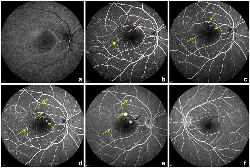
Sanjay et al. Journal of Ophthalmic Inflammation and Infection       (2021) 11:14                                             Page 2 of 5

  A 42-year-old Asian Indian female presented to us with                   arm-retina time of 18 s with multiple hyperfluoroscent
unilateral blurring, in the right eye (OD), 12 days after                  spots seen in the macula which increased in size and in-
COVID-19 infection. Prior to presentation to us, she had                   tensity in later films in an inkblot pattern characteristic of
fever, chills, shortness of breath and cough with general-                 central serous retinopathy in OD. One of the lesion
ized fatigue. Her physician noted that she was afebrile,                   showed a mixed “smoke stack” and “ink blot” appearance
oxygen saturation was 97% with few crepitations in her                     (Fig 3e).
lungs and heart rate was 132/min. Complete blood count                        At the time of ocular presentation, investigations
(CBC) was within normal limits, erythrocyte sedimenta-                     showed serum Ferritin 87.90 U/L (females 11–307), Pro-
tion rate (ESR) 35 mm /hr., random blood sugar (RBS)-                      calcitonin 0.032(< 0.5), mild leucocytosis 11,600 (4500–
118 mg/dl, COVID-19 rapid antigen test was negative.                       11,000), ESR-35 (< 20 mm/hr), CRP 10.7 mg/l(< 3), Im-
  Investigations done at the local hospital a day later                    munoglobulin (Ig) G antibodies to nucleocapsid antigen
showed upper respiratory swab for COVID-19 using real                      of SARS-CoV2 were positive 4.41 (< 1.0 non reactive).
time polymerase chain reaction (RT-PCR) was positive                          Based on the clinical and imaging findings, a diagnosis
and B-beta (Corona virus) CoV specific target gene and                     of unilateral OD multifocal central serous chorioretinopa-
severe acute respiratory syndrome- corona virus 2                          thy (CSCR) was made. In consultation with chest phys-
(SARS-CoV2) specific target gene were detected,                            ician the inhalational and oral steroids were stopped.
WIDAL test negative, urine analysis was within normal                         One month later, there was an improvement in her vi-
limits. Chest X ray of the lung showed bilateral ground                    sion to 20/25 in OD, OCT showed reduction of the sub-
glass opacities (Fig. 1). Based on the records that were                   retinal fluid and the hyper-reflective material and reso-
available with the patient, we in Table 1 show the medi-                   lution of the pigment epithelial detachment (Fig. 4)
cations administered during her stay at a local hospital.                     We would like to report for the first time a case of a
  On ophthalmic examination, her corrected distance                        unilateral multifocal CSCR in an Asian Indian post
visual acuity was 20/40 in OD and 20/20 in the left eye                    COVID-19 treatment.
(OS). The intraocular pressure was 15 and 18 mmHg in                          Published data show that SARS-CoV-2 binds to the
OD/OS respectively. Examination of the anterior seg-                       host cells via the angiotensin converting-enzyme (ACE)
ment was normal in both eyes (BE). Fundus evaluation,                      2 receptor [7]. Endothelial cells become vulnerable when
OD showed absent foveal reflex with serous elevation of                    the ACE 2 receptors are expressed and binding of
the retina with ring reflex at the macula. The OS was                      SARS-CoV-2 may cause systemic endothelial dysfunc-
within normal limits.                                                      tion. All major organs like the lungs, heart, veins, and ar-
  A spectral domain optical coherence tomography (SD-                      teries have higher density ACE 2 receptors. Endothelial
OCT) scan on the Spectralis™ (Heidelberg Engineering,                      dysfunction leads to vasoconstriction, ischemia, tissue
Heidelberg, Germany) of the OD showed hyper-reflective                     edema, and a procoagulant state secondary to endothe-
dots in the posterior vitreous, altered foveal contour with                lial alterations including endothelitis [7].
serous detachment in the macula and with pigment epi-                         The exact pathophysiology of ocular transmission of
thelial detachment (Fig. 2 a). OS was normal (Fig. 2 b).                   the virus remains incompletely understood, although
Fundus fluoroscein angiography (Fig. 3a-e) showed an                       there is preliminary evidence of SARS-CoV-2 being de-
                                                                           tected in ocular secretions. The ocular tropism of the
                                                                           virus and its potential to cause localized ocular disease
                                                                           are worth considering [8].
                                                                              Steroids may be necessary to manage the post COVID-
                                                                           19 systemic manifestations including the cytokine storm.
                                                                              Our patient was administered oral and inhalational
                                                                           steroids during her stay at the local hospital. CSCR oc-
                                                                           curs or is aggravated by administration of corticosteroids
                                                                           irrespective of the route of administration. Steroids when
                                                                           used topically for skin conditions, intra-articular, intra-
                                                                           venous, intramuscular, oral, epidural, intranasal and in-
                                                                           halation are all associated with CSCR [9, 10].
                                                                              It is postulated that the blood retinal barrier may be
                                                                           damaged, with damage to retinal pigment epithelial
                                                                           pump and hyperpermeability of choriocapillaries leading
                                                                           to CSCR.
 Fig. 1 Chest X ray PA view of the lung showing bilateral ground
                                                                              CSCR can develop secondary to exogenous corticoste-
 glass opacities with left lung consolidation during her admission
                                                                           roids several years later.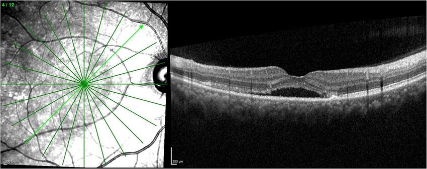
Sanjay et al. Journal of Ophthalmic Inflammation and Infection              (2021) 11:14                                                     Page 3 of 5

Table 1 Shows medications administered to the patient while she was admitted at the local hospital
Route                                          Drug                                                                Dose/duration
Intravenous      Cefoperazone+ Sulbactam                                             1000 mg + 1000 mg for 4 days
Subcutaneous Heparin                                                                 5000 units every 8 h for 3 days
Intravenous      Remdesivir                                                          200 mg loading dose, then 100 mg a day for 4 days
Oral             Dexamethasone                                                       6 mg daily for 7 days
Oral             Azithromycin                                                        500 mg for 7 days
Oral             Doxycycline                                                         100 mg twice daily for 7 days
Oral             Montelukast and Levocetrizine combination                           (10 mg) and (5 mg) for 7 days
Oral             Vitamin C                                                           1 g for 7 days
Oral             Pantoprazole                                                        40 mg once daily for 7 days
Oral             Ivermectin                                                          12 mg once daily for 7 days
Inhalation       Oxygen                                                              2 litres/minute for 7 days
Inhalation       Formoterol fumarate dehydrate and budesonide 200                    (6 mcg) and 9200mcg) twice daily, which was continued even after
                 combination                                                         discharge
Inhalation       Salbutamol rotacaps                                                 four times daily
She had no systemic history of note
At the time of discharge she was switched to oral steroids (methylprednisolone) 16 mg once daily till her presentation to us
Legends: mg milligram; mcg microgram

  Fig. 2 a shows a spectral domain optical coherence tomography scan across the macula of the OD. The white arrow points to a doom shaped
  elevation which is the serous retinopathy. Also in the scan is a smaller doom which represents retinal pigment epithelial detachment. b shows
  the normal scan of OS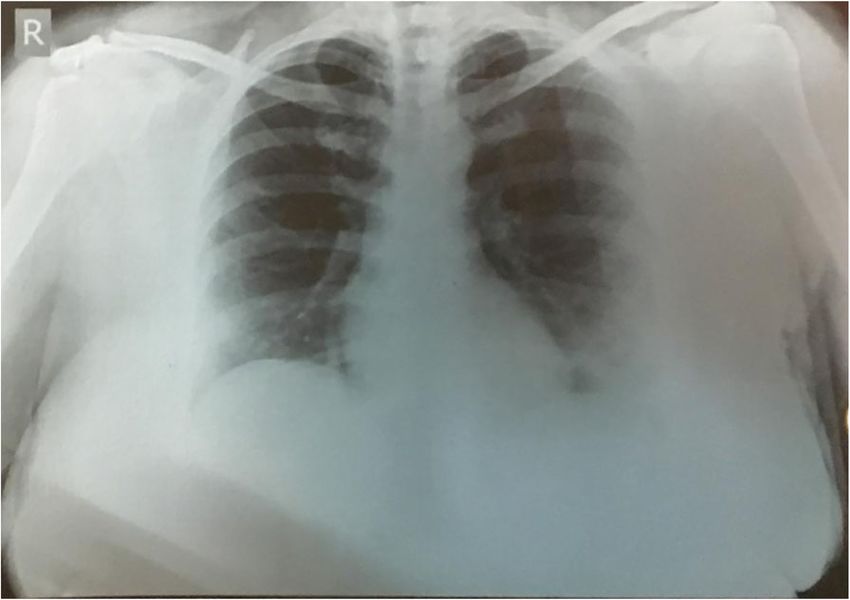
Sanjay et al. Journal of Ophthalmic Inflammation and Infection      (2021) 11:14                                                        Page 4 of 5

 Fig. 3 a-e fundus fluoroscein angiography (FFA) of the OD from early phases a,b to later phases c-e. The yellow arrows point to a pinpoint leak
 initially and increasing in size in later phases. The black arrow with yellow arrow head adjacent to optic disc shows a mixed inkblot and smoke
 stack pattern. f- shows normal left eye

   There may be a temporal correlation between the use                       Postulates on the mechanism of CSCR include vas-
of a corticosteroid nasal spray and the development of                     cular auto-regulation via increased transcription of
CSCR. Posterior sub capsular cataract can occur after                      adrenergic receptors or potentiation of vascular re-
nasal/inhaled steroids.                                                    activity, effects from steroid-induced systemic hyper-
   Cessation of inhalational steroids can lead to reso-                    tension, or a prothrombotic effect. Inhibition of
lution of CSCR, which also happened partially in our pa-                   collagen synthesis in Bruch’s membrane may be an-
tient [11].                                                                other mechanism. The barrier function of retinal pig-
   The choroid has extensive choriocapillaries whose role                  ment epithelium (RPE) may be compromised due to
is to supply oxygen and nutrients to the outer retina                      impaired ion and water transport. The role of the
which has no vascular network. Glucocorticoids possibly                    RPE in CSCR pathogenesis remains poorly under-
enhance the fibroblasts proliferation with compromised                     stood. Increased tissue hydrostatic pressure in the
capillary function leading to their fragility. Corticoste-                 choroid can cause the barrier function of the RPE to
roids may affect choroid, Bruch’s membrane, or the ret-                    be compromised and lead to areas of fluid accumula-
inal pigment epithelium [12].                                              tion between the retina and the RPE which can also

 Fig. 4 shows a spectral domain optical coherence tomography scan across the macula of the OD with reduction of the sub-retinal fluid and the
 pigment epithelial detachment a month later
Sanjay et al. Journal of Ophthalmic Inflammation and Infection               (2021) 11:14                                                            Page 5 of 5

lead to pigment epithelial detachment(s) which also                                Author details
                                                                                   1
represent a form of RPE decompensation [13]                                         Department of Uvea and Ocular Immunology, 121/C, Chord Road, Narayana
                                                                                   Nethralaya, Bangalore, India. 2Department of Retina, 121/C, Chord Road,
  Some refer to the pinpoint areas of leakage seen in                              Narayana Nethralaya, Bangalore, India. 3Chest and Maternity Centre, 878, 5th
acute CSCR as “micro-rips” or “blowouts”.                                          Block, Near Bashyam Circle, Rajaji Nagar Bengaluru, Karnataka 560010, India.
                                                                                   4
  CSCR is a self-limiting condition; various modalities of                          Vikram Hospital, Anne’s College, No.71/1, Millers Road, Bangalore, India.
                                                                                   5
                                                                                    Department of Neuro-ophthalmology, Cornea and Refractive Surgery, 121/C,
treatment have been described in the literature for the                            Chord Road, Narayana Nethralaya, Bangalore, India.
recurrent cases. One month after stopping steroids (in-
halation and oral) there was a significant improvement                             Received: 13 January 2021 Accepted: 3 April 2021

in patient’s visual acuity and SD-OCT findings. Subse-
quently she was lost to follow up.                                                 References
                                                                                   1. World Health Organisation (WHO), COVID-19 dashboard. https://covid19.
                                                                                       who.int/. Accessed 24 Mar 2021.
Conclusion                                                                         2. Acharya S, Diamond M, Anwar S, Glaser A, Tyagi P (2020) Unique case of
We hereby describe a patient with unilateral serous                                    central retinal artery occlusion secondary to COVID-19 disease. IDCases. 21:
                                                                                       e00867. https://doi.org/10.1016/j.idcr.2020.e00867
chorioretinopathy post COVID-19 infection, who devel-                              3. Insausti-García A, Reche-Sainz JA, Ruiz-Arranz C, López Vázquez Á, Ferro-
oped ophthalmic manifestations 12 days after she was                                   Osuna M (2020) Papillophlebitis in a COVID-19 patient: inflammation and
discharged from the hospital, after having tested negative                             hypercoagulable state [published online ahead of printJul 30]. Eur J
                                                                                       Ophthalmol 2020:1120672120947591. https://doi.org/10.1177/112067212094
for COVID-19. Patients with COVID-19 should be                                         7591
warned about possible ophthalmic sequelae even after                               4. Ortiz-Seller A, Martínez Costa L, Hernández-Pons A, Valls Pascual E, Solves
their systemic recovery. Physicians treating COVID-19                                  Alemany A, Albert-Fort M. (2020) Ophthalmic and neuro-ophthalmic
                                                                                       manifestations of coronavirus disease 2019 (COVID-19). Ocul Immunol
should be aware of these important sequelae and refer                                  Inflamm 16; 28: 1285-1289. doi: https://doi.org/10.1080/09273948.2020.18174
the patient to an ophthalmologist for timely                                           97. Epub 2020 Oct 6
intervention.                                                                      5. Sheth JU, Narayanan R, Goyal J, Goyal V (2020) Retinal vein occlusion in
                                                                                       COVID-19: a novel entity. Indian J Ophthalmol 68:2291–2293. https://doi.
Abbreviations                                                                          org/10.4103/ijo.IJO238020
SARS-CoV2: Severe Acute Respiratory Syndrome Corona Virus 2; COVID-                6. Sanjay S, Srinivasan P, Jayadev C, Mahendradas P, Gupta A, Kawali A, Shetty
19: Coronavirus disease 2019; CWS: Cotton wool spots; OD: Right eye;                   R. Post COVID-19 Ophthalmic Manifestations in an Asian Indian Male. Ocul
CBC: Complete blood count; ESR: Erythrocyte sedimentation rate;                        Immunol Inflamm. 2021: 1–6. doi: https://doi.org/10.1080/09273948.2020.1
RBS: Random blood sugar; RT-PCR: Real time polymerase chain reaction;                  870147. Epub ahead of print)
OS: Left eye; SD-OCT: Spectral domain optical coherence tomography;                7. To KF, Lo AW (2004) Exploring the pathogenesis of severe acute respiratory
CSCR: Central serous chorioretinopathy; Ig: Immunoglobulin;                            syndrome (SARS): the tissue distribution of the coronavirus (SARS-CoV) and
ACE: Angiotensin converting-enzyme; RPE: Retinal pigment epithelium                    its putative receptor, angiotensin-converting enzyme 2 (ACE2). J Pathol
                                                                                       203(3):740–743. https://doi.org/10.1002/path.1597
                                                                                   8. Ho D, Low R, Tong L, Gupta V, Veeraraghavan A, Agrawal R. (2020) COVID-
Acknowledgements
                                                                                       19 and the ocular surface: a review of transmission and manifestations. Ocul
Dr. Nikhita Reddy, Department of Retina, Narayana Nethralaya, Bengaluru,
                                                                                       Immunol Inflamm;28:726–734. doi: https://doi.org/10.1080/09273948.2020.1
India
                                                                                       772313. Epub 2020 Jun 16. PMID: 32543262, 5
                                                                                   9. Haimovici R, Gragoudas ES, Duker JS, Sjaarda RN, Eliott D. (1997) Central
Authors’ contributions                                                                 serous chorioretinopathy associated with inhaled or intranasal
Design: S S, B R. Acquisition of data: SS, PBG, DM. Analysis and intrepretation:       corticosteroids. Ophthalmology. Oct;104(10):1653–1660. doi: https://doi.
SS, PBG,PM, AK. Manuscript writing: SS, PBG, DM, PM, AK, RS. Manuscript                org/10.1016/s0161-6420(97)30082-7.
editing: SS, PBG, PM,AK, RS. Intellectual content: SS, PBG, PM, AK, RS. The        10. Tittl MK, Spaide RF, Wong D, Pilotto E, Yannuzzi LA, Fisher YL, Freund B,
authors read and approved the final manuscript.                                        Guyer DR, Slakter JS, Sorenson JA (1999) Systemic findings associated with
                                                                                       central serous chorioretinopathy. Am J Ophthalmol 128(1):63–68. https://doi.
Funding                                                                                org/10.1016/s0002-9394(99)00075-6
None.                                                                              11. Nakatsuka AS, Khanamiri HN, Lam QN, El-Annan J (2019) Intranasal
                                                                                       corticosteroids and central serous Chorioretinopathy: a report and review of
Availability of data and materials                                                     the literature. Hawaii J Med Public Health 78(5):151–154
Available on request                                                               12. Bouzas EA, Karadimas P, Pournaras CJ (2002) Central serous
                                                                                       chorioretinopathy and glucocorticoids. Surv Ophthalmol 47(5):431–448.
Declarations                                                                           https://doi.org/10.1016/S0039-6257(02)00338-7
                                                                                   13. Nicholson B, Noble J, Forooghian F, Meyerle C (2013) Central serous
Ethics approval and consent to participate                                             chorioretinopathy: update on pathophysiology and treatment. Surv
The study was approved by the hospital ethics committee.                               Ophthalmol 58(2):103–126. https://doi.org/10.1016/j.survophthal.2012.07.004
The study was approved by the Narayana Nethralaya Ethics committee, Vide
approval number EC reference NO C/2020/09/09 (virtual). All tenets of the          Publisher’s Note
Helsinki declaration were adhered to.                                              Springer Nature remains neutral with regard to jurisdictional claims in
Patient's written and informed consent was obtained for inclusion in the           published maps and institutional affiliations.
study.

Consent for publication
The patient’s written and informed consent was obtained.

Competing interests
None.
You can also read