Functional characterization of a bioengineered liver after heterotopic implantation in pigs - Nature
←
→
Page content transcription
If your browser does not render page correctly, please read the page content below
ARTICLE
                  https://doi.org/10.1038/s42003-021-02665-2                OPEN

                  Functional characterization of a bioengineered liver
                  after heterotopic implantation in pigs
                  Brett D. Anderson 1,5, Erek D. Nelson 2,5, DongJin Joo2,3, Bruce P. Amiot 2, Aleksandr A. Katane1,
                  Alyssa Mendenhall1, Benjamin G. Steiner1, Aron R. Stumbras1, Victoria L. Nelson1, R. Noelle Palumbo1,
                  Thomas W. Gilbert1, Dominique S. Davidow 1, Jeffrey J. Ross 1 & Scott L. Nyberg 2,4 ✉

                  Organ bioengineering offers a promising solution to the persistent shortage of donor organs.
                  However, the progression of this technology toward clinical use has been hindered by the
1234567890():,;

                  challenges of reconstituting a functional vascular network, directing the engraftment of
                  specific functional cell types, and defining appropriate culture conditions to concurrently
                  support the health and phenotypic stability of diverse cell lineages. We previously demon-
                  strated the ability to functionally reendothelialize the vasculature of a clinically scaled
                  decellularized liver scaffold with human umbilical vein endothelial cells (HUVECs) and to
                  sustain continuous perfusion in a large animal recovery model. We now report a method for
                  seeding and engrafting primary porcine hepatocytes into a bioengineered liver (BEL) scaffold
                  previously reendothelialized with HUVECs. The resulting BELs were competent for albumin
                  production, ammonia detoxification and urea synthesis, indicating the presence of a func-
                  tional hepatocyte compartment. BELs additionally slowed ammonia accumulation during
                  in vivo perfusion in a porcine model of surgically induced acute liver failure. Following explant
                  of the graft, BEL parenchyma showed maintenance of canonical endothelial and hepatocyte
                  markers. Taken together, these results support the feasibility of engineering a clinically scaled
                  functional BEL and establish a platform for optimizing the seeding and engraftment of
                  additional liver specific cells.

                  1 Miromatrix Medical Inc, Eden Prairie, MN, USA. 2 Department of Surgery, Mayo Clinic, Rochester, MN, USA. 3 Department of Surgery, Yonsei University

                  College of Medicine, Seoul, South Korea. 4 William J. von Liebig Center for Transplantation and Clinical Regeneration, Mayo Clinic, Rochester, MN, USA.
                  5
                    These authors contributed equally: Brett D. Anderson, Erek D. Nelson. ✉email: Nyberg.Scott@mayo.edu

                  COMMUNICATIONS BIOLOGY | (2021)4:1157 | https://doi.org/10.1038/s42003-021-02665-2 | www.nature.com/commsbio                                         1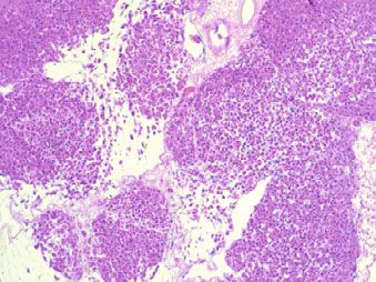
ARTICLE                                                              COMMUNICATIONS BIOLOGY | https://doi.org/10.1038/s42003-021-02665-2

L
      iver transplantation currently represents the only curative             Human umbilical vein endothelial cells (HUVECs) were
      therapy for acute and chronic liver failure; however, access         expanded in 2D tissue culture flasks and seeded into decellular-
      to this treatment is limited by a persistent shortage of donor       ized BEL scaffolds through the suprahepatic inferior vena cava
organs. Whole-organ bioengineering has the potential to fulfill             (sIVC), followed by the portal vein (PV) 24 h later (Fig. 1k, i). We
this unmet need by offering a virtually limitless supply of                previously reported that daily glucose consumption rates (GCR)
bioengineered liver grafts. One of the most promising technolo-            provide a robust, non-invasive metric for monitoring cell
gies for whole-organ bioengineering is perfusion decellularization         proliferation in HUVEC-seeded BEL constructs and is predictive
of whole porcine organs to obtain an acellular scaffold that retains       of successful perfusion outcomes following in vivo inplantation17.
the native extracellular matrix (ECM) composition and tissue               BELs were cultured until a minimum GCR of 50 mg/h was
microenvironments that provide a favorable environment for                 observed (typically 13–16 days following HUVEC seeding) prior
seeding parenchymal cells, and the native vascular networks cri-           to infusing hepatocytes into the scaffold (Fig. 1k, m).
tical to sustaining those parenchymal cells with nutrients and                Primary hepatocytes were isolated from freshly explanted
oxygen. The scaffolds are clinical sized for human transplant and          porcine livers and enriched by differential sedimentation through
can then be repopulated with organ-specific human cells using a             multiple low-speed centrifugation and washing steps to achieve
perfusion-based bioreactor. First applied in a rat cardiac model1,         typical hepatocyte purity of 85-95% as measured by albumin-
perfusion decellularization has since enabled the creation of              positive flow cytometry (Supplementary Fig. S1). Hepatocytes
acellular scaffolds from a variety of whole organs—including,              were seeded through the bile duct and cultured for an additional
heart1, liver2,3, lung4, kidney5,6, and pancreas7,8—all of which           48 h under continuous PV perfusion with media formulated for
have demonstrated an ability to support engraftment of tissue-             the simultaneous culture of endothelial cell and hepatocytes
specific cell types.                                                        (referred to herein as co-culture media; see “Methods”).
   Engraftment of multiple resident liver cell types—including             Hematoxylin and eosin (H&E), as well as immunofluorescence
hepatocytes2,9–11, endothelial cells10,12, and cholangiocytes13—           staining for hepatocyte-specific markers (albumin and fumar-
into perfusion decellularized liver scaffolds has been shown in            ylacetoacetate [FAH]) 48 h post hepatocyte seeding revealed a
small, preclinical models and proof-of-concept attempts to                 clustered distribution of hepatocytes throughout the scaffold
describe a clinical-scale BEL in vitro14–16. As the majority of            parenchyma (Fig. 2a–d), consistent with prior studies that utilized
in vivo BEL functional studies have relied on small animal                 the bile duct for cell infusion9,10,13,18. As we previously
models, they do not recapitulate many of the unique challenges of          reported17, and similar to native liver tissue, endothelial cells
translating whole-organ bioengineering to a clinically relevant            localized within large vessels expressed high levels of CD31, while
scale. The scale-up, cell seeding optimization, and development of         endothelial cells localized within parenchymal capillaries had
bioreactor culture conditions to support the multiple required cell        increased expression of the liver sinusoidal endothelial cell
types remain important hurdles in the development of a clinically          (LSEC) marker LYVE1 and reduced expression of CD31 (Fig. 2e).
translatable BEL.                                                             To characterize the in vitro function of BELs over the course of
   We recently reported the reconstitution of a functional vascular        bioreactor culture, levels of cell-derived soluble factors were
network in a porcine whole liver scaffold with HUVECs, which               quantified in culture media samples from scaffolds seeded with
reproducibly sustained in vivo perfusion for an excess of 10 days          both HUVECs and hepatocytes (co-culture), as well as scaffolds
in a porcine liver transplant model under a steroid-based                  that received only one cell type (HUVEC only and hepatocyte
immunosuppression regimen.17 Building on this success, the                 only) (Fig. 2f). BELs seeded with HUVECs (HUVEC only and co-
current study reports an optimized method for the seeding and              culture) exhibited increasing production of the endothelial cell-
simultaneous culture of HUVECs and primary porcine hepato-                 derived von Willebrand factor (vWF) over time (Fig. 2g).
cytes in decellularized porcine liver scaffolds. The resulting BEL         Importantly, vWF production in co-culture BELs following
constructs resemble native liver tissue with respect to the loca-          hepatocyte seeding was similar to levels observed in HUVEC
lized engraftment of endothelial cells and hepatocytes, detectable         only BEL controls, suggesting that the addition of hepatocytes to
hepatic function during bioreactor culture, and sustained blood            the scaffold did not compromise endothelial cell viability and
perfusion at physiologic pressures in an ex vivo perfusion model.          function. Albumin levels were quantified in media samples from
The current study further shows that these BELs are amenable to            co-culture and hepatocyte only BELs, and there was no
surgical implantation and continuous perfusion in a large animal           appreciable difference in albumin production observed, suggest-
heterotopic liver transplant model, with the maintenance of cell           ing that hepatocyte function not impacted by endothelial cells in
viability and functional marker expression 48 h following                  the scaffold. (Fig. 2h). To further assess key metabolic functions
implantation. Collectively, these studies describe a robust plat-          of hepatocytes in BEL scaffolds, ammonia detoxification and urea
form for the seeding and culture of multiple liver cell types, and         production assays were adapted to bioreactor scale culture based
demonstrate proof of concept for bioengineering a therapeutic              on prior 2D culture and hepatocyte spheroid studies19. In brief,
liver construct at a clinically relevant scale.                            BELs were challenged with fresh co-culture media supplemented
                                                                           with 0.8 mM ammonium chloride 16–20 h following hepatocyte
                                                                           seeding and samples were taken at intervals up to 23 h after the
Results                                                                    challenge for quantification of ammonia and urea (Fig. 2i). As
Characterization of BEL constructs seeded with primary                     expected, HUVEC only BELs showed a steady increase in
endothelial cells and hepatocytes. Freshly explanted porcine               ammonia levels over time due to ongoing cellular metabolism.
livers were decellularized by sequential perfusion with Triton             In contrast, hepatocyte only and co-culture BELs both showed
X-100 and SDS detergent solutions to generate the acellular                substantial ammonia clearance with similar clearance kinetics
scaffolds used in these studies (Fig. 1a–c). Histological staining         although hepatocyte only BELs showed slightly more ammonia
demonstrated complete removal of cellular material from the                clearance (Fig. 2j). Hepatocyte only and co-culture BELs showed
decellularized scaffold (Fig. 1d, e) and retention of the extracellular    similar rates of urea release, while HUVEC only BELs showed
matrix (ECM) proteins collagen I and collagen IV (Fig. 1f–i).              only a slight increase in urea compared to acellular graft controls
Decellularized livers were mounted in custom bioreactors (Fig. 1j)         (Fig. 2k). These assays confirm the ability of hepatocytes to retain
and perfused with antibiotic-free endothelial culture media for            their function in high-density cultures in a decellularized liver
72 h to confirm the sterility of the scaffold (Fig. 1k).                    scaffold. In addition, the data show that endothelial cells cultured

2                                           COMMUNICATIONS BIOLOGY | (2021)4:1157 | https://doi.org/10.1038/s42003-021-02665-2 | www.nature.com/commsbio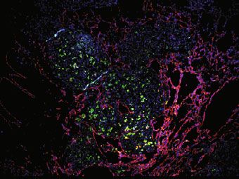
COMMUNICATIONS BIOLOGY | https://doi.org/10.1038/s42003-021-02665-2                                                                                 ARTICLE

             a
                 sIVC   PV BD                    b                        d                          f              COL I      h            COL IV
                                  iIVC                                                                              DAPI                     DAPI

                                 Native Liver
                                                                                          250 μm                      100 μm                    100 μm
                           1. Triton X-100
                           2. SDS                c                        e                          g              COL I      i            COL IV
                                                                                                                    DAPI                     DAPI

                                Decellularized
                                Liver Scaffold
                                                                                          250 μm                      100 μm                    100 μm

              j                                                   k
                           P                                                              Seed HUVECs
                                                                                                                 Seed Hepatocytes
                  GEC                                                                          sIVC PV                 BD
                                 BT
                                                                                Scaffold
                                      PT                                       Qualification
                                               Perfusion
                                             Control System
                                                                      Day -3                     0       1               13-16      (+ 1-3 days)
                                                                    Culture
                                                                    Media                      Endothelial                         Co-culture

             l                                                                             m
                            HUVECs                                                                       Porcine Liver

                                                                 sIVC          PV
                                                                                                                                       BD
                        Culture Expansion

                                                                                                          Enyzmatic
                                                         Decellularized Liver Scaffold                    Digestion
                                                                                                                                   Re-endothelialized
                                                                                                                                         BEL
                                                                       Seed                     Mechanical Separation
                               Harvest                                HUVECs                     and Mesh Filtration    Seed
                                                                                                                     Hepatocytes

Fig. 1 Overview of approach to bioengineering a porcine-derived whole liver construct. a Overview of vessel cannulation and detergent perfusion
methods for whole liver decellularization. b, c Photographs of representative porcine (b) native and (c) decellularized whole livers. d, e Hematoxylin and
eosin staining of histological sections from (d) native and (e) decellularized porcine liver tissue demonstrating the efficient removal of cellular material
while preserving the native tissue architecture. f–i Representative immunofluorescence micrographs from native (f, h) and decellularized (g, i) liver tissue
demonstrating retention of Collagen I and Collagen IV, respectively, in cell-free scaffolds. The absence of DAPI staining in the decellularized scaffolds
demonstrates removal of cellular DNA. j Schematic of perfusion bioreactor design. BELs are suspended in a vessel with culture media. An extended coil of
silicone tubing (GEC) is included in the perfusion circuit to facilitate gas exchange. A bubble trap is positioned directly upstream of the BEL perfusion inlet.
During media perfusion, a pressure transducer provides real-time feedback to a controller which in turn adjusts the flow rate on a peristaltic pump to
maintain a constant perfusion pressure. k Schematic depicting bioreactor culture and cell seeding events. Decellularized scaffolds were pre-qualified in
antibiotic-free media for 3 days prior to HUVEC seedings. HUVEC-seeded liver constructs were cultured in endothelial cell growth media until a 24 h
average glucose consumption rate of 50–90 mg/h was reached (typically 13–16 days), at which point hepatocytes were seeded into the scaffold. BELs were
maintained in co-culture media formulated for simultaneous culture of endothelial cells and hepatocytes for the remainder of the experiment (typically 1–3
additional days). l Overview of HUVEC culture and seeding approach to generate a reendothelialized liver construct. Cells were infused first through the
cannulated iIVC followed by a second cell infusion through the PV 24 h later. m Schematic of porcine donor liver hepatocyte isolation and seeding of
bioengineered liver constructs. Whole porcine livers are enzymatically digested, and dissociated cells were filtered through a series of mesh sieves to
remove large debris and cell aggregates. Hepatocytes were enriched through multiple low speed (70 × g) centrifugation and washing steps. DAPI—4′,6′-
diamidino-2-phenylindole; BEL—bioengineered liver; GEC—gas exchange coil; BT—bubble trap; iIVC—infrahepatic inferior vena cava; PV—portal vein.

in the scaffold did not impede the functionality of hepatocytes in               short-term perfusion with blood, an ex vivo perfusion circuit was
the scaffold in vitro.                                                           created to recirculate porcine blood through BELs (seeded and
                                                                                 cultured as above; [Fig. 2f]) at a regulated target pressure of
Acute blood perfusion studies to assess vascular patency of                      12 mmHg while monitoring flow rates over 30 minutes (Fig. 3a).
BELs. The ability to sustain blood perfusion in vivo is essential to             To mimic the initial graft reperfusion rates experienced in a
the successful development of any bioengineered organ. To                        surgical liver transplant model, initial blood flow through the
determine the ability of BELs in this current study to sustain                   BELs was set to 350 mL/min to fully inflate the graft, and flow

COMMUNICATIONS BIOLOGY | (2021)4:1157 | https://doi.org/10.1038/s42003-021-02665-2 | www.nature.com/commsbio                                                  3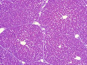
ARTICLE COMMUNICATIONS BIOLOGY | https://doi.org/10.1038/s42003-021-02665-2
a b c CD31 d CD31 e CD31
Albumin FAH LYVE1
DAPI DAPI DAPI
5 cm 250 μm 100 μm 100 μm 100 μm
f g h
Endothelial Media
48 h Post Hep. Seeding (μg/h)
Co-culture Media 80 200
HUVEC Only
vWF production (μg/h)
HUVEC Only Hep. Only
Albumin Production
60 Co-culture
150
Seed
Co-culture HUVECs 40 100
Seed 20 50
Hepatocyte Hepatocytes
Only 0 0
-12-10 -8 -6 -4 -2 0 2
HUVEC
Hepatocyte
Co-culture
Only
Days in Culture
Day 0 1 13-16 +1 +2
Only
Relative to Hepatocyte Seeding
i j k
4.0 5.0
Sample (t = 0, 1, 2, 7, 23 h) Media Media
HUVEC Only HUVEC Only
Ammonia (mmols) Hep. Only 4.0 Hep. Only
3.0
Urea (mmols)
Co-culture Co-culture
3.0
2.0
Ammonia Urea 2.0
Quantification 1.0
1.0
2 L Fresh Media 0.0 0
+ 0.8 mM NH4Cl 0 4 8 12 16 20 24 0 4 8 12 16 20 24
Time (h) Time (h)
Fig. 2 Histological and functional characterization of BEL constructs. a Representative photograph of a BEL seeded with HUVECs and porcine hepatocytes.
b Hematoxylin and eosin staining of representative co-culture BEL tissue sections fixed 48 h after seeding hepatocytes. c–e Immunofluorescent staining of
cell lineage markers in non-serial tissue sections 48 h after seeding hepatocytes: (c) CD31 & albumin; (d) CD31 & FAH; (e) CD31 and LYVE1. f Schematic
depicting seeding and culture timeline for HUVEC only, hepatocyte only, and co-culture BEL constructs used in (g, h, j, k). g vWF production in grafts before
and after hepatocyte seeding. Data from independent HUVEC only (n = 5), hepatocyte only (n = 7), and co-culture (n = 7) BEL constructs are shown. Error
bars denote the mean and standard deviation at each time point. h 24-h average albumin production in co-culture grafts 48 h following hepatocyte seeding.
Data from independent HUVEC only (n = 5), hepatocyte only (n = 7), and co-culture (n = 7) BELs are shown. Error bars denote the mean and standard
deviation. i Schematic of in vitro ammonia clearance and urea production assay. Ammonium chloride is added to the bioreactor media at a concentration of
0.8 mM. Ammonia and urea levels are measured in media samples taken at t = 0, 1, 2 7, and 23 h following the addition of ammonium chloride. Error bars
denote the mean and standard deviation each time point. j, k Ammonia clearance (j) and urea production kinetics (k) following the addition of ammonium
chloride to the bioreactor perfusion media. Data from independent HUVEC only (n = 5), hepatocyte only (n = 7), and co-culture (n = 7) BELs, media only
controls (n = 4) are shown. Error bars denote the mean and standard deviation each time point. BEL—bioengineered liver; FAH—fumarylacetoacetate
hydrolase; CD31—cluster of differentiation 31; LYVE1—lymphatic vessel endothelial hyaluronan receptor 1; vWF—von Willebrand factor.
rates were subsequently allowed to automatically correct to target (Fig. 3b, c), demonstrating the importance of endothelial cells in
a perfusion pressure of 12 mmHg. Additionally, prior to BEL sustaining perfusion through the scaffold. Co-culture BELs
perfusion, the activated clotting time (ACT) of heparinized por- (n = 5) exhibited intermediate flow rate profiles (Fig. 3b, c),
cine blood (ACT > 1500) was titrated to a slightly elevated, phy- presumably due to increased resistance within the scaffold vas-
siologically relevant range of 170–230 (just high enough to inhibit culature from the hepatocyte seeding. Nevertheless, all co-culture
clotting within the in vitro blood circuit components) through the BELs still maintained blood flow (118–293 mL/min) after 30 min
addition of protamine sulfate. To establish benchmarks for of continuous perfusion at physiologic pressures.
optimal perfusion as well as scaffold thrombosis in this system, To further characterize the vascular blood flow through liver
freshly explanted porcine livers (n = 2) and unseeded decel- scaffolds seeded with both HUVECs and hepatocytes, a porcine
lularized liver scaffolds (n = 2) were subjected to continuous ex vivo blood perfusion model was employed whereby the PV,
perfusion in the blood circuit. As expected, native organs sIVC and infrahepatic IVC (iIVC) of anesthetized pigs
exhibited relatively stable flow rates in excess of 300 mL/min by (80–100 kgs) were cannulated and used to establish a perfusion
the end of 30 min, whereas decellularized scaffolds thrombosed circuit through co-culture grafts under physiologic venous flow
within 5 min (Fig. 3b, c). Four out of five HUVEC only BELs (Fig. 3d). Animals were heparinized to an ACT of ~250 to prevent
exhibited relatively stable flow rates (250-350 mL/min) over clot formation in the cannula or tubing. Flow was measured at
30 minutes (similar to freshly explanted livers), while one lost 150–250 mL/min at 30 min and real-time angiography further
flow gradually over the course of perfusion (Fig. 3b, c). Similar to confirmed continuous perfusion and revealed vascular flow
the decellularized scaffolds, hepatocyte only BELs (n = 3) including visualization of capillary beds in the majority of the
experienced a rapid loss in flow within the first five minutes, co-culture BEL (Fig. 3e, Supplementary Movie S1).
4 COMMUNICATIONS BIOLOGY | (2021)4:1157 | https://doi.org/10.1038/s42003-021-02665-2 | www.nature.com/commsbio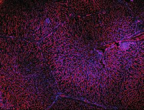
COMMUNICATIONS BIOLOGY | https://doi.org/10.1038/s42003-021-02665-2 ARTICLE
a b c
50 Native (n=2)
Decell (n=2) 400
40
Pressure
Perfusion HUVEC Only (n=5)
(mmHg)
Flow Rate after 30 min
Control System 30 Hepatocyte Only (n=3)
300
20 Co-culture (n=5)
(mL/min)
10
P 200
0
400
PT 100
Flow Rate
300
(mL/min)
PV iIVC 200
0
100
D e
H UV l l
a C
cu te
re
37°C
H e
iv
ep E
C tocy
ltu
ec
0
at
N
0 5 10 15 20 25 30
o-
Time (min)
d e
t=1s t=2s t=3s
IVC
5 cm 5 cm 5 cm
t=6s t = 10 s t = 15 s
PV
80-100 kg
5 cm 5 cm 5 cm
Fig. 3 Acute blood perfusion studies to assess vascular patency in BELs. a Schematic of in vitro blood perfusion circuit. 37 °C porcine blood is perfused at
12 mmHg through the PV with a peristaltic pump and returned to a reservoir through the IVC. b Summary plots of pressures and flow rates measured over
60 minutes during in vitro blood perfusion studies using HUVEC only, hepatocyte only, or co-culture BELs. Freshly explanted (Native) porcine livers and
decellularized scaffolds (Decell) were included as benchmarks for idealized perfusion and rapid thrombosis, respectively. c Violin plots summarizing BEL
flow rates from (b) after 30 min of perfusion. d Schematic of ex vivo blood perfusion model. A synthetic perfusion circuit is established by cannulating the
PV and sIVC within an anesthetized pig. Blood flow is diverted from the animal’s cannulated PV to the BEL PV and returned from the BEL sIVC into the
animal’s cannulated iIVC. e Real-time angiography time lapse imaging following contrast infusion. Imaging was performed after 30 min of continuous blood
perfusion. PV—portal vein; BEL—bioengineered liver; sIVC—suprahepatic inferior vena cava, iIVC—infrahepatic inferior vena cava.
Implantation of co-culture BELs in a large animal model. To was 120–410 mL/min as measured by a Transonic flow probe
assess the ability of co-culture BELs to sustain 48 h of continuous prior to closing the animal. Computed tomography (CT) was
physiologic perfusion and hepatocyte function in vivo, a porcine utilized post-operatively to confirm BEL perfusion and loss of flow
heterotopic liver transplant model of acute liver failure was to the native liver (Fig. 4b, c). Follow up CT imaging at Day 1 and
employed. BELs (48 h post hepatocyte seeding) were situated in a Day 2 confirmed that BEL perfusion was sustained throughout the
heterotopic position in the hepatorenal space and end-to-side remaining duration of the acute implant studies (Fig. 4c).
anastomoses were performed between the BEL PV to the native
PV and BEL sIVC to native iIVC, respectively (Fig. 4a, Supple-
mentary Figs. S4, 5, Supplementary Movie S2). Total portal vein Functional characterization of BELs in Vivo. In addition to
flow was diverted into the BEL by ligating and dividing the native showing sustained perfusion of a hepatocyte/endothelial BEL
PV between the BEL and native liver. The implanted BEL in vivo, the survival and functionality of hepatocytes were
(260 ± 40.8 g) was ~30% the native liver size resulting in a small- assessed. Previous studies have correlated the relationship
for-size syndrome. To reduce portal hypertension while main- between intracranial pressure (ICP) and blood ammonia levels
taining sufficient flow through the implanted BEL, a small por- during acute liver failure19. Therefore, a totally internal intra-
tocaval shunt was created utilizing a 4 mm ringed cranial probe (NEUROVENT-P-tel, Raumedic, Mills River, NC)
polytetrafluoroethylene (PTFE) graft which was anastomosed was implanted 5 days prior to surgery to allow monitoring of the
end-to-side between the recipient’s PV and iIVC. Blood flow intracranial pressure (ICP). Termination of the study was per-
through the portocaval shunt was measured at between 20 and formed when an animal experienced (1) ICP of 20 mmHg or
50 mL/min, or ~10% of the flow through the BEL, after anasto- greater for 2 h, (2) mean arterial pressure (MAP) was 30 or less
mosis and titrated as necessary via banding or ligation of the for 2 h, or (3) 48 h of survival was achieved. Control group ani-
portocaval shunt. mals (n = 2) underwent ICP probe insertion and subsequent
Following BEL perfusion, the native liver was isolated from creation of a total portocaval shunt by directly anastomosing
arterial perfusion by ligating and dividing all structures within the native portal vein to native iIVC before devascularization of the
hepatoduodenal ligament including the common bile duct and the native liver as described above. This resulted in termination at 24
hepatic artery complex. The caudate lobe was devascularized via and 48 h (Fig. 4d). The 24-h survivor was terminated due to a
oversewing and compression with locking suture until cut caudate MAP less than 30, at which point the ICP was 17. The 48-h
parenchyma was cyanotic and did not demonstrate bleeding from survivor had a MAP of 42 and ICP of 16 at the time of termi-
cut edges. Flow through the BEL the first few hours after perfusion nation. Blood ammonia levels in the control animals climbed
COMMUNICATIONS BIOLOGY | (2021)4:1157 | https://doi.org/10.1038/s42003-021-02665-2 | www.nature.com/commsbio 5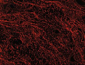
ARTICLE                                                                                   COMMUNICATIONS BIOLOGY | https://doi.org/10.1038/s42003-021-02665-2

               a                                                                                                                     b
                                                                                                                   Native HA
                                                                                                                    Ligation
                                                                 Native
                                                                 Liver

                                                                                                                   Native PV
                                                            Anastomosis                                             Ligation
                                                               Sites
                            BEL

                                      BEL Anastimosis                                 PV Flow
                                                                                    Through BEL
               c
                                                      Post-op                                   Day 1                                               Day 2

               d                                                                                e
                                 100                                                                               1.2
                                               P/C Shunt (n=2)                                                               P/C Shunt (n=2)
                                               BEL (n=3)                                                                     BEL (n=3)
                                                                                                                                                *               **
                   Percent Survival

                                      75
                                                                                                    Ammonia (mM)

                                                                                                                   0.8

                                      50

                                                                                                                   0.4
                                      25

                                       0                                                                           0.0
                                           0      6    12   18 24 30           36    42    48                            0      6    12   18 24 30       36    42   48
                                                             Time (h)                                                                      Time (h)

                        f                                          g                             h                                  CD31 i                     CD31
                                                                                                                                 Albumin                    Albumin
                                                                                                                                    DAPI                       DAPI

                                                                                                    Pre-                                       Post-
                                                      500 μm                         50 μm          Implant                         200 μm     Implant        200 μm
                       j                               CD31        k                  CD31          l                            CYP3A4 m                   CYP3A4
                                                        FAH                            FAH                                         DAPI                       DAPI
                                                       DAPI                           DAPI

                        Pre-                                       Post-                            Pre-                                       Post-
                        Implant                       200 μm       Implant          200 μm          Implant                         200 μm     Implant        200 μm

quickly with levels surpassing 0.5 mM within 15 h of surgery                                        and 48 h (Fig. 4d). The 37-h survivor showed an ICP greater than
(Fig. 4e). In these control animals, there was no ascites accu-                                     20 mmHg with a MAP of 37 mmHg. The 42-h survivor had a low
mulation indicative of negligible portal hypertension as expected                                   MAP secondary to respiratory failure (diagnosed with acute
with the portocaval shunt.                                                                          pulmonary edema and large bilateral pleural effusions on CT)
   In contrast, the experimental group animals (n = 3), having                                      with an ICP of 11. The 48-h survivor reached the end of the study
received co-culture BEL implants, were terminated at 37 h, 42 h,                                    and had a MAP of 38 and ICP of 17. The BEL group also showed

6                                                                COMMUNICATIONS BIOLOGY | (2021)4:1157 | https://doi.org/10.1038/s42003-021-02665-2 | www.nature.com/commsbio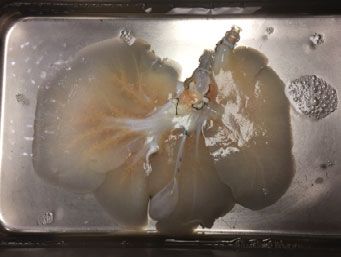
COMMUNICATIONS BIOLOGY | https://doi.org/10.1038/s42003-021-02665-2 ARTICLE
Fig. 4 Heterotopic implantation of co-culture BELs in a large animal model. a Schematic of heterotopic BEL implant surgical model. See text for details.
b Post-operative 3D-reconstruction from CT imaging demonstrating BEL perfusion and devascularization of native liver. BEL is outlined in yellow and native
liver is outlined in green. c Representative axial CT imaging of recipient animal at post-op, 24 h, and 48 h time points. BEL is outlined in yellow. d Kaplan–Meier
curves showing animal survival times within portocaval shunt and BEL implant groups. Symbols are matched to ammonia values in (d). e Post-operative blood
ammonia levels measured in BEL implant recipient animals (n = 3) and portocaval shunt animals (n = 2) over the duration of the experiment. Asterisks (*)
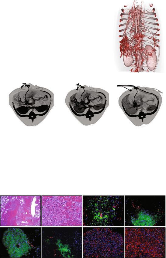
denote data points that were above the upper limit of quantification of the assay (1 mM). f, g Representative histological section of BEL tissue explanted 48 h
post-implant showing viable hepatocytes and endothelialized vasculature. h, i Representative immunostaining BEL tissue (h) pre-implant and (i) explanted
48 h post-implant showing maintenance of CD31 and albumin expression. j, k Representative immunostaining BEL tissue (j) pre-implant and (k) explanted
48 h post-implant showing maintenance of CD31 and FAH expression. l, m Representative immunostaining BEL tissue (l) pre-implant and (m) explanted 48 h
post-implant showing maintenance of CYP3A4 expression. HA—hepatic artery; BEL—bioengineered liver; P/C—portocaval; CT—computed tomography; FAH
—fumarylacetoacetate hydrolase; CYP3A4—cytochrome p450 3A4.
an increase in blood ammonia levels to approximately 0.5 mM, adequately recapitulate the complex microenvironments and
but leveled off or decreased, except for one graft that lost graft functions of native tissues. Perfusion decellularization offers an
perfusion (37-h survivor) that resulted in a rapid increase in elegant solution to this problem by enabling the creation of
ammonia and ICP that prompted termination at 37 h (Fig. 4e). human-scaled whole-organ scaffolds that preserve the integrity of
The ammonia data are suggestive of hepatocyte function and are vascular networks and parenchymal microenvironments that
consistent with the observed ex vivo ammonia clearance. promote robust engraftment and functionality of tissue-specific
However, interpretation of the results of the heterotopic implant cell types.
model may be confounded by the differential portal-systemic The first step towards realizing a clinically relevant BEL is to
pressure between models. ensure long-term patent vascularization of the graft. Multiple
As discussed above, the co-culture BELs had reduced in vitro groups have attempted to address this challenge either by seeding
blood flow at a controlled pressure when compared to HUVEC a decellularized liver graft with endothelial cells2,12,14 or by
only BELs. Combined with the expected small-for-size syndrome modifying native vasculature to inhibit coagulation11,16. How-
of the model and despite the attempt to regulate portal ever, in vivo validation of these approaches has been limited to
hypertension with a small portosystemic shunt, the presence of implantation in rats, and human-scale BELs have been limited to
the increased resistance within the co-culture BEL resulted in in vitro characterization. We recently reported the feasibility of
increased portal hypertension compared with the control group’s using HUVECs to repopulate the vascular matrix of a decel-
portocaval shunt as evidenced by the increased ascites output in lularized porcine liver and achieved up to 20 days of continuous
the experimental group compared to the control group perfusion without systemic anticoagulation in a porcine hetero-
(Supplementary Fig. S3). The presence of portal hypertension topic implant model. We reported on the ability of seeded
within the experimental group, but not the control group, could HUVECs to upregulate sinusoidal endothelial markers and evi-
result in increased ammonia production secondary to mesenteric dence of fenestration formation. We found that the duration of
venous congestion and underestimation of the ammonia patency was limited by the porcine xenogeneic rejection of
clearance function in the experimental group. HUVEC only human endothelial cells17. Importantly, we established glucose
BELs demonstrated mixed results. Based on in vitro flow data, consumption rate as a reliable, non-destructive measure to pre-
this group experienced less portal hypertension than the co- dict patency of a BEL and a critical step in-process quality
culture BEL group, but significantly more than the control measure for BEL candidates for implantation. These observations
portocaval shunt group. This is also supported by the accumula- mark important milestones towards the realization of a clinically
tion of ascites in the HUVEC only BEL group, but not to the relevant BEL, and other engineered organs such as kidney, lung,
extent demonstrated by the co-culture group. Taking these factors and heart.
together, we concluded that portal hypertension is an important Having shown durable patency of BEL vasculature, the next
consideration in BEL development as it may limit the hepatocyte major milestone was to investigate any impact of the introduction
mass that can be seeded per unit volume of the graft. Therefore, of hepatocytes into the BEL on graft patency and the function of
an increased graft size may be needed to increase the hepatocyte the hepatocytes following seeding and implantation. Two pre-
mass while reducing the portal hypertension to decrease vious studies2,11 in rats have shown early indicators of hepatic
ammonia production secondary to mesenteric venous congestion function after seeding rat hepatocytes into a decellularized rat
and provide a better estimation of graft function. BELs retained liver matrix and implanting the resulting graft in a heterotopic
their shape and size (Supplementary Fig. S6), histological position. Uygun et al. demonstrated the persistence of
examination of explanted BELs with Hematoxylin & Eosin hepatocyte-specific markers by immunohistochemical staining,
(H&E) (Fig. 4f, g) and immunostaining for albumin, FAH, CD31, but only evaluated hepatocyte function after 8 h in a renal
CYP3A4, and LYVE1 (Fig. 4h–m, Supplementary S7, Supple- implant site2. Bao et al. showed decreased blood ammonia levels
mentary S8)) pre- and post-implant showed viable porcine as compared to a 90% hepatectomy, but long-term patency
hepatocytes and HUVECs throughout the explanted BEL required chemical modification of the vasculature11. Our previous
suggesting that seeded cells remained viable and phenotypically study showed perfusion of a BEL seeded with only HUVECs for
stable throughout the duration of in vivo perfusion. up to 14 days in pigs, and that duration was limited by a xeno-
geneic immune response to the HUVECs. The current study
shows that the addition of hepatocytes to a clinically scaled BEL
Discussion does not impede perfusion at physiologic pressures in vitro and
Despite recent advances in tissue engineering technologies, the in vivo. Furthermore, engraftment of hepatocytes in the par-
ability to engineer whole-organ systems at a clinically relevant enchymal compartment reconstituted hepatic function within the
scale has remained extremely challenging, largely due to the BEL construct as measured by albumin secretion, urea produc-
inherent difficulties in fabricating scaffolds that provide suitable tion, and ammonia clearance in vitro, and the initial indication of
vascular beds to oxygenate and nourish seeded cells and to ammonia detoxification activity in vivo. Histological examination
COMMUNICATIONS BIOLOGY | (2021)4:1157 | https://doi.org/10.1038/s42003-021-02665-2 | www.nature.com/commsbio 7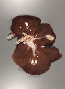
ARTICLE                                                                      COMMUNICATIONS BIOLOGY | https://doi.org/10.1038/s42003-021-02665-2

of co-culture BELs revealed that hepatocytes were appropriately                     cannulated and flushed with sterile saline. Cannulated livers were decellularized by
localized in the parenchymal compartment, while HUVECs                              peristaltic pump (Cole Palmer, 7575-30; 77200-60)-driven vascular perfusion with
                                                                                    1% Triton X-100 followed by 0.6% sodium dodecyl sulfate. Solution flow rates were
localized primarily within large vessels and capillary vasculature.                 automatically regulated by a custom perfusion control system designed to maintain
Both cell types expressed canonical functional markers, and as                      perfusion pressures between 8 and 12 mmHg. Decellularized livers were subse-
previously demonstrated17, HUVECs localized within sinusoidal                       quently disinfected with 1000 ppm peracetic acid, washed with phosphate buffered
capillaries acquired expression of LYVE1, suggesting a level of                     saline, and stored at 4 °C. All aspects of the decellularization process were per-
microenvironment-driven reprogramming toward an LSEC-like                           formed in an ISO 7 cleanroom facility.
phenotype. These results support growing confidence in the
ability to bioengineer a clinically sized liver construct capable of                HUVEC cell culture and seeding of decellularized liver scaffolds. Human
                                                                                    umbilical vein endothelial cells (HUVECs) (Lonza, C2517A) were cultured at 37 °C
revascularization and surgical implantation with sustained per-                     and 5% CO2 in antibiotic-free Endothelial Cell Growth Media (R&D Systems,
fusion and hepatocyte survival in a pig model. This represents a                    CCM027) supplemented with 2% fetal bovine serum (Corning), 50 mg/L ascorbic
major step toward bioengineering transplantable livers for                          acid (Sigma), 1 mg/L hydrocortisone (Sigma), 20 μg/L FGF (R&D Systems), 5 μg/L
clinical use.                                                                       VEGF (R&D Systems), 5 μg/L EGF (R&D Systems), 15 μg/L R3 IGF (Sigma),
                                                                                    1000 U/L heparin (Sigma), and 1.5 μM acetic acid (Sigma). Cells were harvested
   While the current study demonstrates the ability to hetero-                      with 0.25% trypsin-EDTA (Thermo, 25200056) at 90–100% confluency. Decel-
topically implant a BEL with sustained portal flow, hepatocyte                       lularized porcine livers were mounted in bioreactors and perfused with antibiotic-
survival and initial indications of in vivo function, there are                     free cell culture media (37 °C, 5% CO2) for 72 h to confirm the absence of microbial
multiple limitations of the present study we attempted to mini-                     contamination. HUVECs collected at passage 5–9 were infused through the sIVC
mize. First, in this heterotopic large animal implantation model,                   with a syringe (1.2×10⁸ cells in 150 mL culture media). Following 1 h of static
                                                                                    culture to allow for cell attachment within the scaffold, culture media supple-
the BEL graft size was limited by the space available in the                        mented with Penicillin and Streptomycin was perfused through the sIVC at
abdominal cavity while the devascularized native liver was pre-                     12 mmHg. Twenty-four h later, a second inoculum of HUVECs was collected and
sent. This limited our ability to use BELs that were more than                      infused through the PV in the same manner as above. Following seeding, culture
one-third of the average native recipient liver weight and limiting                 media was replaced daily, and volumes were continually adjusted to ensure that
                                                                                    glucose levels remained above 0.3 g/L within a 24 h period. Media perfusion into
the total number of seeded hepatocytes for functional assessment.                   the scaffold was maintained a pressure of at 12 mmHg during culture.
Portal hypertension was consistently observed due to both small-
for-size syndrome and the presence of hepatocytes within the                        Porcine hepatocyte isolation and seeding of BEL scaffolds. Freshly harvested
graft which we showed to increase vascular resistance. In this                      whole livers (400–600 g) were cannulated through the PV, sIVC, and iIVC, and
model, elimination of blood perfusion of the native liver is                        perfused with 5 L of HBSS (Fisher, MT21022CM) to remove residual blood from
achieved through ligation and division of the portal vein and                       the organ followed by 1 L of cold HTK solution (Essential Pharmaceuticals, 25767-
                                                                                    735-24) to minimize ischemic injury to organs during transportation. Livers were
hepatic arteries and oversewing the caudate lobe. The hilar                         then perfused through the PV (500–600 mL/min) with 5 L of HBSS supplemented
structures and hepatogastric ligament were ligated and divided to                   with 2.5 mM EGTA (Sigma, E3889), allowing the first 1 L to drain to waste, and
eliminate all inflow including aberrant vascular anatomy. No                         recirculating the remaining volume for 20 min. Livers were subsequently perfused
collateral branches were detected on CT, but backflow through                        with 2 L of solution comprised of 142 mM NaCl, 6.7 mM KCl, 10 mM HEPES,
the hepatic vein complex was present although remnant liver                         5 mM N-acetyl-L-cysteine, and 1% Penicillin-Streptomycin (Sigma, P4333).
                                                                                    Digestion was initiated with perfusion of 4 L of L-15 media (Fisher, 21083027)
function based on systemic venous backflow is likely to be neg-                      supplemented with 100 mg of Liberase TM (Sigma, 5401127001) and 5 mM CaCl2,
ligible. Finally, this model keeps the devascularized native liver                  allowing the first 500 mL to drain to waste and recirculating the remaining volume
in situ (Supplementary Fig. S9). The dying organ may be detri-                      until livers were soft with visible breakdown of the capsule (20–30 min). After
mental as it is a source of systemic inflammation in a largely                       digestion, 1 L of cold Williams E media (Fisher, RR090071P1) supplemented with
                                                                                    10% FBS (VWR, 97068-085) was poured over the livers and the capsule was gently
anhepatic animal, but also beneficial in that it is a known driver of                pulled apart to release the cell suspension. To eliminate any remaining undigested
hepatocyte proliferation and is contemporarily used clinically                      tissue, cells were filtered through an 8″ wide mesh strainer, followed by a series of
as such.                                                                            mesh sieves (250 μm (VWR, 57334-466), 125 μm (VWR, 57334-474), 70 μm
   Our results provide a justification for pursuing the development                  (Fisher, NCO446099). The filtered cell suspension was brought to a final volume of
                                                                                    2 L with Williams E media supplemented with 10% FBS. Hepatocytes were enri-
of a larger BEL for use in an orthotopic implant model not only to                  ched by low-speed centrifugation (70 × g, 4 °C, 10 min) and washed twice in cold
show increased hepatic function, but also to demonstrate that it is                 William’s E + 10% FBS. Cell viability and yield were quantified by trypan blue dye
possible to support long-term survival of a large animal. Based                     exclusion on a hemocytometer.
upon the present study, the continued development and optimi-                           Following isolation, 2×10⁹ porcine hepatocytes were diluted in 2 L (1×10⁶ cells/mL)
                                                                                    of co-culture media (Williams’ E medium (Gibco) supplemented with 1.5% fetal
zation of a fully functional BEL is warranted. Future work will                     bovine serum (Corning), 50 mg/L ascorbic acid (Sigma), 1 mg/L hydrocortisone
include increasing the size of the BEL to better match the native                   (Sigma), 20 μg/L FGF (R&D Systems), 5 μg/L VEGF (R&D Systems), 5 μg/L EGF
liver and increase the hepatocyte seeding concentration to enable                   (R&D Systems), 15 μg/L R3 IGF (Sigma), 1000 U/L heparin (Sigma), 1.5 μM acetic acid
survival after orthoptic implantation. The use of a larger graft will               (Sigma), 2 mL/L human insulin (Novolin), 3 g/L human albumin (CSL Behring),
remedy two limitations of the current model: low mass of trans-                     150 μg/L linoleic acid (Sigma), 0.1 μM dexamethasone (Sigma), 40 ug/L human
                                                                                    glucagon (Novaplus), 6 mg/L human transferrin (Sigma), 20 ug/L Gly-His-Lys
planted hepatocytes and presence of significant portal hyperten-                     (Sigma), 0.1 μM copper sulfate, 30 nM sodium selenite, 50 pM zinc sulfate, 1 g/L
sion. Orthotopic liver transplantation cannot be performed by the                   L-carnitine (Sigma), 0.2 g/L L-arginine (Sigma), and 10 mg/L glycine (Sigma).
piggy-back technique in swine since a surgical plane of separation                  Hepatocytes were infused through the bile duct of reendothelialized BEL scaffolds
does not exist between the pig’s IVC and parenchyma of its liver,                   (typically 13–16 days following the first HUVEC seeding) with a peristaltic pump at a
                                                                                    rate of 50 mL/min. Hepatocyte-seeded BELs were then returned to continuous media
as occurs in humans. Therefore, our current studies to optimize a                   perfusion through the PV with co-culture media at a pressure of 12 mmHg.
protocol for orthotopic placement of a BEL in a swine model will
use the caval interposition technique of liver transplantation as                   Flow cytometry. Hepatocyte purity post enrichment was quantified by intracel-
originally proposed by Calne. In addition, development of a                         lular anti-pig albumin staining (Bethyl A100-110A, 1:200) facilitated by detection
functional biliary tree will be required for a clinically viable BEL.               with an Alexa Fluor 488 conjugated secondary antibody (Abcam 150129) following
The current results provide confidence in the promise of tissue                      fixation with 4% paraformaldehyde and permeabilization in 0.1% Triton X100.
engineering to provide an alternate supply of donor liver grafts for                Flow cytometry analysis was performed on a BD Accuri C6 Plus instrument and
                                                                                    the resulting data were analyzed using FlowJo 10.
patients suffering from liver failure.
                                                                                    Histological analysis. Tissue samples analyzed in this study were perfused with
Methods                                                                             PBS and fixed with 10% Neutral Buffered Formalin (VWR, 16004-128). Fixed
Decellularization of porcine livers. Whole livers (250-350 g) were explanted from   tissues were paraffin embedded, sectioned, and stained using standard histological
cadaveric pigs. The portal vein, sIVC and iIVC, and common bile duct (BD) were      techniques. Immunofluorescence slides were deparaffinized, rehydrated and

8                                                 COMMUNICATIONS BIOLOGY | (2021)4:1157 | https://doi.org/10.1038/s42003-021-02665-2 | www.nature.com/commsbio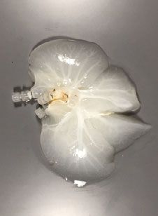
COMMUNICATIONS BIOLOGY | https://doi.org/10.1038/s42003-021-02665-2                                                                                        ARTICLE

retrieval was performed in citrate buffer, pH 6.0 (Abcam AB93678) in a pro-            the animal was allowed to recover for at least five days to allow for local swelling to
grammable decloaker (Biocare, DC2012). Slides were permeabilized with PBS +            subside and to observe for signs of wound infection which would result in elective
 0.05% Tween-20 (Sigma, P9416) and blocked with Sea Block (ThermoFisher,               euthanasia and removal from the study.
37527). Primary antibodies used were Rabbit anti-Collagen I (Abcam, AB34710,               The portocaval shunt procedure was adapted from one described by Lee et al.20.
1:100 dilution), Rabbit anti-Collagen IV (Abcam, AB6586, 1:100 dilution), Mouse        Animals were transitioned to a soft food diet of Ensure and canned dog food (Hills
anti-CD31 (Abcam, AB187377, 1:100 dilution), Rabbit anti-Albumin (Abcam,               Digestive Care a/d) three days prior to surgery and then fasted 16 h prior to the
AB79960, 1:150 dilution)), Rabbit anti-FAH (Abcam, AB83770, 1:100 dilution),           procedure, with access only to water ad lib. Anesthesia was induced via
Rabbit anti-Cytochrome P450 3A4 (Abcam, AB3572, 1:100 dilution) and Rabbit             intramuscular injection of telazol (3.5-5.5 mg/kg) and xylazine (1.5–3.5 mg/kg). IV
anti-LYVE1 (Abcam, AB33682, 1:100 dilution). Secondary antibodies were Goat            administration of 0.9% NaCl was used as necessary for fluid resuscitation and
anti-Mouse Alexa Fluor 488 (ThermoFisher, A11029, 1:500 dilution) and Goat             cefazolin 1 g for surgical prophylaxis. Ketamine (~2 mg/kg/h), midazolam
anti-Rabbit Alexa Fluor 555 (ThermoFisher, A21429, 1:500 dilution). All anti-          (~0.6 mg/kg h) and fentanyl (~0.004 mg/kg/h) were used as necessary as adjuncts.
bodies were diluted in Sea Block. Slides were stained with DAPI (ThermoFisher,         Five hundred mg solumedrol IV was given intravenously for induction
D1306) and mounted using ProLong Antifade Mountant (Thermo, P36961). H&E               immunosuppression. A bladder catheter was placed. After endotracheal intubation,
and immunofluorescence microscopy was performed on an Accuscope 3012                    ventilation was maintained to achieve end-tidal CO2 of 35–40 Torr. Anesthesia was
(H&E) and Zeiss Axioskop 40, respectively.                                             maintained with inhaled isoflurane 0–5%.
                                                                                           A midline laparotomy was performed, and a self-retaining retractor was placed.
                                                                                       Splenectomy was performed via hilar ligation and division taking care to remove
Analysis of cellular metabolites and secreted factors during bioreactor cul-           any splenules and preserve the tail of the pancreas. All ligaments around the liver
ture. Media samples from bioreactors were collected daily and assayed immediately      were taken down and any aberrant vasculature was ligated and divided. A complete
on a CEDEX BioHT analyzer (Roche) to determine levels of glucose, ammonia, and         hepatoduodenal ligament dissection was performed and the HAs, PV and CBD
lactate dehydrogenase activity in the culture media. Measured glucose concentra-       were isolated. The iIVC was mobilized inferiorly to the level of the right renal vein
tions were used to calculate daily consumption rates over a 24 h period prior to       taking care to preserve large local lymphatics. The caudate lobe was devascularized
replenishing bioreactors with fresh media. A separate aliquot of each daily media      with aggressive parenchymal compression using a running, locking 3-0 PDS suture
sample was stored at −80 °C and thawed at the end of each experiment for               until cut parenchymal edges did not demonstrate active bleeding.
quantification of soluble vWF (ThermoFisher, EHVWF) and albumin (Bethyl                     In the control group, a direct portocaval anastomosis was performed. The
Laboratories, A100-110A) by ELISA.                                                     animal was heparinized to a goal ACT of 170–225 s. The iIVC and the PV were
                                                                                       partially clamped and a side-to-side anastomosis 1 cm in diameter was performed.
BEL ammonia clearance kinetics and urea production assays. Sixteen to twenty           After ensuring patency, acute ischemic liver failure was induced by ligation and
hours after seeding hepatocytes, culture media was removed from bioreactors and        division of the HAs, PV upstream from the anastomosis and CBD. This
2 L of co-culture media supplemented with 0.8 mM ammonium chloride. Bior-              represented time zero.
eactor media perfusion was resumed, and media samples were collected in dupli-             In the experimental group, a BEL graft seeded with HUVECs and porcine
cate at t = 0 h, 1 h, 2 h, 7 h, and 23 h. Media ammonia levels were quantified on a     hepatocytes as described above was placed as opposed to a direct portocaval shunt.
CEDEX BioHT, and duplicate frozen samples were assayed in parallel to measure          To reduce portal hypertension from the expected small-for-size syndrome, a 4 mm
urea produced over time (Sigma, MAK0061KT).                                            polytetrafluoroethylene (PTFE) shunt was anastomosed end-to-side between the
                                                                                       recipient’s PV and iIVC. Patency was shown by an increase in PV flow with
                                                                                       conduit clamping as measured by an ultrasonic perivascular flow module
Acute blood perfusion studies. For the in vitro blood perfusion studies, each BEL      (Transonic Systems Inc, Ithaca, NY, USA). Then, two 8 mm diameter, ringed PTFE
was connected to a circuit comprised of silicone tubing, a pressure transducer         prosthetic vascular grafts (W. L. Gore and Associates, Newark, DE, USA) were
(Deltran, DPT-100), and a peristaltic pump (Cole-Palmer, 07522-20). Freshly            anastomosed to the portal vein and iIVC of the BEL using running 6-0 prolene
collected, heparinized porcine blood was warmed to 37 °C and the activated clot-       suture to bolster the anastomoses and provide additional length if needed based on
ting time (ACT) was measured (ITC, Hemochron Response). A solution of pro-             the animals’ anatomy (Fig. S4). Preservation solution was flushed from the liver
tamine sulfate was then gradually added to the blood to neutralize the heparin until   graft using 0.9% NaCl. The liver graft was liberally coated with Tisseel aerosolized
an ACT of 170-220 was reached. 2 L of blood was introduced into the circuit and        fibrin sealant (Baxter Healthcare Co., Deerfield, IL, USA) to provide a physiologic
perfused through the BEL construct at an initial flow rate of 300 mL/min, and then      pseudocapsule and permit handling and retraction as needed during implantation
immediately switched to pressure-dependent flow control targeting a constant            and to provide a bolster against graft fracture and resultant uncontrollable
pressure of 12 mmHg. Flow rates and pressures were recorded over 60 minutes of         hemorrhage secondary to overinflation from portal hypertension. The liver graft
blood perfusion.                                                                       was introduced into the abdomen and placed in the auxiliary position inferior to
    In vivo acute blood studies were performed using domestic swine weighing 80-       the native liver directly anterior to the right adrenal gland. The animal was
100 kg after approval by the Institutional Animal Care and Use Committee               heparinized to a goal ACT of 170-225 sec. The recipient iIVC and portal vein were
(IACUC) at American Preclinical Services (Coon Rapids, MN). Animals were               partially clamped, and end-to-side anastomoses were performed to the BEL’s PTFE
heparinized to a target ACT of 225 s. Recipient vessels: portal vein and iIVC, were    vascular grafts with inflow consisting of the native portal vein flowing to the BEL
cannulated using a 28 F single stage venous cannula (Medtronic). BELs were             portal vein and outflow consisting of the BEL’s iIVC flowing into the recipient’s
connected to portal venous blood flow using PVC (LivaNova) tubing and ¼” luer-          iIVC. The graft’s vasculature was filled with 0.9% NaCl through iIVC, and the graft
lock connectors to achieve functional end-to-side anastomoses between the grafts’      was reperfused by unclamping the inflow and allowing antegrade blood flow to
and recipients’ portal veins and venae cavae. Flow rates were measured using a ½”      vent through the BEL’s sIVC prior to ligation of the BEL sIVC and unclamping of
ultrasonic flow probe (ME 11PXL) connected to a controller box (TS410;                  the outflow anastomosis. PV inflow to the BEL was measured with an ultrasonic
Transonic Systems Inc, Ithaca, NY, USA) and recoded manually. Flow through the         perivascular flow module to ensure flow above 120 mL/min. If blood flow was
BELs was visualized via venogram. Isovue contrast was injected directly into the       below this value, then the PTFE shunt was banded or ligated to increase flow as
perfusion loop upstream of the BEL and images were collected using an OEC 9900         necessary. Hemostasis was achieved with suture ligation or application of topical
Elite mobile C-arm (GE Healthcare).                                                    fibrin sealant). Whole blood transfusion of type-A blood up to 1000 mL was used
                                                                                       as needed to correct for blood loss or hemodynamic instability. Acute ischemic
Heterotopic BEL implantation and portocaval shunt surgeries                            liver failure was induced by ligation and division of the hepatic arteries, PV distal to
ICP probe placement, portocaval shunt and liver implantation procedure. All animal     the anastomosis and common bile duct. This represented time zero.
experiments were performed in accordance with the IACUC at Mayo Clinic                     Once the animals were vitally stable and hemostasis was achieved, an
(Rochester, MN) and American Preclinical Services (Coon Rapids, MN) and all            abdominal drain was placed in the surgical field and connected to bulb suction.
experiments herein were performed in accordance with the guidelines and reg-           Effluent volume and character were recorded throughout the remainder of the
ulations of the committee. Twenty-eight to 36 kg domestic white swine were             study. In the case of two animals, the ultrasonic flow probe was left on the graft
procured from a local USDA-certified (class A) vendor and blood typed for type          portal vein to monitor flow during the monitoring period. The abdomen was closed
AO or A via PCR on buccal swab samples (Zoologix Inc, Chatsworth, CA, USA).            in multiple layers.
    For all animal implant procedures described below, anesthesia was induced via
intramuscular injection of telazol (0.5 mg/Kg) and xylazine (0.2 mg/Kg). IV access
was established for fluid resuscitation with 1 L 0.9% NaCl and administration of        Monitoring and resuscitation. Following surgery, a strict, standardized mon-
cefazolin 1 g for surgical prophylaxis. Extended-release opiate analgesia was          itoring and resuscitation protocol was utilized which involved hourly monitoring of
provided. After endotracheal intubation, ventilation was maintained to achieve end-    vital signs, fluid output, hemodynamic parameters and ICP until death. Sedation
tidal CO2 of 35-40 torr. Anesthesia was maintained with inhaled isoflurane 1-3%.        was maintained with isoflurane 1–3% inhaled. Crystalloid resuscitation of up to
    For ICP probe placement, animals were fasted 16 h prior to the procedure, but      300 mL/h and administration of phenylephrine up to 1 mcg/Kg/min titrating to a
allowed water ad lib. A scalp flap was elevated, and a 4 mm Burr hole was drilled       mean arterial pressure (MAP) of 50 mmHg was permitted. Five percent dextrose
through the frontal bone 1.5 cm lateral to midline and 1 cm superior to the superior   solution was added to crystalloid maintenance fluid to maintain blood glucose of
orbital foramen. The dura mater was punctured bluntly, and a transdermal               60-120 mg/dL. The target body temperature of 37 °C was maintained with a
telemetric intracranial pressure monitor (Raumedic, Helmbrechts, Germany) was          heating blanket. Endpoint was achieved when the animal had two consecutive
introduced into the subdural space. The scalp flap was closed over the monitor and

COMMUNICATIONS BIOLOGY | (2021)4:1157 | https://doi.org/10.1038/s42003-021-02665-2 | www.nature.com/commsbio                                                                 9
ARTICLE COMMUNICATIONS BIOLOGY | https://doi.org/10.1038/s42003-021-02665-2
hourly measurements of MAP < 30 mmHg or ICP > 20 mmHg and was euthanized 13. Hassanein, W. et al. Recellularization via the bile duct supports functional
via pentobarbital overdose. allogenic and xenogenic cell growth on a decellularized rat liver scaffold.
Organogenesis 13, 16–27 (2017).
Animal imaging. Experimental animals were scanned via computer-assisted tomo- 14. Yagi, H. et al. Human-scale whole-organ bioengineering for liver transplantation:
graphy (CT) of the abdomen and pelvis on a SOMATOM Definition VA44A CT a regenerative medicine approach. Cell Transpl. 22, 231–242 (2013).
scanner (Siemens AG, Munich, Germany) post-operatively. 60–90 mL of IV Optiray 15. Tajima, K., Yagi, H. & Kitagawa, Y. Human-scale liver harvest and
350 Ioversol 350 mg/mL was administered at a contrast:saline ratio of 80:20 imme- decellularization for preclinical research. Methods Mol. Biol. 1577, 327–335
diately prior to scanning. Five scans were taken every 15 s to ensure graft patency and (2018).
successful ligation and division of all inflow vessels to the native liver in situ. 16. Bao, J. et al. Hemocompatibility improvement of perfusion-decellularized clinical-
scale liver scaffold through heparin immobilization. Sci. Rep. 5, 10756 (2015).
17. Shaheen, M. F. et al. Sustained perfusion of revascularized bioengineered livers
Biochemical analysis. Blood samples were obtained at time zero and every four
heterotopically transplanted into immunosuppressed pigs. Nat. Biomed. Eng.
hours following induction of acute ischemic liver failure (Supplementary Fig. S2).
4, 437–445 (2019).
Blood ammonia (NH3), albumin (Alb), creatinine (Cre), creatine kinase, aspartate
18. Hassanein, W. et al. Liver scaffolds support survival and metabolic function
aminotransferase (AST), alanine aminotransferase (ALT), total bilirubin (tBil),
of multilineage neonatal allogenic cells. Tissue Eng. Part A 24, 786–793
gamma-glutamyl transferase (GGT), lactate dehydrogenase (LDH) and potassium
(2018).
levels were measured. Blood glucose (Glu) and prothrombin time (PT/INR) were
determined using an automatic point-of-care biochemical analyzer (Abbot Point of 19. Davies, N. A. & Banares, R. A new horizon for liver support in acute liver
Care Inc., Abbott Park, IL, USA). failure. J. Hepatol. 63, 303–305 (2015).
20. Lee, J. H. et al. Functional evaluation of a bioartificial liver support system
using immobilized hepatocyte spheroids in a porcine model of acute liver
Statistics and reproducibility. Independently decellularized, seeded and cultured failure. Sci. Rep. 7, 3804 (2017).
BEL constructs were defined as biological replicates for purpose of all data analyses
conducted in this study. Computed mean values and standard deviations were
shown where applicable. All experiments were repeated at least twice to confirm Author contributions
the reproducibility of the results. All experimental groups were comprised of at B.D.A., D.S.D., J.J.R., S.L.N. designed the study; B.D.A., E.D.N., A.A.K., A.M., B.G.S.,
least two independent biological replicates. D.S.D., T.W.G., J.J.R., S.L.N. analyzed the data; B.G.S. seeded and cultured the BELs;
B.G.S., T.N. and N.P. performed the histological staining; B.D.A. and A.M. performed the
Reporting summary. Further information on research design is available in the Nature bioreactor media assays; E.D.N., D.J.J., B.P.A., S.L.N. performed the surgical procedures;
Research Reporting Summary linked to this article. A.A.K. performed the ex vivo blood perfusion assays; A.R.S. performed the flow cyto-
metry assays; B.D.A. and T.N. drafted the figures; B.D.A., E.D.N., D.S.D., T.W.G., J.J.R.,
and S.L.N. wrote the manuscript
Data availability
Source data for graphs and charts in the main figures are present as Supplementary
Data 1 and any remaining information can be obtained from the corresponding author Competing interests
upon reasonable request. The authors declare the following competing interests: B.D.A., A.A.K., A.M., B.G.S.,
A.R.S., V.L.N., R.N.P., T.W.G., D.S.D., and J.J.R. are employees of Miromatrix Inc.
Miromatrix Inc. is a publicly funded company and owns the exclusive patent rights for
Received: 11 November 2019; Accepted: 8 September 2021; the perfusion decellularization and recellularization technologies utilized in this study.
Published online: 07 October 2021 The remaining authors declare no competing interests.
References Additional information
Supplementary information The online version contains supplementary material
1. Ott, H. C. et al. Perfusion-decellularized matrix: using nature’s platform to
available at https://doi.org/10.1038/s42003-021-02665-2.
engineer a bioartificial heart. Nat. Med. 14, 213–221 (2008).
2. Uygun, B. E. et al. Organ reengineering through development of a
Correspondence and requests for materials should be addressed to Scott L. Nyberg.
transplantable recellularized liver graft using decellularized liver matrix. Nat.
Med. 16, 814–820 (2010). Peer review information Communications Biology thanks the anonymous reviewers for
3. Baptista, P. M. et al. The use of whole organ decellularization for the their contribution to the peer review of this work. Primary Handling Editors: Anam
generation of a vascularized liver organoid. Hepatology 53, 604–617 (2011). Akhtar. Peer reviewer reports are available.
4. Nichols, J. E. et al. Production and transplantation of bioengineered lung into
a large-animal model. Sci Transl Med 10, eaao3926 (2018).
Reprints and permission information is available at http://www.nature.com/reprints
5. Bonandrini, B. et al. Recellularization of well-preserved acellular kidney
scaffold using embryonic stem cells. Tissue Eng. Part A 20, 1486–1498 (2014).
Publisher’s note Springer Nature remains neutral with regard to jurisdictional claims in
6. Song, J. J. et al. Regeneration and experimental orthotopic transplantation of a
published maps and institutional affiliations.
bioengineered kidney. Nat. Med. 19, 646–651 (2013).
7. Huang, Y. B. et al. Comparative decellularization and recellularization of
normal versus streptozotocin-induced diabetes mellitus rat pancreas. Artif.
Organs 43, 399–412 (2019). Open Access This article is licensed under a Creative Commons
8. Guo, Y. et al. Vascularization of pancreatic decellularized scaffold with Attribution 4.0 International License, which permits use, sharing,
endothelial progenitor cells. J. Artif. Organs 21, 230–237 (2018). adaptation, distribution and reproduction in any medium or format, as long as you give
9. Ogiso, S. et al. Efficient recellularisation of decellularised whole-liver grafts appropriate credit to the original author(s) and the source, provide a link to the Creative
using biliary tree and foetal hepatocytes. Sci. Rep. 6, 35887 (2016). Commons license, and indicate if changes were made. The images or other third party
10. Kojima, H. et al. Establishment of practical recellularized liver graft for blood material in this article are included in the article’s Creative Commons license, unless
perfusion using primary rat hepatocytes and liver sinusoidal endothelial cells. indicated otherwise in a credit line to the material. If material is not included in the
Am. J. Transpl. 18, 1351–1359 (2018). article’s Creative Commons license and your intended use is not permitted by statutory
11. Bao, J. et al. Construction of a portal implantable functional tissue-engineered regulation or exceeds the permitted use, you will need to obtain permission directly from
liver using perfusion-decellularized matrix and hepatocytes in rats. Cell the copyright holder. To view a copy of this license, visit http://creativecommons.org/
Transpl. 20, 753–766 (2011). licenses/by/4.0/.
12. Meng, F., Almohanna, F., Altuhami, A., Assiri, A. M. & Broering, D.
Vasculature reconstruction of decellularized liver scaffolds via gelatin-based
re-endothelialization. J. Biomed. Mater. Res. A 107, 392–402 (2019). © The Author(s) 2021, corrected publication 2021
10 COMMUNICATIONS BIOLOGY | (2021)4:1157 | https://doi.org/10.1038/s42003-021-02665-2 | www.nature.com/commsbioYou can also read