Factors influencing upper-most instrumented vertebrae selection in adult spinal deformity patients: qualitative case-based survey of deformity ...
←
→
Page content transcription
If your browser does not render page correctly, please read the page content below
Original Article

Factors influencing upper-most instrumented vertebrae selection
in adult spinal deformity patients: qualitative case-based survey of
deformity surgeons
Sohrab Virk1, Uwe Platz2, Shay Bess3, Douglas Burton4, Peter Passias5, Munish Gupta6,
Themistocles Protopsaltis5, Han Jo Kim1, Justin S. Smith7, Robert Eastlack8, Khaled Kebaish9,
Gregory M. Mundis Jr8, Pierce Nunley10, Christopher Shaffrey11, Jeffrey Gum12, Virginie Lafage1,
Frank Schwab1; International Spine Study Group13
1
Department of Orthopedics, Hospital for Special Surgery, New York, NY, USA; 2Department of spine surgery, Schön Kllink Neustadt, Neustadt,
Germany; 3Denver International Spine Center, Presbyterian St. Luke’s/Rocky Mountain Hospital for Children, Denver, CO, USA; 4Department
of Orthopaedics, University of Kansas Medical Center, Kansas City, KS, USA; 5Department of Orthopedics, NYU Langone Orthopedic Hospital,
New York, NY, USA; 6Department of Orthopaedic Surgery, Washington University, St Louis, MO, USA; 7Department of Neurosurgery,
University of Virginia Medical Center, Charlottesville, VA, USA; 8Scripps Clinic Medical Group Division of Orthopaedic Surgery, La Jolla, CA,
USA; 9Department of Orthopaedic Surgery, Johns Hopkins University School of Medicine, Baltimore, MD, USA; 10Spine Institute of Louisiana,
Shreveport, LA, USA; 11Department of Neurosurgery, Duke University Medical Center, Durham, NC, USA; 12Norton Leatherman Spine Center,
Louisville, KY, USA; 13International Spine Study Group, Denver, Colorado, USA
Contributions: (I) Conception and design: S Virk, F Schwab, V Lafage; (II) Administrative support: S Virk, V Lafage; (III) Provision of study materials
or patients: V Lafage; (IV) Collection and assembly of data: S Virk; (V) Data analysis and interpretation: S Virk; (VI) Manuscript writing: All authors;
(VII) Final approval of manuscript: All authors.
Correspondence to: Sohrab Virk, MD, MBA. 525 E 71st St., Belaire 4E, New York, NY 10021, USA. Email: Sohrab.virk@gmail.com.

                 Background: The decision upper-most instrumented vertebrae (UIV) in a multi-level fusion procedure can
                 dramatically influence outcomes of corrective spine surgery. We aimed to create an algorithm for selection of
                 UIV based on surgeon selection/reasoning of sample cases.
                 Methods: The clinical/imaging data for 11 adult spinal deformity (ASD) patients were presented to 14
                 spine deformity surgeons who selected the UIV and provided reasons for avoidance of adjacent levels. The
                 UIV chosen was grouped into either upper thoracic (UT, T1-T6), lower thoracic (LT, T7-T12), lumbar
                 or cervical. Disagreement between surgeons was defined as ≥3 not agreeing. We performed a descriptive
                 analysis of responses and created an algorithm for choosing UIV then applied this to a large database of ASD
                 patients.
                 Results: Surgeons agreed in 8/11 cases on regional choice of UIV. T10 was the most common UIV in the LT
                 region (58%) and T3 was the most common UIV in the UT region (44%). The most common determinant of
                 UIV in the UT region was proximal thoracic kyphosis and presence of coronal deformity. The most common
                 determinant of UIV in the LT region was small proximal thoracic kyphosis. Within the ASD database (236
                 patients), when the algorithm called for UT fusion, patients fused to TL region were more likely to develop
                 proximal junctional kyphosis (PJK) at 1 year post-operatively (76.9% vs. 38.9%, P=0.025).
                 Conclusions: Our algorithm for selection of UIV emphasizes the role of proximal and regional thoracic
                 kyphosis. Failure to follow this consensus for UT fusion was associated with twice the rate of PJK.

                 Keywords: Upper instrumented vertebra; spinal instrumentation; proximal junctional kyphosis; adult spinal
                 deformity (ASD); long fusion; surgery

                 Submitted May 06, 2020. Accepted for publication Dec 03, 2020.
                 doi: 10.21037/jss-20-598
                 View this article at: http://dx.doi.org/10.21037/jss-20-598

© Journal of Spine Surgery. All rights reserved.                                 J Spine Surg 2021;7(1):37-47 | http://dx.doi.org/10.21037/jss-20-598
38                                                                              Virk et al. UIV selection for thoracolumbar fusion

Introduction                                                     studies (supine radiographs, MRIs, CT scans) and to obtain
                                                                 detailed clinical information on this select group of patients.
The upper-most instrumented vertebrae (UIV) for a spinal
                                                                 The study was conducted in accordance with the Declaration
construct is an important decision for surgeons treating
                                                                 of Helsinki (as revised in 2013). The study was approved by
patients with adult spinal deformity (ASD). The health of
                                                                 Hospital for Special Surgery (IRB #: 2014-357) and informed
adjacent segments to the UIV may determine long-term
                                                                 consent was taken from all the patients. The inclusion criteria
survival of a construct (1). Patients may become impaired
                                                                 for this database was presence of spinal deformity defined as a
from proximal junctional kyphosis (PJK), vertebral fracture,
                                                                 coronal Cobb angle of >20 degrees, sagittal vertical axis (SVA)
limitations of activities of daily living (ADLs) and worse
                                                                 of >5 cm, pelvic tilt (PT) of >25 degrees and/or thoracic
clinic outcomes depending in part on the UIV chosen in
                                                                 kyphosis >60 deg. These 11 patients were presented to
multi-level fusions for ASD (2-5).
                                                                 14 experienced spinal deformity surgeons who were asked
    There are a variety of risks/benefits that are potentially
                                                                 what level they would select as the UIV for correction of
determined by UIV selection. If the top of a construct
                                                                 the spinal deformity. The surgeons were blinded from the
ends in the upper thoracic (UT) spine it allows for more
                                                                 hypothesis of the study and we did not share with results
powerful correction of larger spinal deformities, may reduce
                                                                 amongst surgeons until the drafting of our manuscript. The
the overall rate of proximal junctional failure and enhance
                                                                 selection of these 11 patients was based upon a random
the maintenance of radiographic correction of ASD, but
                                                                 selection of patients that had fusion to the pelvis and up to
it is also may negatively impact ADLs, including personal
                                                                 L1 or higher. Surgeons were presented the cases between
hygiene (5-10). In contrast, when the UIV is in the lower
                                                                 2017–2018.
thoracic (LT) spine, there is less operating room time, less
                                                                     The cases for each patient were summarized in a
blood loss, lower cost, less chance of iliac screw loosening
                                                                 slideshow for the surgeons. Data from the patient’s clinical
and clinical outcomes are still positive (4,11,12).
                                                                 records, including history of present illness (HPI) and
    The goal of the present study is to better understand why
                                                                 pain descriptors such as location, intensity, aggravating
surgeons choose a specific UIV for ASD patients. Typically,
                                                                 or relieving factors (Table 1). Any paresthesia or areas of
clinical experience drives surgeons to either go up to the UT
                                                                 weakness were included. A detailed history of previous
spine versus the LT spine. This decision-making process
                                                                 treatments was included. Physical examination data,
might be unique to each patient and surgeon, however the
                                                                 comprising general and specific spine examination (motor,
authors hypothesized that there is likely a pattern to this
                                                                 sensory exam), were provided to the surgeons (Table 2).
decision making process. We sought to define this decision-
                                                                     Relevant radiographs were included in our presentation
making process through case presentations to a group of
                                                                 for each patient. These included full-length free-
experienced surgeons and to apply this defined pattern of
                                                                 standing antero-posterior and lateral spine radiographs
care prospectively to a group of ASD patients. We further
                                                                 (36” minimum) or full body EOS radiographs. These
hypothesized that patients who had been previously treated
                                                                 radiographs were analyzed using a validated and dedicated
for ASD with a UIV selection consistent with the decision
                                                                 software (Surgimap, Nemaris Inc., USA) (13). Preoperative
making algorithm defined in this study would have a lower
                                                                 supine radiographs were also collected to determine the
rate of PJK compared to patients with a UIV that differed
                                                                 flexibility of the spine. When supine radiographs were not
from the UIV selection algorithm.
                                                                 available, MRI or CT images were used as a substitute to
    We present the following article in accordance with the
                                                                 measure lumbar lordosis or thoracic kyphosis (Figure 1).
STROBE reporting checklist (available at http://dx.doi.
                                                                 Radiographic parameters for spinal alignment investigation
org/10.21037/jss-20-598).
                                                                 were extracted from the analysis and presented to the
                                                                 reviewers including PT and pelvic incidence (PI). Regional
Methods                                                          curvatures were analyzed using the Cobb method and were
                                                                 evaluated at the lumbar level between L1 and S1 (lumbar
Clinical case presentations
                                                                 lordosis) and between T4 and T12 (thoracic kyphosis).
We began our study by first selecting eleven patients from       Global alignment was assessed using the SVA. Radiographs
a single-surgeon database of patients with ASD. Eleven           were also evaluated for accompanying antero or lateral
patients were selected based on previous studies. We used        listhesis. When available, preoperative MRI and CT images
an internal database in order to obtain all pertinent imaging    were also collected. Axial images at each vertebral level

© Journal of Spine Surgery. All rights reserved.                  J Spine Surg 2021;7(1):37-47 | http://dx.doi.org/10.21037/jss-20-598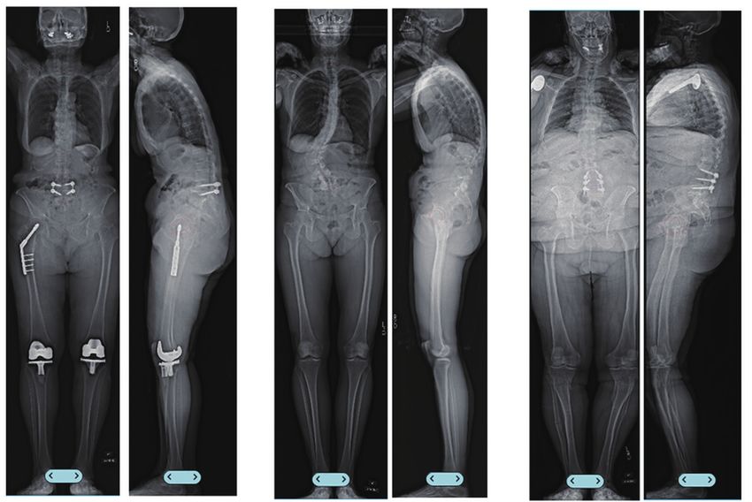
Journal of Spine Surgery, Vol 7, No 1 March 2021                                                                                                   39

 Table 1 Example of relevant demographic data included on the case presentation
 Variables                             Case #1

 Patient information

   Age                                 68 years

   Gender                              F

   Chief complaint                     Chronic low back pain, postural concerns

   Pain                                Deep, 9/10, prolonged with sitting/walking, localized to the lumbar region, radiation to posterior thigh,
                                       uses cane to ambulate

   Past treatment                      Epidural steroid injections with limited relief

 Physical exam information

   General                             Well developed, well nourished

   Orientation                         Oriented to time, place and person

   Peripheral vascular exam            Normal

   Facial sensation                    Intact

   Tongue                              Midline

   Uvula/shoulder shrug                Elevates symmetrically

   Abdomen                             Soft, non-tender, non-distended

   Cervical ROM                        Normal, without pain

   Spurling exam                       Negative bilaterally

   Spine                               Pain with forward flexion, extension and lateral flexion.
                                       Increased thoracic kyphosis with standing

   Upper extremity motor, sensory Normal
   and reflexes

   Lower extremity motor, sensory Motor: normal; sensory/reflexes: normal; toe response: equivocal; straight leg raise: negative;
   and reflexes                   Romberg: negative
 ROM, range of motion.

were assessed for the presence of central spinal stenosis,                     of adjacent vertebral levels. The questionnaire asked (I)
foraminal spinal stenosis and disc-degeneration.                               what would be your UIV level? (II) If the UIV was in the
                                                                               UT, why not the LT? (III) If the UIV was in the LT, why
                                                                               not the UT? (IV) Within the UIV region you selected
Survey to surgeons
                                                                               what were the key determinants against a more proximal
The data for each patient was collected and collated into a                    level? (V) Within the UIV region you selected what were
slideshow. These slideshows included all information that                      the key determinants against a more distal level? Finally,
should be necessary for an operative decision. Each case                       the surgeons were asked to rank the most relevant reason
was followed by a questionnaire. All cases were assumed                        for UIV selection amongst the following reasons: fear of
to have a fusion from at least L1 to the pelvis. This survey                   proximal junctional kyphosis, junctional disease over the
was sent to 14 surgeons to review cases and answer framed                      long term, patient function, trade-off in terms of surgical
questions. The surgeons were asked to pick the best-suited                     time/bleeding and reaching alignment goal, other. All
UIV (any level: cervical, thoracic, or lumbar) for each case.                  surgeons in our study provided complete answers for each
The questionnaire contained 6 questions and encompassed                        of the 11 cases in the survey. Every surgeon that was asked
the choice of the level of UIV and reasons for the avoidance                   to participate in this study had at least 5 years of experience

© Journal of Spine Surgery. All rights reserved.                                 J Spine Surg 2021;7(1):37-47 | http://dx.doi.org/10.21037/jss-20-598
40 Virk et al. UIV selection for thoracolumbar fusion
Table 2 Example of relevant physical data included on the case presentation
Physical exam information Case #1
General Well developed, well nourished
Orientation Oriented to time, place and person
Peripheral vascular exam Normal
Facial sensation Intact
Tongue Midline
Uvula/shoulder shrug Elevates symmetrically
Abdomen Soft, non-tender, non-distended
Cervical ROM Normal, without pain
Spurling exam Negative bilaterally
Spine Pain with forward flexion, extension and lateral flexion. Increased thoracic kyphosis with standing
Upper extremity motor, sensory and Normal
reflexes
Lower extremity motor, sensory and Motor: normal; sensory/reflexes: normal; toe response: equivocal; straight leg raise: negative;
reflexes Romberg: negative
ROM, range of motion.
performing spinal deformity surgery. Each was fellowship a qualitative assessment of reasons for the UIV of
trained in spine surgery. Furthermore, all surgeons included a construct. This algorithm was then applied to a
in this study are members of an international society of prospective database of ASD patients that underwent
specialists devoting time to the advancement/study of an operation for their deformity. This database is a
principles for treating patients with spinal deformity. prospective collection of clinical/radiographic data from
11 centers across the United States. All patients were
enrolled into an Institutional Review Board-approved
Surgeon selection of UIV
protocol by each site. The inclusion criteria for this
The answers from the survey, including UIV selection International database were: patients over 18 years old,
and reasoning behind UIV choice, were collected and presence of spinal deformity defined as a coronal Cobb
organized. We first categorized each UIV chosen into angle of >20 degrees, SVA of >5 cm, PT of >25 degrees
either the UT (T1-T6), LT (T7-T12), lumbar or cervical and/or thoracic kyphosis >60 deg. We only included
region. We defined a disagreement when there were more patients with at least 2 years of follow up information,
than 2 surgeons who differed from the group on UT or were fused to the pelvis, had pre- and post-operative
LT UIV location. The specific level at which the UIV was radiographs to 2 years out from surgery and had no
selected was collected for each surgeon/case. We grouped previous fusion above L1.
descriptive answers together based on common language
used amongst surgeons. Surgeons were blinded to other
Statistical analyses
surgeon’s responses.
The selected UIV was compared to the predicted UIV. A
chi-square analysis was used to compare the rate of PJK in
Development and application of algorithm for UIV
patients fused to the LT spine when the algorithm predicted
selection
better PJK outcomes with UT spine as the UIV. A similar
Once all the answers from surgeons were collected chi-square analysis was done to compare the rate of PJK
and organized, an algorithm was created based on in patients that went to the UT spine when the algorithm
© Journal of Spine Surgery. All rights reserved. J Spine Surg 2021;7(1):37-47 | http://dx.doi.org/10.21037/jss-20-598Journal of Spine Surgery, Vol 7, No 1 March 2021 41
A B C D
T Kypho 1
45.0°
E PT
PI
30.8°
46.4°
LL -18.1°
L4-S1 -35.5°
PI-LL 28.3°
TL 52.7°
TK 69.8°
T1 SPi 9.5°
T1 Slope 57.1°
TPA 40.3°
T1-CL 34.5°
cSVA (C... 30.7mm
2/2 1/2 SVA (C7... 146.3mm
2/2
Figure 1 Example of images included on the case presentation. (A,B) Standing lateral and antero-posterior full-body radiograph; (C)
magnified image of the lateral view of the spine; (D) sagittal T2 MRI through the center of the spine; (E) summary of sagittal radiographic
parameters for this particular case. PT, pelvic tilt; PI, pelvic incidence; LL, lumbar lordosis; TL, thoraco-lumbar kyphosis; TK, thoracic
kyphosis; T1 Spi, T1 spinopelvic incidence; T1 pelvic angle; T1-CL, T1 slope minus cervical lordosis; cSVA, cervical sagittal vertical axis;
SVA, sagittal vertical axis.
stated no change in PJK when going to the LT spine. The regarding the region of the UIV.
definition of PJK was proximal junction sagittal Cobb angle
greater than 10 degrees and proximal junction sagittal
Justifications of UIV region selected
Cobb angle at least 10 degrees greater than preoperative
measurement (14). All statistical analysis was performed The LT region was selected as UIV for a variety of
with SPSS (IBM SPSS, NY). If there was missing data for reasons. The most common reason was small TK (less than
any patient they were excluded from the analysis. 55 degrees) or lack of sagittal deformity in the proximal
thoracic spine. Less common reasons or reasons that
Results were lower in priority for UIV selection for the surgeons
were limitations to function with proximal fusions and
UIV selection summary potential comorbidity of extending a fusion to the UT or
All 14 surgeons responded to all 11 cases and indicated their cervical spine.
desired UIV (Figure 2). The UIV selected at each individual The most common reason for selecting UT was
level is shown in Figure 2. Figure 2 reflects the number of increased kyphosis through the thoracic spine. This led to
surgeons that selected each UIV level. The top three UIV concerns of increased risk of proximal junctional failure.
selected were T10, T11 and T3. The UT, LT, lumbar and Less common or reasons lower in priority for UIV selection
cervical regions were selected in 36.2%, 55.9%, 5.9% and for the surgeons included a desire to achieve a more
2.0% of cases respectively for these 11 cases. In 8/11 cases complete correction, presence of coronal deformity, and age
(73%) there was agreement amongst the spine surgeons of the patient/osteoporosis. In all cases, the most common
© Journal of Spine Surgery. All rights reserved. J Spine Surg 2021;7(1):37-47 | http://dx.doi.org/10.21037/jss-20-59842 Virk et al. UIV selection for thoracolumbar fusion
60
Number of Surgeons selecting UIV 50
40
30
20
10
0
C4 C5 C6 C7 T1 T2 T3 T4 T5 T6 T7 T8 T9 T10 T11 T12 L1
UIV Selected
Figure 2 Number of surgeons that selected a particular UIV for each of the 11 cases. In no situation was T6-T8 selected for the adult spinal
deformity cases. The most commonly selected UIV for UT was T3 and for LT was T10. UIV, upper-most instrumented vertebrae; UT,
upper thoracic; LT, lower thoracic.
reason for selection of the region for UIV was the amount about stopping in the LT due to perceived worse bone
of kyphosis in the thoracic spine. The local selection of UIV health based on radiographs. Also, a subset of surgeons was
was based largely on the local kyphosis above a potential concerned regarding the patient’s overall medical health
UIV selection. to survive a larger surgery to the UT spine. Similarly, the
second case of disagreement was based on the patient’s
relatively younger age and better bone health based on bone
Justifications for individual UIV selected
quality on radiographs which convinced several surgeons
There were common reasons for selecting individual levels that PJK might be avoided by simply fusing to the LT
for the UIV. T1 and T12 were often not selected in order region. Other surgeons were concerned about stopping in
to avoid the cervicothoracic or thoracolumbar junctions, the LT because of the high TK for the case. In the last case
respectively. T6-T8 was avoided because the thoracic apex of disagreement, the surgeons differed on the global sagittal
tended to be at T6-T8. L1 was avoided in one case because alignment of the patient and the risk of PJK perhaps being
of degenerative changes at the L1/L2 level. higher due to BMI of a patient. The radiographs for the
three cases where there was substantial disagreement are
shown in Figure 3.
Reasons for UIV disagreement
In three cases there was substantial disagreement among
Creating an algorithm for UIV selection
surgeons regarding the UIV region. These discrepancies
were between UT and LT regions in all three cases. In one After synthesizing the responses provided by the
case of disagreement there did not appear to be significant 14 surgeons for UIV selection in 11 cases, the group
thoracic deformity, but several surgeons were concerned arrived at an algorithm for selection of UIV. From survey
© Journal of Spine Surgery. All rights reserved. J Spine Surg 2021;7(1):37-47 | http://dx.doi.org/10.21037/jss-20-598Journal of Spine Surgery, Vol 7, No 1 March 2021 43
A B C
1/2 2/2 1/2 2/2 1/2 2/2
Figure 3 AP and lateral standing full-length radiographs of the three cases where there was substantial disagreement between the group of
surgeons evaluating UIV selection. AP, antero-posterior; UIV, upper-most instrumented vertebrae.
responses we felt the most important factor to delineate Discussion
UIV was whether there was significant thoracic coronal
Our study was able to outline the decision-making process
curve. If so, one was obligated to fuse to the UT. The
of 14 surgeons for UIV selection in long spinal constructs
2nd most important consideration from several surgeons
for ASD patients. We found the most important factor in
was whether there was a significant amount of thoracic
determining UIV for patients was the magnitude of thoracic
kyphosis. If there was a large thoracic kyphosis most
kyphosis at the UIV+2 to UIV. Furthermore, there were
surgeons would fuse up to the UT. Deciding which precise
certain favored vertebrae for UIV selection, including T3
level within the UT or LT was based on the Cobb angle for the UT spine T10 for the LT spine.
between the UIV and the UIV+2. The algorithm is shown Our cohort agreed that any severe coronal curve greater
in Figure 4. All surgeons included in this study agreed with than 20 degrees would make it difficult to correct with
our algorithm. a UIV in the LT. Similarly, any patient with an extreme
hyperkyphosis (>50 degrees) was favored to have a UIV in
Applying the algorithm to a retrospective database of ASD the UT. This is because the curve might not be completely
patients corrected with a UIV in the LT or stopping in the LT
may lead to ending a construct at a kyphotic level. When
Out of a database of 1,654 patients, there were 236 patients deciding between individual levels in the UT/LT, local
that met our inclusion criteria. Mean age was 64.1±9.5 years kyphosis at the UIV+2 to UIV dictated the precise level to
old. When the algorithm predicted going up to the UT end the construct.
spine and the surgeon selected a UIV in the LT spine there In one of the cases where there was disagreement
was a statistically significantly higher rate of PJK at 2-year amongst surgeons, there was concern amongst several
follow up (76.9% vs. 38.9%, P=0.025). Conversely, when surgeons regarding the safety of achieving appropriate
the algorithm predicted better outcomes with going to the correction in a medically complex patient. Several reports
LT spine and the surgeon went up to the UT spine, there detail the substantial rate of medical complications for
was no difference in rate of PJK (P>0.05). patients undergoing extensive ASD surgery (15,16).
© Journal of Spine Surgery. All rights reserved. J Spine Surg 2021;7(1):37-47 | http://dx.doi.org/10.21037/jss-20-59844 Virk et al. UIV selection for thoracolumbar fusion
>55°
Thoracic kyphosis (TK)Journal of Spine Surgery, Vol 7, No 1 March 2021 45
does not extend more proximally. Data Sharing Statement: Available at http://dx.doi.
There are several important limitations to the present org/10.21037/jss-20-598
study. We did not examine clinical complication rates amongst
patients. This might have further demonstrated the potential Peer Review File: Available at http://dx.doi.org/10.21037/jss-
problems with aggressive fusions up to UT even if PJK might 20-598.
be avoided. We did not include flexion/extension radiographs
within the survey provided to surgeons. This might have Conflicts of Interest: All authors have completed the ICMJE
shown better flexibility in the lumbar/thoracic spine which uniform disclosure form (available at http://dx.doi.
could have influenced the UIV selection amongst surgeons. org/10.21037/jss-20-598). Dr. SB reports personal fees from
Our analysis was based on responses from a small group of K2M, from Medtronic, personal fees from Nuvasive, from
surgeons. We cannot state that these opinions reflect the larger Orthofix, personal fees from Stryker, outside the submitted
community of spine surgeons as a whole. Our panel, however, work; Dr. DB reports grants from Bioventus, grants from
does constitute a group of surgeons from around the world Depuy, grants from Pfizer, grants from Progenerative
with a broad range of surgical experiences. There may be Medical, outside the submitted work; Dr. PP reports other
inherent biases within our group of deformity surgeons which from Allosource, personal fees from Globus Medical,
we cannot control for within our study design. The authors personal fees from Medicrea, personal fees from Royal
also acknowledge the limitation of using PJK as a measure of Biologics, personal fees from SpineWave, personal fees from
possible poor selection of UIV. PJK may not be symptomatic Terumo, personal fees from Zimmer, outside the submitted
and therefore our results may not accurately reflect clinical work; Dr. MG reports grants from Depuy, personal fees
failure of a UIV level selected but rather only a radiographic from Globus, grants from Innomed, personal fees from
finding for a patient (27). Still, PJK is associated with proximal Johnson and Johnson, personal fees from Medtronic, other
junctional failure and this is why it was selected for analysis. from Proctor and Gamble, personal fees from Wolters
Finally, this is a retrospective review of a database of patients Kluwer Health, outside the submitted work; Dr. TP reports
with cases from surgeons that decided the algorithm of UIV other from Altus, personal fees from Globus Medical,
selection. Therefore, there is a degree of confirmation bias that personal fees from Medicrea, personal fees from Nuvasive,
is inherent in our study design. other from Spine Align, personal fees from Stryker, other
In conclusion, we have provided a simplified algorithm from Torus Medical, outside the submitted work; Dr. HJK
for picking UIV amongst patients requiring surgery for reports personal fees from Alphatec, other from K2M,
ASD. When applied to a retrospective database of patients other from Zimmer, outside the submitted work; Dr. JSS
treated with surgery for ASD, those that did not fuse to the reports other from Alphatec, personal fees from Carlsmed,
UT but rather stopped at the LT spine had higher rates personal fees from Cerapedics, grants from Depuy, other
of PJK than those that followed our algorithm and fused from Nuvasive, personal fees from Styker, other from
to the UT. We recommend close examination of local and Zimmer, outside the submitted work; Dr. RE reports
regional kyphosis when deciding UIV to help avoid possible personal fees from Aesculap, other from Alphatec, personal
development of PJK. fees from Baxter, personal fees from Biederman-Motech,
personal fees from Carevature, other from Globus, other
from Invuity, personal fees from Medtronic, other from
Acknowledgments
Nocimed, personal fees and other from Nuvasive, personal
Funding: The International Spine Study Group (ISSG) fees from Radius, other from Seaspine, personal fees and
is funded through research grants from DePuy Synthes other from SI Bone, other from Spine Innovations, outside
(current), Nuvasive (current), K2M (current), Innovasis the submitted work; Dr. KK reports personal fees and
(past), Biomet (past), and individual donations. other from Depuy, personal fees and other from Orthofix,
other from Strykler, outside the submitted work; Dr. GM
reports personal fees from Carlsmed, other from K2M,
Footnote
other from Nuvasive, personal fees from Seaspine, personal
Reporting Checklist: The authors have completed the fees from Stryker, personal fees from Viseon, outside the
STROBE reporting checklist. Available at http://dx.doi. submitted work; Dr. CS reports grants from Depuy, grants
org/10.21037/jss-20-598 from Globus, grants and other from Medtronic, other from
© Journal of Spine Surgery. All rights reserved. J Spine Surg 2021;7(1):37-47 | http://dx.doi.org/10.21037/jss-20-59846 Virk et al. UIV selection for thoracolumbar fusion
Nuvasive, other from SI Bone, outside the submitted work; fusion level for adult degenerative lumbar scoliosis. Eur
Dr. JG reports grants and other from Accuity, other from Spine J 2013;22:394-401.
Cingulate, personal fees from Depuy, grants from Integra, 4. Kim HJ, Boachie-Adjei O, Shaffrey CI, et al. Upper
personal fees from Intellirod, personal fees from K2M, thoracic versus lower thoracic upper instrumented
personal fees from Mazor, personal fees from Medtronic, vertebrae endpoints have similar outcomes and
grants from Norton Healthcare, other from Nuvasive, complications in adult scoliosis. Spine (Phila Pa 1976)
personal fees from Pfizer, personal fees from Stryker, 2014;39:E795-9.
outside the submitted work; Dr. VL reports personal 5. Sciubba DM, Scheer JK, Smith JS, et al. Which daily
fees from Depuy, personal fees from Globus, other from functions are most affected by stiffness following total
Nuvasive, personal fees from Permanante Medical Group, lumbar fusion: comparison of upper thoracic and
outside the submitted work; Dr. FS reports grants from thoracolumbar proximal endpoints. Spine (Phila Pa 1976)
Depuy, personal fees from Globus, from K2M, personal 2015;40:1338-44.
fees from Medicrea, grants, personal fees and other from 6. Zou L, Liu J, Lu H. Characteristics and risk factors for
Medtronic, grants from Nuvasive, grants from Styker, proximal junctional kyphosis in adult spinal deformity after
grants, personal fees and other from Zimmer, outside the correction surgery: a systematic review and meta-analysis.
submitted work. The other authors have no conflicts of Neurosurg Rev 2019;42:671-82.
interest to declare. 7. Yagi M, Fujita N, Okada E, et al. Fine-tuning the
Predictive Model for Proximal Junctional Failure in
Ethical Statement: The authors are accountable for all Surgically Treated Patients With Adult Spinal Deformity.
aspects of the work and ensuring that questions related Spine (Phila Pa 1976) 2018;43:767-73.
to the accuracy or integrity of any part of the work are 8. Luo M, Wang P, Wang W, et al. Upper Thoracic versus
appropriately investigated and resolved. The study was Lower Thoracic as Site of Upper Instrumented Vertebrae
conducted in accordance with the Declaration of Helsinki (as for Long Fusion Surgery in Adult Spinal Deformity: A
revised in 2013). The study was approved by Hospital for Meta-Analysis of Proximal Junctional Kyphosis. World
Special Surgery (IRB #: 2014-357) and informed consent Neurosurg 2017;102:200-8.
was taken from all the patients. 9. Scheer JK, Osorio JA, Smith JS, et al. Development
of Validated Computer-based Preoperative Predictive
Open Access Statement: This is an Open Access article Model for Proximal Junction Failure (PJF) or Clinically
distributed in accordance with the Creative Commons Significant PJK With 86% Accuracy Based on 510 ASD
Attribution-NonCommercial-NoDerivs 4.0 International Patients With 2-year Follow-up. Spine (Phila Pa 1976)
License (CC BY-NC-ND 4.0), which permits the non- 2016;41:E1328-35.
commercial replication and distribution of the article with 10. Scheer JK, Lafage V, Smith JS, et al. Maintenance of
the strict proviso that no changes or edits are made and the radiographic correction at 2 years following lumbar pedicle
original work is properly cited (including links to both the subtraction osteotomy is superior with upper thoracic
formal publication through the relevant DOI and the license). compared with thoracolumbar junction upper instrumented
See: https://creativecommons.org/licenses/by-nc-nd/4.0/. vertebra. Eur Spine J 2015;24 Suppl 1:S121-30.
11. Banno T, Hasegawa T, Yamato Y, et al. Prevalence and
Risk Factors of Iliac Screw Loosening After Adult Spinal
References
Deformity Surgery. Spine (Phila Pa 1976) 2017;42:E1024-30.
1. Shufflebarger H, Suk SI, Mardjetko S. Debate: 12. Fujimori T, Inoue S, Le H, et al. Long fusion from
determining the upper instrumented vertebra in the sacrum to thoracic spine for adult spinal deformity with
management of adult degenerative scoliosis: stopping at sagittal imbalance: upper versus lower thoracic spine as
T10 versus L1. Spine (Phila Pa 1976) 2006;31:S185-94. site of upper instrumented vertebra. Neurosurg Focus
2. Kim HJ, Bridwell KH, Lenke LG, et al. Proximal 2014;36:E9.
junctional kyphosis results in inferior SRS pain subscores 13. Lafage R, Ferrero E, Henry JK, et al. Validation of a new
in adult deformity patients. Spine (Phila Pa 1976) computer-assisted tool to measure spino-pelvic parameters.
2013;38:896-901. Spine J 2015;15:2493-502.
3. Cho KJ, Suk SI, Park SR, et al. Selection of proximal 14. Glattes RC, Bridwell KH, Lenke LG, et al. Proximal
© Journal of Spine Surgery. All rights reserved. J Spine Surg 2021;7(1):37-47 | http://dx.doi.org/10.21037/jss-20-598Journal of Spine Surgery, Vol 7, No 1 March 2021 47
junctional kyphosis in adult spinal deformity following surgery with fusion from the thoracic spine to the sacrum:
long instrumented posterior spinal fusion: incidence, a comparison of proximal and distal upper instrumented
outcomes, and risk factor analysis. Spine (Phila Pa 1976) vertebrae. J Neurosurg Spine 2013;19:360-9.
2005;30:1643-9. 22. O'Shaughnessy BA, Bridwell KH, Lenke LG, et al. Does
15. Baron EM, Albert TJ. Medical Complications of Surgical a Long-Fusion “T3-Sacrum” Portend a Worse Outcome
Treatment of Adult Spinal Deformity and How to Avoid Than a Short-Fusion “T10-Sacrum” in Primary Surgery
Them. Spine (Phila Pa 1976) 2006;31:S106-18. for Adult Scoliosis? Spine (Phila Pa 1976) 2012;37:884-90.
16. Daubs MD, Lenke LG, Cheh G, et al. Adult Spinal 23. Soroceanu A, Burton DC, Oren JH, et al. Medical
Deformity Surgery: Complications and Outcomes in Patients Complications After Adult Spinal Deformity Surgery:
Over Age 60. Spine (Phila Pa 1976) 2007;32:2238-44. Incidence, Risk Factors, and Clinical Impact. Spine (Phila
17. Schwab FJ, Hawkinson N, Lafage V, et al. Risk factors Pa 1976) 2016;41:1718-23.
for major peri-operative complications in adult spinal 24. Grabel ZJ, Hart RA, Clark AJ, et al. Adult Spinal
deformity surgery: a multi-center review of 953 Deformity Knowledge in Orthopedic Spine Surgeons:
consecutive patients. Eur Spine J 2012;21:2603-10. Impact of Fellowship Training, Experience, and Practice
18. Glassman SD, Alegre GM. Adult spinal deformity in the Characteristics. Spine Deform 2018;6:60-6.
osteoporotic spine: options and pitfalls. Instr Course Lect 25. Lau D, ore CD, Deviren V, et al. 326 The Impact of
2003;52:579-88. Surgeon Experience on Outcomes Following 3-Column
19. Yagi M, Fujita N, Tsuji O, et al. Low Bone-Mineral Osteotomy for Adult Spinal Deformity: Overcoming
Density Is a Significant Risk for Proximal Junctional the Learning Curve and Implementing Better Practices.
Failure After Surgical Correction of Adult Spinal Neurosurgery 2018;65:131.
Deformity. Spine 2018;43:485-91. 26. Hey HWD, Tan KA, Neo CS, et al. T9 versus T10 as
20. Choi JH, Jang JS, Yoo KS, et al. Functional Limitations the upper instrumented vertebra for correction of adult
Due to Stiffness After Long-Level Spinal Instrumented deformity-rationale and recommendations. Spine J
Fusion Surgery to Correct Lumbar Degenerative Flat 2017;17:615-21.
Back. Spine (Phila Pa 1976) 2018;43:1044-51. 27. Cho SK, Shin JI, Kim YJ. Proximal junctional kyphosis
21. Ha Y, Maruo K, Racine L, et al. Proximal junctional following adult spinal deformity surgery. Eur Spine J
kyphosis and clinical outcomes in adult spinal deformity 2014;23:2726-36.
Cite this article as: Virk S, Platz U, Bess S, Burton D, Passias P,
Gupta M, Protopsaltis T, Kim HJ, Smith JS, Eastlack R, Kebaish
K, Mundis GM Jr, Nunley P, Shaffrey C, Gum J, Lafage V,
Schwab F; International Spine Study Group. Factors influencing
upper-most instrumented vertebrae selection in adult spinal
deformity patients: qualitative case-based survey of deformity
surgeons. J Spine Surg 2021;7(1):37-47. doi: 10.21037/jss-20-
598
© Journal of Spine Surgery. All rights reserved. J Spine Surg 2021;7(1):37-47 | http://dx.doi.org/10.21037/jss-20-598You can also read