Evolution of Renal-Disease Factor APOL1 Results in Cis and Trans Orientations at the Endoplasmic Reticulum That Both Show Cytotoxic Effects
←
→
Page content transcription
If your browser does not render page correctly, please read the page content below
Evolution of Renal-Disease Factor APOL1 Results in Cis and
          Trans Orientations at the Endoplasmic Reticulum That Both
          Show Cytotoxic Effects
          Daria Müller,1 Jürgen Schmitz ,2 Katharina Fischer,1 Daniel Granado,1 Ann-Christin Groh,1
          Vanessa Krausel,1 Simona Mareike Lüttgenau,1 Till Maximilian Amelung,1 Hermann Pavenst€adt,1 and
          Thomas Weide*,1
          1
           Internal Medicine D (MedD), Molecular Nephrology, University Hospital of Münster (UKM), Münster, Germany
          2
           Institute of Experimental Pathology, ZMBE, University of Münster, Münster, Germany
          *Corresponding author: E-mail: weidet@uni-muenster.de.

                                                                                                                                                                    Downloaded from https://academic.oup.com/mbe/article/38/11/4962/6330626 by guest on 17 December 2021
          Associate editor: Harmit Malik

          Abstract
          The recent and exclusively in humans and a few other higher primates expressed APOL1 (apolipoprotein L1) gene is
          linked to African human trypanosomiasis (also known as African sleeping sickness) as well as to different forms of kidney
          diseases. Whereas APOL1’s role as a trypanolytic factor is well established, pathobiological mechanisms explaining its
          cytotoxicity in renal cells remain unclear. In this study, we compared the APOL family members using a combination of
          evolutionary studies and cell biological experiments to detect unique features causal for APOL1 nephrotoxic effects.
          We investigated available primate and mouse genome and transcriptome data to apply comparative phylogenetic and
          maximum likelihood selection analyses. We suggest that the APOL gene family evolved early in vertebrates and initial
          splitting occurred in ancestral mammals. Diversification and differentiation of functional domains continued in primates,
          including developing the two members APOL1 and APOL2. Their close relationship could be diagnosed by sequence
          similarity and a shared ancestral insertion of an AluY transposable element. Live-cell imaging analyses showed that both
          expressed proteins show a strong preference to localize at the endoplasmic reticulum (ER). However, glycosylation and
          secretion assays revealed that—unlike APOL2—APOL1 membrane insertion or association occurs in different orienta-
          tions at the ER, with the disease-associated mutants facing either the luminal (cis) or cytoplasmic (trans) side of the ER.
          The various pools of APOL1 at the ER offer a novel perspective in explaining the broad spectrum of its observed toxic
          effects.
          Key words: APOL1, APOL2, endoplasmic reticulum, evolutionary medicine, APOL gene family, APOL phylogeny, APOL
          selection analyses, kidney disease.
Article

          Introduction                                                                          in those infected with Trypanosoma brucei gambiense
          APOL1 (apolipoprotein L1) is the most-studied variant of six                          (Cooper et al. 2017).
          members of conserved human proteins (APOL1-6) and has                                    However, similar to the link between sickle cell anemia and
          been identified as a trypanolytic factor (TLF) by protecting                          human resistance to malaria, two copies of these variants
          humans against African sleeping sickness causing                                      cause an increased risk for numerous renal, predominantly
          Trypanosoma parasites (Hajduk et al. 1989; Raper et al.                               glomerular, diseases (Freedman et al. 2010; Genovese et al.
          1999; Vanhamme et al. 2003; Pays et al. 2014; Weckerle et                             2010; Kasembeli et al. 2015). These include HIV- and
          al. 2016). Trypanosoma brucei rhodesiense and Trypanosoma                             hypertension-associated nephropathy, focal segmental glo-
          brucei gambiense subspecies are resistant to APOL1 and es-                            merulosclerosis, lupus nephritis, membranous nephropathy,
          caped from APOL1 toxicity. By a coevolutionary arms race,                             and—as recently shown—also Covid-19-associated renal fail-
          two variants of APOL1—G1 (S342G/I384M) and G2 (D388N,                                 ure (Tzur et al. 2010; Kopp et al. 2011; Papeta et al. 2011;
          D389Y)—reestablished the APOL1-linked protection against                              Larsen et al. 2014; Velez et al. 2020). These diseases’ common
          these parasites in humans of sub-Saharan African ancestry                             features include proteinuria and rapidly progressive kidney
          (Pays et al. 2014; Capewell et al. 2015; Kruzel-Davila, Wasser,                       disease, which often results in end-stage renal disease
          et al. 2017). The APOL1 variant G2 lyses Trypanosoma brucei                           (Wiggins 2007). Therefore, podocytes—unique postmitotic
          rhodesiense, but not Trypanosoma brucei gambiense, whereas                            cells, essential for the glomerular filtration barrier establish-
          APOL1-G1 is associated with latent, asymptomatic infections                           ment and maintenance—are most likely a primary cellular

          ß The Author(s) 2021. Published by Oxford University Press on behalf of the Society for Molecular Biology and Evolution.
          This is an Open Access article distributed under the terms of the Creative Commons Attribution-NonCommercial License
          (http://creativecommons.org/licenses/by-nc/4.0/), which permits non-commercial re-use, distribution, and reproduction in any
          medium, provided the original work is properly cited. For commercial re-use, please contact journals.permissions@oup.com       Open Access
          4962                          Mol. Biol. Evol. 38(11):4962–4976 doi:10.1093/molbev/msab220 Advance Access publication July 29, 2021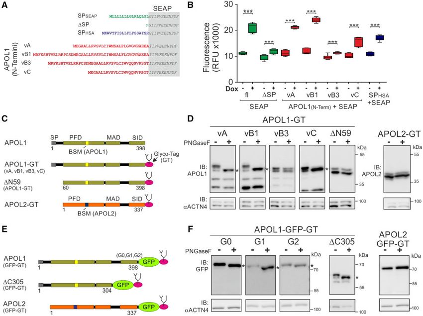
APOL1 Evolution Results in Dual ER Orientations . doi:10.1093/molbev/msab220                                         MBE
target for APOL1-associated renal cytotoxicity (Wiggins 2007;         APOL6 and APOL5 are represented in most placental spe-
Kopp et al. 2020).                                                 cies. However, the mouse genome lacks APOL5. All other
    All APOL family members share high sequence homology.          investigated APOL gene family members are significantly
In a concept initially suggested by Pays and colleagues for        younger and either primate or rodent specific (Page et al.
APOL1 (Perez-Morga et al. 2005; Pays et al. 2006), and ex-         2001; Smith and Malik 2009; Kreit et al. 2015). Our data sug-
tended to all APOL family members by Smith and Malik               gest a rodent-primate APOL6-like ancestor gene origin about
(2009), APOL proteins contain three functional parts: a            70 Ma (fig. 1B, supplementary data SD1–3, Supplementary
pore-forming domain (PFD), a membrane addressing domain            Material online) that—after duplication—diverged indepen-
(MAD), and SRA-interacting-protein-like domain (SID). The          dently into numerous APOL genes in man and mouse. Thus,
PFD of APOLs include Bcl-2 homology 3 (BH3) domain-like            mice (mm: Mus musculus) contain 13 APOL genes that can be
sequence motifs (BSMs) that have been linked to anti- or pro-      divided into mmAPOLD1, mmAPOL6, an mmAPOL7 group
apoptotic features (Wei et al. 2001; Shamas-Din et al. 2011;       (mmAPOL7a, 7b, 7c, and 7e), mmAPOL8, an mmAPOL9 group
Dummer et al. 2015; Adams and Cory 2018). A further com-           (mmAPOL9a, 9b), and an mmAPOL10/11 group

                                                                                                                                    Downloaded from https://academic.oup.com/mbe/article/38/11/4962/6330626 by guest on 17 December 2021
mon feature of the APOL family is that their expression can        (mmAPOL10a, 10b, 11a, 11b). All these genes are clustered
be induced by inflammatory triggers, for example, interferons      on chromosome 15 except mmAPOLD1, which is located on
or TNFa, indicating that they are part of the innate immune        chromosome 6. The human (hs: Homo sapiens) APOLD1 is
system (Vanhollebeke and Pays 2006; Zhaorigetu et al. 2008,        localized on chromosome 12, and human APOL1–6 genes are
2011; Liao et al. 2011; Nichols et al. 2015). APOL1 is only        clustered on chromosome 22 (fig. 1, supplementary data
expressed in humans and a few other higher primates. It is         SD1–3, Supplementary Material online, and Smith and
the only APOL family member containing an N-terminal sig-          Malik 2009).
nal peptide (SP) mediating its secretion into the serum as a          To verify the protein sequence-based reconstruction and
TLF (Duchateau et al. 1997; Pays et al. 2006; Friedman and         derive some information about splitting times of APOL genes
Pollak 2011).                                                      in humans, we aligned the entire genomic sequences of the
    In contrast, numerous studies provide evidence that the        human APOL1–6 cluster and searched for phylogenetically
increased risk for renal diseases is caused by intracellular and   informative transposed elements. These elements were
therefore noncirculating pools of APOL1 (Chen et al. 2015;         inserted before splitting of different gene members.
Heneghan et al. 2015; Khatua et al. 2015; Ma et al. 2017, 2020;    Therefore, orthologous transposons shared among the vari-
Olabisi et al. 2016; Beckerman et al. 2017; Granado et al. 2017;   ous duplicated APOL genes reveal their close evolutionary
Kruzel-Davila, Shemer, et al. 2017; Madhavan et al. 2017; Shah     relationships. Furthermore, based on the age of the inserted
et al. 2019; Uzureau et al. 2020). The observation that reduced    diagnostic elements and their species distribution, they pro-
kidney allograft survival is linked to the donor’s and not the     vide information about the timeframe of duplication and
recipient’s genotype supports this (Reeves-Daniel et al. 2011;     diversification (Kuryshev et al. 2006).
Lee et al. 2012; Freedman et al. 2015, 2016). Of note, APOL2–6        This approach provided an additional perspective on the
lack SPs, suggesting that common or evolutionarily conserved       evolution of APOL gene families and resulted in the identifi-
properties of APOLs are most likely linked to intracellular        cation of two phylogenetically diagnostic, primate-specific
functions.                                                         Alu SINEs (short interspersed elements) in the human
    Thus, considering evolutionary aspects combined with in        APOL gene cluster: AluJ and AluY. The shared diagnostic
silico prediction tools and cell biological experiments may        AluJ element was detected in the intronic region of all inves-
help us obtain more in-depth insights into the pathobiolog-        tigated primates at orthologous positions in APOL1–4 and
ical details of APOL1-associated cell injury.                      was probably inherited from a common primate ancestor
                                                                   about 70 Ma. Only APOL1 and APOL2 shared the diagnostic
                                                                   AluY element in the 50 untranslated region (UTR). The orthol-
Results                                                            ogous AluY transposon was already present in Catarrhine
APOL1 and APOL2 Are the Most Recent Members of a                   primates that diverged about 30 Ma (for dating, see
300-Ma-Old Gene Family                                             Churakov et al. [2010]). Thus, the common AluY motif indi-
We reconstructed the genomic locus of the APOL gene family         cates a close relationship between APOL2 and APOL1.
and included for the first time the gene APOLD1 (from apo-            The APOL1 splice variants correspond to the terminology
lipoprotein L domain containing 1), which shows modest             suggested by Khatua et al. (2015). Figure 2 presents the
homology (30% identity) to the central part of human              cumulated exon composition of APOL1 isoforms compared
APOL6 (BSM and membrane insertion domain/MID) on pro-              with APOL2–4 and includes the positions of diagnostic AluJ
tein level (Regard et al. 2004). The APOLD1 sequence is highly     and AluY insertions which are absent in APOL6, APOL5, and
conserved and shows 88% identity (92% similarity) versus           APOLD1. In addition, our studies revealed traces of a
mouse APOLD1 and 62% identity (75% similarity) versus              MER41G long terminal repeat element in APOL1–4 genes.
APOLD1 from chicken. It is present in all mammals and              They lead to a new alternative transposable element exon
can be traced back 300 Ma to an amniote ancestry. The              (TE exonization) and transcription termination in the
high APOLD1 similarity in deep vertebrate splits indicates         APOL4 gene (fig. 2, supplementary data SD4,
strong functional constraints (fig. 1).                            Supplementary Material online).

                                                                                                                            4963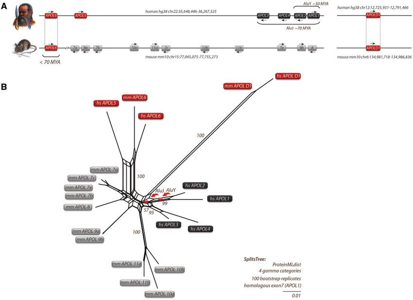
Müller et al. . doi:10.1093/molbev/msab220                                                                                         MBE

                                                                                                                                                    Downloaded from https://academic.oup.com/mbe/article/38/11/4962/6330626 by guest on 17 December 2021
FIG. 1. Local APOL gene family and SplitsTree reconstructions for humans and mice. (A) Genomic location of APOL genes from human and mouse
with genomic coordinates. Arrows indicate gene orientations. APOLD1 evolved early in vertebrates about 300 Ma, whereas APOL6 was probably
inherited by a human–mouse ancestor that lived about 70 Ma. (B) SplitsTree reconstruction of APOL genes based on protein sequences. A similar
tree topology was derived by maximum likelihood and Bayesian tree reconstructions (supplementary data SD1, Supplementary Material online).
The APOL genes of mice (mm for Mus musculus) are indicated in gray boxes. Black boxes represent human APOL genes (hs for Homo sapiens). The
central parallelograms of the reconstruction represent conflicting phylogenetic signals. The tree reconstruction reveals a common origin of mm
and hs APOL6 (red boxes). Bootstrap values are shown for representative branches. Balls indicate the clade-supporting integrations of AluJ and
AluY elements. The human APOL2 and APOL1 genes share an orthologous AluY element. The orthologous AluY transposons were already present
in Catharrine primates (diverged about 30 Ma). The diagnostic AluJ elements were detected at orthologous positions in APOL1-4 in all investigated
primates and were probably inherited from a common primate ancestor of about 70 Ma. The double-slash in the mouse locus indicates the
exclusion of a large genomic region. Human and mouse APOLD1 are located on chromosomes 12 and 6, respectively, and diverged significantly
from other APOL genes.

APOL1 and APOL2 Show Different Expression in                               pre- and postnatal stages. In these organs, APOL1 and APOL2
Human Organs during Development                                            are predominantly expressed at postnatal stages (fig. 3). At
Expression patterns during mammalian organ development                     embryonic and prenatal stages, APOLs exhibit no or only
may provide first hints about the different functions of closely           background expression.
related AluY-positive APOL1 and APOL2 genes. In addition, it                   APOL1 and APOL2 also appear to exhibit organ-specific
may give some information on why only APOL1 is linked to                   expression profiles. In cerebrum and cerebellum, APOL2 is
human diseases, once as a protective TLF and once as a risk                the predominantly expressed APOL gene. This was also ob-
gene for renal diseases. Therefore, we next analyzed the ex-               served in other transcriptome databases (supplementary data
pression patterns of these genes and used a recently estab-                SD5, Supplementary Material online). Its expression increases
lished database of developmental transcriptomes of seven                   during the lifespan, whereas APOL1 is only poorly expressed.
organs (cerebrum, cerebellum, liver, heart, kidney, ovary,                 The predominant APOL gene in the liver is APOL1 (fig. 3,
and testis) across seven species (human, macaque, mouse,                   supplementary data SD5, Supplementary Material online).
rat, rabbit, opossum, and chicken) (Cardoso-Moreira et al.                 Its expression is dramatically induced after birth and is main-
2019). We only analyzed human transcriptomes of cerebrum,                  tained at a very high level (80 reads per kilobase transcript
cerebellum, liver, heart, and kidney because they include both,            per million [RPKM]). APOL2 also exhibits robust postnatal

4964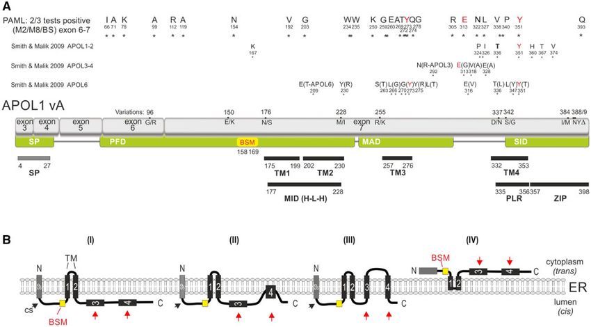
APOL1 Evolution Results in Dual ER Orientations . doi:10.1093/molbev/msab220 MBE
Downloaded from https://academic.oup.com/mbe/article/38/11/4962/6330626 by guest on 17 December 2021
FIG. 2. APOL1 isoforms and related APOL gene members. (A) Gene structures of the four well-characterized APOL1 isoforms vA, vB1, vB3, and vC
(Khatua et al. 2015). Exons for APOL1 vA (gene size 14 kb) are labeled 1–7. White frames indicate UTRs. Black boxes represent leading SPs, gray
boxes the remaining protein-coding exons, and black lines the intronic regions. Start codons are indicated by triangles and stop codons by asterisks.
(B) Cumulative gene structures of all human APOL gene members and isoforms. Red arrows indicate the insertion points of three diagnostic
transposable elements (AluJ merging APOL1-4, MER41G representing alternative exon insertion/exonization in APOL4, AluY indicating a common
ancestry of APOL1-2). Highly diverged sequences are shown by dotted lines (annotated alignments are provided as supplementary data SD4,
Supplementary Material online).
expression, but only approximately one-fourth of that of failed to be secreted and thus remained on the cytoplasmic
APOL1. In heart, APOL1 and APOL2 expressions are signifi- side of the ER. To address this, we first focused on the
cantly upregulated at postnatal stages. Finally, both genes are different N-terminal regions of APOL1 splice variants that
highly expressed in the kidney at postnatal stages and are unique to APOL1 and utterly absent in all other APOLs,
19 weeks postconception. Together, closely related APOL1 including APOL2. In silico analyses (SignalP, TMHMM) using
and APOL2 show different organ-specific expression patterns. the first 60 amino acids of the APOL1 splice variants pre-
dicting putative SPs or hydrophobic transmembrane regions
APOL1–APOL2 Evolutionary Splitting Leads to (TMs) led to ambiguous results. These predictions identified
Different APOL1 Orientations at the ER putative SPs or hydrophobic helixes either only in APOL1
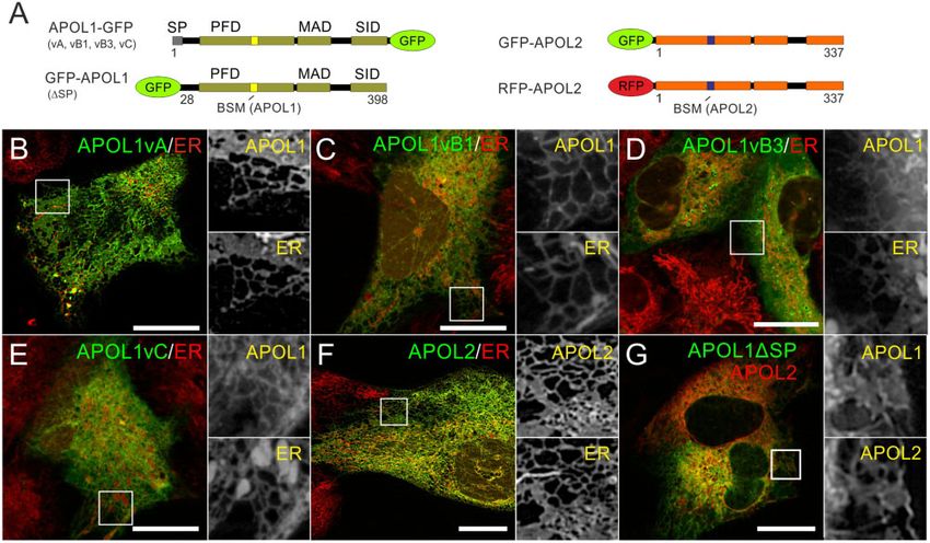
In contrast to APOL2, APOL1 is expressed in different splice vA and vC or only in vA and vB1 (supplementary data SD9,
variants (vA, vB1, vB3, and vC; fig. 2). Therefore, we applied Supplementary Material online). We, therefore, addressed
live-cell imaging to determine the intracellular distributions of this aspect experimentally and replaced the endogenous
GFP-tagged APOL1 splice variants and APOL2 in immortal- SP of the reporter enzyme SEAP (secreted alkaline phospha-
ized podocytes (AB8 cell line, fig. 4), and HEK293T cells (sup- tase) by the different N-termini of APOL1 splice variants
plementary data SD6–8, Supplementary Material online). In and the well-known SP of HSA (human serum albumin)
both cell lines, all GFP-tagged APOL1 and APOL2 proteins as a positive control (fig. 5A and B). Indeed, the SEAP se-
predominantly co-localized with the ER live-cell imaging dye cretion assays (fig. 5B, supplementary data SD9,
(ER Tracker, fig. 4); only in some cells, a meager fraction of Supplementary Material online) demonstrated functional
APOL1 could also be detected co-localizing with the plasma SPs in the N-termini of APOL1 vA, vB1, and vC, similar to
membrane (supplementary data SD6, Supplementary secretion positive controls (SEAP or HSA SPs). Only SEAP
Material online). Thus, binding to ER membranes seemed fused to the N-terminus of APOL1 vB3, and the negative
to be an evolutionarily conserved common feature of control (SEAP without SP) was not secreted. Thus, alterna-
APOL1 and APOL2, as GFP-tagged APOL3 and APOL4 tive splicing of APOL1 results in functional SPs of some but
showed different intracellular distributions (supplementary not all variants.
data SD7 and SD8, Supplementary Material online). APOL2, APOL1 vB3 (fig. 4), and APOL1 in which the SP was
In contrast to APOL2 (and the other APOLs), APOL1 is replaced by GFP (Granado et al. 2017), are targeted to the ER.
the only family member found in the bloodstream, indicat- These data suggest that the APOL1 ER targeting is due to
ing that it is released via the secretory pathway. This structural information, independent of the N-terminal SP.
requires the complete insertion of the nascent APOL1 poly- Therefore, we wondered, how ER targeting could be con-
peptide into the ER lumen mediated by a functional SP on a served among APOL1 and APOL2 proteins and second,
molecular level. We, therefore, wondered whether all APOL1 how—in the case of APOL1—it might be linked to its cyto-
splice variants carry functional SPs or whether some variants toxic effects.
4965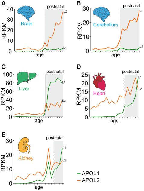
Müller et al. . doi:10.1093/molbev/msab220                                                                              MBE
                                                                      PAGE) when treated with PNGaseF was present only for
                                                                      APOL1 vA, vB1, and vC, demonstrating an N-glycosylation
                                                                      of the C-terminus of these APOL1 splice variants (fig. 5D). The
                                                                      shift was independent of APOL1 SID variants. Thus, APOL1
                                                                      G0 and the RRVs G1 and G2 have the same orientation in the
                                                                      ER lumen (fig. 5E and F and supplementary data SD13,
                                                                      Supplementary Material online). However, PNGaseF treat-
                                                                      ment had no effects on GT-tagged APOL1 vB3, APOL1 lack-
                                                                      ing the complete N-terminus, including the SP (DN59),
                                                                      APOL2 (fig. 5C–F), or APOL1 protein in which the SP (aa1–
                                                                      28) was replaced by an N-terminal GFP-tag (supplementary
                                                                      data SD13, Supplementary Material online). The GT and GFP-
                                                                      tag (GFP-GT) combination fused to APOL1 and APOL2 at the

                                                                                                                                        Downloaded from https://academic.oup.com/mbe/article/38/11/4962/6330626 by guest on 17 December 2021
                                                                      C-terminus led to the same outcomes (fig. 5E and F and
                                                                      supplementary data SD13, Supplementary Material online).
                                                                      Together, these data align with the SEAP assays and demon-
                                                                      strated a luminal (cis) ER localization of the APOL1 SIDs only
                                                                      in splice variants carrying functional SPs.
                                                                         Of note, the APOL1 mutant DC305-GFP-GT, which con-
                                                                      tains an SP but lacks the SID, becomes glycosylated (fig. 5E
                                                                      and F) and localizes to the ER (supplementary data SD14,
                                                                      Supplementary Material online). Furthermore, we also ob-
                                                                      served a luminal orientation and ER localization for a C-ter-
                                                                      minally GFP-GT-tagged APOL1 with an APOL2-BSM, and an
                                                                      APOL1–APOL2 chimera carrying the APOL1 SP, the APOL2
                                                                      PFD and MAD, and the APOL1 SID (supplementary data
FIG. 3. APOL1–APOL2 gene diversification is accompanied by organ-     SD14 and SD15, Supplementary Material online). Thus, our
specific expression. (A) Expression levels of APOL1 (L1, green) and   data show that APOL1 can be expressed in two different
APOL2 (L2, orange) transcriptomes (expressed as RPKM-normalized       orientations at the ER, one fraction (vA, vB1, and vC) with
counts) during human organ development derived from the Evo-          the SID localized inside the ER lumen (cis) and one (vB3) with
devo database. White background indicates embryonic or prenatal       the SID facing the cytoplasm (trans), like APOL2.
stages and gray background postnatal stages of mammalian organ
development (supplementary data SD20, Supplementary Material          APOL1 Shows Cytotoxic Effects in Both Orientations
online).
                                                                      We determined the cytotoxic potential of cis- and trans-ori-
                                                                      entated APOL1 (G0 and RRVs). Both, APOL1-GFP which have
   In silico analyses using full-length APOL1 (vA) and APOL2          luminal orientated SIDs as well as GFP-APOL1 (in which the
sequences suggest the presence of two to five TMs for APOL1           SP was exchanged by GFP), showed reduced cell viability after
(including the SP) and two or three TMs for APOL2 (supple-            24 and 48 h (fig. 6). The reduced cell viability was pronounced
mentary data SD10–12, Supplementary Material online). The             with APOL1 renal risk variants (G1 and G2) but was also
relative positions of the most significant two TMs within the         present with the used African APOL1 G0 haplotype (E150/
primary structure are very similar between APOL1 and                  I228/K255, see Lannon et al. [2019] and supplementary data
APOL2 (supplementary data SD10–12, Supplementary                      SD16, Supplementary Material online). In contrast, APOL2
Material online). We addressed the possible orientations of           overexpression had no toxic effect (fig. 6).
various APOL1 splice variants and APOL2 in the ER to exam-               Since GFP-APOL2 showed the same trans ER orientation as
ine whether their SIDs—which in case of APOL1 carries the             GFP-APOL1 (with the toxic RRV G2), we also investigated the
renal risk variants—faced the luminal (cis) or cytoplasmic side       BSM roles in trans-orientated APOL1 cytotoxicity (and
(trans). For that purpose, we took advantage of an artificial N-      APOL2 nontoxicity). To address this, we modified the
glycosylation tag (GT, Glyco-tag) (Kaup et al. 2011) that only        APOL1 BSM (NIRRLRALADGVQKV) by mutating its
becomes glycosylated in the ER lumen. We fused this tag to            LRALAD core sequence into six alanine residues
the C-termini of various APOL1 splice variants, APOL2, and            (NIRRAAAAAAGVQKV; called B6xAla;) or by changing the
included an APOL1 mutant lacking the first 59 amino acids of          BSM of APOL1 into that of APOL2 (HIRKLRALAEEVEQV;
the N-terminus. This DN59 deletion mutant is very similar to          B2). Strikingly, both modifications led to a loss of APOL1 cy-
APOL2 and even starts with an almost identical amino acid             totoxicity (fig. 6B), demonstrating that APOL1 cytotoxicity in
sequence (fig. 5C). We expressed these GT-tagged APOL pro-            trans orientation requires the BSM. In addition, APOL1 with
teins in HEK293T cells. Lysates of transfected cells were             the APOL2 BSM maintained its ER membrane targeting,
treated with and without PNGaseF, a glycosidase that                  whereas APOL1 with the B6xAla sequence showed a cytoplas-
completely removes N-glycosylation. A shift on sodium                 mic localization (supplementary data SD15, Supplementary
dodecyl sulfate polyacrylamide gel electrophoresis (SDS-              Material online). Chimeric APOL2 proteins, carrying the toxic
4966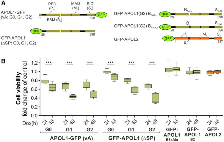
APOL1 Evolution Results in Dual ER Orientations . doi:10.1093/molbev/msab220                                                     MBE

                                                                                                                                                 Downloaded from https://academic.oup.com/mbe/article/38/11/4962/6330626 by guest on 17 December 2021
FIG. 4. APOL1 splice variants and APOL2 are targeted to the ER. (A) Schemes of C-terminally GFP-tagged APOL1 splice variants, GFP-APOL1
(lacking the SP, DSP, aa1-27), and N-terminally GFP- or RFP-tagged APOL2. Live-cell imaging of AB8 podocytes, expressing APOL1-GFP splice
variants vA, vB1, vB3 and vC (B–E), GFP-APOL2 (F), or GFP-tagged APOL1 together with RFP-APOL2 (G). ER membranes are visualized with live-cell
imaging dye ER Tacker. Scale bars: 20 mm.

SID of APOL1-G2 (P2M2S1-G2) or both, the putative pro-                    positively selected sites (fig. 7A). Three of the 27 twice sup-
apoptotic APOL1 BSM and toxic SID (P2B1M2S1-G2), were                     ported sites (Y273, E313, Y351) correspond to positively se-
unable to cause toxic effects (data not shown). Thus, cyto-               lection positions previously identified by Smith and Malik
toxicity is unique to APOL1, present for both ER orientations             (2009). Relaxing the stringency to once significantly detected
but not transferable to cytoplasmic orientated APOL2.                     sites (M2, M8, or 2201) revealed an overlap of 17 of the 23
                                                                          positively selected sites of Smith and Malik (fig. 7A, supple-
                                                                          mentary data SD17, Supplementary Material online).
Positively Selected Sites of APOL1 Are Localized in                       Strikingly, 15 of the 27 positive selected sites are within or
Putative TMs                                                              close to two putative TMs (TM3: aa257–276 and TM4:
Previous work is suggesting that APOL1-linked cytotoxicity                aa332–353, fig. 7, supplementary data SD17 and SD18,
mainly bases on cation channel activity at the plasma mem-                Supplementary Material online).
brane (Thomson and Finkelstein 2015; Olabisi et al. 2016;
Bruno et al. 2017; Giovinazzo et al. 2020; Schaub et al.
2020). This results in novel topology conceptions for                     Discussion
APOL1, in which the SP is followed by up to four additional               Our evolutionary tree reconstructions elucidate an ancestral
TMs (Giovinazzo et al. 2020; Gupta et al. 2020; Scales et al.             APOLD1-like precursor gene as the origin of the entire gene
2020; Schaub et al. 2020). Therefore, we reevaluated codons               family. In addition, our data show almost entirely indepen-
that are under positive selection during APOL1 evolution.                 dent divergences of the APOL gene families in mouse and
These analyses were performed in PAML (Yang 2007; Xu                      human, leading to six further human and 12 further murine
and Yang 2013) and based on a codon alignment of exon 6                   APOL genes (fig. 1, supplementary data SD1–3,
and exon 7 (APOL1 vA equivalents for APOL2–4, supplemen-                  Supplementary Material online, and Page et al. 2001; Smith
tary data SD17, Supplementary Material online). By applying               and Malik 2009; Kreit et al. 2015). A search for phylogeneti-
site-specific models M2 (selection) versus M1 (neutral) and               cally diagnostic primate-specific transposable elements
M8 (selection) versus M7 (neutral), and a branch-site model               revealed two Alu SINEs in the APOL gene family. AluJ is lo-
(labeled 2201), more than 100 potential amino acid sites un-              cated at orthologous positions in APOL1–4, and AluY exclu-
der positive selection were identified (supplementary data                sively is shared by APOL1 and APOL2 genes. The sequence-
SD17, Supplementary Material online). We selected 27 of                   based tree reconstruction argues for a common human–
them with significant support from at least two investigated              mouse APOL6-like ancestor gene 70 Ma that later diverged
models and arranged them with the previously detected                     into the AluJ-orthologous APOL1–4 genes in all investigated
                                                                                                                                        4967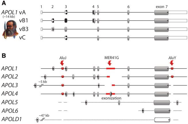
Müller et al. . doi:10.1093/molbev/msab220                                                                                         MBE

                                                                                                                                                    Downloaded from https://academic.oup.com/mbe/article/38/11/4962/6330626 by guest on 17 December 2021
FIG. 5. Different ER insertion orientations accompany APOL1–APOL2 diversification. (A) The endogenous SP of the reporter enzyme SEAP
(secreted alkaline phosphatase, gray box) was replaced by the N-termini of the APOL1 splice variants (vA, vB1, vB3, vC). The endogenous SP of
SEAP (SPSEAP) and the well-known SP of human serum albumin (SPHSA) served as positive, and SEAP lacking its SP (DSP) as negative control. (B)
SEAP secretion assay in HEK293T cells. Fluorescent SEAP secretion was detectable for positive controls (SPSEAP and SPHSA) and splice variants vA,
vB1, and vC. SEAP with the N-termini of APOL1 vB3 and without the SEAP SP (DSP) were not secreted (***P < 0.0001). (C) Scheme: APOL1 splice
variants vA, vB1, vB3, vC, a mutant lacking the complete APOL1 N-terminus (aa1-59; DN59), and APOL2 were fused with an artificial C-terminal N-
glycosylation tag (GT). (D) Western blot analyses of Glyco-tag (GT) APOL proteins (shown in C) before and after PNGaseF glycosidase digestion
showed N-glycosylation and therefore ER luminal localization only for APOL1 vA, vB1, and vC (marked by black asterisks). (E) Scheme: C-terminally
GFP-tagged APOL1 (G0 and renal risk variants G1 and G2), APOL1 lacking C-terminal aa305–398 (DC305), and APOL2. (F) Western blot analysis of
these GFP-GT-tagged proteins (shown in E) before and after PNGaseF glycosidase digestion demonstrates luminal ER localization for these
proteins (asterisks), except for APOL2-GFP-GT.

primates, followed by the appearance of AluY-orthologous                   resistance to all strains of Trypanosoma brucei, except for
APOL1–2 genes in Old World monkeys (Catarrhini). APOL5                     subspecies Trypanosoma brucei rhodesiense and
is among the placental precursors of APOL genes but re-                    Trypanosoma brucei gambiense (Genovese et al. 2010;
moved from the mouse genome. APOLD1, on the other                          Harrington et al. 2014; Cooper et al. 2017; Shukha et al.
hand, predates the mammalian divergence and is still highly                2017; De Simone et al. 2020).
conserved in most vertebrates.                                                In contrast to their different organ-specific expression pat-
    Although APOL2 is found mainly in the brain, the highest               tern, which is most likely due to tissue-specific promotor
expression of APOL1 is in the liver indicating that divergences            activities, APOL1 and APOL2 mainly localize to the ER on
of APOL genes are linked to organ-specific expression patterns             an intracellular level. Taking advantage of live-cell imaging
and cell type-specific roles of the various family members. The            approaches using GFP-tags our data suggest that ER targeting
high expression level of APOL1 in the liver (Shukha et al. 2017)           is a common and, therefore, probably evolutionarily con-
is remarkable as the liver is the chief secretory organ, releasing         served feature of these closely related proteins. However,
many if not most of the blood serum components, including                  this raises the question how the common intracellular local-
APOL1 (wildtype) and HPR (haptoglobin-related protein),                    ization of these proteins is linked to their opposite cytotoxic
which are key components of the TLF that confers innate                    properties.

4968
APOL1 Evolution Results in Dual ER Orientations . doi:10.1093/molbev/msab220 MBE
Downloaded from https://academic.oup.com/mbe/article/38/11/4962/6330626 by guest on 17 December 2021
FIG. 6. APOL1 cytotoxicity is caused by cis- and trans-orientated APOL1 pools. (A) Schemes of APOL1 variants used for cell viability assays. Left
panel: We used APOL1 G0, and renal risk variants G1 and G2 combined with C-terminal GFP (APOL1-GFP, vA aa1–398), and APOL1 fusion
proteins (G0, G1, and G2) in which the APOL1 SP was replaced by an N-terminal GFP-tag (GFP-APOL1 DSP; aa28–398). Right panel: To analyze the
impact of the putative pro-apoptotic “BH3-only motif” (BSM) in APOL1 pools at the outer ER membrane (trans orientation), we replaced the BSM
of GFP-APOL1 by either an Alanine stretch (B6xAla) or the BSM of APOL2 (B2). GFP-tagged APOL2 was used as a control. (B) Cell viability assay of
stable HEK293T cell lines expressing proteins summarized in A: APOL1-expressing cell lines, particularly those with RRVs, exhibited cytotoxic
effects in cis and trans orientations. Toxic GFP-APOL1 (DSP, G2) in which the BSM was replaced by Alanine (GFP-APOL1B6xAla) or the BSM of
APOL2 (GFP-APOL1 B2) and GFP-APOL2 showed no cytotoxicity (***P < 0.0001).
The SEAP secretion and Glyco-tag assays used in this study et al. 2019), mitochondrial fission (Ma et al. 2020), or inter-
demonstrated that APOL1 variants vA, vB1, and vC are lo- actions with Vamp8 (Madhavan et al. 2017) and APOL3
cated inside the ER (cis), whereas those of vB3 and APOL2 are (Uzureau et al. 2020). Moreover, recently Wakashin et al. iden-
faced to the cytoplasmic (trans) side of the ER. In this context, tified the intracellular NLRP12 (NLR family pyrin domain con-
it is worth noting that the SEAP and glycosylation assays are taining 12)—a key regulator of Toll-like receptor signaling—as
strongly in line with the APOL1 splice variant orientations a binding partner of the APOL1 vB3 variant. The data suggest
elucidated by Scales et al. (2020) using differential permeabi- a role in which APOL1 vB3 triggers an enhanced podocyte
lization assays in combination with numerous highly specific damaging by an upregulated inflammatory signaling
antibodies. Thus, evolutionary splitting into APOL1 and (Wakashin et al. 2020).
APOL2 genes resulted in different ER membranes orienta- Trans orientated APOL1 also fits the observation that it
tions, but only for three-fourths of the APOL1 splice variants can bind to phosphoinositides, which are present on the ER’s
(vA, vB1, and vC). This is of substantial relevance for the cytoplasmic leaflet (Wan et al. 2008; Chun et al. 2019).
various discussed APOL1 pathomechanisms, as it is still dis- Together this indicates that SP-less APOL1 vB3, as well as
cussed whether circulating (meaning secreted) (Hayek et al. APOL2 most likely bind peripherally to these lipids (fig. 7B).
2017) or rather intracellular fractions (in cis or trans orienta- The identified BH3-only like sequence motif of APOL pro-
tion) cause APOL1-linked renal diseases (Bruggeman et al. teins points to a similar role in cell programmed death (PCD)
2014; Cheng et al. 2015; Heneghan et al. 2015; Khatua et al. scenarios as shown for other members of the Bcl2-family
2015; Lan et al. 2015; Ma et al. 2017, 2020; Olabisi et al. 2016; (Vanhollebeke and Pays 2006; Wan et al. 2008; Smith and
Beckerman et al. 2017; Granado et al. 2017; Kruzel-Davila, Malik 2009). Some members have been identified as factors
Shemer, et al. 2017; Madhavan et al. 2017; Shah et al. 2019; mediating PCD by directly activating Bax/Bak proteins at the
Uzureau et al. 2020). outer mitochondrial membrane to trigger apoptosis
In a scenario in which APOL1 cytotoxicity requires the SID (Shamas-Din et al. 2011; Dummer et al. 2015; Adams and
facing the cytoplasm (fig. 7B, supplementary data SD18, Cory 2018). Intriguingly, cytoplasmic orientated GFP-APOL1
Supplementary Material online), only cytoplasmic APOL1 in which its BSM is replaced by that of APOL2 loses its cyto-
vB3—or nonprocessed and therefore nonmembrane inte- toxicity (while maintaining ER targeting; fig. 6B, supplemen-
grated APOL1 pools—fulfills this criterion. This is in line tary data SD15, Supplementary Material online). These data
with studies in which APOL1-cytotoxicity was linked to suggest a BSM role as cofactor in APOL1-linked cell death
APOL1 targeting to the outer mitochondrial membrane (Wan et al. 2008; Lan et al. 2015) and are in line with previous
(Ma et al. 2017), the mitochondrial transition pores (Shah studies that observed mitochondrial damaging in cells
4969Müller et al. . doi:10.1093/molbev/msab220 MBE
Downloaded from https://academic.oup.com/mbe/article/38/11/4962/6330626 by guest on 17 December 2021
FIG. 7. APOL1 vA gene structure and regions with positively selected sites. (A) Amino acids (single letter code) and coordinates mark positively
selected sites for APOL1–4 derived from at least two of three PAML models (M2/M8/BS) as large stars. The positively selected sites found by Smith
and Malik (2009) are indicated for APOL1–2, APOL3–4, and APOL6. Red labeled letters show overlapping sites of different studies. The APOL1
variation of alternative amino acids or deletion sites (D) are shown above the exon structure (gray). APOL1 was previously divided into four
functional regions, an SP, a PFD, an MAD, and the SID (green boxes). The PFD also contains a putative pro-apoptotic BH3-only domain sequence
motif (BSM). More recent conceptions suggest an SP (gray bar) and up to four TMs (SP, gray and TM1-4, black bars with their coordinates indicated
as numbers). The first two TMs are also referred to as membrane insertion domain (MID) or helix–turn–helix region (H-L-H). The TM4 overlaps
with a pore-lining region (PLR), followed by leucine-zipper domain (ZIP). (B) Model of possible orientations of APOL1 (and APOL2) at the ER. The
presence or absence of the SP determines the orientation of APOL1 splice variants and of APOL2 at the ER. Functional SPs result in a luminal (cis)
ER localization of the APOL1 C-terminus. If APOL1 functions as an ion pore, this most likely requires two (I), “two and a half” (II), or four (III) TMs
(black boxes). During evolution, positively selected amino acids accumulate in regions within, or close to, the putative TM3 and TM4 (red arrows).
APOL1 vB3 and APOL2, that lack functional SPs (IV), are most likely bound to the cytoplasmic leaflet of the ER membrane (trans). Since cis as well
as trans pools of APOL1 show cytotoxic effects—that are pronounced in the case of renal risk variants—our data suggest the presence of different
cellular pathomechanisms.
overexpressing APOL1 (Ma et al. 2017; Shah et al. 2019).Our In an alternative concept, APOL1 forms an active cation
data elucidated functional SPs for APOL1 vA, vB1 and vC and, channel by four TMs, including the central membrane in-
therefore, a luminal orientation of these splice variants’ C- sertion domain (MID, aa177–228, TM1, and TM2), a pore-
termini. Strikingly, such a cis orientation agrees with scenarios lining region (PLR, aa335–356, or TM4), and an additional
in which APOL1’s cytotoxic effects are caused by ion channel TM3 between MID and PLR (aa257–276, see fig. 7 and
activity at the plasma membrane (Thomson and Finkelstein Giovinazzo et al. 2020; Schaub et al. 2020). In this model,
2015; Olabisi et al. 2016; Bruno et al. 2017; Giovinazzo et al. changes of the intracellular pH-values appear to be crucial
2020; Schaub et al. 2020), or where APOL1 is acting as a for APOL1 membrane insertion and channel function. This
paracrine (meaning secreted) factor (Hayek et al. 2017). includes the possibility that proper formation of a third TM
The DC305-GFP-GT mutant, which lacks the APOL1 SID may require the C-terminal SID (aa305–398), indicating that
but contains the SP of variant A and the helix–turn–helix only the full-length APOL1 vA can form four TMs. However,
loop of the MID (aa177–228) showed a luminal orientation. in every case, the suggested APOL1 topology concepts are in
These data indicate the presence of a central membrane in- line with our glycosylation (and secretion) assays. Most pos-
sertion domain composed of two juxtaposed membrane itively selected sites during APOL1 evolution are close to or
spanning helices (TM1 and TM2) and suggest that—at least even within the novel postulated TM3 and TM4 (fig. 7A,
in this mutant—the region between aa257 and aa276 is un- supplementary data SD16, Supplementary Material online).
able to form a third membrane spanning helix (TM3). From Remarkably, positively selected sites also include Y351. This
this point of view, the DC305 mutant supports a current tyrosine is crucial for the pH-dependent gating of APOL1-
topology concept in which APOL1—in addition to the linked ion channel activity (Schaub et al. 2020) or part of a
SP—contains two TMs and a “half-spanning loop” (fig. 7 putative half-spanning loop, suggested by Gupta et al.
and Gupta et al. 2020). (2020).
4970APOL1 Evolution Results in Dual ER Orientations . doi:10.1093/molbev/msab220 MBE
The different APOL1-pathomechanisms are finally a con- address how a renal risk is turned into APOL1-linked kidney
sequence of alternative splicing of the APOL1 gene. Indeed, diseases.
alternatively spliced transcripts of a gene can regulate various
biological processes, and their evolution often led to func- Materials and Methods
tional innovation (Bush et al. 2017). Interestingly, the start
codon and 50 GT splicing site of alternative exon 2 of APOL1 Phylogenetic Tree and Gene Reconstruction
(APOL1 vB1; fig. 2A) that constitute one of the two SP initi- We extracted all known human and mouse APOL genes using
ation codes of APOL1 are present in most primate species the UCSC Gene Sorter (https://genome.ucsc.edu, last
representatives of APOL1–4 genes (supplementary data SD4, accessed August 2, 2021). The APOL gene family’s gene archi-
Supplementary Material online), but the expression is re- tecture was derived from both species following the graphi-
stricted to APOL1. Apart from that, the alternative SP from cally visualized genome assemblies of the UCSC Genome
exon 3 of APOL1 (APOL1 vA and APOL1 vC) is also repre- Browser (fig. 1A). To reconstruct the evolutionary history of
sented and expressed for APOL4 (fig. 2A and B; supplemen- APOL genes, we assembled all amino acid sequences from the
Downloaded from https://academic.oup.com/mbe/article/38/11/4962/6330626 by guest on 17 December 2021
tary data SD4, Supplementary Material online). In this large homologous exon 7 (corresponding to APOL1 vA; fig.
context, it is worth mentioning that the rapid evolution of 2A) in PhyDE (http://www.phyde.de, last accessed August 2,
SPs correlates to a relaxed selection on nonsynonymous and 2021). The alignment was optimized using the MUSCLE mul-
synonymous sites in a gene (Li et al. 2009). Thus, gene dupli- tiple sequence aligner PhyDE plugin (Edgar 2004). SplitsTree4
cation goes ahead with relaxation from selective constraints (version 4.15.1, Huson and Bryant 2006) was used to recon-
and may explain the evolution of the SPs of APOL1 after struct the phylogenetic tree of APOL genes. We used the
divergence from APOL2. Therefore, it is tempting to speculate Character Method ProteinMLdist under the JTT reconstruc-
that the ratio between APOL1 splice variants is part of ongo- tion model and four gamma categories to derive the gene
ing evolution. affiliations shown in figure 1. SplitsTree reconstructs a con-
Under normal conditions, high and efficient APOL1 secre- sensus network that visualizes potential data conflicts by
tion may result in low levels of nonprocessed, luminal-faced parallelograms. Next, we performed a bootstrap analysis of
fractions of APOL1 at the ER, thereby most likely preventing a random subset of the data with 100 replicates. When 100%
damaging of the secreting cells. However, an upregulation of of the resampled data sets showed the same split (suggesting
solid phylogenetic signals in the original data set), the related
APOL1, triggered by inflammatory cytokines, may cause an
branch was labeled with 100 in the consensus network. The
overload of APOL1 pools at the ER in cis and trans orienta-
tree topology was verified by applying a maximum likelihood
tions (Nichols et al. 2015). Thus, ER targeting combined with
analysis in MEGA7 (JTT matrix-based model, discrete Gamma
high expression levels at the ER could be a key determinant in
distribution applying four gamma categories, 100 bootstrap
APOL1-linked cytotoxicity, particularly in postmitotic cells
replications), and applying a Bayesian inference in MrBayes
like podocytes with a very limited self-renewal capacity 3.2 (prset aamodelpr¼fixed Jones, lset rates¼invgamma
(Wen et al. 2018). Ngammacat ¼ 4, 200.000 generations, burnin 200) (supple-
Recent studies elucidated high APOL1 expression levels as mentary data SD1–3, Supplementary Material online).
an essential cofactor for APOL1-linked cytotoxicity (O’Toole
et al. 2018; Datta et al. 2020). Furthermore, increasing evi-
dence identified ER stress per se as an aggravating or Detecting Clade-Supporting Insertions of
disease-promoting factor for podocyte diseases (Cybulsky Transposable Elements
2013, 2017). Thus, APOL1 pools at the ER combined with To obtain additional information about the relationship and
their potential to cause ER-linked disturbances might age of different APOL genes in humans, we extracted intron,
have—as yet—underestimated potentials in explaining why exon, and UTR genomic regions of the human APOL genes.
To detect diagnostic TE insertions shared among the primate
SP-lacking as well as SP-containing APOL1 (especially of the
APOL genes, we aligned all six conserved APOL genes from
G1 and G2 RRVs) induce the observed pleiotropic toxic
various primate representatives (human, orangutan, rhesus
effects in in vitro and in vivo models.
monkey, baboon, squirrel monkey, mouse lemur). To identify
APOL1’s pathobiological mechanisms explaining its cyto-
transposed elements, we ran a local RepeatMasking (www.
toxicity in renal cells are still controversially discussed. This
repeatmasker.org). We extracted Alu SINEs, which were then
study focusses on the evolutionary history of the APOL gene
inspected for insertion homology using a genomic alignment
family in mouse and human. In addition, it provides experi-
from the different members of the APOL gene family (sup-
mental data that both cis (luminal) as well as trans (cytoplas-
plementary data SD4, Supplementary Material online). Such
mic) orientations of APOL1 splice isoforms could be diagnostic elements are inserted before splitting various gene
responsible for the wide spectrum of pathogenic mechanisms members, and after that, they represent informative pres-
proposed for podocyte damage leading to kidney disease. ence/absence markers of relationship at their homologous
However, APOL1-linked cytotoxicity depends on many fac- gene positions.
tors, including APOL1 expression levels, the haplotype, the Alu SINEs are primate-specific and the most abundant
presence of renal risk variants, its intracellular localization, and transposed elements in primates, thus uniquely suitable as
most likely also on the ratio between cis and trans orientated clade markers to analyze genes and species’ evolution
APOL1 pools. Hence, further studies will be necessary to (Churakov et al. 2010). Comparative analyses of the
4971Müller et al. . doi:10.1093/molbev/msab220 MBE
presence/absence patterns of the identified TEs in various sequencing. Details concerning constructs are summarized in
primate species enabled us to determine their evolutionary supplementary data SD19, Supplementary Material online.
time of insertion. Primer sequences are available from DM and TW.
Detecting Positive Selection Sites Cell Lines and Transient Transfection
To detect positively selected sites within protein-coding HEK293T cells were cultured in standard DMEM medium
sequences, we 1) used the PAML site specific model (model (Thermo Fisher Scientific) containing 10% fetal calf serum
¼ 0, NSsites ¼ 0 1 2 7 8) and a single Newick user tree, and 2) (FCS) and 1% antibiotics and L-glutamine (penicillin/strepto-
the branch-site model (model ¼ 2, NSsites ¼ 2, x ¼ 0 (es- mycin) at 37 C under a 5% CO2 atmosphere. Human im-
timated), initial x ¼ 1 labeled 2201 vs. x ¼ 1 (predetermined mortalized AB8/13 podocytes (Saleem et al. 2002) were
neutral), initial x ¼1 labeled 2211) (supplementary data cultured in standard RPMI 1640 medium (Sigma-Aldrich)
SD16, Supplementary Material online). The PAML incorpo- containing 10% FCS, 1% supplements, and 1% antibiotics
rated Bayes Empirical Bayes analysis determined the proba- (penicillin/streptomycin) at 33 C under 5% CO2 atmosphere
Downloaded from https://academic.oup.com/mbe/article/38/11/4962/6330626 by guest on 17 December 2021
bility of positive selection for each codon position (fig. 7). (Schulze et al. 2017). Transient transfections were performed
via calcium-phosphate-based precipitation (Schulze et al.
Constructs and Cloning 2014), or via Lipofectamine 2000 as described in the manu-
Expression cassettes encoding EGFP-APOL1 G0 lacking the SP facturer’s specifications (Thermo Fisher Scientific).
(EGFP-DSP APOL1, aa28–398) or encoding full-length APOL1
G0 with a C-terminal EGFP-tag have been described earlier Generation of Stable Cell Lines
(Granado et al. 2017). Standard site-directed mutagenesis was Stable cell lines were generated as previously described
used to obtain cDNAs encoding for APOL1 G1 (S342G/ (Schulze et al. 2014; Wennmann et al. 2014). Briefly, lentivirus
I384M) and G2 (D388N389Y) renal risk variants. Human production was performed in HEK293T cells transiently trans-
APOL2, APOL3, and APOL4 cDNAS were cloned using a hu- fected with psPAX2 and pMD2.G helper plasmids and mod-
man cDNA library derived from AB8 podocytes (Duning et al. ified pINDUCER21-Puro-plasmids (Meerbrey et al. 2011)
2008) and cloned into pEGFP-N1 (BD Clontech), a modified encoding the APOL proteins. The virus-containing superna-
pEGFP-C2 (encoding RFP instead of EGFP), or pENTR-vectors tant was collected and filtered through a 0.45-mm sterile filter
according to the manufacturer’s instructions (Thermo Fisher (EMD Millipore). The virus-containing medium was added to
Scientific). For glycosylation analysis, a Glycotag-tag1.4 tail HEK293T and AB8 podocyte cells using one volume of fresh
(AAAAAANATVAAASGDVWDI) (Kaup et al. 2011) was DMEM medium and one volume of the virus-containing fil-
fused to the C-terminal part of different APOL constructs trate supplemented with polybrene (final concentration 8 mg/
via polymerase chain reaction (PCR). Furthermore, APOL2– ml). After 24 h, the virus-containing medium was replaced by
APOL1 chimeric constructs, as well as APOL1/2 BH3 mutants, fresh medium, and cells were regenerated for a further 24 h.
were generated using overlapping primers combined with in Transduced cells were selected using puromycin (4 mg/ml,
combination with SOE PCR techniques. All APOL constructs, HEK293T; 2 mg/ml, AB8). All established stable cell lines
deletion constructs, mutants, Glyco-variants, and chimeric were tested for inducible overexpression of APOL proteins
constructs were cloned into pENTR or pENTR-EGFP vectors by western blot and immunofluorescence analysis.
and shuttled into a modified pINDUCER21-Puro destination
vector as previously described (Schulze et al. 2014). The Cell Viability Assays
pSEAP2-Control vector from Clontech (Takara Cat. No. Real-time cell viability measurements were performed via the
631717) served as a template for the different SEAP APOL1 RealTime-Glo MT Cell Viability Assay (Promega) according to
splice variant constructs. For the N-termini, different primers the manufacturer’s instructions. Briefly, 10,000 HEK293T cells
were designed whereby the different APOL1 N-termini and in 50 ml medium were seeded onto 96-well white bottom
the HSA SP were fused to SEAP during PCR, or the SP of SEAP plates (Nunc). After adherence, 50 ml 2 RealTime-Glo en-
was deleted (SEAP, SEAPDSP, SEAP-vA-cSPcs, SEAP-vC-cSPcs, zyme-substrate mix diluted in medium was added to each
SEAP-HSA-SP). Via restriction cloning, the amplified DNA was well either with or without 125 ng/ml doxycycline (Granado
ligated into a Gateway pDONR221 vector (Invitrogen Cat. No. et al. 2017). The luminescence was measured using a 37 C
12536017) and labeled as pENTR after insertion. For prewarmed microplate reader (TECAN) at the indicated time
APOL1vB1 the pENTR-SEAP-APOL1vA, and for APOL1vB3 points. For each cell line, three independent experiments
the pENTR-SEAP-APOL1vC, constructs served as templates (N ¼ 3), including three technical replicates for each N
for PCR. The DNA was subsequently cloned into a pENTR- (n ¼ 3), were measured.
pDONOR via restriction cloning. All SEAP constructs were
shuttled in a doxycycline-inducible pInducer21-Puro destina- Western Blot Analyses
tion vector (Schulze et al. 2014). pME-mTagBFP-CAAX was a Western blot analyses were performed as previously de-
gift from Nicholas Cole (addgene No. 75149; Don et al. 2017). scribed (Schulze et al. 2014, 2017). Briefly, cell lysates were
The BFP-CAAX cassette was shuttled into a pQCXIH desti- boiled for 5 min, and equal volumes were separated via SDS-
nation vector via Gateway LR reaction. The mCherry-Sec61b PAGE using 8–15% gels (Bio-Rad). Separated proteins were
construct (addgene No. 49155) was kindly provided by Gia then transferred to a PVDF membrane (EMD Millipore) and
Voeltz (Zurek et al. 2011). All constructs were verified by DNA incubated in blocking buffer (5% skim milk powder dissolved
4972APOL1 Evolution Results in Dual ER Orientations . doi:10.1093/molbev/msab220 MBE
in TBS containing 0.05% Tween-20 [TBS-T]) for 1 h at room Armenteros et al. 2019). APOL gene expression patterns in
temperature. The monoclonal GFP antibody (JL-8; #632380) human organs were analyzed using a database and an evo-
was provided by Takara Bio Europe (Clontech). The antibod- devo application tool (https://apps.kaessmannlab.org/evode-
ies against APOL1 (HPA018885), APOL2 (HPA001078), and b- voapp, last accessed August 2, 2021) recently provided by
Tubulin (#T8328) were purchased from Sigma-Aldrich, the Cardoso-Moreira et al. (2019)
antibody against a-Actinin 4 (#ALX-210–356) was from
Enzo/AlexisPrimary antibodies were used 1:1,000 in TBS-T Colocalization Analysis
with 5% bovine serum albumin (BSA) and incubated at 4 Analysis was performed by using ImageJ. To analyze colocal-
C overnight. The membrane was then washed three times ization between APOL1-EGFP or EGFP-APOL1 and the mem-
with TBS-T and incubated with horseradish peroxidase- brane, labeled with pQXCIH-BFP-CAAX, a mask Image was
coupled secondary antibodies (Jackson Immunoresearch) di- created. Therefore, the blue channel was duplicated; a thresh-
luted 1:3,000 in blocking buffer for 30–45 min at room tem- old called Moments was applied and manually adjusted to
perature. After three further wash steps with TBS-T, the signal remove the background signal. The Coloc2 PlugIn was used to
Downloaded from https://academic.oup.com/mbe/article/38/11/4962/6330626 by guest on 17 December 2021
was detected using the Lumi-Light chemiluminescence de- measure the Pearson Correlation Coefficient (PSF 3.0;
tection reagent (Roche). Pearson’s R-value [no threshold]). GraphPad PRISM software
was used to visualize the correlation coefficient analysis.
Live-Cell Imaging and Microscopy Pearson R-values 0.4 were considered as weak or no corre-
For live-cell imaging, cells expressing fluorophore-coupled lation, >0.4 untilMüller et al. . doi:10.1093/molbev/msab220 MBE
were homogenized by shearing force and then sonicated for Bruggeman LA, O’Toole JF, Ross MD, Madhavan SM, Smurzynski M, Wu
10 min. After removing cell debris and nuclei via centrifuga- K, Bosch RJ, Gupta S, Pollak MR, Sedor JR, et al. 2014. Plasma apo-
lipoprotein L1 levels do not correlate with CKD. J Am Soc Nephrol.
tion for 15 min at 4 C and 10,000 g, supernatants were 25(3):634–644.
stored at 20 C or directly used for PNGaseF digestion. Bruno J, Pozzi N, Oliva J, Edwards JC. 2017. Apolipoprotein L1 confers pH-
switchable ion permeability to phospholipid vesicles. J Biol Chem.
PNGaseF Digestion 292(44):18344–18353.
The membrane orientations of APOLs were elucidated via Bush SJ, Chen L, Tovar-Corona JM, Urrutia AO. 2017. Alternative splicing
and the evolution of phenotypic novelty. Philos Trans R Soc Lond B
glycosylation signals of their C-terminal GT-tag. To confirm Biol Sci. 372(1713):20150474.
glycosylation, RIPA lysates were digested with PNGaseF Capewell P, Cooper A, Clucas C, Weir W, MacLeod A. 2015. A co-
according to the manufacturer’s instructions (New England evolutionary arms race: trypanosomes shaping the human genome,
Biolabs). Laemmli buffer was added to the lysates, and for a humans shaping the trypanosome genome. Parasitology 142(Suppl
digestion control, the origin lysate was mixed with an equal 1):S108–S119.
Cardoso-Moreira M, Halbert J, Valloton D, Velten B, Chen C, Shao Y,
volume of a 50:50 mixture of Laemmli buffer and water.
Downloaded from https://academic.oup.com/mbe/article/38/11/4962/6330626 by guest on 17 December 2021
Liechti A, Ascenç~ao K, Rummel C, Ovchinnikova S, et al. 2019. Gene
expression across mammalian organ development. Nature
Supplementary Material 571(7766):505–509.
Supplementary data are available at Molecular Biology and Chen TK, Choi MJ, Kao WHL, Astor BC, Scialla JJ, Appel LJ, Li L, Lipkowitz
MS, Wolf M, Parekh RS, et al. 2015. Examination of potential modi-
Evolution online. fiers of the association of APOL1 alleles with CKD progression. Clin J
Am Soc Nephrol. 10(12):2128–2135.
Author Contributions Cheng D, Weckerle A, Yu Y, Ma L, Zhu X, Murea M, Freedman BI, Parks
D.M. performed most of the experiments, supported by K.F., JS, Shelness GS. 2015. Biogenesis and cytotoxicity of APOL1 renal risk
variant proteins in hepatocytes and hepatoma cells. J Lipid Res.
D.G., A.C.G., V.K., S.M.L., and T.M.A. Evolutionary studies were 56(8):1583–1593.
mainly done by J.S. D.M., K.F., A.C.G., J.S., and T.W. analyzed Chun J, Zhang JY, Wilkins MS, Subramanian B, Riella C, Magraner JM,
the data. D.M., H.P., and T.W. designed the study. D.M., J.S., Alper SL, Friedman DJ, Pollak MR. 2019. Recruitment of APOL1
and T.W. prepared the manuscript. All authors approved the kidney disease risk variants to lipid droplets attenuates cell toxicity.
final manuscript. Proc Natl Acad Sci U S A. 116(9):3712–3721.
Churakov G, Grundmann N, Kuritzin A, Brosius J, Makałowski W,
Acknowledgments Schmitz J. 2010. A novel web-based TinT application and the chro-
nology of the primate Alu retroposon activity. BMC Evol Biol. 10:376.
We thank Beate Surmann, Truc Van Le, and Kristin Doctor for [PMC][10.1186/1471-2148-10-376] [21126360]
excellent technical support, all laboratory members for fruit- Cooper A, Ilboudo H, Alibu VP, Ravel S, Enyaru J, Weir W, Noyes H,
ful discussions, and Dr Gia Voeltz for plasmids. Moreover, we Capewell P, Camara M, Milet J, et al. 2017. APOL1 renal risk variants
have contrasting resistance and susceptibility associations with
thank Dr Beate Vollenbröker and Dr Daniela Braun for the African trypanosomiasis. Elife 6.
critical reading of the manuscript. Finally, we thank the mem- Corpet F. 1988. Multiple sequence alignment with hierarchical clustering.
bers of EvoPAD for the inspiring discussions of APOL evolu- Nucleic Acids Res. 16(22):10881–10890.
tion. The work was supported by grants from the German Cybulsky AV. 2013. The intersecting roles of endoplasmic reticulum
Research Foundation to T.W. (Grant No. DFG WE 2550/3-1) stress, ubiquitin-proteasome system, and autophagy in the patho-
genesis of proteinuric kidney disease. Kidney Int. 84(1):25–33.
and J.S. (Grant No. DFG SCHM 1469/10-1). K.F and T.M.A. [PMC][10.1038/ki.2012.390] [23254900]
were supported by the Medical Faculty of the University of Cybulsky AV. 2017. Endoplasmic reticulum stress, the unfolded protein
Muenster (Medizinerkolleg Münster, MedK grant numbers response and autophagy in kidney diseases. Nat Rev Nephrol.
No. 18-80022 and No. 16-0087, respectively). The work con- 13(11):681–696. [CrossRef][10.1038/
tains parts of the PhD thesis of D.M. and the MD theses of K.F. nrneph.2017.129][PMC][28970584]
Datta S, Kataria R, Zhang JY, Moore S, Petitpas K, Mohamed A, Zahler N,
and T.M.A. Pollak MR, Olabisi OA. 2020. Kidney disease-associated APOL1 var-
iants have dose-dependent, dominant toxic gain-of-function. J Am
Data Availability Soc Nephrol. 31(9):2083–2096. [PMC][10.1681/ASN.2020010079]
All relevant data are available within this article, the [32675303]
De Simone G, Pasquadibisceglie A, Polticelli F, di Masi A, Ascenzi P. 2020.
Supplementary Material online, or from the corresponding Haptoglobin and the related haptoglobin protein: the N-terminus
author upon request. makes the difference. J Biomol Struct Dyn. 1–10. doi: 10.1080/
07391102.2020.1837675.
Don EK, Formella I, Badrock AP, Hall TE, Morsch M, Hortle E, Hogan A,
References Chow S, Gwee SSL, Stoddart JJ, et al. 2017. A Tol2 gateway-
Adams JM, Cory S. 2018. The BCL-2 arbiters of apoptosis and their compatible toolbox for the study of the nervous system and neu-
growing role as cancer targets. Cell Death Differ. 25(1):27–36. rodegenerative disease. Zebrafish 14(1):69–72.
Almagro Armenteros JJ, Tsirigos KD, Snderby CK, Petersen T N, Winther Duchateau PN, Pullinger CR, Orellana RE, Kunitake ST, Naya-Vigne J,
O, Brunak S, von Heijne G, Nielsen H. 2019. SignalP 5.0 improves O’Connor PM, Malloy MJ, Kane JP. 1997. Apolipoprotein L, a new
signal peptide predictions using deep neural networks. Nat human high density lipoprotein apolipoprotein expressed by the
Biotechnol. 37(4):420–423. pancreas. Identification, cloning, characterization, and plasma distri-
Beckerman P, Bi-Karchin J, Park ASD, Qiu C, Dummer PD, Soomro I, bution of apolipoprotein L. J Biol Chem. 272(41):25576–25582.
Boustany-Kari CM, Pullen SS, Miner JH, Hu C-AA, et al. 2017. Dummer PD, Limou S, Rosenberg AZ, Heymann J, Nelson G, Winkler CA,
Transgenic expression of human APOL1 risk variants in podocytes Kopp JB. 2015. APOL1 kidney disease risk variants: an evolving land-
induces kidney disease in mice. Nat Med. 23(4):429–438. scape. Semin Nephrol. 35(3):222–236.
4974You can also read