Epidemiologic and Etiological Features of Korean Patients With Behçet's Disease
←
→
Page content transcription
If your browser does not render page correctly, please read the page content below
pISSN: 2093-940X, eISSN: 2233-4718
                                Journal of Rheumatic Diseases Vol. 28, No. 4, October, 2021
                                https://doi.org/10.4078/jrd.2021.28.4.183                                                       Review Article

Epidemiologic and Etiological Features of Korean Patients
With Behçet’s Disease
                            1                                            2
Soo Hyun Choi, BA , Do-Young Kim, M.D., Ph.D.
1
Tulane University School of Medicine, New Orleans, LA, USA, 2Department of Dermatology and Cutaneous Biology Research Institute, Yonsei
University College of Medicine, Seoul, Korea

Behçet’s disease (BD) is a multisystem disease in which environmental factors provoke an adverse immune response in patients
with genetic susceptibility towards BD, subsequently leading to a cascade of dysregulated inflammation throughout the body.
It is particularly prevalent in regions spanning the ancient Silk Road, including Korea, where the first known case of BD was re-
ported in 1961. We summarize the history, epidemiology, and clinical presentation of BD in Korea, highlighting the clinical
tendencies that are particularly seen in the Korean BD population as compared to European populations. Analysis of epidemio-
logic trends over the past three decades in Korea shows a decreasing prevalence of complete BD and a higher prevalence of
intestinal BD. We also discuss the ever-evolving understanding of the pathogenesis of BD, noting the complex interplay among
genetics, environment, and immunology. The HLA-B51 allele is the most significant known genetic risk factor in developing
BD. We also discuss more recently studied associations between BD and immune factors such as IL-10, IL-23R-IL-12RB2,
IL-1A-IL-1B, CCR1, ERAP1, and the GIMAP cluster, the last of which has been found to have an association with BD specifically
in Korea. Environmental factors such as pollution and microbials are often the inciting event in developing BD, as they trigger
an imbalanced immune response in genetically susceptible individuals, one that has been often found to exhibit an aberrant
Th1/Th17 response. There would be value to further studying the pathogenesis and clinical characteristics of Korean BD. (J
Rheum Dis 2021;28:183-191)

Key Words. Behçet syndrome, HLA-B51, Epidemiology, Genome-wide association studies

                       INTRODUCTION                                                      ical characteristics of BD in the Mediterranean/Middle
                                                                                         East versus East Asia, including Korea. In this review, we
  Behçet’s disease (BD) is a multisystemic, inflammatory                                 will discuss the history and clinical manifestations of BD
disease with a chronic, relapsing course. It is charac-                                  in Korea, as well as the genetic and environmental factors
terized by a variety of clinical manifestations, including                               that play a role in the pathogenesis of the disease.
recurrent oral and genital ulcers, inflammatory skin le-
sions, and involvement of ocular, vascular, articular, gas-                                          HISTORICAL BACKGROUND
trointestinal, and neurologic systems. The prevalence of
BD is higher in regions of the ancient Silk Road, spanning                                The earliest reports of BD in Korean literature were two
from the Mediterranean to East Asia. Populations in this                                 cases described by the ophthalmologist Dr. Joo in 1961
region exhibit a higher frequency of the HLA-B51 allele,                                 [1]. A subsequent case report in 1962 described four cas-
which is the most important genetic susceptibility factor                                es of BD, one with severe systemic manifestations of the
in the development of BD. Despite this common genetic                                    disease [2]. Anecdotal cases have continued to be re-
component, there are significant differences in the clin-                                ported in Korean literature from that point on.

Received:August 17, 2021, Accepted:August 25, 2021
Corresponding to:Do-Young Kim http://orcid.org/0000-0002-0194-9854
                 Department of Dermatology and Cutaneous Biology Research Institute, Yonsei University College of Medicine, 50-1 Yonsei-ro,
                 Seodaemun-gu, Seoul 03722, Korea. E-mail:dykim@yuhs.ac

Copyright ⓒ 2021 by The Korean College of Rheumatology.
This is an Open Access article, which permits unrestricted non-commerical use, distribution, and reproduction in any medium, provided the original work is properly cited.

                                                                                  183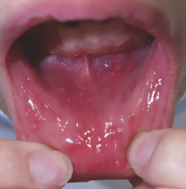
Soo Hyun Choi and Do-Young Kim

  On November 10th, 1983, the Behçet’s Disease Specialty           a higher prevalence of gastrointestinal (GI) BD and rela-
Clinic—the first of its kind in Korea—opened at the                tively fewer patients with vascular and CNS symptoms
Severance Hospital of Yonsei University College of                 [8,9]. The ISG criteria showed 58% sensitivity when used
Medicine as a joint effort among the departments of                with Korean BD patients, a sharp decline from the 92%
Dermatology, Ophthalmology, and Otorhinolaryngology                sensitivity in the ISG study sample [10]. In this context,
[3]. Korean researchers joined the international BD soci-          the Japanese diagnostic criteria, which includes GI in-
ety and presented the first set of compiled domestic data          volvement as a diagnostic criteria, may be useful in the
at the 4th International Conference on BD in London on             Korean clinical setting [11]. There have been efforts to es-
September 5∼6th, 1985. Finally, on March 4th, 1999, a              tablish a more precise diagnostic criteria for BD in Korea,
meeting was held regarding the organization of a Korean            prioritizing GI symptoms over vascular and CNS manifes-
academic society for BD [4]. It was attended by 35                 tations [12,13]. In short, physicians should understand
physicians. Professor Sungnack Lee was appointed as the            the benefits and limitations of the available criteria for BD
first president of the Korean Study Group for BD (which            diagnosis, and their adaptation in clinical practice should
was renamed as the Korean Society for BD [KSBD] later),            be met with careful prioritization and consideration of
and the first Annual Academic Meeting of KSBD was held             each criteria’s respective characteristics (Table 1) [14].
on October 2nd, 1999, with presentations from lecturers
Drs. Colin G. Barnes (UK) and Shigeaki Ohno (Japan)                         CLINICAL MANIFESTATIONS
[4]. Finally, the 9th International Conference on BD was
held on May 27th to 29th, 2000, in Seoul, Korea [5]. As of           BD is a disease characterized by widespread, multi-
August 2021, KSBD (President, Prof. Eun-So Lee) has                system inflammation, and it manifests itself in various or-
held 21 annual meetings.                                           gan systems. Mucocutaneous manifestations are key
                                                                   markers of BD, and their recognition may allow for early
      DIAGNOSTIC CRITERIA FOR BD                                   diagnosis and intervention. Oral ulcers are nearly ubiq-
                                                                   uitous across all cases and classified into three types 1)
  At present, a final diagnosis of BD is made based on clin-       minor, 2) major, and 3) herpetiform ulcers (Figure 1).
ical presentation, as there are no laboratory markers of           Genital ulcers are similar to oral ulcers in appearance and
high diagnostic value. The International Study Group               course but tend to be less recurrent. Various cutaneous
(ISG)’s diagnostic criteria for BD published in 1990 is the        presentations occur in BD patients, and multiple con-
most widely recognized and used criteria [6]. It was fur-          current skin lesion types are often observed in a single pa-
ther revised in 2014 as the International Criteria for BD          tient (Figure 2) [15]. Papulopustular lesions and eryth-
(ICBD) to increase diagnostic sensitivity, refining criteria       ema nodosum-like panniculitis are the classic cutaneous
for vascular and central nervous system (CNS) involve-             symptoms. Less common lesions, such as superficial
ment [7]. However, epidemiological data in Korea shows             thrombophlebitis, Sweet’s syndrome-like dermatosis,

Table 1. Comparison of diagnostic criteria for BD
                Sign/symptom                        ISG criteria (1990)     Japanese criteria (1987)    ICBD criteria (2014)
        Oral aphthosis                                     ◼⃞                         ◎                     O (2 points)
        Genital aphthosis                                  O                          ◎                     O (2 points)
        Skin lesions                                       O                          ◎                     O (1 point)
        Ocular lesions                                     O                          ◎                     O (2 points)
        Positive pathergy test                             O                          ×                     O (1 point)*
        Joint involvements                                 ×                          O                         ×
        Epididymitis                                       ×                          O                         ×
        Intestinal involvement                             ×                          O                         ×
        Neurological manifestations                        ×                          O                     O (1 point)
        Vascular manifestations                            ×                          O                     O (1 point)
 ⃞ : required symptom, ◎: major symptom. BD: Behçet’s disease, ICBD: International Criteria for BD, ISG: International Study
Group. *Optional in ICBD criteria. Adapted from the article of Kirino and Nakajima (Intern Med 2019;58:1199-207) [14].

184                                                                                  J Rheum Dis Vol. 28, No. 4, October, 2021
Epidemiologic and Etiological Features of Korean Patients With Behçet’s Disease
Figure 1. Clinical features of recurrent oral aphthous ulcers. (A) Minor ulcers, (B) major ulcers, (C) herpetiform ulcers. Ulcers ≤1
cm are considered minor ulcers while larger ulcers are considered major ulcers. Multiple scattered ulcers of several millimeters are
considered herpetiform ulcers. Major ulcers may cause mucosal scarring and persist several weeks or longer.
Figure 2. Cutaneous manifes-
tations of Behçet’s disease (BD).
(A) Erythema nodosum-like le-
sion (arrow) and concurrent
papulopustular lesions (white
arrowheads) in lower leg. (B, C)
Papulopustular lesion on trunk
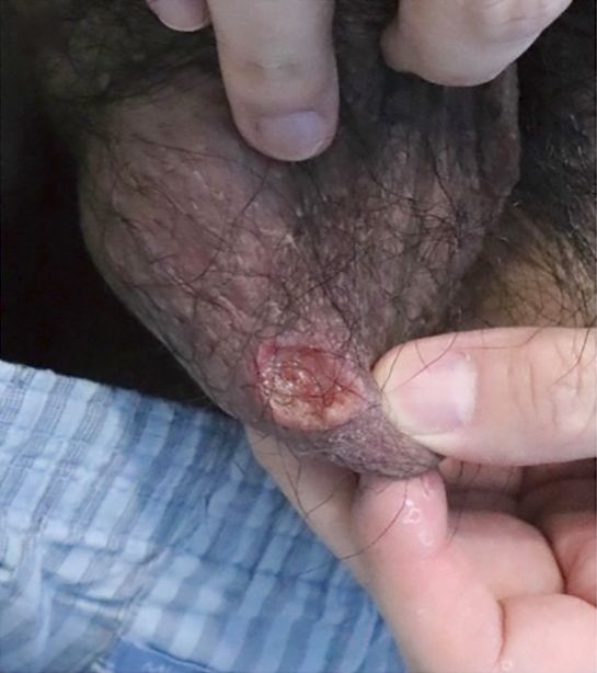
(B) and a scrotal ulcer in a male
patient with BD.
pyoderma gangrenosum, and erythema multiforme-like symptoms. Intestinal BD exhibits characteristic ulcers in
lesions are also seen. Ocular manifestations of disease the gastrointestinal tract, clinical assessment of which is
vary depending on site of involvement and include irido- guided by endoscopy. Endoscopic guidelines for in-
cyclitis, keratitis, episcleritis, scleritis, vitritis, and classic testinal BD diagnosis were recently established by an ex-
posterior uveitis, including retinal vasculitis and optic pert group of gastroenterologists in Korea [20]. The clin-
neuritis. Severe involvement of the posterior chamber is ical course of intestinal BD during the first 5 years are var-
particularly related to visual morbidity. A nationwide iable, but the majority of Korean patients with intestinal
analysis revealed that BD was an important clinical risk BD exhibit remission or mild clinical activity at the 5 year
factor for blindness in Korean patients with non-anterior mark [21]. Volcano-shaped ulcers, higher C-reactive pro-
uveitis [16]. tein levels, a history of postoperative steroid therapy, and
Articular, vascular, neurological, and intestinal involve- the presence of intestinal perforations detected by pathol-
ment is less common but also lead to significant morbid- ogy are poor prognostic factors in intestinal BD [22].
ity and mortality in patients with BD. Vascular BD lesions Recent studies suggest the overall severity of BD in
can involve both arteries and veins, but deep vein throm- Korea tends to be milder than BD at-large. Our chrono-
bosis of the lower extremities is the most common pre- logical analysis of a large, single-center in Korea over the
sentation [17]. One of the largest cohorts on neuro-BD in last three decades shows decreased prevalence of the ma-
Korea showed brainstem manifestations (43.9%) to be jor features of BD as well as an increased age of initial dis-
the most common, followed by multifocal (32.7%) and ease presentation [8]. The study additionally showed a
spinal cord (12.2%) manifestations [18,19]. About 27% clinical evolution of BD that is decreasingly of the symp-
of patients in this analysis exhibited a progressive disease tomatically complete type, being characterized by less oc-
course, thus highlighting the necessity of careful ular involvement and increased intestinal involvement.
long-term follow-up in BD patients with neurological Similarly, analysis of BD patients’ ophthalmology clinic
www.jrd.or.kr 185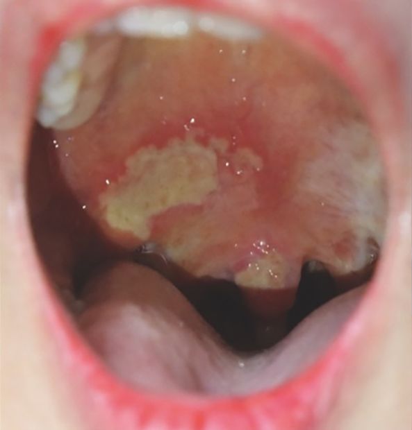
Soo Hyun Choi and Do-Young Kim

visits over the past two decades revealed that recent BD       in two parts, 1) genetic factors and 2) environmental fac-
patients experience significant less ocular involvement        tors and immunopathogenesis. It will do so while high-
and a better visual prognosis [23]. This clinical pattern of   lighting recent updates on the Korean BD patient
decreasing complete-type BD and increasing GI-type BD          population.
was similarly identified in Japanese patients [24]. This
may be due to improved hygiene practices preventing ad-        Genetics
verse microbial-immune interactions, greater public              Human leukocyte antigen (HLA)-B51 is believed to be
awareness of BD, and improved healthcare resources.            the strongest risk factor for BD. Meta-analysis on
                                                               HLA-B51/B5 in BD from 78 independent studies re-
      EPIDEMIOLOGY OF BD IN KOREA                              vealed that HLA–B51/B5 allele carriers have an increased
                                                               risk of developing BD compared to non-carriers with a
  Three epidemiological studies using the national health      pooled odds ratio (OR) of 5.78 (95% CI 5.00∼6.67) [29].
database gave recent reports regarding the estimated           Subgroup analysis of BD throughout East Asia showed
prevalence of BD in Korea. The Health Insurance Review         OR 5.18 (4.15∼6.47), suggesting HLA–B51 as a con-
& Assessment (HIRA) data from 2011∼2015 reported               sistent risk factor for development of BD across various
an estimated prevalence of Korean BD of 32.8∼35.7 per          ethnicities. This analysis fails to isolate HLA-B51 from
100,000 population [25]. Analysis from the Korean              the HLA-B5, HLA-B52 split antigen, the latter of which is
National Health Insurance Service Claims Database from         not related to BD susceptibility, and thus likely represents
2006 to 2015 reported the mean prevalence of BD in             a small underestimation of the true genetic contribution
Korea to be 26.195 patients per 100,000 population [26].       of HLA-B51 in the development of BD. A strong gene in-
  Given these estimated prevalence reports and the total       teraction was recently found between HLA-B51 and
population of Korea (51.71 million in 2019), there can be      ERAP1, a gene encoding for an aminopeptidase that is
estimated to be 13,500 to 18,500 registered BD cases           primarily responsible for final peptide trimming in the
nationwide. Despite this rough estimate, the prevalence        endoplasmic reticulum. HLA-B51 is therefore considered
of BD in Korea is not fully known, as reflected in the dis-    a key mediator of the aberrant inflammation in the
crepancies amongst various nationwide dataset analyses.        ‘MHC-I-opathy’ of BD [30,31].
Based on the increasing trend of registered BD patients in       The reported prevalence of HLA-B51 in Korean patients
the HIRA dataset, Kim et al. [25] anticipates a higher         with BD varies depending on inclusion criteria or the spe-
prevalence of BD, 36.9 (95% confidence interval [CI]           cialties of enrolling institutes. Overall, 40.8%∼55.7% of
35.0∼39.0) to 44.7 (95% CI 40.2∼49.6), between the             BD shows HLA-B51 positivity in Korea [32,33]. Similarly,
years 2016 and 2025. Conversely, Lee et al. [26] reported      a recent analysis on Japanese BD registered in the
the incidence of BD in Korea to have decreased from            Ministry of Health, Labour and Welfare of Japan showed
7.474 cases in 2006 to less than 2.6 cases per 100,000         44.5% of BD patients, diagnosed based on the ICBD cri-
population in 2015. Similarly, Jun et al. [27] reported that   teria, were HLA-B51-positive [34]. In this study,
the annual incidence of BD had decreased from 8.15 to          HLA-B51-positive patients in Japan had a higher risk for
1.51 per 100,000 population based on the HIRA dataset          ocular lesions (OR 1.59, 95% CI: 1.37∼1.84) and a lower
from 2004 to 2017. Prevalence reports should thus be           risk for genital ulcers (OR 0.72, 95% CI: 0.62∼0.84;
cautiously interpreted, given the multiple conclusions of      p<0.001) and GI symptoms (OR 0.65, 95% CI: 0.55∼
various analyses.                                              0.77). Of note, HLA-B51 positivity has been found to
                                                               have a much stronger association with non-in-
                  PATHOGENESIS                                 testinal-type BD than intestinal BD in both Korean and
                                                               Japanese patients [35], suggesting that intestinal BD may
 The exact pathogenesis of BD remains largely elusive.         have a distinctive immune-genetic profile, particularly in
However, it is generally accepted that the initial BD pre-     Far East Asia [36].
sentation is incited by environmental factors, such as in-       To date, many genome-wide association studies (GWAS)
fectious agents of pollution, acting on patients of genetic    have suggested IL-10, IL-23R-IL-12RB2, IL-1A-IL-1B,
susceptibility [28]. With this background in mind, this        CCR1, and ERAP1 as additional susceptibility genes for
review will briefly summarize the complex etiology of BD       BD [30,37,38]. Independent GWAS and linkage analysis

186                                                                              J Rheum Dis Vol. 28, No. 4, October, 2021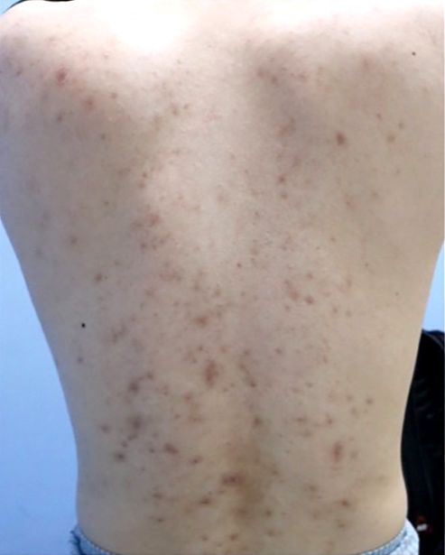
Epidemiologic and Etiological Features of Korean Patients With Behçet’s Disease

reproduced the association of BD and IL-23R-IL-12RB2                a relatively high-risk allele in the Japanese population,
in the Korean population but not between BD and IL-10               specifically within the HLA-B*51-negative population
or ERAP1 [39,40]. Notably, the GIMAP cluster, which is              (OR 4.02, 95% CI: 2.29∼7.05) [44]. Given this finding,
involved in T-cell survival, was identified as a novel sus-         the role of HLA-A in the immunopathogenesis of Korean
ceptibility loci for Korean BD, thus suggesting that aber-          BD should be further studied and elucidated. Studies on
ration of the T-cell response may contribute to the devel-          genetic predisposition of BD in Korean population is
opment of Korean BD. This association between the                   summarized in Table 2 [39,40,42,43,45-49].
GIMAP cluster and BD has not been replicated in
European populations [41], suggesting that varying ge-              Environmental factors and immune response
netic predispositions of geographically-different pop-               The environmental factors triggering BD development
ulations may induce different immunopathogenic pro-                 include microorganisms and environmental pollutants
files of BD. Interestingly, single nucleotide polymorphisms         [28]. Among infectious agents, common bacteria such as
in IL17A had a positive association with the development            Streptococcus sanguinis and viruses such as herpes sim-
of intestinal BD in the Korean population, suggesting a             plex virus (HSV) have been widely investigated. As im-
close relationship between genetically predisposing fac-            proved oral hygiene practices are correlated with an im-
tors and ethnicity-specific disease features [42]. HLA-A*26         proved disease course [50], it is evident that oral com-
has also been identified as an independent-risk HLA al-             mensals have a direct or indirect role in the pathogenesis
lele in Korean BD [43]. It has also been determined to be           of BD. Notably, streptococcal 65-kDa heat shock protein

Table 2. Summary of studies on genetic predisposition of BD in Korean population
  Author        Year                    Method                   Patient number                       Outcome
Kwon et al.     2019    Genotyping for 20 targeted             98 BD patients m.16812A>C and m.16183A>C are more
 [45]                    mitochondrial SNPs                      196 controls    frequently observed in BD patients than controls
Kang et al.     2017    Genotyping for SNPs in                  369 BD patients IL23R-IL12RB2 intergenic SNPs rs1495965,
  [39]                   IL23R-IL12RB2, IL10, STAT4,             2000 controls    rs1495966, rs4655535 significantly associated
                         and ERAP1                                                with BD risk
Kang et al.     2014    Genotyping of 24 SNPs in JAK1,         223 BD patients No association with SNPs in IL-10 mediated
 [40]                    STAT3, and TYK2                         222 controls    signaling pathways (JAK1, TYK2, STAT3)
Lee et al.      2013    GWAS using Affymetrix genome-wide      379 BD patients Association of BD to GIMAP locus on chromosome
 [46]                    human SNP array 6.0                     800 controls    7q36, as well as an association with four SNPs:
                                                                                 rs1522596 in GIMAP4, rs10266069 and
                                                                                 rs10256482 in GIMAP2, and rs2286900 in
                                                                                 GIMAP1
Kim et al.      2012     Genotyping for SNPs in IL17A, IL23R,                   TT genotype of IL17A rs8193036 and GG+GT
 [42]                      and STAT4                                             genotype of IL23R rs1884444 associated with
                                                                                 development of intestinal BD
Kang et al.     2011     Genotyping for HLA-A locus            223 BD patients HLA-A*02:07, A*26:01, and A*30.04 increased
 [43]                                                            1,398 controls risk for BD; HLA-A*33:03 decreased risk for BD
Park et al.     2009     Investigation of polymorphisms of     285 BD patients CTLA4-1661 GG and CTLA4-1722 TC genotypes
 [47]                      promoter region and exon 1 of         287 controls    significantly higher in BD patients than controls.
                           CTLA4 gene                                            CTLA4-1722 CC genotype significantly lower in
                                                                                 BD patients with ocular symptoms
Kim et al.      2006    Analysis of three polymorphisms in     99 BD patients Allele 3 and genotype allele3/allele 3 of
 [48]                     SLC11A gene (5'promoter (GT)n,         98 controls     5'-promoter (GT)n in SLC11A1 gene had
                          D543N, A318V)                                          significantly lower risk of developing BD than
                                                                                 control
Park et al.     2002    Investigation of MICA polymorphism in 108 BD patients MICA*A6, rather than HLA-B51 positivity, is more
 [49]                     relation to HLA-B51 positivity and BD 204 controls     strongly associated with Korean BD patients
                          clinical manifestations
BD: Behçet’s disease.

www.jrd.or.kr                                                                                                                187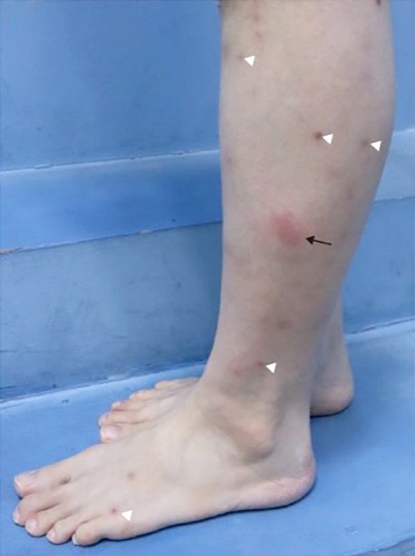
Soo Hyun Choi and Do-Young Kim

(HSP) from oral S. sanguinis has been reported to be an        Increased chemotaxis, phagocytosis and production of re-
important trigger in the pathogenesis of BD [51]. Our          active oxygen species from hyperactivated neutrophils
group demonstrated that the S. sanguinis HSP, GroEL            may cause endothelial dysfunction and the subsequent
protein, is the target of a serum anti-endothelial IgA anti-   vasculitis seen in BD [62]. Neutrophil extracellular traps
body, suggesting that molecular mimicry between bacte-         also promote thrombosis by activating macrophages
rial and host proteins may activate autoreactive lympho-       [63]. Skewed macrophage differentiation favoring the M1
cytes and lead to autoantibody production in the develop-      type was also reported in BD lesions and experimental an-
ment of BD [52].                                               imal models [64,65].
  Studies have identified HSV-1 in the active mucosal le-        Overproduction of inflammatory cytokines by innate
sions of patients with BD via polymerase chain reaction        immune cells causes a hyperactive Th1- and Th17-medi-
[53,54]. Inoculation of HSV-1 (KOS strain) in the ears of      ated immune response in BD. As discussed in the section
ICR mice successfully induced a BD-like animal model,          on genetic pathogenesis, IL23R dysregulation is asso-
which is now the most widely-tried mouse model in              ciated with BD development. The disease-protective var-
translational studies [55,56]. Notably, HSV-inoculated         iant, IL23R R381Q, has been found to be associated with
mice showed higher incidence of BD-related symptoms            reduced IL-23-dependent IL-17 production [66]. Additionally,
under conventional conditions than those under specific        peripheral blood monocytes in BD patients tend to facili-
pathogen free conditions (15.0% vs. 2.2%), suggesting          tate T-cell differentiation into Th1 and Th17 [67]. Thus,
that environmental stressors and a subsequently altered        both genetic predisposition and the corresponding innate
microbiome play pivotal roles in provoking the patho-          immune response are responsible for the Th1/Th17-
genic inflammation induced by the anti-HSV immune re-          skew in the T-cell activity seen in BD.
sponse seen in BD [57]. Environmental pollutants are             In short, environmental factors may trigger an innate
thought be significant contributors in the development of      immune reaction in patients with BD that exhibits an
BD [28,58]. This is with the exception of smoking nic-         aberrant Th1/Th17 response, subsequently leading to in-
otine which, based on recent epidemiological analysis us-      flammation and disease progression.
ing a nationwide database, may be a potential protective
factor [59].                                                                      CONCLUSION
  Pathogenic BD phenotypes are the consequence of an
aberrant immune response induced by both genetic pre-           The pathogenesis and clinical presentations of BD in
disposition and environmental factors. Immune re-              Korea is an ever-evolving area of understanding. Careful
sponses are largely classified into innate and adaptive        attention to the multifactorial genetic, immunologic, and
responses.                                                     environmental factors at play allows clinicians to make
  A few BD-risk alleles found in the Korean population,        more precise BD management and screening recom-
such as KLRC4 [30] and MICA [49], support the role of          mendations, as well as informs future areas of study.
natural killer (NK) cells in the development of BD [60].       Analysis reports on nationwide health datasets have been
NK cells, innate cytotoxic cells, regulate the function of     variable in regard to BD prevalence trends, and there
other immune cells, including dendritic cells and T-cells.     would be value in systematically collecting and analyzing
KLRC4 may act as a receptor for the identification of          trends of BD in Korea over time. Further studies identify-
MHC class I molecules. Although the exact role of NK           ing risk factors and pathogenetic mechanisms specific to
cells in BD remain unclear, an alteration in a subset of NK    Korean BD would help refine management in the large
cells may lead to the imbalance in the Th1 response and        number of BD patients in Korea.
immunoregulatory role of NK cells [61].
  BD patients exhibit high intrinsic activation of neu-                    CONFLICT OF INTEREST
trophils, which are involved in the vasculitic infiltration
of BD lesions. High levels of proinflammatory cytokines         No potential conflict of interest relevant to this article
and chemokines, such as IL-8, tumor necrosis factor-al-        was reported.
pha, interferons, granulocyte macrophage colony-stim-
ulating factor (GM-CSF)/G-CSF, and CXCL-8 are closely
related with the activation of neutrophils in BD [60].

188                                                                              J Rheum Dis Vol. 28, No. 4, October, 2021

Epidemiologic and Etiological Features of Korean Patients With Behçet’s Disease

          AUTHOR CONTRIBUTIONS                                            from 2002 to 2013. Ocul Immunol Inflamm 2020 Apr 15
                                                                          [Epub]. DOI:10.1080/09273948.2020.1746352.
                                                                    17.   Lee NH, Bae M, Jin M, Chung SW, Lee CW, Jeon CH.
 D.Y.K. conceived and planned the outline of review.                      Characterization of venous involvement in vasculo-Behçet
S.H.C. and D.Y.K. reviewed literatures and wrote the                      disease. Korean J Thorac Cardiovasc Surg 2020;53:381-6.
manuscript.                                                         18.   Kim SW, Kim TG, Oh J, Kim DY, Choi YC, Kim SM, et al.
                                                                          Clinical and radiographic characteristics of neuro-Behçet’s
                                                                          disease in South Korea. J Clin Neurol 2019;15:429-37.
                      REFERENCES                                    19.   Yoon DL, Kim YJ, Koo BS, Kim YG, Lee CK, Yoo B.
                                                                          Neuro-behçet’s disease in South Korea: clinical character-
 1. Joo CR. Two cases of Behçet’s syndrome. J Cathol Med Coll             istics and treatment response. Int J Rheum Dis 2014;17:
    1961;5:393-400.                                                       453-8.
 2. Kim D. Behcet’s syndrome: a report of four cases including      20.   Cheon JH, Kim ES, Shin SJ, Kim TI, Lee KM, Kim SW, et al.
    one with extreme systemic manifestations. J Korean Med                Development and validation of novel diagnostic criteria for
    Assoc 1962;5:52-58.                                                   intestinal Behçet’s disease in Korean patients with ileoco-
 3. Bang D, Lee ES, Lee S. Behçet’s disease. Paper presented at:          lonic ulcers. Am J Gastroenterol 2009;104:2492-9.
    22nd World Congress of Dermatology; 2011 May 24-29;             21.   Jung YS, Cheon JH, Park SJ, Hong SP, Kim TI, Kim WH.
    Seoul, Korea.                                                         Clinical course of intestinal Behcet’s disease during the first
 4. Bang D, Lee ES, Sohn S, Kim DY, Cho S, Choi MJ. Behçet’s              five years. Dig Dis Sci 2013;58:496-503.
    disease in Korea. Seoul, Hanuri Publishing Co., 2013.           22.   Jung YS, Yoon JY, Lee JH, Jeon SM, Hong SP, Kim TI, et al.
 5. International Society for Behçet’s Disease. 8-9th Interna-            Prognostic factors and long-term clinical outcomes for sur-
    tional Conference on Behçet’s Disease; 1998 Oct 7-9, 2000             gical patients with intestinal Behcet’s disease. Inflamm
    May 27-29; Emilia, Italy, Seoul, Korea. Seoul: Design Mecca           Bowel Dis 2011;17:1594-602.
    Publishing Co.; 2000.                                           23.   Chung YR, Lee ES, Kim MH, Lew HM, Song JH. Changes in
 6. Criteria for diagnosis of Behçet’s disease. International             ocular manifestations of Behçet disease in Korean patients
    Study Group for Behçet’s Disease. Lancet 1990;335:                    over time: a single-center experience in the 1990s and
    1078-80.                                                              2000s. Ocul Immunol Inflamm 2015;23:157-61.
 7. International Team for the Revision of the International        24.   Kirino Y, Ideguchi H, Takeno M, Suda A, Higashitani K,
    Criteria for Behçet’s Disease (ITR-ICBD). The International           Kunishita Y, et al. Continuous evolution of clinical pheno-
    Criteria for Behçet’s Disease (ICBD): a collaborative study           type in 578 Japanese patients with Behçet’s disease: a retro-
    of 27 countries on the sensitivity and specificity of the new         spective observational study. Arthritis Res Ther 2016;18:
    criteria. J Eur Acad Dermatol Venereol 2014;28:338-47.                217.
 8. Kim DY, Choi MJ, Cho S, Kim DW, Bang D. Changing clin-          25.   Kim JN, Kwak SG, Choe JY, Kim SK. The prevalence of
    ical expression of Behçet disease in Korea during three deca-         Behçet’s disease in Korea: data from Health Insurance
    des (1983-2012): chronological analysis of 3674 hospi-                Review and Assessment Service from 2011 to 2015. Clin
    tal-based patients. Br J Dermatol 2014;170:458-61.                    Exp Rheumatol 2017;35 Suppl 108(6):38-42.
 9. Bang D, Lee JH, Lee ES, Lee S, Choi JS, Kim YK, et al.          26.   Lee YB, Lee SY, Choi JY, Lee JH, Chae HS, Kim JW, et al.
    Epidemiologic and clinical survey of Behcet’s disease in              Incidence, prevalence, and mortality of Adamantiades-
    Korea: the first multicenter study. J Korean Med Sci 2001;            Behçet’s disease in Korea: a nationwide, population-based
    16:615-8.                                                             study (2006-2015). J Eur Acad Dermatol Venereol 2018;
10. Davatchi F, Sadeghi Abdollahi B, Chams-Davatchi C,                    32:999-1003.
    Shahram F, Shams H, Nadji A, et al. The saga of diagnostic/     27.   Jun JB, Kim HJ, Kazmi SZ, Kang T, Kim KB, Kang MJ, et al.
    classification criteria in Behcet’s disease. Int J Rheum Dis          Significant decline in the incidence of Behcet’s disease in
    2015;18:594-605.                                                      South Korea: a nationwide population-based study (2004-
11. Mizushima Y. [Revised diagnostic criteria for Behçet’s dis-           2017). Arthritis Care Res (Hoboken) 2020 Aug 8 [Epub].
    ease in 1987]. Ryumachi 1988;28:66-70. Japanese.                      DOI:10.1002/acr.24408.
12. Chang HK, Lee SS, Bai HJ, Lee YW, Yoon BY, Lee CH, et al.       28.   Cho SB, Cho S, Bang D. New insights in the clinical under-
    Validation of the classification criteria commonly used in            standing of Behçet’s disease. Yonsei Med J 2012;53:35-42.
    Korea and a modified set of preliminary criteria for Behçet’s   29.   de Menthon M, Lavalley MP, Maldini C, Guillevin L, Mahr A.
    disease: a multi-center study. Clin Exp Rheumatol 2004;22             HLA-B51/B5 and the risk of Behçet’s disease: a systematic
    (4 Suppl 34):S21-6.                                                   review and meta-analysis of case-control genetic association
13. Chang HK, Kim SY. Survey and validation of the criteria for           studies. Arthritis Rheum 2009;61:1287-96.
    Behcet’s disease recently used in Korea: a suggestion for       30.   Kirino Y, Bertsias G, Ishigatsubo Y, Mizuki N, Tugal-Tutkun
    modification of the International Study Group criteria. J             I, Seyahi E, et al. Genome-wide association analysis identi-
    Korean Med Sci 2003;18:88-92.                                         fies new susceptibility loci for Behçet’s disease and epistasis
14. Kirino Y, Nakajima H. Clinical and genetic aspects of                 between HLA-B*51 and ERAP1. Nat Genet 2013;45:202-7.
    Behçet’s disease in Japan. Intern Med 2019;58:1199-207.         31.   McGonagle D, Aydin SZ, Gül A, Mahr A, Direskeneli H.
15. Lee ES, Bang D, Lee S. Dermatologic manifestation of                  ‘MHC-I-opathy’-unified concept for spondyloarthritis and
    Behçet’s disease. Yonsei Med J 1997;38:380-9.                         Behçet disease. Nat Rev Rheumatol 2015;11:731-40.
16. Oh BL, Lee JS, Lee EY, Lee HY, Yu HG. Incidence and risk        32.   Ryu HJ, Seo MR, Choi HJ, Baek HJ. Clinical phenotypes of
    factors for blindness in uveitis: a nationwide cohort study           Korean patients with Behcet disease according to gender,

www.jrd.or.kr                                                                                                                      189
Soo Hyun Choi and Do-Young Kim
age at onset, and HLA-B51. Korean J Intern Med 2018; SLC11A1 gene polymorphisms in Korean patients with
33:1025-31. Behçet’s disease. Scand J Rheumatol 2006;35:398-401.
33. Chang HK, Kim JU, Cheon KS, Chung HR, Lee KW, Lee IH. 49. Park SH, Park KS, Seo YI, Min DJ, Kim WU, Kim TG, et al.
HLA-B51 and its allelic types in association with Behçet’s Association of MICA polymorphism with HLA-B51 and dis-
disease and recurrent aphthous stomatitis in Korea. Clin ease severity in Korean patients with Behcet’s disease. J
Exp Rheumatol 2001;19(5 Suppl 24):S31-5. Korean Med Sci 2002;17:366-70.
34. Mizuki Y, Horita N, Horie Y, Takeuchi M, Ishido T, Mizuki 50. Karacayli U, Mumcu G, Simsek I, Pay S, Kose O, Erdem H,
R, et al. The influence of HLA-B51 on clinical manifestations et al. The close association between dental and periodontal
among Japanese patients with Behçet’s disease: a nation- treatments and oral ulcer course in behcet’s disease: a pro-
wide survey. Mod Rheumatol 2020;30:708-14. spective clinical study. J Oral Pathol Med 2009;38:410-5.
35. Han M, Jung YS, Kim WH, Cheon JH, Park S. Incidence and 51. Kaneko F, Oyama N, Yanagihori H, Isogai E, Yokota K,
clinical outcomes of intestinal Behçet’s disease in Korea, Oguma K. The role of streptococcal hypersensitivity in the
2011-2014: a nationwide population-based study. J Gastro- pathogenesis of Behçet’s disease. Eur J Dermatol 2008;18:
enterol 2017;52:920-8. 489-98.
36. Kim SW, Jung YS, Ahn JB, Shin ES, Jang HW, Lee HJ, et al. 52. Cho SB, Zheng Z, Ahn KJ, Choi MJ, Cho S, Kim DY, et al.
Identification of genetic susceptibility loci for intestinal Serum IgA reactivity against GroEL of Streptococcus san-
Behçet’s disease. Sci Rep 2017;7:39850. guinis and human heterogeneous nuclear ribonucleopro-
37. Remmers EF, Cosan F, Kirino Y, Ombrello MJ, Abaci N, tein A2/B1 in patients with Behçet disease. Br J Dermatol
Satorius C, al. Genome-wide association study identifies 2013;168:977-83.
variants in the MHC class I, IL10, and IL23R-IL12RB2 re- 53. Kim DY, Cho S, Choi MJ, Sohn S, Lee ES, Bang D.
gions associated with Behçet’s disease. Nat Genet 2010;42: Immunopathogenic role of herpes simplex virus in Behçet’s
698-702. disease. Genet Res Int 2013;2013:638273.
38. Mizuki N, Meguro A, Ota M, Ohno S, Shiota T, Kawagoe T, 54. Lee S, Bang D, Cho YH, Lee ES, Sohn S. Polymerase chain re-
et al. Genome-wide association studies identify IL23R- action reveals herpes simplex virus DNA in saliva of pa-
IL12RB2 and IL10 as Behçet’s disease susceptibility loci. tients with Behçet’s disease. Arch Dermatol Res 1996;288:
Nat Genet 2010;42:703-6. 179-83.
39. Kang EH, Kim S, Park MY, Choi JY, Choi IA, Kim MJ, et al. 55. Sohn S, Lee ES, Bang D. Learning from HSV-infected mice
Behçet’s disease risk association fine-mapped on the IL23R- as a model of Behçet’s disease. Clin Exp Rheumatol 2012;
IL12RB2 intergenic region in Koreans. Arthritis Res Ther 30(3 Suppl 72):S96-103.
2017;19:227. 56. Sohn S, Lee ES, Bang D, Lee S. Behçet’s disease-like symp-
40. Kang EH, Choi JY, Lee YJ, Lee EY, Lee EB, Song YW. Single toms induced by the Herpes simplex virus in ICR mice. Eur
nucleotide polymorphisms in IL-10-mediated signalling J Dermatol 1998;8:21-3.
pathways in Korean patients with Behçet’s disease. Clin Exp 57. Islam SMS, Ryu HM, Sayeed HM, Sohn S. Interrelationship
Rheumatol 2014;32(4 Suppl 84):S27-32. of stress, environment, and herpes simplex virus type-1 on
41. Ortiz-Fernández L, Conde-Jaldón M, García-Lozano JR, Behçet’s disease: using a mouse model. Front Immunol
Montes-Cano MA, Ortego-Centeno N, Castillo-Palma MJ, 2021;12:607768.
et al. GIMAP and Behçet disease: no association in the 58. Bai YC, Wang CY, Lin CL, Lai JN, Wei JC. Association be-
European population. Ann Rheum Dis 2014;73:1433-4. tween air pollution and the risk of uveitis: a nationwide,
42. Kim ES, Kim SW, Moon CM, Park JJ, Kim TI, Kim WH, et al. population-based cohort study. Front Immunol 2021;12:
Interactions between IL17A, IL23R, and STAT4 poly- 613893.
morphisms confer susceptibility to intestinal Behcet’s dis- 59. Lee YB, Lee JH, Lee SY, Lee JH, Yu DS, Han KD, et al.
ease in Korean population. Life Sci 2012;90:740-6. Association between smoking and Behçet’s disease: a na-
43. Kang EH, Kim JY, Takeuchi F, Kim JW, Shin K, Lee EY, et al. tionwide population-based study in Korea. J Eur Acad
Associations between the HLA-A polymorphism and the Dermatol Venereol 2019;33:2114-22.
clinical manifestations of Behcet’s disease. Arthritis Res 60. Tong B, Liu X, Xiao J, Su G. Immunopathogenesis of
Ther 2011;13:R49. Behcet’s disease. Front Immunol 2019;10:665.
44. Nakamura J, Meguro A, Ishii G, Mihara T, Takeuchi M, 61. Yamaguchi Y, Takahashi H, Satoh T, Okazaki Y, Mizuki N,
Mizuki Y, et al. The association analysis between HLA-A*26 Takahashi K, et al. Natural killer cells control a T-helper 1
and Behçet’s disease. Sci Rep 2019;9:4426. response in patients with Behçet’s disease. Arthritis Res
45. Kwon M, Yoo SJ, Yoo IS, Kim J, Kang SW, Choi IA, et al. Ther 2010;12:R80.
Associations of mitochondrial deoxyribonucleic acid poly- 62. Emmi G, Becatti M, Bettiol A, Hatemi G, Prisco D, Fiorillo
morphisms with Behçet’s disease in the Korean population. C. Behçet’s syndrome as a model of thrombo-inflammation:
Arch Rheumatol 2019;34:211-9 the role of neutrophils. Front Immunol 2019;10:1085.
46. Lee YJ, Horie Y, Wallace GR, Choi YS, Park JA, Choi JY, et 63. Li L, Yu X, Liu J, Wang Z, Li C, Shi J, et al. Neutrophil ex-
al. Genome-wide association study identifies GIMAP as a tracellular traps promote aberrant macrophages activation
novel susceptibility locus for Behcet’s disease. Ann Rheum in Behçet’s disease. Front Immunol 2021;11:590622.
Dis 2013;72:1510-6. 64. Nakano H, Kirino Y, Takeno M, Higashitani K, Nagai H,
47. Park KS, Baek JA, Do JE, Bang D, Lee ES. CTLA4 gene poly- Yoshimi R, et al. GWAS-identified CCR1 and IL10 loci con-
morphisms and soluble CTLA4 protein in Behcet’s disease. tribute to M1 macrophage-predominant inflammation in
Tissue Antigens 2009;74:222-7. Behçet’s disease. Arthritis Res Ther 2018;20:124.
48. Kim SK, Jang WC, Park SB, Park DY, Bang KT, Lee SS, et al. 65. Islam SMS, Sohn S. HSV-induced systemic inflammation as
190 J Rheum Dis Vol. 28, No. 4, October, 2021Epidemiologic and Etiological Features of Korean Patients With Behçet’s Disease
an animal model for Behçet’s disease and therapeutic ceptor 4 gene TLR4 in Behçet disease. Proc Natl Acad Sci U
applications. Viruses 2018;10:511. S A 2013;110:8134-9.
66. Kirino Y, Zhou Q, Ishigatsubo Y, Mizuki N, Tugal-Tutkun I, 67. Ahn Y, Hwang JH, Zheng Z, Bang D, Kim DY. Enhancement
Seyahi E, et al. Targeted resequencing implicates the fami- of Th1/Th17 inflammation by TRIM21 in Behçet’s disease.
lial Mediterranean fever gene MEFV and the toll-like re- Sci Rep 2017;7:3018.
www.jrd.or.kr 191You can also read