Emergence of Mucormycosis: A Therapeutic Challenge for COVID-19 in Pakistan - Scholars Middle East Publishers
←
→
Page content transcription
If your browser does not render page correctly, please read the page content below
Saudi Journal of Pathology and Microbiology
                                                                                                 Abbreviated Key Title: Saudi J Pathol Microbiol
                                                                                            ISSN 2518-3362 (Print) |ISSN 2518-3370 (Online)
                                                                                  Scholars Middle East Publishers, Dubai, United Arab Emirates
                                                                                                 Journal homepage: https://saudijournals.com

                                                                                                                          Review Article

Emergence of Mucormycosis: A Therapeutic Challenge for COVID-19
in Pakistan
Rabia Kanwar1*, Tariq Munir1, Hafiz Khurram Shurjeel3, Aman Ullah2, Muhammad Danish4, Saad Zafar1, Awais
Aleem1, Muhammad Basit Husnain Haider1, Sajida Mustafa1
1
  Institute of Microbiology, University of Agriculture, Faisalabad 38000, Pakistan
2
  Faculty of Life science and Technology, Kunming university of Science and Technology, Kunming 650500 China
3
  University of Sargodha, University Road, Sargodha, Punjab, Pakistan
4
  Services Institute of Medical Sciences (SIMS), Jail Rd, Shadman, Lahore Punjab, Pakistan

DOI: 10.36348/sjpm.2021.v06i10.007                                | Received: 29.08.2021 | Accepted: 04.10.2021 | Published: 18.10.2021
*Corresponding author: Rabia Kanwar

Abstract
Black fungus is opportunistic pathogens that may cause life-threating infection inimmunocompromised patients. The
mucormycosis associated with COVID-19 is now become a serious health concern around the globe, including several
Asian countries. In Pakistan mucormycosis fatalities are now being found among COVID-19 individuals. Individuals
with diabetes, malnourishment, Cancer, organ transplantation, active tuberculosis, Liver diseases, chronic respiratory
diseases, HIV, AIDS and asthma are more Susceptible to infection. Diabetes mellitus patients are at more risk of
mortality infection of this fungus. To counteract mucromycosis in patients, rapid and precise diagnostic facilities, medical
assistance, and a quick yet coordinated approach are all suggested.
Keywords: Black fungus, Immunocomprised patients, COVID-19.
Copyright © 2021 The Author(s): This is an open-access article distributed under the terms of the Creative Commons Attribution 4.0 International
License (CC BY-NC 4.0) which permits unrestricted use, distribution, and reproduction in any medium for non-commercial use provided the original
author and source are credited.

                                                                             infections. The phylum Glomeromycota includes
INTRODUCTION                                                                 opportunistic fungus such as cutaneous mucormycosis,
           Meanwhile, as the world tries to deal with the                    which affects those with weakened immune systems
devastation brought on by COVID-19, a new threat had                         [5]. More than 90% of people with COVID-19
also emerged: the "black fungus." There are 1,063,125                        associated mucromycosis (CAM) have diabetes,
confirmed COVID-19 cases in Pakistan, with 23,797                            according to a research conducted worldwide (6).
fatalities, making it one of the severely affected                           Immunocompromised persons, such as COVID-19
countries by the pandemic [1]. Global epidemiological                        patients, diabetics, people taking steroids, and those
surveillance showed that mucormycosis infections are                         with cancer and organ transplants, are more susceptible
on the rise, especially high prevalence in Asian region                      to infection (7). COVID-19 has been reported in several
[2]. Even before the COVID-19 pandemic, Pakistan                             studies to link with significant mortality in diabetic
have reported      high    incidence of    mucormycosis                      ketoacidosis (DKA) patients, and these research
infections approximately 14/100,000, despite the lack                        indicated that DKA is a frequent and serious
of sufficient information to determine the true burden of                    complication for patients with COVID-19 [8].
fungus illnesses in Pakistan [3]. Some mucormycosis
fatalities are now being found among COVID-19                                         Not only can the virus damage the immune
individuals at various hospitals in Pakistan, which is in                    system of COVID-19 patients, but the treatment
line with the trend in Pakistan's neighboring nation,                        regimen utilized for severe cases (e.g. steroids) can also
India [4].                                                                   decrease their immunological reaction. COVID-19
                                                                             patients hospitalized to critical care units getting
          Mucormycosis or Black fungus is an emerging                        oxygen therapy may come into contact with humidifiers
and fatal fungal infection lead to severe infection in                       in the ward, increasing their moisture exposure and so
immunocomprised patients mostly related to diabetes                          rendering them more vulnerable to a fungal infection.
mellitus. According to infectious site mucormycosis can                      According to the International Diabetes Federation's
be divided into six forms: pulmonary, rhino orbital                          figures, 8.9% of Indian adults are diabetic, totaling
cerebral, gastrointestinal, cutaneous and invasive                           roughly 77 million people [9]. This review emphasize
Citation: Rabia Kanwar et al (2021). Emergence of Mucormycosis: A Therapeutic Challenge for COVID-19 in Pakistan. Saudi                     363
J Pathol Microbiol, 6(10): 363-368.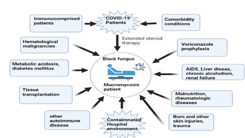
Rabia Kanwar et al; Saudi J Pathol Microbiol, Oct, 2021; 6(10): 363-368
the origin and occurrence of mucormycosis, its                               present incutaneous type.The most typical infection
associated illnesses, its course in immunocompromised                        locations includes sinuses (39%), disseminated (23%);
and COVID-19 afflicted patients, and the multiple risk                       lungs (24%), and skin (19%) [18]. Initially the fungus
factors and their impact on multiple organs, as well as                      invade the blood vessel causing thrombosis and tissue
the difficulties in overcoming this infection. As with                       infarction. Angioinvasion occurs when fungal spores
the COVID-19 outbreak and increased pressure on                              come      into    touch     with     endothelial    cells.
healthcare system, this study will also provide a broad                      Moreinteractions with these cells' receptors leads to cell
data foundation for optimum treatment results and                            injury and proliferation of Fungus [19]. Fungi are often
prevention of this fungal infection.                                         eliminated through polymorphonuclear phagocytes in
                                                                             healthy individuals. As a result, fungal growth is
Mucormycosis                                                                 common in people who have defects in this process.
         Mucormycosis or zygomycosis is a deadly but                         Furthermore, Mucorales can be resistant to these
occasional fungal disease caused by a mould’s family                         processes, leaving them highly virulent [20].
known as mucormycetes. Mucormycosis, often known
as "deadly black fungus" is a potentially fatal                              Mucormycosis-associated Risk factor
infection from the subphylum Mucoromycotina, order                                    Mucormycosis has been linked to a number of
Mucorales [10, 11]. After Aspergillus, Mucorales are                         underlying factors that predispose a person to
the most prevalent fungus discovered in haematological                       infection. COVID-19, diabetes, organ and stem cell
malignancies, solid organ transplantation and stem cell                      transplantation, haematological diseases, trauma,
transplantation [12]. Mucormycosis is caused by 11                           burns, metabolic acidosis, steroidal usage, broad-
genus and 27 species of Mucorales [13]. Rhizopus is                          spectrum antibiotics, malnutrition, Cancer, voriconazole
the most prevalent genus that causes mucormycosis,                           use are some of these variables [21]. Figure 1 shows
after Mucor and Lichtheimia. Mucorales may be found                          the association between COVID-19 and black
in a variety of environments, including soil, decayed                        fungus and several risk factors for onset of
food, dust and Manure [14].                                                  mucormycosis. Among the various types of
                                                                             mucormycosis infection, ROCM was linked to the
         Mucormycosis was firstly documented in                              existence of diabetes and cutaneou infections was more
1855, when the first true human instance of the disease                      common in trauma patients, and in organ
was discovered. Furbringer discovered pulmonary                              transplantation , pulmonary,        intestinal,      and
mucormycosis in 1876 in a cancer patient with a                              disseminated         types       commonly         found.
hemorrhagic infarct in right lung which was filled with                      Furthermore, haematological malignancies appeared in
fungus hyphae and spores in Germany. Mucormycosis                            the disseminating form [22]. Mucormycosis is more
was discovered in an autopsy in 1956. Mucormycosis is                        common in people with diabetic ketoacidosis because
mostly transmitted by inhalation of fungal spores, the                       of innate immunity in such people causes
ingestion of contaminated food items and the infusion                        polymorphonuclear phagocytes to kill the fungus. The
of fungus directly into skin abrasions or wounds [15].                       sinuses were the most afflicted area in diabetic
                                                                             individuals, following pulmonary areas [23].
          Furthermore, outbreaks of mucormycosis
might       be      nosocomial related      to       the                              During the neutropenia stage of the illness,
contaminating ventilation      systems,        medical                       those with haematological malignancies were prone to
devices, hospital discards such as hospital clothes                          mucormycosis. Mucormycosis in haematological
(linens), bandages and       so         on         (16).                     malignancies is caused by chemotherapy and use of
Immunocompromised people are at most risk to be                              voriconazole, which is used in aspergillosis treatment
effected by mucormycosisas their immune systems are                          [24]. Mucormycosisfound more prevalent among
unable to combat the fungus. It is mostly identified by                      peoplehaving acute leukaemia instead in those with
diagnostic examination of a biopsy taken from the                            someother kinds of cancer. The major modalities for
infected site (17). According to infectious site                             prevention of mucormycosisamong individuals having
mucormycosis can be divided into six forms:                                  haematological malignancies were avoiding exposures
pulmonary, rhino-orbital cerebral, gastrointestinal,                         to environment, while therapeutic options included
cutaneous and invasive infections. The phylum                                surgery, antifungal therapy, and neutropenia reversal.
Glomeromycota includes opportunistic fungus such as                          Soil as well as decaying organic matter such as leaves,
cutaneous mucormycosis, which affects those with                             rotten wood, compost piles, and animal feces are among
weakened immune systems. Among mucormycosis                                  the places where mucorales may be found. Because of
causing species.                                                             the wide range of locations in which mucorales may be
                                                                             found, people are constantly being exposed to
        Rhizopus     cause    ROCM.      Meanwhile,                          them. Basic hygiene measures can help protect against
Cunninghamellafound in two forms either pulmonary or                         mucormycosis [25].
disseminating and Saksenaea andApophysomyces were

© 2021 |Published by Scholars Middle East Publishers, Dubai, United Arab Emirates                                                   364
Rabia Kanwar et al; Saudi J Pathol Microbiol, Oct, 2021; 6(10): 363-368
Fig-1: Association between COVID-19 and black fungus in contrast to several risk factors for onset of mucormycosis.
Several risk factors that lead to the development of mucormycosis, includes comorbidity conditions, diabetes mellitus, and
haematological malignancies HIV and other diseases, immunocompromised persons, voriconazole. Furthermore,
immunosuppressive therapies such as stem cell therapy and organ transplantation render people more prone to the disease.
Exposure of spores due to unhygienic Practices leads to mucromycosis.
Transplantation treatments have also been therapeutic settings, circumstances are created to avoid
identified as one of the mucormycosis risk factors. the formation of mucormycosis.
However, the disease's occurrence varies depending on
the type of organs donated. Because patients of Mucormycosis and COVID-19: a complicated
transplantation treatments mostly given immune- relationship
suppressants and large dosage of steroids, they are most COVID-19 has brought a slew of new diseases
susceptible to the fungal infection. Furthermore, and difficulties around the globe [29].COVID-19
corticosteroids decrease macrophages and neutrophils, symptoms include an increase in body temperature,
impairing the body's capacity to fight infection [26]. hypoxia, osmolarity and shortness of breath [30].
Individuals who use steroids are likewise considered Recently, COVID-19 healed individuals have been
at high-risk. Patients undergoing stem cell treatment are troubled with a very new illness known as
also given voriconazole, which, when administered Mucormycosis disease. Mucormycosismay rapidly
prophylactically, reduces the incidence of spread and invade the sinuses and lungs before moving
mucormycosis [27]. on to the intra-orbital and cerebral spaces parts of the
body. Complication of mucormycosis in COVID-19
Deferoxamine treatment and iron infected person have been demonstrated in figure 2.The
overload which is used for the treatment of people primary signs of COVID-19 offer an ideal setting
having diabetic ketoacidosis, renal failure with haemo- for fungus to grow and flourish within the human body.
dialysis are at double risk of getting mucormycosis. Diabetics, people on systemic corticosteroids, patients
However, deferoxamine treatment increases the risk of of neutropenia, stem cell transplant, hematologic
mucormycosis in individuals. The iron eliminated by malignancies, and immune-compromised persons are
the medication is utilised by the fungus to proliferate, all prone to mucormycosis [31]. Diabetes may enhance
creating a suitable environment for their development. COVID-19 associated morbidity and mortality by the
Mucormycosis is not only seen in people with chronic following mechanisms: i) decreased viral clearance,
illnesses; it may also be seen in those who have had ii) reduction in T-cell activity, iii) increased cytokine
surgery, most likely after utilizing contaminated goods storm iv) immuno-suppression [32]. In COVID-19
(28). Because invasive mucormycosisbecomes more patients, hyperglycemia exacerbates the cytokine storm
common in clinical settings, it is very critical to sustain via disrupting endothelial cells, resulting in multi-organ
a sterile, patient-safe atmosphere. Furthermore, caution destruction. The acidic environment and increased
should be exercised when aiding chronic patients. In amounts of free ferric ions promote the development of
© 2021 |Published by Scholars Middle East Publishers, Dubai, United Arab Emirates 365Rabia Kanwar et al; Saudi J Pathol Microbiol, Oct, 2021; 6(10): 363-368
mucorales in diabetic ketoacidosis. These conditions COVID-19 are typically treated with high doses of
favor the invasion and effective attachment steroids, as well as extensive use of oxygen masks with
of fungal hyphae inside the body. Persons with chronic ventilators, which cause them more vulnerable to
diabetes who have foot ulcers are at risk for this mucormycosis. Steroids lower both inflammatory
infection because any damaged skin tissue is an response and immune response , where the synthesis of
accessible entrance point for this fungus. Furthermore, white blood cells (WBCs) as well as T-helper cells is
COVID-19 therapy is still in its early stages [33]. To reduced, allowing any foreign material to infiltrate and
counteract the effects of SARS-CoV-2 infection, totally destroy the immune system inside the host cell.
patients are given high doses of steroids which Furthermore, these hormones may cause an unregulated
decreases inflammation of the lungs and may help limit release of sugar, allowing themucorales to proliferate,
the damage done to the body by the cytokine storm. reproduce, and invade at a rapid speed [34].
However, patients infected with this novel strain of
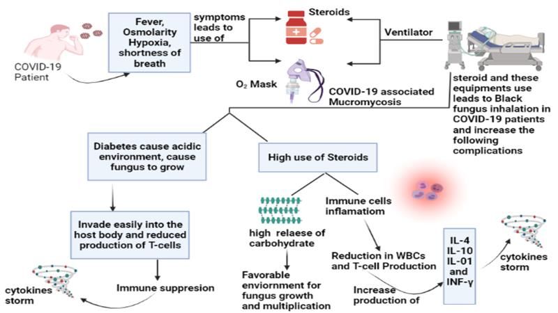
Fig-2
Figure 2: COVID-19 associated hypertension was positive for COVID-19 have
Mucormycosis (CAM): Because of dysregulation mucormycosis co-infection. He was treated with
of immune system, a COVID-19-infected person is amphotericin B for fungal infection and broad-spectrum
more susceptible to mucormycosis and may be given antibiotics for bacterial co-infections [36].
immune suppressant medications that inhibit the body's
phagocytic cells from fighting the black fungus at an Severe COVID-19 infection also raise levels
ideal level. COVID-19 additionally raises the level of of pro-inflammatory cytokines such as IL-1, IL-6, and
iron in the blood, which the fungus requires to grow and TNF-α, while lowering the levels of CD4 INF-γ, CD4
multiply, making the patient more susceptible to and CD8 cells. This increases the risk of co-infection,
infection. Persons infected with COVID-19 are including mucormycosis. The use of low-quality
frequently given oxygen treatment. Contamination in all oxygen cylinders, a polluted and humid hospital
these devices can act as a source of infection for environment, tap water in humidifiers, and an overuse
mucormycosis. COVID-19 patients are at increased risk of antibiotics can all contribute to the spread
for this disease due to the steroid treatment they get. of mucormycosis infection [37]. According to a study
in India, systemic corticosteroids were used to treat
COVID-19 associated Mucormycosis in Pakistan uncontrolled diabetes patients who also had rhino-
An observational study have been conducted orbital mucormycosis and were positive for COVID-19
in a Hospital in Karachi, Pakistan on PCR confirmed [38]. An observational research conducted in Pakistan
COVID-19 cases of adult patients in July 2020 to May, found a 15.6% fungus infection rate in patients with
2021. Mucormycosis was identified by a combination proven COVID-19 who needed ICU hospitalization
of clinical, microbiologic, radiographic and histological [39]. Pakistan will face a significant challenge in
examinations [35]. An old patient with a combination of prioritizing mucormycosis surveillance, prognosis, and
diseases including diabetes, heart disease and management along with a rigorous COVID-19
© 2021 |Published by Scholars Middle East Publishers, Dubai, United Arab Emirates 366Rabia Kanwar et al; Saudi J Pathol Microbiol, Oct, 2021; 6(10): 363-368
infection. Combating cutaneous mucormycosis in mucormycosis in India and Pakistan: a serious
underdeveloped nations is complicated by inadequate cause for concern during the ongoing COVID-19
laboratory facilities and a lack of competence, making it pandemic. The American Journal of Tropical
difficult to keep reliable statistics of disease incidence Medicine and Hygiene, 1(aop).
[40]. 8. Goldman, N., Fink, D., Cai, J., Lee, Y. N., &
Davies, Z. (2020). High prevalence of COVID-19-
CONCLUSION associated diabetic ketoacidosis in UK secondary
In conclusion, COVID-19 care. Diabetes research and clinical practice, 166,
potentially associated with an increased risk of 108291.
subsequent bacterial and fungal infections, due to the 9. Sachdev, S. S., Chettiankandy, T. J., Gaikwad, R.,
result of immunologic dysregulation. In addition, the Suryawanshi, S., & Yaduwanshi, K. (2021).
unregulated use of steroids, broad-spectrum COVID-19 associated Mucormycosis: The call for
antibiotics and monoclonal antibodies, as part of the dental practitioners. Clinical Dentistry (0974-
COVID-19 treatment might lead to fungal illnesses or 3979), 15(6).
aggravate pre-existing fungal diseases. The increasing 10. Chegini, Z., Didehdar, M., Khoshbayan, A.,
threat of deadly re-emerging infectious illnesses like Rajaeih, S., Salehi, M., & Shariati, A. (2020).
mucormycosis, significantly risk public health security. Epidemiology, clinical features, diagnosis and
An increase in mucormycosis infections has prompted a treatment of cerebral mucormycosis in diabetic
surge in demand for the antifungal medication like patients: a systematic review of case reports and
amphotericin B, the only recommended and effective case series. Mycoses, 63(12), 1264-1282.
therapy. Expanding laboratory testing capacity is 11. Chibucos, M. C., Soliman, S., Gebremariam, T.,
critical in light of the COVID-19 pandemic. This will Lee, H., Daugherty, S., Orvis, J., ... & Bruno, V.
allow for better epidemiological surveillance, and M. (2016). An integrated genomic and
awareness and preventative actions can help reduce the transcriptomic survey of mucormycosis-causing
strain on our health care system. Importantly, early and fungi. Nature communications, 7(1), 1-11.
accurate diagnostic facilities, as well as therapy and 12. Jeong, W., Keighley, C., Wolfe, R., Lee, W. L.,
management of immunocompromised COVID-19 Slavin, M. A., Kong, D. C. M., & Chen, S. A.
patients, should be developed to help and avoid CAM (2019). The epidemiology and clinical
battle. manifestations of mucormycosis: a systematic
review and meta-analysis of case reports. Clinical
REFERENCES Microbiology and Infection, 25(1), 26-34.
1. Coronavirus, W. H. O. (2021). Dashboard| WHO 13. Gomes, M. Z., Lewis, R. E., & Kontoyiannis, D. P.
Coronavirus (COVID-19) Dashboard with (2011). Mucormycosis caused by unusual
Vaccination Data. mucormycetes, non-Rhizopus,-Mucor, and-
2. Prakash, H., & Chakrabarti, A. (2019). Global Lichtheimia species. Clinical Microbiology
epidemiology of mucormycosis. Journal of Reviews, 24(2), 411-445.
Fungi, 5(1), 26. 14. Chow, V., Khan, S., Balogun, A., Mitchell, D., &
3. Jabeen K, Farooqi J, Mirza S, Denning D, Zafar A. Mühlschlegel, F. A. (2015). Invasive rhino-orbito-
(2017). Serious fungal infections in Pakistan. cerebral mucormycosis in a diabetic patient–the
European Journal of Clinical Microbiology & need for prompt treatment. Medical mycology case
Infectious Diseases, 36(6):949-56 reports, 8, 5-9.
4. Asri, S., Akram, M. R., Hasan, M. M., Asad Khan, 15. Reid, G., Lynch III, J. P., Fishbein, M. C., & Clark,
F. M., Hashmi, N., Wajid, F., & Ullah, I. (2021). N. M. (2020, February). Mucormycosis.
The risk of cutaneous mucormycosis associated In Seminars in respiratory and critical care
with COVID‐19: A perspective from Pakistan. The medicine (Vol. 41, No. 01, pp. 099-114). Thieme
International Journal of Health Planning and Medical Publishers.
Management. 16. Rammaert, B., Lanternier, F., Zahar, J. R.,
5. Singh, A. K., Singh, R., Joshi, S. R., & Misra, A. Dannaoui, E., Bougnoux, M. E., Lecuit, M., &
(2021). Mucormycosis in COVID-19: a systematic Lortholary, O. (2012). Healthcare-associated
review of cases reported worldwide and in mucormycosis. Clinical Infectious
India. Diabetes & Metabolic Syndrome: Clinical Diseases, 54(suppl_1), S44-S54.
Research & Reviews. 17. Ramanathan, S., Kate, S., Kembhavi, S.,
6. John, T. M., Jacob, C. N., & Kontoyiannis, D. P. Cheriyalinkal Parambil, B., Kc, A., Bhat, V., ... &
(2021). When uncontrolled diabetes mellitus and Banavali, S. (2020). A Retrospective Analysis of
severe COVID-19 converge: the perfect storm for Invasive Fungal Diseases (IFD) of the Central
mucormycosis. Journal of Fungi, 7(4), 298. Nervous System in Children With Lymphoid
7. Ghazi, B. K., Rackimuthu, S., Wara, U. U., Mohan, Malignancies. Journal of pediatric
A., Khawaja, U. A., Ahmad, S., ... & Essar, M. Y. hematology/oncology, 42(4), e202-e206.
(2021). Rampant increase in cases of
© 2021 |Published by Scholars Middle East Publishers, Dubai, United Arab Emirates 367Rabia Kanwar et al; Saudi J Pathol Microbiol, Oct, 2021; 6(10): 363-368
18. Skiada, A., Pavleas, I., & Drogari-Apiranthitou, M. 30. Balachandar, V., Kaavya, J., Mahalaxmi, I., Arul,
(2020). Epidemiology and diagnosis of N., Vivekanandhan, G., Bupesh, G., ... & Mohana
mucormycosis: an update. Journal of Fungi, 6(4), Devi, S. (2020). COVID-19: A promising cure for
265. the global panic. Sci Total Environ, 725, 138277.
19. Spellberg, B., Edwards Jr, J., & Ibrahim, A. (2005). 31. Ahmadikia, K., Hashemi, S. J., Khodavaisy, S.,
Novel perspectives on mucormycosis: Getso, M. I., Alijani, N., Badali, H., ... & Rezaie, S.
pathophysiology, presentation, and (2021). The double‐edged sword of systemic
management. Clinical microbiology reviews, 18(3), corticosteroid therapy in viral pneumonia: A case
556-569. report and comparative review of influenza‐
20. Ibrahim, A. S., & Kontoyiannis, D. P. (2013). associated mucormycosis versus COVID‐19
Update on mucormycosis pathogenesis. Current associated mucormycosis. Mycoses.
opinion in infectious diseases, 26(6), 508. 32. Balachandar, V., Mahalaxmi, I., Subramaniam, M.,
21. Dantas, K. C., Mauad, T., de André, C. D. S., Kaavya, J., Kumar, N. S., Laldinmawii, G., ... &
Bierrenbach, A. L., & Saldiva, P. H. N. (2021). A Cho, S. G. (2020). Follow-up studies in COVID-19
single-centre, retrospective study of the incidence recovered patients-is it mandatory?. Science of the
of invasive fungal infections during 85 years of Total Environment, 729, 139021.
autopsy service in Brazil. Scientific reports, 11(1), 33. Kar, P., Kumar, V., Vellingiri, B., Sen, A., Jaishee,
1-10. N., Anandraj, A., ... & Subramaniam, M. D.
22. Sarvestani, A. S., Pishdad, G., & Bolandparvaz, S. (2020). Anisotine and amarogentin as promising
(2013). Predisposing factors for mucormycosis in inhibitory candidates against SARS-CoV-2
patients with diabetes mellitus; an experience of 21 proteins: a computational investigation. Journal of
years in southern iran. Bulletin of Emergency & Biomolecular Structure and Dynamics, 1-11.
Trauma, 1(4), 164. 34. Kinoshita, M., Sato, K., Vellingiri, B., Green, S. J.,
23. Khatri, A., Chang, K. M., Berlinrut, I., & Wallach, & Tanaka, M. (2021). Inverse association between
F. (2021). Mucormycosis after Coronavirus disease hypertension treatment and COVID-19 prevalence
2019 infection in a heart transplant recipient–case in Japan. International Journal of Infectious
report and review of literature. Journal of Medical Diseases.
Mycology, 101125. 35. Nasir, N., Farooqi, J., Mahmood, S. F., & Jabeen,
24. Shadrivova, O. V., Burygina, E. V., & Klimko, N. K. (2021). COVID-19 associated mucormycosis: a
N. (2019). Molecular diagnostics of mucormycosis life-threatening complication in patients admitted
in hematological patients: a literature with severe to critical COVID-19 from
review. Journal of Fungi, 5(4), 112. Pakistan. Clinical Microbiology and Infection.
25. Raut, A., & Huy, N. T. (2021). Rising incidence of 36. Shakir, M., Maan, M. H. A., & Waheed, S. (2021).
mucormycosis in patients with COVID-19: another Mucormycosis in a patient with COVID-19 with
challenge for India amidst the second wave?. The uncontrolled diabetes. BMJ Case Reports
Lancet. Respiratory Medicine. CP, 14(7), e245343.
26. Almyroudis, N. G., Sutton, D. A., Linden, P., 37. Gangneux, J. P., Bougnoux, M. E., Dannaoui, E.,
Rinaldi, M. G., Fung, J., & Kusne, S. (2006). Cornet, M., & Zahar, J. R. (2020). Invasive fungal
Zygomycosis in solid organ transplant recipients in diseases during COVID-19: We should be
a tertiary transplant center and review of the prepared. Journal de mycologie medicale, 30(2),
literature. American Journal of 100971.
Transplantation, 6(10), 2365-2374. 38. Sen, M., Lahane, S., Lahane, T. P., Parekh, R., &
27. Lionakis, M. S., Lewis, R. E., & Kontoyiannis, D. Honavar, S. G. (2021). Mucor in a viral land: a tale
P. (2018). Breakthrough invasive mold infections of two pathogens. Indian journal of
in the hematology patient: current concepts and ophthalmology, 69(2), 244.
future directions. Clinical Infectious 39. Nasir, N., Farooqi, J., Mahmood, S. F., & Jabeen,
Diseases, 67(10), 1621-1630. K. (2020). COVID‐19‐associated pulmonary
28. Mahalaxmi, I., Jayaramayya, K., Venkatesan, D., aspergillosis (CAPA) in patients admitted with
Subramaniam, M. D., Renu, K., Vijayakumar, P., severe COVID‐19 pneumonia: An observational
... & Vellingiri, B. (2021). Mucormycosis: An study from Pakistan. Mycoses, 63(8), 766-770.
opportunistic pathogen during COVID- 40. Yousaf, A., Khan, F. M. A., Hasan, M. M., Ullah,
19. Environmental Research, 111643. I., & Bardhan, M. (2021). Dengue, measles, and
29. Mahalaxmi, I., Kaavya, J., Mohana Devi, S., & COVID-19: a threefold challenge to public health
Balachandar, V. (2021). COVID‐19 and olfactory security in Pakistan. Ethics, Med Public
dysfunction: A possible associative approach Heal, 100704(10.1016).
towards neurodegenerative diseases. Journal of
cellular physiology, 236(2), 763-770.
© 2021 |Published by Scholars Middle East Publishers, Dubai, United Arab Emirates 368You can also read