DYNAMICAL MODELING OF MIR 34A, MIR 449A, AND MIR 16 REVEALS NUMEROUS DDR SIGNALING PATHWAYS REGULATING SENESCENCE, AUTOPHAGY, AND APOPTOSIS IN ...
←
→
Page content transcription
If your browser does not render page correctly, please read the page content below
www.nature.com/scientificreports
OPEN Dynamical modeling of miR‑34a,
miR‑449a, and miR‑16 reveals
numerous DDR signaling pathways
regulating senescence, autophagy,
and apoptosis in HeLa cells
Shantanu Gupta 1,6*, Pritam Kumar Panda2,6, Ronaldo F. Hashimoto 1,6,
Shailesh Kumar Samal3, Suman Mishra4, Suresh Kr. Verma2, Yogendra Kumar Mishra 5
&
Rajeev Ahuja2*
Transfection of tumor suppressor miRNAs such as miR-34a, miR-449a, and miR-16 with DNA damage
can regulate apoptosis and senescence in cancer cells. miR-16 has been shown to influence autophagy
in cervical cancer. However, the function of miR-34a and miR-449a in autophagy remains unknown.
The functional and persistent G1/S checkpoint signaling pathways in HeLa cells via these three
miRNAs, either synergistically or separately, remain a mystery. As a result, we present a synthetic
Boolean network of the functional G1/S checkpoint regulation, illustrating the regulatory effects of
these three miRNAs. To our knowledge, this is the first synthetic Boolean network that demonstrates
the advanced role of these miRNAs in cervical cancer signaling pathways reliant on or independent of
p53, such as MAPK or AMPK. We compared our estimated probability to the experimental data and
found reasonable agreement. Our findings indicate that miR-34a or miR-16 may control senescence,
autophagy, apoptosis, and the functional G1/S checkpoint. Additionally, miR-449a can regulate just
senescence and apoptosis on an individual basis. MiR-449a can coordinate autophagy in HeLa cells in a
synergistic manner with miR-16 and/or miR-34a.
MicroRNAs (miRNAs) are well-characterized master regulators of gene expression that play a critical role in
fundamental biological p rocesses1. Recent research suggests that manipulating miRNA expression may reflect
DNA damage challenges (e.g., radiation, chemotherapy, and reactive oxygen species (ROS)). Veena et al.2 have
discovered that Phosphofurin acidic cluster sorting protein 1 (PACS1) expression is increased in HeLa cells2 (see
Fig. 1). Additionally, they found that microRNA-34a (miR-34a) and/or microRNA-449a (miR-449a) targeting
PACS1 can promote DNA damage response (DDR) in Hela cells. Indeed, overexpression of PACS1 reduces the
amounts of phosphorylated histone H2AX (H2AX), hence enhancing the checkpoint’s barrier.
In this scenario, miR-34a and/or miR-449a targeting PACS1 can boost H2AX production in HeLa cells. Addi-
tionally, it has been demonstrated that increased expression of Cdc25A and Wip1 leads to the advancement of
HeLa cancer3,4. As a result, MicroRNA-16 (miR-16) is being highlighted as a potential inhibitor of Wip1/Cdc25A
activity in cervical c ancer3,5. Pothof et al.3 previously established that overexpression of miR-16 binds Cdc25A
and inhibits the G1/S checkpoint in HeLa cells. Similarly, miR-16 targeting Cdc25A and/or Wip1 modulates
H2AX expression and enhances DDR pathways in cervical cancer3,6.
It is widely established that all of these miRNAs (miR-34a, miR-449a, and miR-16) may control various signal-
ing pathways involved in DNA damage-induced cell cycle arrest, senescence, and apoptosis. Despite this, current
1
Instituto de Matemática e Estatística, Departamento de Ciência da Computação, Universidade de São Paulo,
Rua do Matão 1010, São Paulo, SP 05508‑090, Brazil. 2Condensed Matter Theory Group, Materials Theory
Division, Department of Physics and Astronomy, Uppsala University, Box 516, 751 20 Uppsala, Sweden. 3Unit of
Immunology and Chronic Disease, Institute of Environmental Medicine, Karolinska Institutet, 17177 Stockholm,
Sweden. 4School of Biotechnology, KIIT University, Bhubaneswar 751024, India. 5Mads Clausen Institute,
NanoSYD, University of Southern Denmark, Alsion 2, 6400 Sønderborg, Denmark. 6These authors contributed
equally: Shantanu Gupta, Pritam Kumar Panda and Ronaldo F. Hashimoto. *email: shantanubrasil1@gmail.com;
rajeev.ahuja@physics.uu.se
Scientific Reports | (2022) 12:4911 | https://doi.org/10.1038/s41598-022-08900-y 1
Vol.:(0123456789)
www.nature.com/scientificreports/
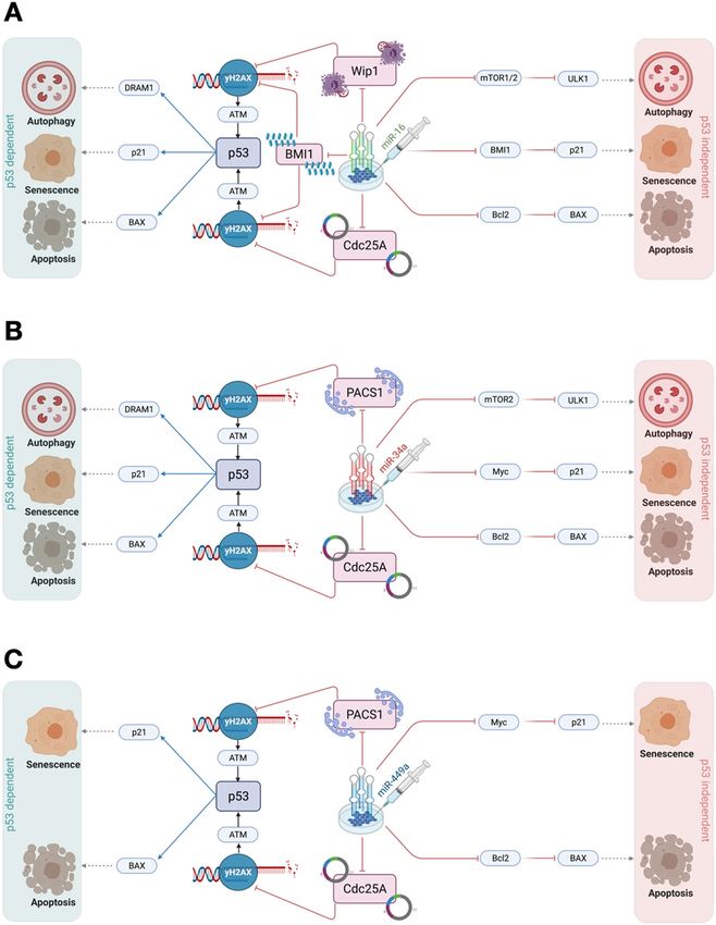
Figure 1. Schematic illustration depicting the role of microRNAs (miR-34a, miR-449a, and miR-16) in Cancer.
Alterations throughout miRNA biogenesis can affect the availability of target mRNA of PACS1 and RICTOR
regulated by miR-34a, miR-449a, and miR-16, respectively. miRNA genes are transcribed by RNA polymerase
II to produce the large primary transcripts pri-miRNAs. The pre-miRNAs are processed by another RNase
III enzyme Dicer to a ~ 20–22-nucleotide miRNA/miRNA* duplex. After the duplex is unwound, the mature
miRNA is incorporated into a protein complex termed RISC. A miRNA-loaded RISC mediates gene silencing
via mRNA cleavage and degradation on the complementarity between the miRNA and the targeted mRNA
transcript. In addition, miRNAs may function as ligands to directly bind with Toll-like receptors (TLR),
triggering downstream signaling pathways. Methyltransferase-like 3 (METTL3) is recently discovered to
methylate pri-miRNAs, marking them for recognition and processing by DGCR to yield mature miRNA.
research shows that these miRNAs may potentially play a role in autophagy signaling. Huang et al.7 demonstrated
the involvement of miR-16 in the activation of autophagy by targeting mTOR-2 within this framework. Addition-
ally, they revealed that overexpression of miR-16 in HeLa cells inhibits G1 arrest and death7.
Similarly, Rathod et al.8 established that miR-34a targets mTOR-2 directly. When glioma and glioma stem
cell lines were compared to normal brain tissue, they discovered that miR-34a expression was reduced while the
AKT-mTOR pathway was elevated8. Additionally, they established that mTOR-2 and miR-34a had a negative
connection and revealed that miR-34a was specifically targeting mTOR-2 in matched cell lines8. Furthermore,
Torossian et al.9 shown that miR-34a is required for the regulation of autophagic and apoptotic cell death in
lymphoma cells by directly targeting BCL2 e xpression9.
miRNAs are key participants in the autophagy process, which progresses by initiating autophagy, proceeds
with developmental stages, and ends by the degradation stage that has accumulated during autophagy10. miR-
NAs have been shown to affect ATGs (autophagy-related genes) and associated regulator expression processes,
including induction, vesicle nucleation, phagophore assembly, lysosomal fusion, and d egradation10.
The PI3K-AKT-mTOR, TP53-mTOR, and Ca2+-AMPK-mTOR pathways are among the upstream nutrient
and energy signals involved in autophagy induction regulation, and certain miRNAs have been shown to modify
these signals to cause phagophore induction11–13. Under genotoxic stress, TP53 and HMGB1 form a complex
that has a reciprocal inhibitory function, controlling SIRT1-mTOR signaling downstream. Via the Ca2+-AMPK-
mTOR pathway, calcium-metabolizing enzymes including transient receptor potential melastatin 3 (TRPM3)
and Drosophila inositol 1,4,5-triphosphate kinase 2 (IP3K2) regulate autophagy i nitiation13.
In HeLa cells, expressing a wild-type p53 protein, they are rendered deficient functional p53 due to the expres-
sion of the human papillomavirus (HPV) protein E614, which accelerates p53 degradation14. It is an assump-
tion that the accumulation of p53 may not control a functional G1/S checkpoint (due to HPV proteins)15.
However, increasing evidence suggests that the p53 pathway is important and may induce the G1/S checkpoint
in DDR14,16,17. In addition to the p53 pathway, other signaling pathways such as ATM, AMPK, and MAPK are
linked in the regulation of cell fate determination (senescence, autophagy, and apoptosis) at the G1/S checkpoint
in HeLa cells17,18.
In this context, the study of Veena et al.2 and Huang et al.7 provided evidence that transcription of these miR-
NAs (miR-16, miR-34a, and miR-449a) are associated with the induction of autophagy, apoptosis, and senescence
at the functional and stable G1/S checkpoint in HeLa cells. However, the precise molecular mechanisms of these
miRNAs (miR-16, miR-34a, and miR-449a) synergistically or individually in cervical cancer are still unclear.
Therefore, we present a synthetic Boolean model for the functional G1/S checkpoint in HeLa cells involving the
Scientific Reports | (2022) 12:4911 | https://doi.org/10.1038/s41598-022-08900-y 2
Vol:.(1234567890)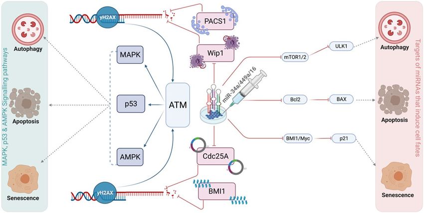
www.nature.com/scientificreports/

                                  Figure 2.  Synthetic gene regulatory network for the functional G1/S checkpoint. Arrows denote activations
                                  and hammer-head arcs represent inhibitions, respectively. Dashed hammer-head arcs represent targets of
                                  miRNA-34a, miR-449a and miR-16. The yellow rectangular nodes represent miRNAs and the red one denotes
                                  transfected miRNAs which induces DNA damage response in HeLa cells. The network outputs are in orange
                                  rectangular nodes represents proliferation or autophagy or apoptosis or senescence.

                                  Figure 3.  The wild-type state of the synthetic network and experimental verified perturbations. WT, miR-16
                                  E1 along with miR-449a KO and miR-34a KO, mTORC2 KO, mTORC1 KO, miR-449a E1 together with miR-16
                                  KO and miR-34a KO, miR-34a E1 along with miR-449a KO and miR-16 KO, and at the last, PACS1 KO. E1
                                  represents gain-of-function (GoF). Whereas, KO represents loss-of-function (LoF) perturbations corresponding
                                  to referential experiments. The left-most column shows levels of Input (Transfected_miRNAs), highlighted
                                  in orange color and the right-most column presents the model outputs: Proliferation, Autophagy, Apoptosis
                                  and Senescence. Each line represents a single stable state or fixed point corresponding to the input. White cells
                                  denote a null i.e., “inactive” value, whereas black cells denote activation means “active” (value 1), respectively.

                                  p53, AMPK, and MAPK pathways (see Fig. 2). To our knowledge, this is the first study considering the role of
                                  three miRNAs (miR-34a, miR-449a, and miR-16) in the three major DDR signaling pathways (p53, MAPK, and
                                  AMPK) in cervical cancer.

                                  Results
                                  The network construction and its fixed points. "Transfection of miRNAs" is a single input to the
                                  model, represented by a rectangle node in red (see Fig. 2). MiRNAs are shown as yellow rectangular nodes (miR-
                                  16, miR-34a, and miR-449a). Each miRNA’s target is shown by a dashed hammer-head arc, whereas arrows indi-
                                  cate activation and hammer-head arcs indicate inhibition, respectively. Proliferation, autophagy, apoptosis, and
                                  senescence are represented by the model outputs in orange-colored rectangular nodes. The network is composed
                                  of 32 proteins and three miRNAs, which are connected by 145 direct interactions (see Fig. 2).
                                      The network simulations generate four stable states or fixed points for the wild-type case (WT), each of which
                                  is associated with a distinct phenotype, as seen in Fig. 3. The first state is a proliferative state (on behalf of the
                                  input: Transfected-miRNA = "Inactive"), implying that no G1/S arrest occurs since only cell cycle promoters such
                                  as CDK46/CycD, CDK2/CycE, and Cdc25A and HVP E6 and PACS1 are activated. The remaining three states
                                  are the result of cell cycle arrest events (also known as tristable dynamics), i.e., they are triggered by a single
                                  input: transfected miRNA = "active." The second and third states are associated with cellular death are apoptosis

Scientific Reports |   (2022) 12:4911 |                https://doi.org/10.1038/s41598-022-08900-y                                                     3

                                                                                                                                                 Vol.:(0123456789)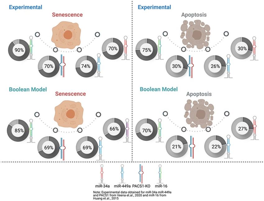
www.nature.com/scientificreports/
Stimulus/perturbations Response/phenotype References
7
Transfection of miR-16 Induction of autophagy, apoptosis and senescence
7
Knockdown (KO) of mTORC2 Autophagy
7
Knockdown (KO) of mTORC1 Induction of autophagy, apoptosis and senescence
2
Transfection of miR-449a Induction of apoptosis and senescence
2
Transfection of miR-34a Induction of apoptosis and senescence
2
Knockdown (KO) of PACS1 Induction of apoptosis and senescence
Synergistic regulation between miR-34 E1 and miR-449a E1 Induction of senescence, autophagy and apoptosis ?
Synergistic regulation between miR-16 E1 and miR-449a E1 Induction of autophagy, apoptosis and senescence ?
Synergistically overexpression (E1) of miR-449a/miR-34a/miR-16 Inhibits proliferation on the functional and stable G1/S checkpoint ?
Targeting of Cdc25A by miR-16/miR-34a/miR-449a Repression of proliferation through the induction of DNA Damage Response pathways ?
Repression of proliferation through the induction of autophagy along with apoptosis and senes-
Knockdown (KO) of PACS1 ?
cence
Transfection of miR-34a Inhibition of proliferation and induction of senescence, autophagy and apoptosis ?
Table 1. Agreement between proposed Synthetic Boolean model and experimental data from the literature
in HeLa cells. Ectopic expression (E1) represents gain of function (GoF) (or transfection of miRNA) and
Knockdown (KO) represents loss of function (LoF) of the corresponding molecule. Cases for which no
experimental data were found are indicated by ‘?’.
and autophagy, respectively. The second state reflects the autophagy phenotype induced by ULK1/Beclin1 acti-
vation in conjunction with DRAM1, whereas, the third stage denotes apoptotic cell death, as long as Caspase3
and DRAM1 are activated, except for ULK1/Beclin1. The fourth stable state depicts the senescence phenotype
caused by p53-A and p21 activation.
To determine if these miRNA transfections affected the tristable dynamics, we have performed perturbations
of miR-16. We found that ectopic expression of miR-16 (E1) inhibits proliferation and promotes autophagy,
apoptosis, and senescence (see in Fig. 3). These findings imply that miR-16 has an effect on the tristable dynamics,
which is consistent with Huang et alexperimental’s observation of the three phenotypes in HeLa cell c ultures7.
miR‑449a acts synergistically with miR‑34a/miR‑16 to regulate the autophagy signaling path‑
way. To determine how miR-34a and miR-449 transfection can influence senescence and apoptosis in HeLa
cells, as indicated by Veena et al.2. See Fig. 3, we examine each miRNA independently, which means we knock
down (KO) miR-16 in conjunction with miR-34a to elucidate miR-449a’s regulatory role. To elucidate the role
of miR-34a in the regulation of senescence and apoptosis, we conducted the same thing as previously described,
i.e. we knocked down miR-449a in conjunction with miR-16. Following that, we overexpressed both miRNAs
(miR-34 E1 and miR-449a E1) in combination with miR-16 knockdown. Finally, we performed a single per-
turbation of PACS1 by performing a knockdown (KO) of PACS1. We found out that perturbations of miR-34a
overexpression (E1) controls senescence, apoptosis, and autophagy in HeLa cells, whereas, overexpression (E1)
of miR-449a regulates senescence and apoptosis. Following that, we co-expressed miR-34a and miR-449a E1
to examine their synergistic effects. We observed that miR-449a, in combination with miR-34a, may regulate
senescence, autophagy, and apoptosis in HeLa cells following DNA damage at the G1/S checkpoint. Similarly, we
overexpressed (E1) miR-16 and miR-449a simultaneously. MiR-449a, in combination with miR-16, was shown
to control senescence, autophagy, and apoptosis, as well as a functioning and persistent G1/S checkpoint in HeLa
cells. Additionally, we did a PACS1 knockdown (KO) perturbation and discovered that PACS1 KO controls
senescence, apoptosis, and autophagy in HeLa cells. (Supplementary File S1).
Furthermore, Veena and colleagues’ w ork2 was unable to demonstrate the synergistic regulation of miR-34a
and miR-449a in HeLa cells. In this regard, we discovered that each miRNA may control senescence and apop-
tosis separately (which is entirely by the study of Veena et al.2). Additionally, our findings indicate that miR-449a
may act in concert with miR-34a or miR-16 to modulate the autophagy signaling pathway. However, miR-449a
and miR-34a share a common t arget19, except for mTOR-28, and inhibiting mTOR1/2 expression can induce
autophagy in HeLa cells via the ULK1/Beclin1 pathway. Huang et al.7 shown that miR-16 targeting mTORC-2
triggers autophagy in Hela cells in this manner. Thus, our findings are consistent with those of Veena et al.2 and
Huang et al.7.
The two experimental investigations mentioned before performed as significant references for developing the
model. The technique for developing the model was to align it precisely with the knowledge supplied by each
study; for example, one study focused exclusively on one miR-167, ignoring miR-449a/miR-34a. While the other
research elucidates the functions of miR-34a and miR-449a but not of miR-162. Additionally, the exact functions
of these molecules in the HeLa cell model were explored. To do this, we examined the specific regulatory role
of miR-16 in the context of miR-34a and/or miR-449a loss of function (LoF) and similarly examined miR-16 in
the context of miR-34a and/or miR-449a loss of function (LoF). For further information, see Fig. 3 and Table 1.
When the model is used in conjunction with experimental research, it delivers similar findings as we expected
in our work. Then, we proced further to broaden its perturbations, which are currently unknown in the HeLa
cell literature as indicated by the question mark (?) in Table 1. For instance, our model predicts that synergistic
coordination between miR-34a E1/miR-449a E1 or miR-16 E1/miR-449a E1 can govern HeLa cell senescence,
autophagy, and apoptosis. In the second case, transfection of all miRNAs (miR-16 E1/miR-449a E1/miR-34a E1)
Scientific Reports | (2022) 12:4911 | https://doi.org/10.1038/s41598-022-08900-y 4
Vol:.(1234567890)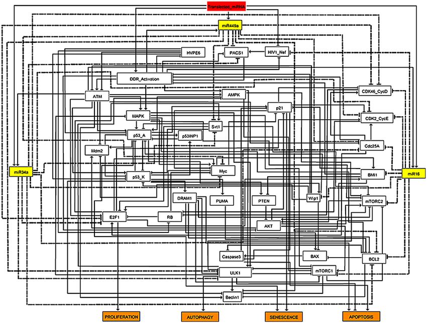
www.nature.com/scientificreports/
Figure 4. Cross-validation through the experimental studies. Upperside: cell fate decisions such as senescence
and apoptosis indicate its corresponding experimental study2,7. Whereas, down-side: cell fate decisions such
as senescence and apoptosis signify the single node model perturbation. Each circle represents the phenotype
percentage that was observed in each experimental study2,7. Whereas, the model perturbation for each molecule
was obtained through the Monte Carlo simulations (10,000 runs). miR-34a, miR-449a, and miR-16 represent
Gain-of-Function (GoF). Whereas, knockdown (KO) represents Loss-of-Function (LoF). For more detail see
"Cross-validation through the experimental studies" section.
combined can cause a functioning and persistent G1/S checkpoint. Thirdly, silencing Cdc25A in the presence of
miR-16 and/or miR-449a and/or miR-34a can reduce proliferation and induce DNA damage response (DDR)
pathways, which can activate a functional and persistent G1/S checkpoint as well as autophagy, apoptosis, and
senescence. In the fourth case, silencing/knockdown PACS1 results in autophagy, apoptosis, and senescence.
Finally, miR-34a can be used to influence senescence, autophagy, and apoptosis in HeLa cells. Table 1 and Sup-
plementary File S1 include further information.
Cross‑validation through the experimental studies. We estimated in silico the possibilities of each
phenotype for the wild-type case when the input (transfection of miRNAs) of the model is ON. We applied the
Monte Carlo algorithm in GINsim with 10.000 runs (see section "Methods"). For the WT case, we obtained 60%
for senescence, 25% for apoptosis, and 15% for autophagy.
Additionally, to analyze single node perturbation probabilities, as Veena et al.2 and Huang et al.7 did in their
respective experimental studies we have overexpressed miR-34a and/or miR-449a. They evaluated the perfor-
mance of these miRNAs by transfection, demonstrating that they accelerated senescence and apoptosis in HeLa
cells via PACS1 knockdown (KO). As such, they examine the involvement of miR-449a and/or miR-34a in HeLa
cell senescence or apoptosis. Thus, we assessed the probabilities of cell fates such as senescence and apoptosis
(Fig. 4) and compared them to those observed in cultures treated for 48 h with miR-34a and/or miR-449a
transfection from a subset of HeLa c ells2. Huang et al.7 then employed miR-16 overexpression and knockdown
(KO) and investigated the role of this miRNA in the induction of cell fates in HeLa cells by camptothecin (CPT).
The MTT test and flow cytometry analysis were used to determine the percentages of cells with senescent and
apoptotic phenotypes, respectively. Thus, using our Boolean model, we developed predictions for cell fates
such as apoptosis and senescence (Fig. 3) and normalized them to the proportion of Hela cells seen after 36 h
of treatment with 10 uM CPT. Additionally, transfection of miR-16 induces G1/S checkpoint arrest when cells
are treated for 4 h with hydroxyurea (HU) and propidium iodide (PI). Notably, our model coincides with the
findings of Veena et al.2 and Huang et al.7, Fig. 4.
Integration of miR‑34a/miR‑449a/miR‑16 on the phenotypic stabilization at the G1/S check‑
point. Subsequently, we examined whether the combination of these miRNAs had an effect on phenotype
stabilization at the G1/S checkpoint. To do so, we performed a single perturbation, in which we over-expressed
(E1) all of these miRNAs simultaneously (miR-34a E1 + miR-449a E1 + miR-16 E1) and compared it to combined
cases of miRNAs, such as (miR-34a E1 + miR-449 E1), (miR-34a E1 + miR-16 E1) and (miR-449a E1 + miR-16
E1). We perturbed the gain of function (GoF) in each example and ran Monte Carlo simulations with 10,000
runs (see section "Methods"). This allowed us to investigate all conceivable interactions between these miRNAs
(Fig. 5). We observed that miR-34a E1/miR-449a E1 combined action generated the most prominent apoptotic
phenotype and decreased the autophagic phenotype. MiR-34a E1/miR-16 E1 acting in concert enhanced the
autophagic phenotype. MiR-16 E1/miR-449a E1 regulation improves the senescent phenotype in cells but has
a little effect on autophagy. Finally, a combination of all three miRNAs induces an increase in the number of
senescent cells, increases the autophagic phenotype, and decreases the apoptotic phenotype.
Scientific Reports | (2022) 12:4911 | https://doi.org/10.1038/s41598-022-08900-y 5
Vol.:(0123456789)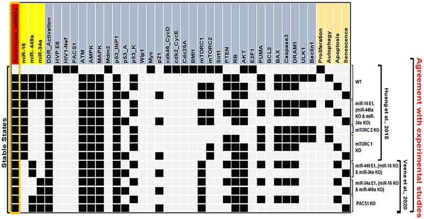
www.nature.com/scientificreports/
Figure 5. Integration of miR-34a, miR-449a, and miR-16 on the phenotypic stabilization in HeLa cells. The
First synergy case between overexpression (E1) of miR-34a E1/miR-449a E1. The second synergy case between
miR-34 E1/miR-16 E1 overexpression (E1). Whereas, the third case between miR-449a E1/miR-16 E1. At the
last, all these miRNAs overexpressed (E1) together i.e., miR-34a E1/miR-449a E1/miR-16 E1. Each particular
color bar denotes its corresponding phenotype. For each case, we have run 10.000 Monte Carlo simulations. In
addition, the first three cases are compared with the last case of synergy. For more detail see "Integration of miR-
34a/miR-449a/miR-16 on the phenotypic stabilization at the G1/S checkpoint" section.
Figure 6. The p53-independent signaling pathways. Gain of function (GoF) determines overexpression (E1)
while loss of function (LoF) describes the knockout (KO) of the component corresponding to the model. The
stable states were characterized for several scenarios: MAPK E1, AMPK E1 along with p53 KO, ATM KO, and
ATM E1. White cells indicate a zero value, while black/red/purple/orange and blue cells indicate activation
(value 1). The left-hand side highlighted in orange box presents the status of the input transfected miRNAs and
the right-hand side shows the outputs of the model such as proliferation, autophagy, apoptosis, and senescence.
Per line describes a single stable state or fixed point analogous to the input. The first three steady states or fixed
points belong to the gain-of-function (GoF) of MAPK as well as loss-of-function (LoF) of p53 and the next
three steady states describe the gain-of-function (GoF) of AMPK along with the loss-of-function (LoF) of p53:
defines alternative signaling pathways in the G1/S arrest. The knockout (KO) of ATM inhibits arrest. Whereas,
the gain-of-function (GoF) of ATM suppresses proliferation and induces the identified arresting phenotypes
such as autophagy, apoptosis, and senescence.
ATM: the main controller of multiple DNA damage response signaling pathways. It is widely
established that the p53 pathway is a critical regulator of cell fate decisions in DDR20. Numerous studies pub-
lished recently have suggested that the MAPK or AMPK signaling pathways play a substantial role (independent
of p53) in inducing cell fate in cervical cancer21,22. As a result, we inquire if miR-16, miR-34a, and miR-449a
transfection may control additional signaling pathways such as MAPK or AMPK (in the absence of p53) (see
Fig. 6). We observed that overexpression (E1) of either MAPK or AMPK may control autophagy, apoptosis, and
senescence at the functional G1/S checkpoint. Our findings concur with those of Zhong et al.23 and Law et al.24,
indicating that alternative pathways such as MAPK and/or AMPK may be responsible for cell destiny in HeLa
cells in a p53-independent manner. Additionally, the analysis revealed that these perturbations are consistent
with the Taji et al. study’s25 observation of an interchangeable double-phenotype state (senescence + autophagy)
in HeLa c ells25. Thus, our data indicate that transfection of miR-16, miR-34a, and miR-449a into Hela cells may
activate the G1/S checkpoint (with or without p53) via numerous signaling pathways such as AMPK or MAPK.
Scientific Reports | (2022) 12:4911 | https://doi.org/10.1038/s41598-022-08900-y 6
Vol:.(1234567890)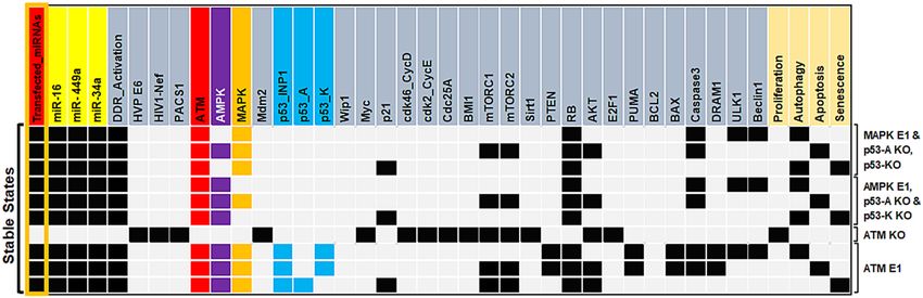
www.nature.com/scientificreports/
Moreover, current research indicates that ATM can control senescence, autophagy, and apoptosis, as well as
various DNA damage signaling pathways. For example, Liang et al.26 had shown that the ATM is necessary to
trigger autophagy and apoptosis in HeLa cells via the MAPK pathway. Furthermore, they found that silencing
ATM abolishes autophagy and apoptosis while promoting cancer development whereas, overexpression of ATM
suppresses proliferation. Beauvarlet et al.27, revealed that ATM is essential to regulate the balance of senescence,
autophagy, and apoptosis in HeLa cells via the ATM/AMPK pathway (independent of p53). Sanli et al.28 revealed
that the ATM/AMPK pathway may control autophagy and apoptosis in HeLa cells in the absence of p53. On
the other side, ATM deficiency increases the p roliferation28. We tested it, as seen in Fig. 6, by knocking down
ATM and observed that knocking down ATM increases the proliferation, while overexpression of ATM inhibits
proliferation and regulates autophagy, apoptosis, and senescence in HeLa cells at the G1/S phase. Our findings
are highly consistent with those of these investigations26–28. To summarize, ATM is the primary regulator of
DDR’s various signaling pathways (Fig. 6).
Discussion
In this study, we have investigated the molecular mechanisms by considering the performance of miR-16, miR-
34a, and miR-449a individually or synergistically at the functional G1/S checkpoint in HeLa cells (see Fig. 2).
Recently, Veena et al.2 found that PACS1, which is the direct target of miR-34a and miR-449a is upregulated in
HeLa cells and promotes cancer progression while observing the decreased expression of miR-34a and miR-
449a in the HeLa cells, simultaneously. They have also reported that the transfection of miR-34a and/or miR-
449a inhibits cancer proliferation by the induction of senescence and apoptosis in HeLa cells. The aforemen-
tioned study shows that manipulation of miR-34a and miR-449a can modulate senescence and apoptosis in a
p53-dependant manner in HeLa cells. Previously, Huang et al.7 demonstrated that the transfection of miR-16
suppressed cell proliferation and promoted autophagy, apoptosis, and senescence by targeting mTOR at the G1/S
checkpoint in HeLa cells.
The synergistic coordination among the miR-16, miR-34a, and miR-449a in the functional G1/S checkpoint
and the molecular mechanisms involving these miRNAs in cell fate decisions for HeLa cells is challenging. For
that, a Boolean model was constructed based on the available experimental d ata2,7 in the HeLa cells. In the lack
of transfection of miRNAs, the model predicts the only proliferative state, which is consistent with experimental
studies2,7. In the presence of miRNAs (transfection of miRNAs), the model predicts autophagy, apoptosis, and
senescence, which is agreeable with Huang et al.7 (See Fig. 3).
Likewise, we demonstrated the regulatory role of individual miRNAs or synergistically (with two or more
miRNAs) in HeLa cells’ functioning G1/S checkpoint. To do this, we examined each miRNA’s perturbations,
including ectopic (E1) and knockdown (KO) expression (see inTable 1). Individual transfections of miR-16 or
miR-34a can elicit tristable dynamics (autophagy, apoptosis, and senescence) as well as a functioning and sta-
ble G1/S checkpoint, whereas miR-449a regulates only bistability (senescence and apoptosis) but not autophagy.
Transfection of miR-449a in combination with miR-16 and/or miR-34a can induce tristable dynamics in a syner-
gistic way (for more details see Table 1 and Supplementary File S1). Indeed, Veena and colleagues’ investigation
did not demonstrate a synergistic regulation between miR-34a and miR-449a in HeLa c ells2. We found out that
each miRNA (miR-34a and miR-449a) may control senescence and apoptosis separately. Additionally, our data
indicated that miR-34a controls the autophagy signaling pathway by targeting mTOR alone (or in conjunction
with miR-449a/miR-16). Similarly, miR-449a can regulate the autophagy signaling pathway in conjunction with
miR-34a or miR-16. Indeed, miR-449a and miR-34a share common targets19, but miR-34a has a unique target in
mTOR-28. It has been demonstrated that silencing mTOR1/2 expression enhances the induction of autophagy by
ULK1/Beclin1 in HeLa c ells29. Similarly, Huang et al.7 demonstrated that miR-16-mediated silencing of mTORC-2
controls autophagy in Hela cells. Thus, our findings are consistent with those of Veena et al.2 and Huang et al.7.
Additionally, after the model cooperates with experimental research, we opted to increase its perturbations
in HeLa cells that are still unknown. For example, our model predicts that synergistic co-expression of miR-34a
E1/miR-449a E1 or miR-16 E1/miR-449a E1 can influence autophagy, apoptosis, and senescence in HeLa cells.
In the second case, co-transfection of all miRNAs (miR-16 E1/miR-449a/miR-34a E1) can establish a function-
ing and persistent G1/S checkpoint. The third prediction is that silencing Cdc25A in the presence of miR-16
and/or miR-449a and/or miR-34a can inhibit proliferation and thus the induction of DNA Damage Response
pathways, which may result in the activation of a functional and stable G1/S checkpoint, as well as autophagy,
apoptosis, and senescence. In the fourth case, silencing PACS1 results in autophagy, apoptosis, and senescence.
Veena et al. showed that knocking down PACS1 promoted apoptosis and senescence in HeLa cells. In light
of this, we hypothesize that knocking down PACS1 can influence autophagy as well as senescence and apopto-
sis. Furthermore, we performed cross-validation on each experimental trial. We ran a Monte Carlo simulation
(10.000 times) and compared the results to those of Veena et al.2 and Huang et al.7. These findings are entirely
consistent with the findings of these investigations2,7. (see Fig. 4). Additionally, we studied three unique instances
of synergistic overexpression (E1) between these miRNAs (miR-34a E1/miR-449a E1), (miR-449a E1/miR-16
E1), and (miR-34a E1/miR-16 E1), and compared them to the case of all three miRNAs combined (miR-34a E1/
miR-449a E1/miR-16 E1). This enables us to deduce the specific effect of those miRNAs acting in synergy on
phenotypic stability. The results are shown in Fig. 5. Each instance of the synergistic relationship illustrates the
distinct consequences of phenotypes. For instance, miR-34a E1/miR-449a E1 increased apoptosis and decreased
autophagy. MiR-34a E1/miR-16 E1 co-expression increased the autophagic phenotype. Mutual regulation of
miR-449a E1/ miR-16 E1 increases senescent phenotypes in cells but has a little effect on autophagy. Finally,
integration of all three miRNAs (miR-34a E1 + miR-449a E1 + miR-16 E1) results in an increase in senescent cells,
an increase in the autophagic phenotype, and a decrease in the apoptotic phenotype (Fig. 5).
Scientific Reports | (2022) 12:4911 | https://doi.org/10.1038/s41598-022-08900-y 7
Vol.:(0123456789)www.nature.com/scientificreports/
Likewise, in the absence of p53, transfection of miR-16, miR-34a, and miR-449a can influence phenotypes
such as autophagy, apoptosis, and senescence. Notably, we inferred that the MAPK and/or AMPK signaling
pathways can modulate the functional G1/S checkpoint (when the p53 pathway is not functional in the cells).
Additionally, we observed that ATM is required for the functioning G1/S checkpoint in HeLa cells, as well as for
the regulation of autophagy, apoptosis, and senescence, as proposed by Liang et al.26 (see Fig. 6). Thus, our find-
ings suggest that manipulating these miRNAs can activate the functional G1/S checkpoint, as well as autophagy,
apoptosis, and senescence, via MAPK or AMPK pathways (independent of p53). For more detail see Fig. 7. Thus,
this is the first and most comprehensive work to explore the involvement of three miRNAs (miR-34a, miR-449a,
and miR-16) at the G1/S checkpoint and to demonstrate that these miRNAs may influence numerous DDR
signaling pathways in cervical cancer (see in Fig. 8).
In conclusion, our model agrees with experimental results concerning the individual effect of miRNA(s)
in cell fate decisions in HeLa cells. We shed light on a very complex landscape between these three miRNAs.
Furthermore, our testable predictions abolished HeLa cancer progression through the induction of functional
and stable G1/S checkpoint. Therefore, our synthetic approach may contribute to the inhibition of tumor growth
and proliferation in cancer cells through the transfection of these miRNAs.
Methods
Collection of the public databases/tool and structure of the Gene Regulatory Network in HeLa
cells. Developing a gene regulatory network concerning miRNAs (miR-34a, miR-449a, and miR-16). We
have only used PubMed studies and databases such as BIOGRID 3.5 (https://thebiogrid.org/)30. The focus was to
identify genes or proteins that were targeted by these miRNAs (Fig. 2) such as Cyclin-dependent kinases 4 and 6
complexes/CyclinD1 (CDK4/6-Cyclin D), Cyclin-dependent kinase 2/CyclinE2 (CDK2/Cyclin E), Cell division
cycle 25A (Cdc25A), E2F1, and BCL2, are the experimentally verified common targets of miR-34a31, miR-449a32
and miR-1633. In addition, Myc, Sirt1 and PACS1 are unique targets of miR-34a2,34 and miR-449a2,32, except
miR-16. Whereas, different targets of miR-16 are BMI1, Wip1, and mTOR-1/233. Whereas, the unique target of
miR-34a is mTOR 2 8. For that, we have used public databases like; TARGET SCAN HUMAN 7.2 (http://www.
targetscan.org/vert_72/)35.
GINsim 3.0.0b was used for the construction and simulation of the Boolean model and visualization of the
results36. Which is a Java-based software and is freely available to researchers (http://www.ginsim.org/downl
oads)36. GINsim algorithms recognize all the attractors for the wild-type case (unpertubated Boolean model) as
well as for various mutant situations. The model file is available in the "Code Availability" section.
PubMed literature based boolean network, rules and simulations. The Boolean method is based
on the characterization of a regulatory graph, where an individual node defines a molecule and each directed
edge (or arc) signifies an activation or inhibition among two nodes. Nodes are Boolean variables that only con-
sider “0” and “1” i.e., “active” and “inactive” values. Based on the description of the biochemical information,
each node in the network is assigned a logical rule, which determines its activation level concerning the position
of its regulators37.
A Boolean network of miRNAs (miR-34a, miR-449a, and miR-16) was generated by translating the biological
interactions described in the gene regulatory network (Fig. 2) into Boolean rules. These Boolean rules regulating
nodes are based on the biochemical literature from PubMed and are available in Supplementary Table S1. Clas-
sical Boolean operators were used to write these rules "AND, "OR" and "NOT". Attractors are the main outcome
of simulations using a Boolean network. The dynamical performance of a Boolean model can be interpreted by
a state transition graph (STG). In this graph, each node describes the state of the network variables and the arcs
signify transitions between these states. The STG serves all possible trajectories that one initial state can drive to
a final state. Terminal nodes that have no outgoing edges are called stable states (or fixed points) while a set of
transitions trapped among a fixed group of states in the STG defines a cyclic state. For the updates of states, asyn-
chronous updates were considered38, which has the potential to describe non-deterministic behavior observed in
molecular networks. Moreover, this approach allows in silico gain-of-function (GoF) or loss-of-function (LoF)
perturbations, we force node values to be “active” or “inactive”, respectively, to examine the effect of particular
nodes on network dynamics and the resulting p henotype36,37.
miRNAs‑mediated molecular mechanisms at the G1/S checkpoint in HeLa. Cell-fate determina-
tion such as apoptosis, senescence or autophagy occur at cell cycle checkpoints20. As the G1/S checkpoint is p53
pathway-dependent and it is caused via DNA Damage, we added a new layer of complexity to the operative G1/S
regulatory network based on the works by Xie et al.39 and Huang et al.7 who considered DNA-damage induced
autophagy in Hela cells. Here we briefly outline the central direct molecular interactions known in the literature
that constitute our synthetic regulatory network.
The studies of Veena et al.2 and Huang et al.7 used miR-34a and miR-449a and miR-16 transfection to induce
autophagy, apoptosis, and senescence. Transfection of miRNAs leads to a G1/S checkpoint activation for sup-
pressing Wip1, Cdc25A, BMI1 and PACS12,3,6,40. Then in our synthetic network, we used all these as an input
signal (see Fig. 2). In addition, miR-34a, miR-449a, and miR-16 transfection induce a DNA-damage response
in Hela cells activating the ATM/p53 pathway2,7. which are involved in the induction of senescence, apoptosis,
and autophagy2,7. The targets of miR-34a, miR-449a and miR-16 in the network are presented in what follows.
DNA double-strand breaks generated by radiomimetic chemicals or reactive oxygen species (ROS), drugs or
ionizing radiation can stimulate the ATM and p53 pathways41. ATM or p53 can directly induce miR-34a42,43 and
stimulated p53 triggers the transcription of the E3 ubiquitin-protein ligase (Mdm2) which is its own negative
regulator44. Whereas, miR-449a can be induced by DNA D amage45. As in our previous models (for more details
Scientific Reports | (2022) 12:4911 | https://doi.org/10.1038/s41598-022-08900-y 8
Vol:.(1234567890)www.nature.com/scientificreports/
Figure 7. Comprehensive molecular mechanisms of transfection of miRNAs on tumor growth and proliferation in HeLa
cells. (A) Transfection of miR-16-induced DNA damage in cells by targeting Wip16 and C dc25A3. Once DNA damage is
triggered in cells it may regulate p53-dependent or independent molecular mechanisms at the functional G1/S c heckpoint20.
miR-16 directly targets mTOR1/2 induced autophagic cell death7,60. Whereas miR-16 controls the senescent phenotype by
targeting BMI1, which regulates p21 expression61. Whereas, miR-16 regulates apoptotic cell death by targeting Bcl2, which
triggers BAX/Caspase e xpression62. On the other hand, transfection of miR-16 regulates cell fate through the p 5346. miR-
16 accelerated the p53 pathway and then, p53 induces autophagic cell death through D RAM163. Senescence by induction
of p2120 while apoptotic through induction of BAX/Caspase20. (B) Transfection of miR-34-induced DNA damage in cells
by targeting PACS1 and C dc25A2,64. Once DNA damage is triggered in cells it may control p53-dependent or independent
molecular mechanisms at the functional G1/S checkpoint. miR-34 directly targets mTOR2 induced autophagic cell d eath8.
Whereas miR-34a regulates the senescent phenotype by targeting M yc65, which regulates p21 expression. Whereas, miR-34a
cl266, which triggers BAX/Caspase expression. On the other hand, transfection of
rules apoptotic cell death by targeting B
miR-34 coordinates cell fate through the p5334. miR-34 stimulated the p53 pathway and then, p53 induces autophagic cell
death through D RAM163. Senescence by induction of p 2120 while apoptotic through the introduction of BAX/Caspase20. (C)
Transfection of miR-449a-induced DNA damage in cells by targeting PACS1 and C dc25A2,45. Once DNA damage is generated
in cells it may control p53-dependent or independent molecular mechanisms at the functional G1/S c heckpoint2. miR-449a
controls the senescent phenotype by targeting M yc67, which modulates p21 expression. Whereas, miR-449a rules apoptotic
68
cell death by targeting Bcl2 , which triggers BAX/Caspase expression. On the other hand, transfection of miR-449a regulates
cell fate through the p532. miR-449a activated the p53 pathway and then, p53 induces senescence by the activation of p 2120,
while triggers the apoptosis by the activation of BAX/Caspase20. On the right, p53-independent molecular mechanisms of cell
fate decisions such as autophagy, senescence, and apoptosis. On the left, p53-dependent molecular mechanisms of cell fate
determination (autophagy, senescence, and apoptosis). Red-hammer head arrows represent inhibitions, while blue arrows
indicate activation, respectively.
Scientific Reports | (2022) 12:4911 | https://doi.org/10.1038/s41598-022-08900-y 9
Vol.:(0123456789)www.nature.com/scientificreports/
Figure 8. Schematic illustration depiciting the role of three miRNAs (miR-34a, miR-449a, and miR-16) at the
G1/S checkpoint which can modulate multiple DDR signaling pathways in HeLa cells.
see46–51), based on the several phosphorylation events that p53 possesses, it is described by two variables: p53-A
and p53-K. P53-A serves p53 phosphorylated at serine 15 and serine 20, whereas p53-K describes additional
phosphorylation at serine 46, in which case p53 becomes an inducer of apoptosis. P21 (Cyclin-dependent kinase
inhibitor 1A), Wip1, and tumor protein p53 inducible nuclear protein 1 (TP53INP1) are initiated by p53-A and
p53-K. ATM serine/threonine kinase (ATM) phosphorylates p53 to the p53-A form that initiates transcription of
Wip1, which in turn inactivates ATM6. Similarly, the apoptosis controller Bcl-2-binding component 3 (BBC3, also
called PUMA), DRAM1, and the BCL2 associated X (BAX) are initiated by p53-K. In the model, the apoptotic
phenotype entails activation of Caspase 3 through the BAX which is ruled by PUMA and DRAM1.Whereas, p21
henotype52. It recognized that the targeting of mTOR Complex 1
activation is correlated with the senescence p
(mTORC1) and mTOR Complex 2 (mTORC2) can begin the autophagy p henotype53. mTOR directly represses
the Unc-51-like kinase 1 (ULK1) protein complex, which plays a fundamental role in provoking autophagy53,54.
Interference of the mTORs expression intensifies ULK1/Beclin-1 activity leading to autophagy i nduction55.
DRAM1 is needed to induced apoptosis via direct activation of BAX56. On the other hand, DRAM1 represses
mTORs expression, which indirectly activates ULK1/ Beclin-157. Thus, p53 coordinates apoptotic/autophagic
phenotypes39. CDK4/6-Cyclin D and CDK2/Cyclin E are the foremost cell cycle regulators that boost the G1 to
S phase transition58.
Based on the main interactions above we defined our Boolean model of the G1/S checkpoint regulation in
Hela cells (Fig. 2).
Expected performances of tumor suppressor miRNAs in HeLa cell line. Simulations using the
miRNAs (miR-34a, miR-449a, and miR-16) in the Boolean model should describe the cancer biology of the cell.
Indeed, cancer cells can be uncomplicated as they either remain in a proliferative state due to downregulation
of miR-34a, miR-449a and miR-16 i.e., upregulation of PACS1 or AKT/mTOR pathway (in the absence of DNA
damage or treatment). Whereas in the case of DNA damage i.e., when DNA damage is present in cancer cells,
upregulation of these tumors suppressors miR-34a, miR-449a, and miR-16 can inhibit the tumor growth and
proliferation through the induction of senescence and/or cell deaths such as apoptotic or autophagic at the G1/S
checkpoint. Therefore, at least four fixed points/stable states (attractors) are expected.
Statistical information. GINsim 3.0.0b is a powerful tool36 and recently introduced three different algo-
rithms to estimate their reachability probabilities: Monte Carlo, Avatar, and Firefront. In this work, we have
used "Monte Carlo simulation" because of its ability to define the reachability probabilities of the Boolean model
attractors under an asynchronous updating scheme59. Monte Carlo is just as fast and effective as Avatar or Fire-
front for estimating the probabilities of reaching steady states. Avatar and Firefront are more valuable for dealing
with cyclic attractors. However, since no cyclic attractors were identified in our Boolean network simulations.
So, we chose Monte Carlo over Avatar and Firefront. Therefore, GINsim allows the estimation of probabilities
to reach specific attractors. In this work, we adopted Monte Carlo simulations with ’exact exit probabilities’ and
compared them with clinical outcomes2,7.
Data availability
All data needed to evaluate the conclusions in the paper are present in the paper and/or the Supplementary
Materials.
Scientific Reports | (2022) 12:4911 | https://doi.org/10.1038/s41598-022-08900-y 10
Vol:.(1234567890)www.nature.com/scientificreports/
Code availability
The model code is available in the Github repository: https://github.com/pritampanda15/Dynamic-modeling-
of-miR-34a-miR-449a-and-miR-16......HeLa-cells.
Received: 7 September 2021; Accepted: 2 March 2022
References
1. Wang, Y. & Taniguchi, T. MicroRNAs and DNA damage response. Cell Cycle 12, 32–42 (2013).
2. Veena, M. S. et al. Dysregulation of hsa-miR-34a and hsa-miR-449a leads to overexpression of PACS-1 and loss of DNA damage
response (DDR) in cervical cancer. J. Biol. Chem. 295, 17169–17186 (2020).
3. Pothof, J. et al. MicroRNA-mediated gene silencing modulates the UV-induced DNA-damage response. EMBO J. 28, 2090–2099
(2009).
4. Wang, H.-Y. et al. Knockdown of Wip1 enhances sensitivity to radiation in HeLa cells through activation of p38 MAPK. Oncol.
Res. 22, 225–233 (2014).
5. Choi, D. W. et al. WIP1, a homeostatic regulator of the DNA damage response, is targeted by HIPK2 for phosphorylation and
degradation. Mol. Cell 51, 374–385 (2013).
6. Shreeram, S. et al. Wip1 phosphatase modulates ATM-dependent signaling pathways. Mol. Cell 23, 757–764 (2006).
7. Huang, N. et al. MiR-15a and miR-16 induce autophagy and enhance chemosensitivity of Camptothecin. Cancer Biol. Ther. 16,
941–948 (2015).
8. Rathod, S. S., Rani, S. B., Khan, M., Muzumdar, D. & Shiras, A. Tumor suppressive miRNA-34a suppresses cell proliferation and
tumor growth of glioma stem cells by targeting Akt and Wnt signaling pathways. FEBS Open Bio 4, 485–495 (2014).
9. Torossian, A. et al. Blockade of crizotinib-induced BCL2 elevation in ALK-positive anaplastic large cell lymphoma triggers
autophagy associated with cell death. Haematologica 104, 1428–1439 (2019).
10. Shan, C. et al. The emerging roles of autophagy-related microRNAs in cancer. Int. J. Biol. Sci. 17, 134–150 (2021).
11. Hall, D. P. et al. TRPM3 and miR-204 establish a regulatory circuit that controls oncogenic autophagy in clear cell renal cell car-
cinoma. Cancer Cell 26, 738–753 (2014).
12. He, C. & Klionsky, D. J. Regulation mechanisms and signaling pathways of autophagy. Annu. Rev. Genet. 43, 67–93 (2009).
13. Chowdhari, S. & Saini, N. Gene expression profiling reveals the role of RIG1 like receptor signaling in p53 dependent apoptosis
induced by PUVA in keratinocytes. Cell Signal 28, 25–33 (2016).
14. Hietanen, S., Lain, S., Krausz, E., Blattner, C. & Lane, D. P. Activation of p53 in cervical carcinoma cells by small molecules. PNAS
97, 8501–8506 (2000).
15. Al-Mohanna, M. A. et al. p53 is dispensable for UV-induced cell cycle arrest at late G1 in mammalian cells. Carcinogenesis 22,
573–578 (2001).
16. Gao, J. et al. The anticancer effects of ferulic acid is associated with induction of cell cycle arrest and autophagy in cervical cancer
cells. Cancer Cell Int. 18, 102 (2018).
17. Hsieh, W. T. et al. Latex of euphorbia antiquorum induces apoptosis in human cervical cancer cells via c-Jun N-terminal kinase
activation and reactive oxygen species production. Nutr. Cancer 63, 1339–1347 (2011).
18. Chen, Y.-H. et al. Metformin induces apoptosis and inhibits migration by activating the AMPK/p53 axis and suppressing PI3K/
AKT signaling in human cervical cancer cells. Mol. Med. Rep. 23, 1–1 (2021).
19. Lizé, M., Klimke, A. & Dobbelstein, M. MicroRNA-449 in cell fate determination. Cell Cycle 10, 2874–2882 (2011).
20. Hafner, A., Bulyk, M. L., Jambhekar, A. & Lahav, G. The multiple mechanisms that regulate p53 activity and cell fate. Nat. Rev. Mol.
Cell Biol. 20, 199–210 (2019).
21. Li, J., Jiang, P., Robinson, M., Lawrence, T. S. & Sun, Y. AMPK-beta1 subunit is a p53-independent stress responsive protein that
inhibits tumor cell growth upon forced expression. Carcinogenesis 24, 827–834 (2003).
22. Reinhardt, H. C., Aslanian, A. S., Lees, J. A. & Yaffe, M. B. p53 deficient cells rely on ATM and ATR-mediated checkpoint signaling
through the p38 MAPK/MK2pathway for survival after DNA damage. Cancer Cell 11, 175–189 (2007).
23. Zhong, W. et al. Activation of the MAPK11/12/13/14 (p38 MAPK) pathway regulates the transcription of autophagy genes in
response to oxidative stress induced by a novel copper complex in HeLa cells. Autophagy 10, 1285–1300 (2014).
24. Law, B. Y. K. et al. Hernandezine, a novel AMPK activator induces autophagic cell death in drug-resistant cancers. Oncotarget 7,
8090–8104 (2016).
25. Taji, F. et al. Autophagy induction reduces telomerase activity in HeLa cells. Mech. Ageing Dev. 163, 40–45 (2017).
26. Liang, N. et al. ATM pathway is essential for ionizing radiation-induced autophagy. Cell. Signal. 25, 2530–2539 (2013).
27. Beauvarlet, J. et al. Modulation of the ATM/autophagy pathway by a G-quadruplex ligand tips the balance between senescence
and apoptosis in cancer cells. Nucleic Acids Res. 47, 2739–2756 (2019).
28. Sanli, T., Steinberg, G. R., Singh, G. & Tsakiridis, T. AMP-activated protein kinase (AMPK) beyond metabolism: A novel genomic
stress sensor participating in the DNA damage response pathway. Cancer Biol Ther 15, 156–169 (2014).
29. Xu, T., Sun, D., Chen, Y. & Ouyang, L. Targeting mTOR for fighting diseases: A revisited review of mTOR inhibitors. Eur. J. Med.
Chem. 199, 112391 (2020).
30. Chatr-Aryamontri, A. et al. The BioGRID interaction database: 2017 update. Nucleic Acids Res. 45, D369–D379 (2017).
31. Zhang, L., Liao, Y. & Tang, L. MicroRNA-34 family: A potential tumor suppressor and therapeutic candidate in cancer. J. Exp. Clin.
Cancer Res. 38, 53 (2019).
32. Yong-Ming, H. et al. miR-449a: A potential therapeutic agent for cancer. Anticancer Drugs 28, 1067–1078 (2017).
33. Aqeilan, R. I., Calin, G. A. & Croce, C. M. miR-15a and miR-16-1 in cancer: Discovery, function and future perspectives. Cell
Death Differ. 17, 215–220 (2010).
34. Slabáková, E., Culig, Z., Remšík, J. & Souček, K. Alternative mechanisms of miR-34a regulation in cancer. Cell Death Disease 8,
e3100 (2017).
35. Agarwal, V., Bell, G. W., Nam, J.-W. & Bartel, D. P. Predicting effective microRNA target sites in mammalian mRNAs. Elife 4,
e05005 (2015).
36. Naldi, A. et al. Logical modeling and analysis of cellular regulatory networks with GINsim 3.0. Front Physiol 9, 646 (2018).
37. Abou-Jaoudé, W. et al. Logical modeling and dynamical analysis of cellular networks. Front. Genet. 7, 94 (2016).
38. Silveira, D. A., Gupta, S. & Mombach, J. C. M. Systems biology approach suggests new miRNAs as phenotypic stability factors in
the epithelial-mesenchymal transition. J. R. Soc. Interface 17, 20200693 (2020).
39. Xie, X., Le, L., Fan, Y., Lv, L. & Zhang, J. Autophagy is induced through the ROS-TP53-DRAM1 pathway in response to mitochon-
drial protein synthesis inhibition. Autophagy 8, 1071–1084 (2012).
40. Patel, N. et al. miR-15a/miR-16 down-regulates BMI1, impacting Ub-H2A mediated DNA repair and breast cancer cell sensitivity
to doxorubicin. Sci. Rep. 7, 4263 (2017).
41. Fernandez, A. et al. Epigenetic mechanisms in DNA double strand break repair: A clinical review. Front. Mol. Biosci. 8, 653 (2021).
Scientific Reports | (2022) 12:4911 | https://doi.org/10.1038/s41598-022-08900-y 11
Vol.:(0123456789)www.nature.com/scientificreports/
42. Salzman, D. W. et al. miR-34 activity is modulated through 5′-end phosphorylation in response to DNA damage. Nat. Commun.
7, 10954 (2016).
43. Suzuki, H. I. et al. Modulation of microRNA processing by p53. Nature 460, 529 (2009).
44. Bar-Or, R. L. et al. Generation of oscillations by the p53-Mdm2 feedback loop: A theoretical and experimental study. Proc. Natl.
Acad. Sci. 97, 11250–11255 (2000).
45. Mao, A. et al. MicroRNA-449a enhances radiosensitivity by downregulation of c-Myc in prostate cancer cells. Sci. Rep. 6, 27346
(2016).
46. Gupta, S., Silveira, D. A. & Mombach, J. C. M. Towards DNA-damage induced autophagy: A Boolean model of p53-induced cell
fate mechanisms. DNA Repair 96, 102971 (2020).
47. Gupta, S., Silveira, D. A. & Mombach, J. C. M. Modeling the role of microRNA-449a in the regulation of the G2/M cell cycle
checkpoint in prostate LNCaP cells under ionizing radiation. PLoS ONE 13, e0200768 (2018).
48. Gupta, S., Silveira, D. A. & Mombach, J. C. M. ATM/miR-34a-5p axis regulates a p21-dependent senescence-apoptosis switch in
non-small cell lung cancer: A Boolean model of G1/S checkpoint regulation. FEBS Lett. 594, 227–239 (2020).
49. Silveira, D. A., Gupta, S. & Mombach, J. C. P53/E2F1/miR-25 axis regulates apoptosis induction in glioblastoma cells: A qualitative
model. J. Phys. Complex (2020).
50. Gupta, S., Silveira, D. A., Barbé-Tuana, F. M. & Mombach, J. C. M. Integrative data modeling from lung and lymphatic cancer
predicts functional roles for miR-34a and miR-16 in cell fate regulation. Sci. Rep. 10, 1–8 (2020).
51. Gupta, S., Hashimoto, R. F. Dynamical analysis of a boolean network model of the Oncogene role of lncRNA ANRIL and lncRNA
UFC1 in non-small cell lung cancer. Biomolecules 12(3), 420. https://doi.org/10.3390/biom12030420 (2022).
52. Carnero, A. Markers of cellular senescence. In Cell senescence pp. 63–81. Springer (2013).
53. Kim, Y. C. & Guan, K.-L. mTOR: A pharmacologic target for autophagy regulation. J. Clin. Investig. 125, 25–32 (2015).
54. Alers, S., Löffler, A. S., Wesselborg, S. & Stork, B. Role of AMPK-mTOR-Ulk1/2 in the regulation of autophagy: Cross talk, shortcuts,
and feedbacks. Mol. Cell. Biol. 32, 2–11 (2012).
55. Nazio, F. et al. mTOR inhibits autophagy by controlling ULK1 ubiquitylation, self-association and function through AMBRA1
and TRAF6. Nat. Cell Biol. 15, 406 (2013).
56. Crighton, D. et al. DRAM, a p53-induced modulator of autophagy, is critical for apoptosis. Cell 126, 121–134 (2006).
57. Lu, T. et al. DRAM1 regulates autophagy and cell proliferation via inhibition of the phosphoinositide 3-kinase-Akt-mTOR-
ribosomal protein S6 pathway. Cell Commun. Signal. 17, 28 (2019).
58. Bertoli, C., Skotheim, J. M. & De Bruin, R. A. Control of cell cycle transcription during G1 and S phases. Nat. Rev. Mol. Cell Biol.
14, 518 (2013).
59. Mendes, N. D. et al. Estimating attractor reachability in asynchronous logical models. Front. Physiol. 9, 1161 (2018).
60. Singh, Y., Garden, O. A., Lang, F. & Cobb, B. S. MicroRNA-15b/16 enhances the induction of regulatory T cells by regulating the
expression of rictor and mTOR. J. Immunol. 195, 5667–5677 (2015).
61. Kitadate, A. et al. MicroRNA-16 mediates the regulation of a senescence–apoptosis switch in cutaneous T-cell and other non-
Hodgkin lymphomas. Oncogene 35, 3692 (2016).
62. Cimmino, A. et al. miR-15 and miR-16 induce apoptosis by targeting BCL2. Proc. Natl. Acad. Sci. 102, 13944–13949 (2005).
63. Zajkowicz, A., Gdowicz-Kłosok, A., Krześniak, M., Ścieglińska, D. & Rusin, M. Actinomycin D and nutlin-3a synergistically
promote phosphorylation of p53 on serine 46 in cancer cell lines of different origin. Cell Signal 27, 1677–1687 (2015).
64. Sun, F. et al. Downregulation of CCND1 and CDK6 by miR-34a induces cell cycle arrest. FEBS Lett. 582, 1564–1568 (2008).
65. He, X. et al. MiR-34a modulates ionizing radiation-induced senescence in lung cancer cells. Oncotarget 8, 69797 (2017).
66. Hermeking, H. The miR-34 family in cancer and apoptosis. Cell Death Differ 17, 193–199 (2010).
67. Feliciano, A., Sánchez-Sendra, B., Kondoh, H. & LLeonart ME,. MicroRNAs regulate key effector pathways of senescence. J. Aging
Res. 2011, e205378 (2011).
68. Wei, B., Song, Y., Zhang, Y. & Hu, M. microRNA-449a functions as a tumor-suppressor in gastric adenocarcinoma by targeting
Bcl-2. Oncol. Lett. 6, 1713–1718 (2013).
Acknowledgements
SNIC and HPC2N have been acknowledged for providing computational resources. The schematic figures were
created with BioRender.com.
Author contributions
S.G, and P.K.P designed and conceptualized the study, S.G, P.K.P and R.F.H analyzed the results and wrote the
manuscript; P.K.P, S.M designed all the illustrations; S.G. verified and provided feedback to the illustrations;
S.K.S, S.M, S.K.V, R.A and Y.K.M provided critical feedback and edited the manuscript. All authors approved
the final version of the manuscript.
Funding
Open access funding provided by Uppsala University. S.G. and R.F.H. acknowledge financial support from the
São Paulo Research Foundation (FAPESP), Grant No. 2021/09070-8 and 2015/22308-2, P.K.P, Y.K.M and R.A.
acknowledge the financial support from the Swedish Research Council (VR Grant No. 2016-06014).
Competing interests
The authors declare no competing interests.
Additional information
Supplementary Information The online version contains supplementary material available at https://doi.org/
10.1038/s41598-022-08900-y.
Correspondence and requests for materials should be addressed to S.G. or R.A.
Reprints and permissions information is available at www.nature.com/reprints.
Publisher’s note Springer Nature remains neutral with regard to jurisdictional claims in published maps and
institutional affiliations.
Scientific Reports | (2022) 12:4911 | https://doi.org/10.1038/s41598-022-08900-y 12
Vol:.(1234567890)You can also read