DNAJC30 disease-causing gene variants in a large Central European cohort of patients with suspected Leber's hereditary optic neuropathy and optic ...
←
→
Page content transcription
If your browser does not render page correctly, please read the page content below
Vision science
Original research
J Med Genet: first published as 10.1136/jmedgenet-2021-108235 on 28 January 2022. Downloaded from http://jmg.bmj.com/ on March 5, 2022 by guest. Protected by copyright.
DNAJC30 disease-causing gene variants in a large
Central European cohort of patients with suspected
Leber’s hereditary optic neuropathy and optic atrophy
Sinja Kieninger ,1 Ting Xiao ,1 Nicole Weisschuh,1 Susanne Kohl,1 Klaus Rüther,2
Peter Michael Kroisel,3 Tobias Brockmann,4 Steffi Knappe,4 Ulrich Kellner,5,6
Wolf Lagrèze,7 Pascale Mazzola,8 Tobias B Haack ,8,9 Bernd Wissinger,1
Felix Tonagel10
► Additional supplemental ABSTRACT angiography. In addition, peripapillary microan-
material is published online Background Leber’s hereditary optic neuropathy giopathy is often observed at the beginning of
only. To view, please visit
the journal online (http://dx. (LHON) has been considered a prototypical the disease. Central or cecocentral scotomas are
doi.org/10.1136/jmedgenet- mitochondriopathy and a textbook example for maternal another typical finding.2 3 In the chronic phase,
2021-1 08235). inheritance linked to certain disease-causing variants best-corrected visual acuity in patients with LHON
in the mitochondrial genome. Recently, an autosomal nearly always decreases to 20/200 or below. The
For numbered affiliations see
recessive form of LHON (arLHON) has been described, temporal quadrant or all quadrants of the optic disc
end of article.
caused by disease-causing variants in the nuclear are pale, and optical coherence tomography shows
Correspondence to encoded gene DNAJC30. the thinning of the retinal nerve fibre layer in the
Dr Felix Tonagel, Centre for Methods and results In this study, we screened corresponding quadrants.3 4 Penetrance is reduced
Ophthalmology, University of the DNAJC30 gene in a large Central European in LHON but rarely related to the ‘mutation load’
Tübingen, Tübingen, Germany; cohort of patients with a clinical diagnosis of LHON since mtDNA variants are usually homoplasmic in
F elix.Tonagel@med.uni-
or other autosomal inherited optic atrophies (OA). LHON families.5 Rather certain mtDNA haplo-
tuebingen.de
We identified likely pathogenic variants in 35/1202 goups are predominantly observed in patients
SK and TX contributed equally. patients, corresponding to a detection rate of 2.9%. with LHON (eg, haplogroup J in patients with
The previously described missense variant c.152A>G;p. the m.11778G>A and the m.14484T>C variant)
Received 20 September 2021 (Tyr51Cys) accounts for 90% of disease-associated
Accepted 7 January 2022 and are thought to increase the risk of visual loss.6
alleles in our cohort and we confirmed a strong founder In addition, penetrance is about 3–5 times higher
effect. Furthermore, we identified two novel pathogenic in males.7 8 Differences in the exposure to toxic
variants in DNAJC30: the nonsense variant c.610G>T;p. factors (eg, tobacco or alcohol consumption), the
(Glu204*) and the in-frame deletion c.230_232del;p. presence of an X linked susceptibility factor and the
(His77del). Clinical investigation of the patients with protective role of oestrogens have been proposed to
arLHON revealed a younger age of onset, a more play a role in this gender bias.7–10 Abuse of alcohol
frequent bilateral onset and an increased clinically and cigarettes is known to worsen the symptoms
relevant recovery compared with LHON associated with and prognosis.11 12 Idebenone, a synthetic coen-
disease-causing variants in the mitochondrial DNA. zyme Q10 analogue initially developed for the
Conclusion This study expands previous findings on
treatment of Alzheimer’s disease, has been proved
arLHON and emphasises the importance of DNAJC30
safe and efficient in rescuing of visual acuity (VA)
in the genetic diagnostics of LHON and OA in European
in patients with LHON,13 14 although spontaneous
patients.
visual recovery is sometimes also observed in non-
treated patients with LHON.15 16
Three point mutations in the mitochondrial
INTRODUCTION genome (m.11778G>A in MT-ND4, m.3460G>A
Leber’s hereditary optic neuropathy (LHON, in MT-NDI and m.14484T>C in MT-ND6) account
OMIM:535000), first reported by Theodore Leber for about 90%–95% of the LHON disease cases and
in 1871,1 is the most common disease linked to have been shown to cause dysfunction of complex
© Author(s) (or their mitochondrial DNA (mtDNA) variants and a text- I (CI) in the mitochondrial respiratory chain, a
employer(s)) 2022. Re-use book example for maternal inheritance.2 decrease of ATP synthesis and the increased produc-
permitted under CC BY-NC. No Onset of LHON is usually in the second or third tion of reactive oxygen species, eventually leading
commercial re-use. See rights
decade of life, but manifestations in childhood or to death of retinal ganglion cells.2 17 In addition,
and permissions. Published
by BMJ. at an older age have also been observed. Symptoms some rare mtDNA variants have been recurrently
include initial unilateral acute or subacute painless associated with LHON.18 19
To cite: Kieninger S, Xiao T, Recently, Stenton et al reported that certain vari-
vision loss with involvement of the second eye after
Weisschuh N, et al.
J Med Genet Epub ahead of a few weeks to months, accompanied by dyschro- ants in the nuclear gene DNAJC30 result in an auto-
print: [please include Day matopsia. In the acute phase of LHON, fundus somal recessive inherited form of LHON (arLHON,
Month Year]. doi:10.1136/ examination shows a papillary hyperaemia of the OMIM:619382). Notably, a strong geographic
jmedgenet-2021-108235 optic disc without leakage in fluorescein fundus accumulation of DNAJC30- linked arLHON was
Kieninger S, et al. J Med Genet 2022;0:1–8. doi:10.1136/jmedgenet-2021-108235 1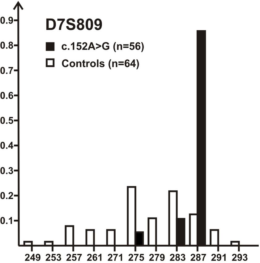
Vision science
reported with >85% of their 29 families originating from Eastern family members. Biallelism for compound heterozygous variants
J Med Genet: first published as 10.1136/jmedgenet-2021-108235 on 28 January 2022. Downloaded from http://jmg.bmj.com/ on March 5, 2022 by guest. Protected by copyright.
Europe (Russia, Ukraine, Poland, Romania).20 The patients with was assessed by allelic cloning. In brief, a fragment harbouring
arLHON manifested similar symptoms to classical LHON asso- both variants was amplified by PCR from patient’s genomic DNA
ciated with disease-causing variants in the mtDNA (mtLHON), using standard protocols and the resulting PCR product was
except one female patient who presented with Leigh syndrome. cloned into a pMiniT2.0 vector using the PCR Cloning Kit (New
The DNAJC30 protein is a chaperone protein of CI interacting England Biolabs, Frankfurt, Germany). Plasmid DNA isolated
with mitochondrial complex V to promote ATP synthesis, and is from individual clones were then used for Sanger sequencing.
mainly expressed in neurons.20 21
Given the unexpected report of pathogenic variants in a Founder effect analysis for the DNAJC30: c.152A>G variant
nuclear gene to cause LHON, we performed a retrospective Microsatellite marker analysis was done by PCR amplification
screening of DNAJC30 in a large series of genetically unsolved with fluorescence-labelled primers using Qiagen Multiplex PCR
Central European patients with LHON, and patients diagnosed Kit reagents (Qiagen, Hilden, Germany) and PCR conditions
with inherited optic atrophy (OA). We compiled and compared as recommended by the supplier. PCR fragments were sepa-
the prevalence, patients’ demography and clinical findings with rated on an ABI 3130xl capillary sequencer (Applied Biosyste-
those linked to mtDNA variants. ms—Thermo Fisher Scientific) along with a ROX-500 length
standard and fragment sizes were determined using GeneMapper
SUBJECTS AND METHODS Software (Applied Biosystems—Thermo Fisher Scientific). PCR
Study cohort primer sequences are provided in online supplemental table 1.
In this study, 1202 patients (1197 index patients and 5 affected
family members) were retrospectively selected for DNAJC30 Clinical investigations
screening, including 800 patients with a clinical diagnosis of Patients underwent ophthalmological examination according
LHON and 402 patients with OA. The cohort comprised 769 to the clinical standards of the recruiting centres, including VA
male and 431 female patients (1.8:1), the gender of two patients measurement, slit lamp examination, perimetry and indirect
is unknown. Genomic DNA samples of patients were collected ophthalmoscopy. Colour vision was examined using Ishihara
between 1992 and 2021 at the Institute for Ophthalmic plates and Farnsworth-Munsell Dichotomous D-15 test. VA is
Research, University Clinics Tübingen, Germany. Prior to this given in logMAR. A clinically relevant recovery (CRR) of visual
study, patients had undergone routine LHON or OA diagnostic impairment was defined as improvement of VA of 0.2 logMAR
or research-based genetic testing in which no likely pathogenic or greater.
variants could be detected.
RESULTS
Identification of DNAJC30 variants (Sanger sequencing) Genetic findings
Screening of the entire single exon DNAJC30 gene Sanger sequencing of 1202 LHON and OA genetically unsolved
(NM_032317.3) was performed using Sanger sequencing. Exon patients identified 35 individuals from 32 families with homo-
1 and parts of the flanking untranslated region of DNAJC30 zygous or compound heterozygous, putatively pathogenic
were amplified by PCR from genomic DNA using forward DNAJC30 variants (table 1). This corresponds to a detection rate
primer 5′−3′: ggcacccggtttttatgtc, and reverse primer 5′−3′: of 2.9% in the entire cohort. More specifically, 29/800 (3.6%) of
gcagggggagtacagttcct. PCR products were purified by treatment the clinically diagnosed LHON cases and 6/402 (1.5%) OA cases
with ExoSAP-IT reagent (Thermo Fisher Scientific, Darmstadt, of our cohort were identified to carry putatively disease-causing
Germany) and Sanger sequencing was performed using BigDye DNAJC30 variants. Of the 35 patients, 30 were males and 5 were
Cycle Sequencing V.1.1 chemistry (Thermo Fisher Scientific). females (ratio 6:1), demonstrating apparent male predominance
Sequencing products were separated by capillary electropho- in DNAJC30-linked arLHON given a male:female ratio of 1.8:1
resis on an ABI 3130xl Genetic Analyzer (Applied Biosystems— in the entire cohort. Segregation analysis could be performed in
Thermo Fisher Scientific). eight patients from five families, allelic cloning was conducted
The obtained sequences of exon 1 and the flanking intronic in three patients from three families carrying two heterozygous
regions were analysed using Sequencing Analysis V.5.2 (Applied variants. Segregation and allelic cloning confirmed the expected
Biosystems—Thermo Fisher Scientific) and the SeqMan II recessive mode of inheritance and true biallelism.
(DNASTAR, Madison, Wisconsin, USA) software. Missense vari- The most prevalent disease-associated variant identified in our
ants were evaluated for their pathogenic potential using the web- study was the c.152A>G;p.(Tyr51Cys) variant already reported
based tools MutationTaster (http://www.mutationtaster.org/) by Stenton et al.20 Additionally, two novel most likely patho-
and PolyPhen- 2 (http://genetics.bwh.harvard.edu/pph2/). Vari- genic variants were identified: a nonsense variant c.610G>T;p.
ants were filtered for an allele frequency of G;p.(Tyr51Cys) was
variants in a homozygous or compound heterozygous state with found in 30 patients with LHON (n=26) and OA (n=4) in
a second likely pathogenic variant were considered. Likely patho- homozygous state (figure 2D, online supplemental table 2). The
genic variants were finally validated through additional Sanger nonsense variant c.610G>T;p.(Glu204*) was observed in three
sequencing on independent DNA samples where available. unrelated patients with LHON in compound heterozygous state
with the common c.152A>G;p.(Tyr51Cys) variant, as confirmed
Confirmation of biallelism by allelic cloning (figure 2A B, online supplemental table 2).
Segregation analysis by means of Sanger sequencing was And finally, two affected siblings clinically diagnosed with OA
performed depending on availability of DNA of additional carry the deletion c.230_232del in homozygous state (figure 2C,
2 Kieninger S, et al. J Med Genet 2022;0:1–8. doi:10.1136/jmedgenet-2021-108235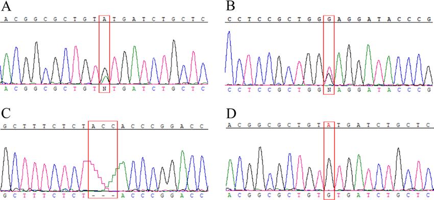
Vision science
J Med Genet: first published as 10.1136/jmedgenet-2021-108235 on 28 January 2022. Downloaded from http://jmg.bmj.com/ on March 5, 2022 by guest. Protected by copyright.
Table 1 Patients with bi-allelic variants in DNAJC30
Patient Gender Clinical diagnosis Variant Allele status Method Segregation analysis Family relation
LHON 59 (1316) M LHON c.152A>G;p.(Tyr51Cys) Hom SS Yes Brother of patient LHON 59 (1824)
LHON 59 (1824) M LHON c.152A>G;p.(Tyr51Cys) Hom SS Yes Brother of patient LHON 59 (1316)
LHON 84 M LHON c.152A>G;p.(Tyr51Cys) Hom SS NA NA
LHON 96 M LHON c.152A>G;p.(Tyr51Cys) Compd het SS Allelic cloning NA
c.610G>T;p.(Glu204*)
LHON 210 M LHON c.152A>G;p.(Tyr51Cys) Hom SS NA NA
LHON 238 F LHON c.152A>G;p.(Tyr51Cys) Hom SS NA NA
LHON 246 M LHON c.152A>G;p.(Tyr51Cys) Hom SS NA NA
LHON 286 F LHON c.152A>G;p.(Tyr51Cys) Hom SS NA NA
LHON 347 M LHON c.152A>G;p.(Tyr51Cys) Hom SS NA NA
LHON 377 M LHON c.152A>G;p.(Tyr51Cys) Hom SS NA NA
LHON 380 M LHON c.152A>G;p.(Tyr51Cys) Hom SS NA NA
LHON 466 M LHON c.152A>G;p.(Tyr51Cys) Hom SS NA NA
LHON 507 F LHON c.152A>G;p.(Tyr51Cys) Hom SS Yes NA
LHON 526 M LHON c.152A>G;p.(Tyr51Cys) Hom SS NA NA
LHON 573 M LHON c.152A>G;p.(Tyr51Cys) Compd het SS Allelic cloning NA
c.610G>T;p.(Glu204*)
LHON 582 M LHON c.152A>G;p.(Tyr51Cys) Hom SS NA NA
LHON 600 M LHON c.152A>G;p.(Tyr51Cys) Hom SS NA NA
LHON 606 M LHON c.152A>G;p.(Tyr51Cys) Hom SS NA NA
LHON 612 M LHON c.152A>G;p.(Tyr51Cys) Hom SS NA NA
LHON 749 (12040) M LHON c.152A>G;p.(Tyr51Cys) Hom SS Yes Twin of patient LHON 749 (14508)
LHON 749 (14508) M LHON c.152A>G;p.(Tyr51Cys) Hom SS Yes Twin of patient LHON 749 (12040)
LHON 760 M LHON c.152A>G;p.(Tyr51Cys) Hom SS Yes NA
LHON 785 F LHON c.152A>G;p.(Tyr51Cys) Hom SS NA NA
LHON 895 M LHON c.152A>G;p.(Tyr51Cys) Hom SS NA NA
LHON 1076 M LHON c.152A>G;p.(Tyr51Cys) Hom SS NA NA
LHON 1088 M LHON c.152A>G;p.(Tyr51Cys) Hom SS NA NA
LHON 1089 F LHON c.152A>G;p.(Tyr51Cys) Hom SS NA NA
LHON 1129 M LHON c.152A>G;p.(Tyr51Cys) Hom SS NA NA
LHON 1149 M LHON c.152A>G;p.(Tyr51Cys) Compd het SS Allelic cloning NA
c.610G>T;p.(Glu204*)
OAK 317 M OA c.152A>G;p.(Tyr51Cys) Hom SS NA NA
OAK 559 (19776) M OA c.230_232del;p.(His77del) Hom SS Yes Brother of patient OAK 559 (31530)
OAK 559 (31530) M OA c.230_232del;p.(His77del) Hom WGS Yes Brother of patient OAK 559 (19776)
OAK 627 M DOA c.152A>G;p.(Tyr51Cys) Hom SS NA NA
OAK 715 M OA c.152A>G;p.(Tyr51Cys) Hom SS NA NA
OAK 767 M DOA c.152A>G;p.(Tyr51Cys) Hom WGS NA NA
M, male; F, female; LHON, Leber′s hereditary optic neuropathy; OAK, optic atrophy/Kjer type; DOA, dominant optic atrophy; OA, optic atrophy; Hom, homozygous; Compd het,
compound heterozygous; SS, Sanger sequencing; WGS, whole genome sequencing; NA, not available.
online supplemental table 2). In 15 additional patients, only a (Arg98Pro). All variants were predicted to be disease-causing.
single heterozygous variant could be identified: 12 patients However, no second potential pathogenic variant to fulfil the
with the common missense variant c.152A>G;p.(Tyr51Cys) requirements for an autosomal recessive mode of inheritance
and 3 cases with additional missense variants: c.292T>C;p. was identified in these patients.
(Tyr98His), c.494A>G;p.(Asp165Gly) and c.278G>C;p. Given the high prevalence of the c.152A>G;p.(Tyr51Cys)
variant in our cohort, we investigated a potential founder effect
for this variant. For that purpose, we genotyped five microsatel-
lite markers covering a region 2.26 Mb on chromosome 7q11.23
in close vicinity to DNAJC30 (online supplemental table 1).
Analysis of the allele spectrum at D7S809, the marker closest to
DNAJC30 locus (~80 kb telomeric), revealed a strong bias for
a single marker allele (287 bp allele) on chromosomes bearing
the c.152A>G variant (85%) in comparison with non-mutant
Figure 1 Scheme of the DNAJC30 protein domains and location of the controls (12.5%) (figure 3). This strongly suggests a founder
variants. Variant p.(Tyr51Cys), p.(His77del), p.(Pro78Ser) and p.(Leu101Gln) effect and a common, eventually a single ancestral mutation
are located in the J domain. The variant p.(Glu204*) is located upstream event. For other more distant markers, the linkage between the
of the transmembrane domain. Novel variants detected in our study are c.152A>G variant in DNAJC30 and the respective marker is
indicated in red. much more ‘eroded’ (online supplemental table 1), arguing for a
Kieninger S, et al. J Med Genet 2022;0:1–8. doi:10.1136/jmedgenet-2021-108235 3
Vision science
Follow-up data were available for 20 patients. The median age at
J Med Genet: first published as 10.1136/jmedgenet-2021-108235 on 28 January 2022. Downloaded from http://jmg.bmj.com/ on March 5, 2022 by guest. Protected by copyright.
onset of the disease was 18.5 years (range 9.5–45.1). All patients
showed involvement of both eyes. Bilateral onset was observed
in 40% (n=8). In cases with bilateral onset, a median of 3.5
weeks (range 1–17) elapsed between the first eye and the second
eye. Initial papillary microangiopathy (figure 4) was common
and occurred in 94.1% (n=16). In the course of the disease,
mostly temporally accentuated papillary atrophy was observed
in 91.7% (n=22). Visual field defects (figure 4) were central or
cecocentral in 96.6% (n=28). Colour vision disturbances were
observed in 68.8% (n=11) and were non-specific. Median VA
Figure 2 Representative electropherograms of detected pathogenic at nadir was 1.3 logMAR (1.9–0.7) and 0.5 logMAR (1.9–0) at
sequence variants. The upper sequence digits represent the wild-type last visit. CRR was seen in 45% (n=9) at a median of 19 months
sequence. The mutant sequence corresponds to the lower sequence (range 1–58) after onset. Final VA in patients with CRR was 0.15
digits. The red box highlights the position of the variant. (A, B) Patient logMAR (0.8–0) and 1.0 logMAR (range 1.9–0.2) in patients
LHON 96, LHON 573 and LHON 1149 carry the compound heterozygous without CRR. Only one patient received idebenone over a
variants c.152A>G and c.610G>T. (C) Patient OAK 559 (19776) and period of 6 months. He developed a CRR even before the start
his brother OAK 559 (31530) carry the 3 bp deletion c.230_232del in of idebenone therapy.
homozygous state. (D) Thirty patients carry the missense variant c.152A>G
in homozygous state. LHON, Leber’s hereditary optic neuropathy.
DISCUSSION
To the surprise of many researchers, Stenton et al recently
high age of the variant which is consistent with the high relative
reported that variants in the nuclear gene DNAJC30 can cause an
prevalence of the variant in the European population (0.001457
autosomal recessive form of LHON.20 We here report the results
and 0.004765 in non-Finnish Europeans and Finnish Europeans,
of a first independent replication study confirming the exis-
respectively; gnomAD V.2.1.1).
tence of arLHON associated with bi-allelic variants in DNAJC30
overall. Taking advantage of a very large cohort of patients clini-
Clinical findings cally diagnosed with LHON or OA but still unsolved with respect
The cumulated demographic and clinical findings in the 35 to the genetic aetiology, we identified 35 patients from 32 fami-
patients with bi-allelic DNAJC30 variants are compiled in table 2
lies with putatively disease-causing homozygous or compound
and online supplemental table 2. The vast majority of patients
heterozygous variants in DNAJC30. While Stenton et al reported
(>85%) were of Central European origin (ie, Germany and
a strong geographic accumulation of DNAJC30-linked arLHON
Austria). Clinical data were available for 28 of the 35 patients.
in Eastern Europe (Russia, Ukraine, Poland, Romania),20 our
study which included in its majority German patients or patients
with residency in Germany demonstrates that DNAJC30-linked
arLHON is not a regional peculiarity and not uncommon in
Central Europe.
How does its frequency compare to that of mtDNA variant-
linked LHON? The 35 patients from 32 families with DNAJC30
variants are opposed by 346 patients from 265 families with one
of the common mtDNA variants (m.11778G>A; m.3460G>A;
m.14484T>C) in our database. Thus, the relative proportion
of patients with LHON in our entire database based on known
genetic aetiology is: 66.4% (m.11778G>A), 16% (m.3460G>A),
9.8% (m.14484T>C) and 7.7% (bi-allelic DNAJC30 variants).
This ranks DNAJC30-linked arLHON behind the three common
mtDNA variants but in its prevalence not far below that of the
m.14484T>C variant in our population.
The previously reported DNAJC30 variant c.152A>G;(p.
Tyr51Cys) is also by far the most common variant in our patients
with arLHON and accounts for 90% of all disease alleles. This
is due to a founder effect as we demonstrated by microsatel-
lite marker analysis and corroborating the SNP-based analysis in
Eastern European patients by Stenton et al. The reduced fraction
of common alleles for microsatellite markers more distant to
DNAJC30 (online supplemental table 1) argues for an old age of
the c.152A>G;(p.Tyr51Cys) variant consistent with its consid-
Figure 3 The c.152A>G variant is a founder variant. Allele spectrum erable frequency in the European population. This frequency
(x-axis=allele size, y-axis=relative frequency) of marker D7S809 on may also explain the occurrence of subjects being single hetero-
chromosomes bearing DNAJC30: c.152A>G variant (black bars) in zygous carriers of the c.152A>G;(p.Tyr51Cys) variant in our
comparison to non-mutant chromosomes from controls (white bars). The large cohort, although we cannot fully rule out the possibility of
287 bp allele is strongly over-represented on disease-linked chromosomes missed disease-linked variants in remote parts of the gene or in
(85% vs 12.5% on control chromosomes) indicating a founder effect. distant regulatory sequences.
4 Kieninger S, et al. J Med Genet 2022;0:1–8. doi:10.1136/jmedgenet-2021-108235Vision science
J Med Genet: first published as 10.1136/jmedgenet-2021-108235 on 28 January 2022. Downloaded from http://jmg.bmj.com/ on March 5, 2022 by guest. Protected by copyright.
Table 2 Aetiology and clinical results of patients with DNAJC30-associated LHON
No. of patients/mean or median (range) Percentage of documented cases (%)
Average age of onset, years (range) 18.5 (9.5–45.1) NA
Female 5 14.3
Origin
Central European 30 85.7
Eastern-E urope 2 5.7
Turkey 2 5.7
Arabia 1 2.8
Follow-up, weeks (range) 246 (3–1291) NA
Presentation
Bilateral* 28 100
Unilateral* 0 0
Onset
Bilateral* 8 40
Unilateral* 12 60
Onset of subsequential eye*, weeks (n, range) Median 3.5 (10, range 1–17) NA
Initial papillary hyperaemia
Absent* 5 29.4
Present* 12 70.6
Initial peripapillary microangiopathy
Absent* 1 5.9
Present* 16 94.1
Papillary atrophy
Absent* 2 8.3
Present* 22 91.7
Temporal quadrant 15 68.2
Global quadrant
2 8.3
Fraction not specified 5 20.8
Visual field defects
Central and cecocentral* 28 96.6
Others* 1 3.4
Colour vision disturbance
Absent* 2 12.5
Unspecific* 11 68.8
Protan/Deutan* 1 6.3
Tritan* 2 12.5
Median VA
At nadir (n, range) 1.3 (37, 1.9–0.7) NA
Of all patients at last visit (n, range) 0.5 (42, 1.9–0) NA
Of CRR patients at last visit (n, range) 0.15 (18, 0.8–0) NA
Of non-CRR patients at last visit (n, range) 1.0 (20, 1.9–0.2) NA
Interval onset—nadir*, weeks (n, range) 7.5 (12, 2–28) NA
CRR
Absent* 11 55
Present* 9 45
Interval onset—CRR*, months (n, range) 19 (9, 1–58) NA
VA is given in logMAR. All individual eyes were included in the calculation of the median VA. CRR was defined as an increase of at least 0.2 logMAR.
*Clinical parameters which were not available from all patients. Therefore, the sum of patients in the different parameter categories isVision science
J Med Genet: first published as 10.1136/jmedgenet-2021-108235 on 28 January 2022. Downloaded from http://jmg.bmj.com/ on March 5, 2022 by guest. Protected by copyright.
Figure 4 Exemplary clinical findings of DNAJC30-associated arLHON-affected patients. (A) Cecocentral visual field defect in patient LHON 1088.
(B) Papillary hyperaemia (white arrows) and peripapillary microangiopathy (black arrows) shortly after onset of the disease in patient LHON 1089.
(C) Temporally accentuated papillary atrophy occurring in the further course of the disease (in patient OAK 767). (D) Optical coherence tomography scan
of the RNFL showing a temporal decrease of thickness in OAK 767. arLHON, autosomal recessive inherited form of LHON; LHON, Leber’s hereditary optic
neuropathy.
and resulting in the accumulation of low functional CI. Variant mtLHON, occurrence of CRR varies according to the disease-
p.(Glu204*) is located upstream of the transmembrane domain causing variant type and publication, ranging from 4% to 25%
and is expected to result in a shortened protein. Although not for m.3460G>A and m.11778G>A and from 37% to 58% for
experimentally tested for DNAJC30, we would not expect that m.14484T>C.23 In our study, a CRR was observed in 45% of
mutant transcripts undergo nonsense- mediated mRNA decay the patients for which follow-up data were available. The young
due to the single exon structure of the gene. average age of our patients may have had an influence on this, as
The phenotypic and clinical characteristics of patients with it has been reported that a younger age at onset may be associated
arLHON are similar in the basic features to those of mtLHON. with a better prognosis.25 The young age of onset, more frequent
These include the increased incidence in males compared with bilateral onset and more frequent occurrence of CRR compared
females at a ratio of 6:1 (male:female ratio in the entire cohort is with mtLHON are basically consistent with and corroborate the
1.8:1), the initial peripapillary microangiopathy, the cecocentral findings of Stenton et al,20 who also studied a cohort of patients
visual field defects and also the optic atrophy that develops in with arLHON.
the medium term. No extra-ocular manifestation were found in In conclusion, our findings confirm that variants in the nuclear
our DNAJC30-linked patients with arLHON.20 21 encoded gene DNAJC30 are causative for an autosomal reces-
Although relevant clinical parameters were not available sively inherited form of LHON clinically similar and overlap-
for all patients in this retrospective study, the large number of ping with the presentation of classical mtDNA variant-associated
patients enabled us to give insights into the clinical character- LHON. Moreover, patients with DNAJC30 variants are not an
istics and phenotypic spectrum of DNAJC30-linked arLHON. Eastern European peculiarity but do represent a decent portion of
While the age of onset of mtLHON is given as 19–29 years in the entire LHON patient population in Central Europe. Further-
previous publications,23 our patients were considerably younger more, we expanded the genetic variant spectrum of DNAJC30.
with a mean age of 18.5 years. A bilateral onset in mtLHON was Therefore, our study strongly emphasises that DNAJC30 should
described as 25%24; in arLHON it was present in 40% in our be included in prospective genetic diagnostics of patients with
cohort. If the eyes were affected consecutively, an interval of 8 any form of hereditary optic neuropathies.
weeks for mtLHON was reported in previous studies,23 25 but
it was much shorter, on average only 3.5 weeks in our patients Author affiliations
with arLHON. For clinical management and patients’ prog- 1
Molecular Genetics Laboratory, Institute for Ophthalmic Research, Centre for
nosis, the appearance of a CRR is of particular interest: In Ophthalmology, University of Tübingen, Tübingen, Germany
6 Kieninger S, et al. J Med Genet 2022;0:1–8. doi:10.1136/jmedgenet-2021-108235Vision science
2
J Med Genet: first published as 10.1136/jmedgenet-2021-108235 on 28 January 2022. Downloaded from http://jmg.bmj.com/ on March 5, 2022 by guest. Protected by copyright.
Facharztpraxis für Augenheilkunde, Berlin-Mitte, Germany 5 Jacobi FK, Leo-Kottler B, Mittelviefhaus K, Zrenner E, Meyer J, Pusch CM, Wissinger
3
Diagnostic & Research Institute of Human Genetics, Diagnostic & Research Centre B. Segregation patterns and heteroplasmy prevalence in Leber’s hereditary optic
for Molecular BioMedicine, Medical University of Graz, Graz, Austria neuropathy. Investig Ophthalmol Vis Sci 2001;42:1208–14.
4
Department of Ophthalmology, Universitätsmedizin Rostock, University of Rostock, 6 Hudson G, Carelli V, Spruijt L, Gerards M, Mowbray C, Achilli A, Pyle A, Elson J, Howell
Rostock, Germany N, La Morgia C, Valentino ML, Huoponen K, Savontaus M-L, Nikoskelainen E, Sadun
5
Zentrum für Seltene Netzhauterkrankungen, AugenZentrum Siegburg, MVZ AA, Salomao SR, Belfort R, Griffiths P, Yu-Wai-Man P, de Coo RFM, Horvath R, Zeviani
Augenärztliches Diagnostik- und Therapiecentrum Siegburg GmbH, Siegburg, M, Smeets HJT, Torroni A, Chinnery PF. Clinical expression of Leber hereditary optic
Germany neuropathy is affected by the mitochondrial DNA-haplogroup background. Am J Hum
6
RetinaScience, Bonn, Germany Genet 2007;81:228–33.
7
Eye Centre, Medical Centre - University of Freiburg, Faculty of Medicine, University 7 Carelli V, d’Adamo P, Valentino ML, La Morgia C, Ross-Cisneros FN, Caporali L,
of Freiburg, Freiburg, Germany Maresca A, Loguercio Polosa P, Barboni P, De Negri A, Sadun F, Karanjia R, Salomao
8 SR, Berezovsky A, Chicani F, Moraes M, Moraes Filho M, Belfort R, Sadun AA,
Institute of Human Genetics and Applied Genomics, University of Tübingen,
Tübingen, Germany D’Adamo P, Polosa PL, Filho MM. Parsing the differences in affected with LHON:
9 genetic versus environmental triggers of disease conversion. Brain 2016;139:e17.
Centre for Rare Diseases, University of Tübingen, Tübingen, Germany
10 8 Giordano C, Montopoli M, Perli E, Orlandi M, Fantin M, Ross-Cisneros FN, Caparrotta
Centre for Ophthalmology, University of Tübingen, Tübingen, Germany
L, Martinuzzi A, Ragazzi E, Ghelli A, Sadun AA, d’Amati G, Carelli V. Oestrogens
ameliorate mitochondrial dysfunction in Leber’s hereditary optic neuropathy. Brain
Acknowledgements We would like to thank all patients and families and
2011;134:220–34.
recruiting clinicians for participation and contribution to the Tübingen long-term
9 Shankar SP, Fingert JH, Carelli V, Valentino ML, King TM, Daiger SP, Salomao SR,
study on the genetic basis of inherited optic neuropathies. Specifically, we would
Berezovsky A, Belfort R, Braun TA, Sheffield VC, Sadun AA, Stone EM. Evidence for a
like to thank Beate Leo-Kottler, Tübingen, Dr Rosemarie Richter, Berlin, Dr Änne
novel X-linked modifier locus for Leber hereditary optic neuropathy. Ophthalmic Genet
Petzschmann, Berlin, Professor Andreas Gal, Hamburg, Dr Eckhard Roth, Düsseldorf,
2008;29:17–24.
Professor Dr Rolf Winter, Hannover, Dr Bernhard Jurklies, Essen, Dr Friedmar R. Kreuz,
10 Yu J, Liang X, Ji Y, Ai C, Liu J, Zhu L, Nie Z, Jin X, Wang C, Zhang J, Zhao F, Mei S,
Dresden, Dr Ch. Büning, Kassel, Professor Dr Lutz E. Pillunat, Dresden, Dorothea
Zhao X, Zhou X, Zhang M, Wang M, Huang T, Jiang P, Guan M-X. PRICKLE3 linked
Wand, Halle-Wittenberg.
to ATPase biogenesis manifested Leber’s hereditary optic neuropathy. J Clin Invest
Contributors Acquisition of clinical data: FT, KR, PK, TB, SKn, UK, WL. Acquisition 2020;130:4935–46.
of genetic data: SKi, TX, NW, SK, PM, TH, BW. Writing of original draft: SKi, TX, FT, 11 Cullom ME, Heher KL, Miller NR, Savino PJ, Johns DR. Leber’s hereditary optic
BW. Review and editings: SKi, TX, FT, BW, SKo, NW, KR, PK, TB, SKn, UK, WL, PM, TBH. neuropathy masquerading as tobacco-alcohol amblyopia. Arch Ophthalmol
Guarantor of this study: FT 1993;111:1482–5.
12 Amaral-Fernandes MS, Marcondes AM. Miranda PM do AD, Maciel-Guerra at,
Funding This work was supported in parts by grants of the Waldtraut and Sieglinde
Sartorato El. mutations for Leber hereditary optic neuropathy in patients with alcohol
Hildebrand Foundation, and the ERA-Net E-Rare program. TX is the fellow of and
and tobacco optic neuropathy. Mol Vis 2011;17:3175–9.
supported by the Chinese Scholarship Council.
13 Zhao X, Zhang Y, Lu L, Yang H. Therapeutic effects of idebenone on Leber hereditary
Competing interests None declared. optic neuropathy. Curr Eye Res 2020;45:1315–23.
Patient consent for publication Not applicable. 14 Catarino CB, von Livonius B, Priglinger C, Banik R, Matloob S, Tamhankar MA, Castillo
L, Friedburg C, Halfpenny CA, Lincoln JA, Traber GL, Acaroglu G, Black GCM, Doncel C,
Ethics approval This study involves human participants and was approved by Fraser CL, Jakubaszko J, Landau K, Langenegger SJ, Muñoz-Negrete FJ, Newman NJ,
the institutional review board of the Ethics Committee of the University Hospital of Poulton J, Scoppettuolo E, Subramanian P, Toosy AT, Vidal M, Vincent AL, Votruba M,
Tübingen under the study numbers 112/2001, 598/2011BO1 and 637/2017BO1. Zarowski M, Zermansky A, Lob F, Rudolph G, Mikazans O, Silva M, Llòria X, Metz G,
Participants gave informed consent to participate in the study before taking part. Klopstock T. Real-World clinical experience with idebenone in the treatment of Leber
Provenance and peer review Not commissioned; externally peer reviewed. hereditary optic neuropathy. J Neuroophthalmol 2020;40:558–65.
15 Johns DR, Smith KH, Miller NR. Leber’s hereditary optic neuropathy. clinical
Data availability statement All data relevant to the study are included in the manifestations of the 3460 mutation. Arch Ophthalmol 1992;110:1577–81.
article or uploaded as supplementary information. not applicable. 16 Moon Y, Kim US, Han J, Ahn H, Lim HT. Clinical and optic disc characteristics
Supplemental material This content has been supplied by the author(s). of patients showing visual recovery in Leber hereditary optic neuropathy. J
It has not been vetted by BMJ Publishing Group Limited (BMJ) and may not Neuroophthalmol 2020;40:15–21.
have been peer-reviewed. Any opinions or recommendations discussed are 17 Yu-Wai-Man P, Votruba M, Burté F, La Morgia C, Barboni P, Carelli V. A
solely those of the author(s) and are not endorsed by BMJ. BMJ disclaims all neurodegenerative perspective on mitochondrial optic neuropathies. Acta
liability and responsibility arising from any reliance placed on the content. Neuropathol 2016;132:789–806.
Where the content includes any translated material, BMJ does not warrant the 18 Achilli A, Iommarini L, Olivieri A, Pala M, Hooshiar Kashani B, Reynier P, La Morgia C,
accuracy and reliability of the translations (including but not limited to local Valentino ML, Liguori R, Pizza F, Barboni P, Sadun F, De Negri AM, Zeviani M, Dollfus
regulations, clinical guidelines, terminology, drug names and drug dosages), and H, Moulignier A, Ducos G, Orssaud C, Bonneau D, Procaccio V, Leo-Kottler B, Fauser S,
is not responsible for any error and/or omissions arising from translation and Wissinger B, Amati-Bonneau P, Torroni A, Carelli V. Rare primary mitochondrial DNA
adaptation or otherwise. mutations and probable synergistic variants in Leber’s hereditary optic neuropathy.
PLoS One 2012;7:e42242.
Open access This is an open access article distributed in accordance with the 19 Peverelli L, Catania A, Marchet S, Ciasca P, Cammarata G, Melzi L, Bellino A, Fancellu
Creative Commons Attribution Non Commercial (CC BY-NC 4.0) license, which R, Lamantea E, Capristo M, Caporali L, La Morgia C, Carelli V, Ghezzi D, Bianchi
permits others to distribute, remix, adapt, build upon this work non-commercially, Marzoli S, Lamperti C. Leber’s hereditary optic neuropathy: a report on novel mtDNA
and license their derivative works on different terms, provided the original work is pathogenic variants. Front Neurol 2021;12:657317.
properly cited, appropriate credit is given, any changes made indicated, and the use 20 Stenton SL, Sheremet NL, Catarino CB, Andreeva NA, Assouline Z, Barboni P, Barel
is non-commercial. See: http://creativecommons.org/licenses/by-nc/4.0/. O, Berutti R, Bychkov I, Caporali L, Capristo M, Carbonelli M, Cascavilla ML, Charbel
Issa P, Freisinger P, Gerber S, Ghezzi D, Graf E, Heidler J, Hempel M, Heon E, Itkis YS,
ORCID iDs Javasky E, Kaplan J, Kopajtich R, Kornblum C, Kovacs-Nagy R, Krylova TD, Kunz WS, La
Sinja Kieninger http://orcid.org/0000-0002-8656-3239 Morgia C, Lamperti C, Ludwig C, Malacarne PF, Maresca A, Mayr JA, Meisterknecht
Ting Xiao http://orcid.org/0000-0001-9853-9474 J, Nevinitsyna TA, Palombo F, Pode-Shakked B, Shmelkova MS, Strom TM, Tagliavini F,
Tobias B Haack http://orcid.org/0000-0001-6033-4836 Tzadok M, van der Ven AT, Vignal-Clermont C, Wagner M, Zakharova EY, Zhorzholadze
NV, Rozet J-M, Carelli V, Tsygankova PG, Klopstock T, Wittig I, Prokisch H. Impaired
complex I repair causes recessive Leber’s hereditary optic neuropathy. J Clin Invest
REFERENCES 2021;131:1–12.
1 Leber T. Ueber hereditäre und congenital-angelegte Sehnervenleiden. Graefe's Arhiv 21 Tebbenkamp ATN, Varela L, Choi J, Paredes MI, Giani AM, Song JE, Sestan-Pesa M,
für Ophthalmologie 1871;17:249–91. Franjic D, Sousa AMM, Liu Z-W, Li M, Bichsel C, Koch M, Szigeti-Buck K, Liu F, Li Z,
2 Amore G, Romagnoli M, Carbonelli M, Barboni P, Carelli V, La Morgia C. Therapeutic Kawasawa YI, Paspalas CD, Mineur YS, Prontera P, Merla G, Picciotto MR, Arnsten AFT,
options in hereditary optic neuropathies. Drugs 2021;81:57–86. Horvath TL, Sestan N. The 7q11.23 protein DNAJC30 interacts with ATP synthase and
3 La Morgia C, Carbonelli M, Barboni P, Sadun AA, Carelli V. Medical management of links mitochondria to brain development. Cell 2018;175:e23:1088–104.
hereditary optic neuropathies. Front Neurol 2014;5:1–7. 22 Kampinga HH, Craig EA. The Hsp70 chaperone machinery: J proteins as drivers of
4 Newman NJ, Lott MT, Wallace DC. The clinical characteristics of pedigrees of functional specificity. Nat Rev Mol Cell Biol 2010;11:579–92.
Leber’s hereditary optic neuropathy with the 11778 mutation. Am J Ophthalmol 23 Yu-Wai-Man P, Turnbull DM, Chinnery PF. Leber hereditary optic neuropathy. J Med
1991;111:750–62. Genet 2002;39:162–9.
Kieninger S, et al. J Med Genet 2022;0:1–8. doi:10.1136/jmedgenet-2021-108235 7Vision science
J Med Genet: first published as 10.1136/jmedgenet-2021-108235 on 28 January 2022. Downloaded from http://jmg.bmj.com/ on March 5, 2022 by guest. Protected by copyright.
24 Meyerson C, Van Stavern G, McClelland C. Leber hereditary optic neuropathy: current 25 Riordan-Eva P, Sanders MD, Govan GG, Sweeney MG, Da Costa J, Harding AE. The
perspectives. Clin Ophthalmol 2015;9:1165–76. clinical features of Leber’s hereditary optic neuropathy defined by the presence of a
pathogenic mitochondrial DNA mutation. Brain 1995;118 (Pt 2:319–37.
8 Kieninger S, et al. J Med Genet 2022;0:1–8. doi:10.1136/jmedgenet-2021-108235You can also read