Curcumin ameliorates ischemic stroke injury in rats by protecting the integrity of the blood brain barrier
←
→
Page content transcription
If your browser does not render page correctly, please read the page content below
EXPERIMENTAL AND THERAPEUTIC MEDICINE 22: 783, 2021

              Curcumin ameliorates ischemic stroke injury in rats by
                protecting the integrity of the blood‑brain barrier
                      SHUGUANG WU1*, TING GUO2*, WENXUAN QI3, YUYU LI3, JIE GU3, CUI LIU3,
                       YUEHONG SHA4, BAOCHENG YANG3,5, SHUQUN HU3,5 and XUEMEI ZONG3,5

       1
        Department of Anesthesiology, The Second Affiliated Hospital of Zhejiang University School of Medicine, Hangzhou,
         Zhejiang 313000; 2Department of Neurology, Fujian Medical University Union Hospital, Fuzhou, Fujian 350001;
             3
               Jiangsu Provincial Institute of Health Emergency, Xuzhou Medical University, Xuzhou, Jiangsu 221002;
                 4
                   Department of Emergency, First People's Hospital, Pizhou, Jiangsu 221300; 5Emergency Center of
                     The Affiliated Hospital of Xuzhou Medical University, Xuzhou, Jiangsu 221002, P.R. China

                                     Received September 30, 2020; Accepted April 16, 2021

                                                   DOI: 10.3892/etm.2021.10215

Abstract. The blood‑brain barrier (BBB) is critical for proper       Introduction
cerebral homeostasis and its dysfunction during ischemic
stroke can result in significant neurological injury. The major      Stroke, a common neurological disorder associated with
goal of the present study was to identify whether curcumin           a high risk of disability and mortality (1), disrupts the
pretreatment possessed protective effects on BBB integrity           blood‑brain barrier (BBB), where it can induce neuronal
during the 24 h of acute ischemic brain injury. To investigate the   injury through brain edema and inflammation (2). There is
protective effects of curcumin, male Sprague‑Dawley rats were        increasing evidence that inflammation contributed to the
divided into multiple groups, including sham, middle cerebral        progression of ischemia‑induced secondary brain damage,
artery occlusion/reperfusion (MCAO/R) vehicle and curcumin           leading to aggravated dysregulation of the BBB, as well as
pretreated MCAO/R groups. The effects of curcumin were               cerebral edema (3,4). Thus, identification of novel drugs
measured by analyzing neurological deficits, infarct size, BBB       to protect against ischemic stroke and to understand the
permeability and expression levels of permeability‑related           underlying mechanisms involved is important to combat this
proteins in the brain. It was found that curcumin pretreatment       pathology (5).
significantly improved neurological scores, decreased infarct            The BBB consists of a variety of cell types and acts as a
size, and protected synaptic remodeling of hippocampal               border between the brain and the blood circulating through
neurons and upregulated the protein expression level of tight        the body (6). It is well‑known that BBB impairment is a conse‑
junction proteins, ZO‑1, occludin and claudin‑5 in ischemic          quence of ischemic stroke (7,8). Controlling the BBB during
rat brains. Furthermore, curcumin pretreatment before stroke         stroke has both neuronal and vascular protective effects (9).
was shown to downregulate the phosphorylation of NF‑κ B              Previous studies have suggested that NF‑κ B and MMP‑9,
and MMP‑9, which are central mediators of inflammation.              which are central mediators of inflammation, play pivotal roles
The results from the present study indicated that curcumin           in inflammatory damage following experimental ischemic
pretreatment ameliorated ischemic stroke injury by protecting        stroke (10,11).
BBB integrity and synaptic remodeling, as well as inhibiting             There are numerous types of MMPs. The MMP that
inflammatory responses.                                              damages the BBB following stroke is gelatinase MMP‑9 (12).
                                                                     Under normal, steady state conditions, MMP‑9 protein expres‑
                                                                     sion levels in endothelial cells are low and are present as
                                                                     inactive zymogens. In animals experiencing cerebral ischemia
                                                                     and reperfusion, inflammatory mediators are upregulated and
Correspondence to: Dr Xuemei Zong or Professor Shuqun Hu,            activate leukocytes, which can secrete MMP‑9 (13). When
Jiangsu Provincial Institute of Health Emergency, Xuzhou Medical
                                                                     MMP‑9 protein levels in the endothelial cells are elevated,
University, 99 Huaihai Road, Xuzhou, Jiangsu 221002, P.R. China
                                                                     extracellular matrix components and substrates in the BBB are
E‑mail: xuemeizong@gmail.com
E‑mail: hushuqun88@126.com                                           damaged, primarily through degradation of type IV collagen.
                                                                     Structural damage to the BBB will increase permeability and
*
    Contributed equally                                              result in vascular‑derived brain edema, and toxic substances
                                                                     are introduced into the BBB and harm brain tissue (14).
Key words: curcumin, ischemic stroke, blood‑brain barrier,           Notably, NF‑κ B directly regulates the transcription of MMP‑9,
inflammation, tight junction                                         and inhibition of NF‑κ B activity reduces the protein expres‑
                                                                     sion level of MMP‑9, as well as inflammation (15,16). Reduced
                                                                     MMP‑9 protein expression in endothelial cells, as well as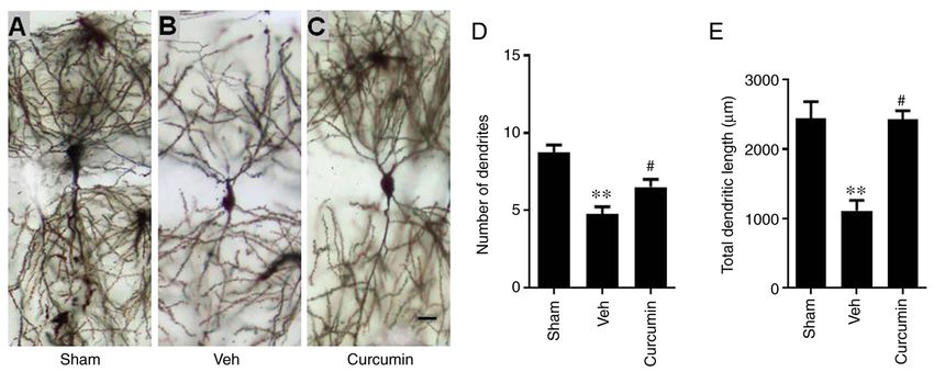
2 WU et al: CURCUMIN PROTECTS BLOOD-BRAIN FROM STROKE
decreased inflammation, reduces BBB disruption and cerebral the more severe the symptoms of neural power defciency.
injury caused by ischemic stroke (17). Neurological functions were evaluated 24 h, and 3 and 7 days
Curcumin, a pleiotropic agent extracted from the rhizome following MCAO/R using a modified Neurological Severity
of Curcuma longa (18), exerts pharmacological effects against Scale (mNSS), as previously described (26). This scale includes
stroke via its anti‑oxidative and anti‑inflammatory action (19). measurement of balance, reflex, sensory and motor skills. The
Previous research has revealed that curcumin could pass mNSS is similar to the contralateral neglect tests performed
through the BBB and significantly decreased water content, for humans. Neurological functions were measured on a scale
infarct size and BBB leakage in a middle cerebral artery from 0 to 18, where normal was scored as 0 and maximum
occlusion/reperfusion (MCAO/R) rat model (20‑23). The deficit was scored as 18. A mNSS score of 0 was indicative of
post‑ischemic neuroprotective effects of curcumin have been the sham group.
investigated; however, the potential mechanisms behind these
effects are not fully known. Measuring infarct size. The rats were anesthetized 24 h
The aim of the present study was to assess how curcumin post‑MCAO/R and 2 mm coronal slices were prepared. The
affected brain injury, the BBB status and the protein expres‑ issues were stained at 37˚C for 30 min using 2% solution 2, 3,
sion level of phosphorylated (p)NF‑κ Bp65 and MMP‑9 in a 5‑triphenyltetrazolium chloride (TTC; Sigma‑Aldrich; Merck
rat MCAO/R stroke model. We hypothesized that pretreatment KGaA) diluted in PBS. Following which, the tissues were fixed
with curcumin would protect BBB integrity after MCAO/R using 4% paraformaldehyde in a 4˚C refrigerator overnight.
in the brain via its anti‑inflammatory effects, including the Red staining was associated with undamaged regions after
attenuation of pNF‑κ Bp65 and MMP‑9 in endothelial cells MCAO/R and white stained areas revealed regions experi‑
and increase the expression level of tight junction proteins. encing an infarct. The relative infarction volume percentage
(RIVP) was calculated using the following formula:
Materials and methods RIVP=total infarct area/total area x100%. Infarct size was
quantified using ImageJ software 1.8.0 (National Institutes of
Animal and establishment of the stroke model. A total of Health).
90 3‑month‑old male Sprague‑Dawley (SD) rats, weighing
250‑280 g were provided by Xuzhou Medical University Rapid golgi staining. The brains were removed from the
(Jiangsu, China). The rats were housed in a tempera‑ rats and processed using the rapid Golgi staining kit (FD
ture‑controlled (22±2˚C) setting, including 55±10% humidity Neurotechnologies, Inc.), based on the instructions provided
and a 12 h light/dark cycle with free access to food and water. by the manufacturer. Briefly, serial sections (100‑µm) from
All rats were sufficiently adapted to their environments before the hippocampus were prepared on a freezing microtome
subjected to surgery. The MCAO/R stroke model used in the and dehydrated in an ascending absolute ethanol series
present study was established as previously described (24). (50, 70 and 90%), washed in xylene and mounted in neutral
Briefly, rats were intraperitoneally anesthetized with ketamine balsam. Next, 5 pyramidal neurons extracted from each rat (3
(60 mg/kg) and xylazine (5 mg/kg). The ventral midline neck rats/group; 20 brain sections from each group) were measured
was cut to expose the right common (CCA), right internal (ICA) from area CA1 of the hippocampus. A camera lucida drawing
and right external (ECA) carotid arteries. The ECA branches tube, attached to an Olympus BX51 microscope (x400;
were then ligated. Next, a 4‑0 nylon monofilament containing Olympus Corporation) was used to select neurons for analyses.
a round tip (Guangzhou Jialing Biotechnology Co., Ltd.) was To analyze the neurons, the soma center was considered as
inserted along the CCA into the ICA. This was performed the reference dot. Total dendritic length and number were
until resistance was detected. Approximately 90 min following measured every 50 µm.
MCAO surgery, the monofilament was removed, followed by
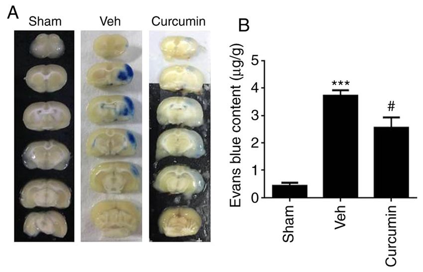
reperfusion for 24 h, and 3 and 7 days post‑MCAO. The exact Evaluation of BBB permeability. To measure BBB perme‑
same surgery was performed in the sham group rats; however, ability, Evans blue (EB) dye was used as a tracer, as previously
filament insertion was not performed. described (27). Briefly, the rats were injected with 2% EB solu‑
The SD rats were randomized into three individual groups, tion diluted in normal saline through the tail vein (2 ml/kg of
including the sham (n=30), the MCAO/R with vehicle (n=30) body weight; Sigma‑Aldrich; Merck KGaA) 24 h post‑surgery.
and the MCAO/R plus curcumin groups (n=30). Curcumin The EB dye was allowed to circulate for 2 h. Next, the rats were
(300 mg/kg; Sigma‑Aldrich; Merck KGaA) was dissolved anesthetized and transcardially perfused using 0.9% sodium
in 2% dimethyl sulfoxide (vehicle) and intraperitoneally chloride. The brains were removed, divided into left and right
administered 30 min prior to MCAO/R surgery (25). All the hemispheres, then the right hemisphere was immersed in
experiments were performed following guidelines written by formamide (10 ml/kg; Sigma‑Aldrich; Merck KGaA) at 60˚C
the Institutional Animal Care and Use Committee of China. for 24 h. Cortical proteins were formamide extracted and
The studies were also approved by the Ethics Committee centrifuged (2,500 x g) for 10 min at 4˚C. A total of 1 ml
for the Use of Experimental Animals at Xuzhou Medical supernatant was measured in a spectrophotometer at 620 nm
University (assurance nos. 2015‑46 and 2015‑47). to compare EB content in the brain tissue with standard EB
solution.
Assessing neurological deficits. The modified Neurological
Severity Scale (mNSS) has a total score of 18 points and is Immunofluorescence. Following brain perfusion, the tissues
divided into 4 parts as follows: Motion, sensation, balance, and were further post‑fixed overnight in 4% paraformaldehyde and
reflex. The score of normal rats was 0. The higher the score, 4˚C refrigerator, then dehydrated in 30% sucrose. Cerebral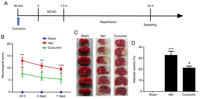
EXPERIMENTAL AND THERAPEUTIC MEDICINE 22: 783, 2021                                                              3

Figure 1. Curcumin pretreatment ameliorates MCAO/R‑induced neurological impairment and early brain injury in rats. (A) Schematic representing the
experimental protocol used for measuring the effects of curcumin pretreatment on brain injury. (B) Neurological Severity Scale scores in the different rat
groups were evaluated at 24 h, and 3 and 7 days following reperfusion. (C) Rat brains were sliced and stained using 2,3,5‑triphenyltetrazolium chloride 24 h
following MCAO/R and (D) the percentage of the relative infarct size was calculated. The data are presented as the mean ± SEM. n= 6. **P
4                                        WU et al: CURCUMIN PROTECTS BLOOD-BRAIN FROM STROKE

Figure 2. Protective effects of curcumin on hippocampus neuronal remodeling against MCAO/R. Representative images of golgi stained hippocampus pyra‑
midal neurons in the (A) sham, (B) vehicle‑treated MCAO/R and (C) curcumin‑treated MCAO/R groups. Scale bar, 10 µm. Quantitative analyses of the
(D) numbers of dendrites and (E) total dendritic length. The data are presented as the mean ± SEM. n=6. **P
EXPERIMENTAL AND THERAPEUTIC MEDICINE 22: 783, 2021                                                        5

Figure 4. Curcumin reduces MCAO/R‑induced protein expression levels of pNF‑κ Bp65, NF‑κ Bp65 and MMP‑9. Representative western blots and densito‑
metric quantifications of (A) pNF‑κ Bp65 and NF‑κ Bp65, and (B) MMP‑9 24 h after MCAO/R. The data are presented as the mean ± SEM. n=3, **P
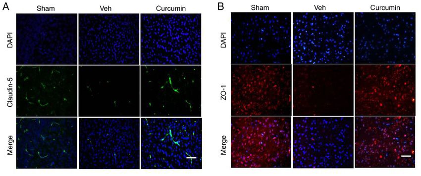
6                                        WU et al: CURCUMIN PROTECTS BLOOD-BRAIN FROM STROKE

Figure 5. Curcumin reverses the attenuation of tight junction proteins claudin‑5 and ZO‑1 induced by MCAO/R. Representative in situ expression
of (A) claudin‑5 (green) and (B) ZO‑1 (red), and merged with DAPI counter staining (blue) in the peri‑infarct region from sham‑operated, MCAO/R
vehicle and curcumin treated animals. Scale bar, 50 µm. Veh, MCAO/R vehicle group; MCAO/R, middle cerebral artery occlusion/reperfusion;
DAPI, 4',6‑diamidino‑2‑phenylindole.

Figure 6. Curcumin inhibits the degradation of tight junction proteins. Representative western blots and quantitative analyses showing the protein expression
levels of (A) occludin, (B) ZO‑1 and (C) claudin‑5 in the rat brain tissues from the 3 experimental groups. The data are presented as the mean ± SEM. n=3.
**
  P
EXPERIMENTAL AND THERAPEUTIC MEDICINE 22: 783, 2021                                                7

proteins in the peripheral blood, resulting in an increase of      the data. YS and BY collected the data. YS, BY, SH and XZ
water in the cell space and formation of vascular‑derived brain    performed data analysis and/or wrote part of the paper. SW,
edema. There is an introduction of toxic substances into the       XZ and SH confirm the authenticity of all the raw data. All
BBB and damage to brain tissue, which destroys the integrity       authors read and approved the final manuscript.
of the vascular structure (43,46,47). Previously, the asso‑
ciation between MMP‑9 and ischemic brain injury has gained         Ethics approval and consent to participate
interest (46,47). When an inflammatory reaction occurs in the
body, activated white blood cells secrete substances that can      All the experiments were performed following guidelines
be toxic, including IL‑8, TNF, MMPs, nitric oxide and reac‑        written by the Institutional Animal Care and Use Committee
tive oxygen species. Among these, MMP‑9 is a key protein,          of China. The studies were also approved by the Ethics
which causes damage to the BBB. Inhibition of MMP‑9 has            Committee for the Use of Experimental Animals at Xuzhou
been reported to prevent damage to the brain by maintaining        Medical University (assurance nos. 2015‑46 and 2015‑47).
BBB integrity in elderly humans (48). In the present study,
curcumin pretreatment was found to significantly reduce            Patient consent for publication
MMP‑9 protein expression levels induced following MCAO/R.
This is consistent with previous reports showing that MMP‑9        Not applicable.
inhibition mediated BBB protection in a mouse model of
ischemic stroke (16). Therefore, it is reasonable to conclude      Competing interests
that curcumin pretreatment could reduce BBB damage at least
via the reduction of MMP‑9 protein expression level to protect     The authors declare that they have no competing interests.
against damage from cerebral ischemia.
    The BBB is composed of highly selective tight junc‑            References
tions between endothelial cells, made primarily of the
membrane‑associated accessor y proteins, including                  1. Bai X, Zhang YL and Liu LN: Inhibition of TRIM8
                                                                       restrains ischaemia‑reperfusion‑mediated cerebral injury by
occludin, ZO‑1 and claudin‑5 (4,49,50). The results from               regulation of NF‑κ B activation associated inflammation and
the present study revealed that pretreatment with curcumin             apoptosis. Exp Cell Res 388: 111818, 2020.
blocked the decrease of tight junction protein expression,          2. Li W, Suwanwela NC and Patumraj S: Curcumin prevents reper‑
                                                                       fusion injury following ischemic stroke in rats via inhibition of
which is instrumental in maintaining BBB integrity and                 NF‑κ B, ICAM‑1, MMP‑9 and caspase‑3 expression. Mol Med
function.                                                              Rep 47: 4710‑4720, 2017.
    In summary, the present study revealed that pretreat‑           3. Wang L, Geng J, Qu M, Yuan F, Wang Y, Pan J, Li Y, Ma Y,
                                                                       Zhou P, Zhang Z and Yang GY: Oligodendrocyte precursor cells
ment with curcumin prior to stroke could inhibit the central           transplantation protects blood‑brain barrier in a mouse model of
pro‑inflammatory mediator NF‑κ B, reduce the protein expres‑           brain ischemia via Wnt/β‑catenin signaling. Cell Death Dis 11:
sion level of MMP‑9 and attenuate BBB damage, indicating               9, 2020.
                                                                    4. Beard RS Jr, Reynolds JJ and Bearden SE: Hyperhomocysteinemia
its neuroprotective effects. These data provide novel targets to       increases permeability of the blood‑brain barrier by NMDA
investigate its protective effects underlying cerebral ischemic        receptor‑dependent regulation of adherens and tight junctions.
                                                                       Blood 118: 2007‑2014, 2011.
injury and provide a new direction to determine therapeutics        5. Yang E, Cai Y, Yao X, Liu J, Wang Q, Jin W, Wu Q, Fan W,
for brain insults by restoring the BBB.                                Qiu L, Kang C and Wu J: Tissue plasminogen activator disrupts
                                                                       the blood‑brain barrier through increasing the inflammatory
                                                                       response mediated by pericytes after cerebral ischemia. Aging
Acknowledgements                                                       (Albany NY) 11: 10167‑10182, 2019.
                                                                    6. Cao C, Zhou J, Wu X, Qian Y, Hong Y, Mu J, Jin L, Zhu C and
Not applicable.                                                        Li S: Activation of CRHR1 contributes to cerebral endothelial
                                                                       barrier impairment via cPLA2 phosphorylation in experimental
                                                                       ischemic stroke. Cell Signal 66: 109467, 2020.
Funding                                                             7. Liu ZJ, Liu W, Liu L, Xiao C, Wang Y and Jiao JS:
                                                                       Curcumin protects neuron against cerebral ischemia‑induced
                                                                       inflammation through improving PPAR‑Gamma function.
This research was supported by the Jiangsu Provincial                  Evid Based Complement Alternat Med 2013: 470975, 2013.
Commission of Health and Family Planning, Genera Programs           8. Zhang DD, Jin C, Zhang YT, Gan XD, Zou MJ, Wang YY,
(grant. no. H201527), Open Project Program of Jiangsu Key              Fu WL, Xu T, Xing WW, Xia WR and Xu DG: A novel IL‑1RA‑PEP
                                                                       fusion protein alleviates blood‑brain barrier disruption after
Laboratory of Anesthesiology (grant no. KJS1704) and Jiangsu           ischemia‑reperfusion in male rats. J Neuroinflammation 15: 16,
Social Development Foundation (grant no.BE2017641).                    2018.
                                                                    9. Guo P, Jin Z, Wu H, Li X, Ke J, Zhang Z and Zhao Q: Effects
                                                                       of irisin on the dysfunction of blood‑brain barrier in rats after
Availability of data and materials                                     focal cerebral ischemia/reperfusion. Brain Behav 9: e01425,
                                                                       2019.
The datasets used and/or analyzed during the current study are     10. Bai X, Zhang X, Chen L, Zhang J, Zhang L, Zhao X, Zhao T
available from the corresponding author on reasonable request.         and Zhao Y: Protective effect of naringenin in experimental
                                                                       ischemic stroke: Down‑regulated NOD2, RIP2, NF‑κ B, MMP‑9
                                                                       and up‑regulated claudin‑5 expression. Neurochem Res 39:
Author's contributions                                                 1405‑1415, 2014.
                                                                   11. Zhang J, Fu B, Zhang X, Chen L, Zhang L, Zhao X, Bai X,
                                                                       Zhu C, Cui L and Wang L: Neuroprotective effect of bicyclol
XZ and SH conceived the design of the study. SW and TG                 in rat ischemic stroke: Down‑regulates TLR4, TLR9, TRAF6,
performed the experiments and analyzed the data. TG, WQ,               NF‑κ B, MMP‑9 and up‑regulates claudin‑5 expression. Brain
YL, JG and CL performed the experiments and analyzed                   Res 1528: 80‑88, 2013.
8 WU et al: CURCUMIN PROTECTS BLOOD-BRAIN FROM STROKE
12. Vandooren J, Van Damme J and Opdenakker G: On the structure 32. Jiang J, Wang W, Sun YJ, Hu M, Li F and Zhu DY:
and functions of gelatinase B/matrix metalloproteinase‑9 in Neuroprotective effect of curcumin on focal cerebral ischemic rats
neuroinflammation. Prog Brain Res 214: 193‑206, 2014. by preventing blood‑brain barrier damage. Eur J Pharmacol 561:
13. Turner RJ and Sharp FR: Implications of MMP9 for blood brain 54‑62, 2007.
barrier disruption and hemorrhagic transformation following 33. Wang C, Yang YH, Zhou L, Ding XL, Meng YC and Han K: Curcumin
ischemic stroke. Front Cell Neurosci 10: 56, 2016. alleviates OGD/R‑induced PC12 cell damage via repressing CCL3
14. Riabinska A, Zille M, Terzi MY, Cordell R, Nieminen‑Kelhä M, and inactivating TLR4/MyD88/MAPK/NF‑κB to suppress inflam‑
Klohs J and Piña AL: Pigment epithelium‑derived factor mation and apoptosis. J Pharm Pharmacol 72: 1176‑1185, 2020.
improves paracellular blood‑brain barrier integrity in the normal 34. Zong X, Wu S, Li F, Lv L, Han D, Zhao N, Yan X, Hu S and
and ischemic mouse brain. Cell Mol Neurobiol 40: 751‑764, 2020. Xu T: Transplantation of VEGF‑mediated bone marrow mesen‑
15. Song Y, Yang Y, Cui Y, Gao J, Wang K and Cui J: Lipoxin A4 chymal stem cells promotes functional improvement in a rat
methyl ester reduces early brain injury by inhibition of the nuclear acute cerebral infarction model. Brain Res 1676: 9‑18, 2017.
factor Kappa B (NF‑κ B)‑dependent matrix metallopeptidase 9 35. Yang Y and Rosenberg GA: Blood‑brain barrier breakdown in acute
(MMP‑9) pathway in a rat model of intracerebral hemorrhage. and chronic cerebrovascular disease. Stroke 42: 3323‑3328, 2011.
Med Sci Monit 25: 1838‑1847, 2019. 36. Gerace E, Scartabelli T, Pellegrini‑Giampietro DE and
16. Ludewig P, Sedlacik J, Gelderblom M, Bernreuther C, Landucci E: Tolerance induced by (S)‑3,5‑dihydroxyphenylglycine
Korkusuz Y, Wagener C, Gerloff C, Fiehler J, Magnus T and postconditioning is mediated by the PI3K/Akt/GSK3β signal‑
Horst AK: Carcinoembryonic antigen‑related cell adhesion ling pathway in an in vitro model of cerebral ischemia.
molecule 1 inhibits MMP‑9‑mediated blood‑brain‑barrier Neuroscience 433: 221‑229, 2020.
breakdown in a mouse model for ischemic stroke. Circ Res 113: 37. Eghbaliferiz S, Farhadi F, Barreto GE, Majeed M and
1013‑1022, 2013. Sahebkar A: Effects of curcumin on neurological diseases: Focus
17. Li XF, Zhang XJ, Zhang C, Wang LN, Li YR, Zhang Y, He TT, on astrocytes. Pharmacol Rep 72: 769‑782, 2020.
Zhu XY, Cui LL and Gao BL: Ulinastatin protects brain against 38. Nery‑Flores SD, Mendoza‑Magaña ML, Ramírez‑Herrera MA,
cerebral ischemia/reperfusion injury through inhibiting MMP‑9 Ramírez‑Vázquez JJ, Romero‑Prado MMJ, Cortez‑Álvarez CR
and alleviating loss of ZO‑1 and occludin proteins in mice. Exp and Ramírez‑Mendoza AA: Curcumin exerted neuroprotection
Neurol 302: 68‑74, 2018. against ozone‑induced oxidative damage and decreased NF‑κ B
18. Ding R, Feng L, He L, Chen Y, Wen P, Fu Z, Lin C, Yang S, activation in rat hippocampus and serum levels of inflammatory
Deng X, Zeng J and Sun G: Peroxynitrite decomposition catalyst cytokines. Oxid Med Cell Longev 2018: 9620684, 2018.
prevents matrix metalloproteinase‑9 activation and neurovas‑ 39. Kodali M, Hattiangady B, Shetty GA, Bates A, Shuai B and
cular injury after hemoglobin injection into the caudate nucleus Shetty AK: Curcumin treatment leads to better cognitive and mood
of rats. Neuroscience 297: 182‑193, 2015. function in a model of Gulf War Illness with enhanced neurogen‑
19. Han L, Liu DL, Zeng QK, Shi MQ, Zhao LX, He Q, Kuang X and esis, and alleviation of inflammation and mitochondrial dysfunction
Du JR: The neuroprotective effects and probable mechanisms of in the hippocampus. Brain Behav Immun 69: 499‑514, 2018.
Ligustilide and its degradative products on intracerebral hemor‑ 40. Lambertsen KL, Biber K and Finsen B: Inflammatory cytokines
rhage in mice. Int Immunopharmacol 63: 43‑57, 2018. in experimental and human stroke. J Cereb Blood Flow Metab 32:
1677‑1698, 2012.
20. Kumari A, Singh DK, Dash D and Singh R: Intranasal curcumin 41. Kar F, Hacioglu C, Senturk H, Donmez DB, Kanbak G and Uslu S:
protects against LPS‑induced airway remodeling by modulating Curcumin and LOXblock‑1 ameliorate ischemia‑reperfusion
toll‑like receptor‑4 (TLR‑4) and matrixmetalloproteinase‑9 induced inflammation and acute kidney injury by suppressing the
(MMP‑9) expression via affecting MAP kinases in mouse model. semaphorin‑plexin pathway. Life Sci 256: 118016, 2020.
Inflammopharmacology 27: 731‑748, 2019. 42. Tang X, Sun L, Wang G, Chen B and Luo F: RUNX1: A regulator
21. Wang YF, Gu YT, Qin GH, Zhong L and Meng YN: Curcumin of NF‑κ B signaling in pulmonary diseases. Curr Protein Pept
ameliorates the permeability of the blood‑brain barrier during Sci 19: 172‑178, 2017.
hypoxia by upregulating heme oxygenase‑1 expression in brain 43. Zhu H, Dai R, Fu H and Meng Q: MMP‑9 upregulation is
microvascular endothelial cells. J Mol Neurosci 51: 344‑351, 2013. attenuated by the monoclonal TLR2 antagonist T2.5 after
22. Yavarpour‑Bali H, Ghasemi‑Kasman M and irzadeh M: oxygen‑glucose deprivation and reoxygenation in rat brain
Curcum in‑loaded nanopa r ticles: A novel therapeutic microvascular endothelial cells. J Stroke Cerebrovasc Dis 28:
strategy in treatment of central nervous system disorders. 97‑106, 2019.
Int J Nanomedicine 14: 4449‑4460, 2019. 44. Zhu H, Dai R, Zhou Y, Fu H and Meng Q: TLR2 ligand
23. Tsai YM, Chien CF, Lin LC and Tsai TH: Curcumin and Pam3CSK4 regulates MMP‑2/9 expression by MAPK/NF‑κ B
its nano‑formulation: The kinetics of tissue distribution signaling pathways in primary brain microvascular endothelial
and blood‑brain barrier penetration. Int J Pharm 416: 331‑338, 2011. cells. Neurochem Res 43: 1897‑1904, 2018.
24. Wang Y, Luo J and Li SY: Nano‑curcumin simultaneously 45. Yang SL, Chen LJ, Kong Y, Xu D and Lou YJ: Sodium nitroprus‑
protects the blood‑brain barrier and reduces M1 microglial acti‑ side regulates mRNA expressions of LTC4 synthesis enzymes
vation during cerebral ischemia‑reperfusion injury. ACS Appl in hepatic ischemia/reperfusion injury rats via NF‑kappaB
Mater Interfaces 11: 3763‑3770, 2019. signaling pathway. Pharmacology 80: 11‑20, 2007.
25. Bavarsad K, Barreto GE, Hadjzadeh MA and Sahebkar A: 46. Zhang S, An Q, Wang T, Gao S and Zhou G: Autophagy‑
Protective effects of curcumin against ischemia‑reperfusion and MMP‑2/9‑mediated reduction and redistribution of
injury in the nervous system. Mol Neurobiol 56: 1391‑1404, 2018. ZO‑1 contribute to hyperglycemia‑increased blood‑brain
26. Xue X, Wang H and Su J: Inhibition of MiR‑122 decreases barrier permeability during early reperfusion in stroke.
cerebral ischemia‑reperfusion injur y by upregulating Neuroscience 377: 126‑137, 2018.
DJ‑1‑phosphat ase a nd tensin homologue deleted on 47. Chang JJ, Emanuel BA, Mack WJ, Tsivgoulis G and
chromosome 10 (PTEN)/Phosphonosinol‑3 kinase (PI3K)/AKT. Alexandrov AV: Matrix metalloproteinase‑9: Dual role
Med Sci Monit 26: e915825, 2020. and temporal profile in intracerebral hemorrhage. J Stroke
27. Guo T, Wang Y, Guo Y, Wu S, Chen W, Liu N and Geng D: 1,25‑D3 Cerebrovasc Dis 23: 2498‑2505, 2014.
protects from cerebral ischemia by maintaining BBB permeability 48. Liu WC, Wang X, Zhang X, Chen X and Jin X: Melatonin supple‑
via PPAR‑γ activation. Front Cell Neurosci 12: 480, 2018. mentation, a strategy to prevent neurological diseases through
28. Danielson M, Reinsfelt B, Westerlind A, Zetterberg H, Blennow K maintaining integrity of blood brain barrier in old people. Front
and Ricksten SE: Effects of methylprednisolone on blood‑brain Aging Neurosci 9: 165, 2017.
barrier and cerebral inflammation in cardiac surgery‑a random‑ 49. Xiong D, Deng Y, Huang B, Yin C, Liu B, Shi J and
ized trial. J Neuroinflammation 15: 283, 2018. Gong Q: Icariin attenuates cerebral ischemia‑reperfusion injury
29. Jiao H, Wang Z, Liu Y, Wang P and Xue Y: Specific role of through inhibition of inflammatory response mediated by NF‑κB,
tight junction proteins claudin‑5, occludin, and ZO‑1 of the PPARα and PPARγ in rats. Int Immunopharmacol 30: 157‑162, 2016.
blood‑brain barrier in a focal cerebral ischemic insult. J Mol 50. Sun J, Guo W, Ben Y, Jiang J, Tan C, Xu Z, Wang X and Bai C:
Neurosci 44: 130‑139, 2011. Preventive effects of curcumin and dexamethasone on lung transplanta‑
30. Sikora E, Scapagnini G and Barbagallo M: Curcumin, inflamma‑ tion‑associated lung injury in rats. Crit Care Med 36: 1205‑1213, 2008.
tion, ageing and age‑related diseases. Immun Ageing 7: 1, 2010.
31. Huang L, Chen C, Zhang X, Li X, Chen Z, Yang C, Liang X, This work is licensed under a Creative Commons
Zhu G and Xu Z: Neuroprotective effect of curcumin against Attribution-NonCommercial-NoDerivatives 4.0
cerebral ischemia‑reperfusion via mediating autophagy and International (CC BY-NC-ND 4.0) License.
inflammation. J Mol Neurosci 64: 129‑139, 2018.You can also read