Methoxychlor Hepatotoxicity and Trials of Camel Milk Restoration
←
→
Page content transcription
If your browser does not render page correctly, please read the page content below
Asian Research Journal of Current Science

                           3(1): 24-35, 2021; Article no.ARJOCS.415

          Methoxychlor Hepatotoxicity and Trials of Camel
                                        Milk Restoration
                            Eman E. Elsharkawy1*, Eman M. Shaker2, Neveen A. El-Nisr3
                                                              and Nahed, M. Wahba3
  1
   Department of Forensic Medicine and Toxicology, Faculty of Veterinary Medicine, Assuit University,
                                                                                              Egypt.
             2
               Department of Food Hygiene, Faculty of Veterinary Medicine, Sohag University, Egypt.
                                                   3
                                                    Animal Health Research Institute, Assuit, Egypt.

                                                                                        Authors’ contributions

           This work was carried out in collaboration among all authors. Author EEE designed the study,
          performed the statistical analysis, wrote the protocol and wrote the first draft of the manuscript.
      Authors EMS and NAEN managed the analyses of the study. Author NAEN managed the literature
                                            searches. All authors read and approved the final manuscript.

                                                                                       Received 01 March 2021
  Original Research Article                                                             Accepted 07 May 2021
                                                                                        Published 11 May 2021

ABSTRACT

 The present study was carried out to investigate the restoration effect of camel's milk against
 methoxychlor induced liver toxicity. The unique characters of camel’s milk make it used extensively
 in the field of medicine as anti-microbial, anti-diabetic and as a hepatoprotective agent.
 Methoxychlor is an environmental contaminant, which is widely used as a pesticide in many
 countries, has been shown to induce hepatotoxicity in rat. MXC caused a significant increase in
 serum transaminases (AST and ALT), and alkaline phosphatase, while MXC induced a significant
 reduction in total protein and albumin levels. MXC significantly inhibited lipid peroxidation and
 markedly enhanced glutathione in liver homogenate. Pathological damages as degeneration and
 coagulative necrosis of the hepatocytes were established in liver. Newly formed bile ducteules
 denotes neoplastic changes in the portal tract with abnormal mitotic pattern were associated with
 the long term exposure. The present study concluded that camel milk treatment may play a
 protective role against methoxychlor -induced liver damage in rats.

Keywords: Hepatotoxicity; methoxychlor; AST; ALT; oxidative stress; camel milk.

1. INTRODUCTION                                           might represent such a potential candidate. CM
                                                          is different from other ruminant milk; it is lower in
Recently, the interest concerned with using of            cholesterol, protein and sugar, but higher in
alternative medicines for the treatment of hepatic        minerals, vitamins, and insulin [1,2]. It also
disease has been arisen. Camel’s milk (CM)                contains a relatively large amount of
_____________________________________________________________________________________________________

*Corresponding author: Email: medicine1971@yahoo.com, eman.elsharkawy@vet.au.edu.eg;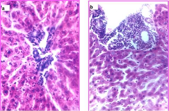
Elsharkawy et al.; ARJOCS, 3(1): 24-35, 2021; Article no.ARJOCS.415

polyunsaturated fatty acids and linoleic acids,             oxidative stress reported in our previous studies
which are essential for human nutrition [3].                [17,9]. Discovering the restoration effect of
Additionally, CM exhibits a wide range of                   camel’s milk against hepatotoxic effect was the
biological activities; antimicrobial, antioxidative,        main reason beyond the conduction of the
antithrombotic, antihypertensive, and immuno-               current experiment which aimed to investigate
modulatory effect [4,5]. This might be associated           the protective effect of camel’s milk against
with the unique composition of camel's milk:                methoxychlor induced liver toxicity.
indeed, the content of immunemodulatory
proteins, fatty acids, important minerals and               2. MATERIALS AND METHODS
vitamins allows camel's milk to be potentially
used as an anti-inflammatory, antidiabetic,                 2.1 Chemicals
hepato-protective and cardio-protective food
[6,7].                                                      Camel’s milk samples were collected daily early
                                                            in the morning from camel farm. Milk was
Methoxychlor is one of the environmental                    collected from camels by hand milking. The
contaminants which is widely used as a pesticide            samples were collected in sterile screw bottles
in many countries that was developed to replace             and kept in cool boxes until transported to the
dichloro-diphenyl-trichloroethane (DDT), and its            laboratory. The rats were given this fresh milk
chemical name is 1,1,1-trichloro-2,2-bis (p –               (100 mL/24 h/cage) as such without any further
methoxy phenyl) ethane. It has been reported                treatment.
that methoxychlor undergo hepatic microsomal
mono oxygenase mediated activation and the                  Methoxychlor                (1,1,1-trichloro-2,2-bis
resultant reactive metabolites possibly free                [methoxyphenyl] ethane, Approx 95% was
radicals    bind    covalently   to  microsomal             purchased from Sigma (St. Louis, Mo., USA).
components and induce liver damage [8,9].                   MXC was dissolved in corn oil (1:100). Reduced
Antioxidants/free radical scavengers, and                   glutathione (GSH) antioxidant enzyme and lipid
sulfhydryl containing compounds inhibit covalent            peroxide thiobarbituric acid reacting substances
binding of methoxychlor in human liver                      (TBARS) were measured using commercial test
microsomes, suggesting that the reactive                    kits supplied Bio-diagnostics (Bio- diagnostics,
intermediate is a free radical [10]. It has also            Cairo, Egypt). All other chemicals used in the
been reported that human cytochrome P-450                   experiment were of analytical grade.
enzymes responsible for conversion of MXC into
its major metabolites, the mono-o-demethylated              2.2 Animals and Treatment
derivatives and CYP1A2, have been shown to
play predominant role in this reaction [11]. The            A total of 100 adult female Sprague Dawley rats,
ability of cytochrome P-450 system to induce                4 to 6 weeks old, weighed about 100–150 gm
reactive oxygen species (ROS) has been                      were used in all experiments. They were
reported [12]. ROS are formed in both                       obtained from the Laboratory Animal House,
physiological and pathological conditions in                Assiut University, Egypt. The animals were
mammalian tissues, due to their high reactivity             housed in plastic cages on wood chips for
they may interact with biomolecules inducing                bedding and acclimated for 10 days before
oxidative stress [13]. Free radicals/ROS                    starting the experiment. All animals were housed
generated in tissues subcellular compartments               in standard cages (6 rats/cage), feeding with
are efficiently scavenged by the antioxidant                standard laboratory diet and tap water ad libitum.
defense system, which constitutes antioxidant               The rats were housed at 24-25 ‘C and humidity
enzyme such as superoxide dismutase, catalase,              (65%) and in daily dark/light cycle. The studies
and glutathione reductase and glutathione                   were conducted in accordance with the principles
peroxidase.      Under     normal   physiological           and procedures outlined in the National Institute
conditions free radicals/ROS are generated in               of Health of USA (NIH) guide for the Care and
subcellular compartments of liver which are                 Use of the Laboratory Animals [18].
subsequently scavenged by the antioxidant
defense system of the corresponding cellular                2.3 Experimental Design
compartments [14]. The organs production of
free radicals and dis-function of the antioxidant           The experiment is divided to two stages: First
defense system have been reported upon                      stage for 6 months and the second stage for 12
exposure to toxic chemicals [15,16]. MXC                    months. In both stages, rats were randomly
induced several organs damage due to the                    divided into four groups of twenty-five animals

                                                       25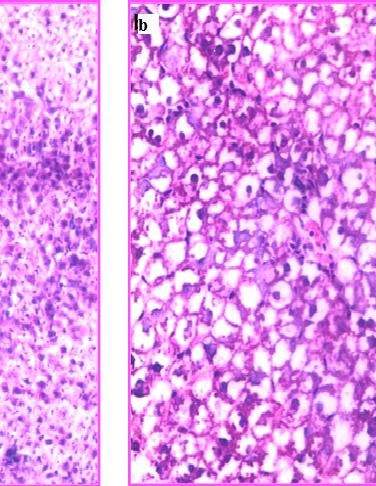
Elsharkawy et al.; ARJOCS, 3(1): 24-35, 2021; Article no.ARJOCS.415

each as follows: MXC -treated group received an            Na OH, 1.5 ml of 0.8% aqueous solution of
oral dose of MXC 200 mg/ kg b.w, twice/ week,              thiobarbituric acid and 0.2 ml liver homogenate
by gavages for 6 or 12 months. This dose was               (20% in 1.15% K Cl). The mixture was made up
selected according to Murono et al. [19]. MXC              to 4.0 ml with distilled water and kept in boiling
plus camel’s milk - treated group received                 water bath for 60 min. After cooling with tap
(100 ml/24 h/cage) as their sole source of                 water, the mixture was centrifuged at 2500g for
drinking for 6 or 12 months. Camel milk -treated           10 min. The supernatant was taken out and the
group was received daily a dose of                         intensity of pink color was measured at 532 nm
(100 ml/24 h/cage) as their sole source of                 on a spectrophotometer. TBARS were quantified
drinking, for 6 or12 months. This dose was used            using an extinction coefficient of 1.56 - 105 M1
according to the studies of Althnaian et al. [20].         cm1 and expressed as nmol of TBARS per mg
Control group received a daily oral dose of 2 ml           protein.
corn oil.
                                                           2.5.3 Estimation of reduced glutathione in
2.4 Sample Collections                                           liver

After 6 and 12 months of MXC exposure, female              GSH in the liver was assayed by the method
rats were anesthetized with CO, and decapitated.           described by Sun et al. [24]. The fresh tissues
Trunk blood was collected after decapitation and           were immediately homogenized in ice-cold 0.02
allowed to clot at 4°C. Sera were collected and            M EDTA solution. Aliquots of tissue homogenate
stored at -80°C to determine of serum total                were treated with a 50% w/v trichloroacetic acid
protein as well as liver function enzyme activities        while shaking, kept for 15 min and centrifuged.
(ALT, AST, and ALP). Meanwhile, the abdominal              After supernatant fractions were mixed with Tris
cavity was dissected immediately; the livers were          buffer (pH 8.9) and DTNB, absorbance at 412
separated for the histopathological examination.           nm was measured. Reduced glutathione was
                                                           used as an external standard. GSH levels were
2.5 Methodology                                            expressed as l mol/g tissue.

2.5.1 Biochemical assays                                   2.5.4 Determination of protein

Serum was used to determine total protein and              Protein concentrations were measured by the
albumin by colorimetric method according to                method of Bradford [25], using bovine serum
Doumas, [21]. The serum samples were assayed               albumin as a standard. Protein concentration
for aspartate transaminase (AST), alanine                  used for the concentration of reduced glutathione
transaminase (ALT), alkaline phosphatase (ALP)             and lipid peroxidation TBARS and can be
according to Rec, [22].                                    expressed as activity per milligram of protein by
                                                           dividing the units by milliliter of protein
2.5.1.1 Preparation of liver homogenate                    concentration.

Liver tissue homogenate was prepared according             2.6 Histopathological Examination
to the instruction of the kits. Briefly, 500 mg of
hepatic tissues was homogenized in 10 mL ice-              Liver specimens were fixed with 10%
cold phosphate buffer saline (50 mM, pH 7.4).              formaldehyde and processed routinely for
The homogenate mixture was centrifuged at                  paraffin embedding technique. Embedded
3800 × g (4 °C) for 15 min. The supernatant was            tissues were sectioned at 5mµ and stained with
used for measurement of reduced glutathione                hematoxylin and eosin (H&E) [26] for routine
dismutase     ((TBARS)),      thiobarbituric  acid         histopathological examination. They were then
((TBARS)),                                                 examined under the light microscope.

2.5.2 Estimation of lipid peroxidation in liver            2.7 Statistical Analysis

A breakdown product of lipid peroxidation,                 The data were analyzed using one-way ANOVA
thiobarbituric acid reacting substances (TBARS)            for all the experiments. Statistically significant
was measured by the method described by                    differences were determined using the Dunnett’s
Rungby and Ernst, [23]. In brief, the reaction             test for comparing to the vehicle-treated control
mixture consisted 0.2 ml of 8.1% SDS, 1.5 ml of            or the Bonferroni test for multiple comparisons.
20% acetic acid solution adjusted to pH 3.5 with           Graph Pad Prism graphing and the analysis

                                                      26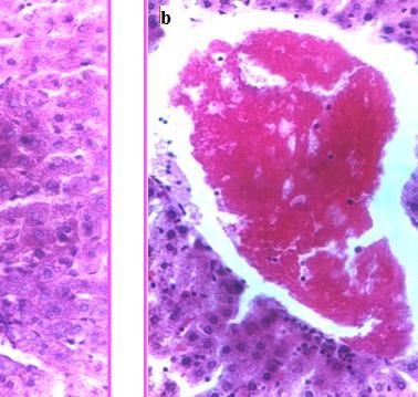
Elsharkawy et al.; ARJOCS, 3(1): 24-35, 2021; Article no.ARJOCS.415

software (version 4a; Graph Pad Software, Inc.,              rats. These alterations were obtained in both first
San Diego, CA) was used for all statistical                  (6 months) and second stages (12 months) of the
analyses. A statistically significant difference was         experiment. However, there was not a significant
confirmed at P < 0. 05.                                      difference between the control and camel milk
                                                             treated rats.
3. RESULTS
                                                             3.3 Histopathology
3.1 Biochemical Assays
                                                             At the first stage of the experiment, after 6
A significant reduction in the serum of total                months of exposure, the liver of rats treated with
protein and albumin concentration (g\dl) was                 MXC showed degeneration of the hepatocytes
obtained in both MXC and MXC plus camel milk                 which changed to coagulative necrosis
treated groups in the comparison with the control            associated        with      cellular   infiltration   of
at (P< 0.05), after 6 and 12 months of exposure.             mononuclear cells type Fig. (5.a). At the end of
On the other hand, a significant elevation in                this stage, there was severe fatty degeneration of
serum ALT, AST and ALP levels (U/I) were                     the hepatocytes with focal area of kupffer cell
recorded in MXC and MXC plus camel milk                      proliferation Fig. (5.b). In the second stage of the
treated rates compared with the control at (P <              experiment, after 12 months of exposure, the
0.05), after 6 and 12 months of exposure. There              liver tissue was severely damaged and there are
was a significant difference between MXC -                   apoptotic changes including condensation,
treated and MXC plus camel milk-treated                      increase esinophillia of cytoplasm shrinkage of
groups in total protein, albumin, and ALT, AST               the nucleus associated with area of cellular
and ALP serum levels in the first and second                 reaction Fig. (6.a). The hepatic blood vessels
stages of the experiment. The control and camel              were firstly congested and surrounded with
milk-treated rats had        equivalent serum                leukocytic       infiltration     (lymphocytes      and
concentrations of all previous parameters                    macrophages) and the large blood vessel filled
(Tables 1 and 2).                                            with prteinous material Fig. (6.b). The portal
                                                             areas showed some newly formed bile ducteules
3.2 The Oxidative            Status      of     Liver        denotes neoplastic changes in the portal tract
    Homogenate                                               associated with coagulation to cytoplasm and
                                                             cellular infiltration Fig. (7.a). The hepatocytes
As shown in Figs. 1 & 2 liver GSH levels,                    were disarranged, dissociated, along with some
expressed as (U/mg protein), were significantly              mitotic patterns. The blood vessels and bile ducts
lower in the MXC and MXC plus camel milk                     were dilated and highly infiltrated with
treated groups than in the control group at (P<              lymphocytes Fig. (7.b). The liver of rats treated
0.05). On the other hand, the liver TBARS                    with MXC and milk in the first and second stages
concentration, expressed as (U/mg protein), in               showed only mild degenerative changes in the
MXC and MXC plus camel milk treated groups                   hepatocytes and mild connective tissue
were significantly higher than in the control group          proliferation around the blood vessels and bile
at (P < 0.05) Figs. 3 & 4. There is a significant (p         ducts Fig. (8.a). The liver of rats treated with milk
< 0.0 5) difference in the activities of GSH and             only showed normal hepatocytes, blood vessels
TBARS levels in MXC plus camel milk treated                  and bile ducts Fig. (8.b).

    Table 1. The effect of the MXC and camel milk on the serum biochemical parameters in
                                 exposed groups for 6 months

 Groups         Tp            ALB             Globulin          AST                ALT            ALP
                 g\dl         g\dl            g\dl              U\l               U\l             U\l
 MXC            5.36±0.2*c     3.20±0.3*c     2.21±0.2 * c      301 ± 27.9 *bc    99.7 ±9.7*bc    786 ± 102.5 * bc
 Camel milk     5.82±0.3*c     3.49±0.1*c     2.34±0.3 * c      185 ± 19.8 *ac    67.9± 6.8 *ac   582 ± 29.8* ac
 MXC+
 Camel milk     6.70±0.4ab    3.72±0.2 ab     2.98±0.4 ab       112 ± 16.3 ab     37.8 ± 5.3 ab   386 ± 47.6 ab
 Control        7.56±o.1         4.23±0.2     3.33±0.1          127 ± 13.5        43.1 ± 6.1      385 ± 42.5
 Data are expressed as means ± S.D. of twenty animals per group. *denotes P < 0.05 as compared to control group,
      a. Denotes P < 0.05 as compared to MXC- group. b. Denotes P < 0.05 as compared to MXC+CM -group.
                   c. Denotes P < 0.05 as compared to CM – group (One- way ANOVA/Duncan)

                                                        27
Elsharkawy et al.; ARJOCS, 3(1): 24-35, 2021; Article no.ARJOCS.415

    Table 2. The effect of the MXC and camel milk on the serum biochemical parameters in
                                 exposed groups for 12 months

Groups                                  Tp             ALB             Globulin           AST               ALT             ALP
                                        g\dl           g\dl            g\dl               U\l               U\l             U\l
MXC                                     6.47±0.2* bc   3.42±0.44*c     2.24±0.33*bc       136.26±48.7 *bc   80.5± 6.7 *bc   89.12±10*bc
Camel milk                              6.81±0.1*a     3.94±0.34*c     2.34±0.23*ac       131.82±20.6 *ac   57.6± 3.8 *ac   78.88±21* ac
MXC +
Camel milk                              6.92±0.2a      4.19±0.23ab     2.94±0.32ab        117.02± 22.6 ab   48.4 ± 3.3 ab   56.13±5 ab
Control                                 7.56 ±0.3      4.23±0.30       3.35±0.43          112.30±24.5       42.0 ± 3.1      55.43±3
  Data are expressed as means ± S.D. of twenty animals per group. *denotes P < 0.05 as compared to control group,
       a. Denotes P < 0.05 as compared to MXC- group. b. Denotes P < 0.05 as compared to MXC+CM -group.
                    c. Denotes P < 0.05 as compared to CM – group (One- way ANOVA/Duncan)

                                   40

                                   35

                                                                                               28.6
       GSH levels (U/mg protein)

                                   30                                                                       27.3
                                                                                                      ab
                                   25
                                                                         19.2
                                   20
                                                                                *ac
                                   15                   12.5
                                                               *bc
                                   10

                                   5

                                   0
                                                                                      1
                                                                     Expermintal treated groups

                                                           MXC        MXC+CM              CM      Control

   Fig. 1. The effect of long exposure to MXC for 6 months on GSH levels and the restoration
                                       effect of Camel milk
Data are expressed as means ± S.D. of twenty animals per group. *denotes P < 0.05 as compared to control group, a denotes
P < 0.05 as compared to MXC- group. b denotes P < 0.05 as compared to MXC+CM -group. C denotes P < 0.05 as compared
                                       to CM – group (One- way ANOVA/Duncan)

4. DISCUSSION                                                                        camel milk could bring a significant decrease in
                                                                                     activities of these enzymes when compared to
In the present study, the chronic exposure of rats                                   MXC exposed groups. The increased serum
to MXC was associated with significant reduction                                     levels of hepatic markers have been attributed to
in the levels of serum total protein and albumin.                                    the liver injury, because these enzymes are
Moreover, the activities of serum marker                                             place in cytoplasmic area of the cell and are
enzymes (AST, ALT and ALP) were found                                                released into circulation in case of cellular
elevated markedly in rats treated with MXC.                                          damage [20]. At this point, our study supports
These changes were not observed in control rat                                       that liver damage is induced by MXC
samples. The present study revealed that                                             administration. As a matter of fact, the elevation
treatment with camel milk alone did not increase                                     in transaminases are encountered in conditions
the activities of serum AST, ALT and ALP levels.                                     causing hepatocellular damage, loss of functional
In addition, the simultaneous treatment with                                         integrity of cell membrane, and necrosis such as

                                                                                28
Elsharkawy et al.; ARJOCS, 3(1): 24-35, 2021; Article no.ARJOCS.415
in chemically induced liver injury and elevation in documented that the beneficial effects of
enzymes [27]. The rise in serum AST and ALT is fermented camel milk against cardiotoxicity
more specific and predominant in the liver injury. induced by carbon tetrachloride in mice.
The modulations in transaminases are also
influenced by the degree of hepatic These studies declared that the protective effect
decomposition related to cell necrosis [28]. A of camel milk against these toxicants induced
significant increase in ALP could occur in oxidative stress is due to its antioxidant
parenchymal liver disorders such as hepatitis properties. Camel milk was found to contain high
and cirrhosis, and striking elevation is concentrations of vitamins A, B2, C and E and is
encountered with extrahepatic biliary tract very rich in magnesium and other trace
(mechanical) obstruction or with intrahepatic elements, these vitamins act as antioxidants and
(functional cholestasis) [29]. Our have been found to be useful in preventing
histopathological findings confirmed toxicant-induced tissue injury [1,34,35]. Camel
hepatocellular damage where, on microscopic milk decreased (p < 0.05) MXC induced elevated
examination the livers in MXC-treated groups enzyme levels in tested groups, indicating the
revealed severe pathological damages such as: protection of structural integrity of hepatocytes
sinusoidal dilatation, congestion of central vein, cell membrane or regeneration of damaged liver
leukocytes and lymphocytes infiltration. Several cells [36]. The increase of albumin concentration
studies have provided a considerable support for after treatment with camel milk may be attributed
evidencing the protective effects of camel milk on to the decrease in lipid peroxidation processes
liver damage [30,31]. The anticytoxic and and increase in the activities of plasma protein
antigenotoxic effects of CM constituents against thiols as a result of treatment with camel
the genotoxic effects of chemicals as cisplatin milk in both animal and human [37,
anti-tumor agent are being investigated by 38,31]. Moreover, the results obtained from
Salwa and Lina [32]. Also, Hamed et al. [33] the liver histopathological analysis were in
35
30.6
30
27.3
ab
25
GSH levels (U/mg protein)
20
15.2
15
*ac
9.12
10
5
0
1
Expermintal treated groups
MXC MXC+CM CM Control
Fig. 2. The effect of long exposure to MXC for 12 months on GSH levels and the restoration
effect of Camel milk
Data are expressed as means ± S.D. of twenty animals per group. *denotes P < 0.05 as compared to control group, a denotes
P < 0.05 as compared to MXC- group. b denotes P < 0.05 as compared to MXC+CM -group. C denotes P < 0.05 as compared
to CM – group (One- way ANOVA/Duncan).
29Elsharkawy et al.; ARJOCS, 3(1): 24-35, 2021; Article no.ARJOCS.415
70
TBARS levels (nmol/g protein)
60 56.2
50 46.6
*bc
40 *ac 33.2 32.8
30
ab
20
10
0
1
Expermintal treated groups
MXC MXC+CM CM Control
Fig. 3. The effect of long exposure to MXC for 6 months on TBARS levels and the restoration
effect of Camel milk
Data are expressed as means ± S.D. of twenty animals per group. *denotes P < 0.05 as compared to control group, a denotes
P < 0.05 as compared to MXC- group. b denotes P < 0.05 as compared to MXC+CM -group. C denotes P < 0.05 as compared
to CM – group (One- way ANOVA/Duncan)
100
90
TBARS levels (nmol/g protein)
80 76.2
70
*bc
60
48.6
50
40 *ac 32.8
30.2
30
bc
20
10
0
1
Expermintal treated groups
MXC MXC+CM CM Control
Fig. 4. The effect of long exposure to MXC for 12 months on TBARS levels and the restoration
effect of Camel milk
Data are expressed as means ± S.D. of twenty animals per group. *denotes P < 0.05 as compared to control group, a denotes
P < 0.05 as compared to MXC- group. b denotes P < 0.05 as compared to MXC+CM -group. C denotes P < 0.05 as compared
to CM – group (One- way ANOVA/Duncan)
30Elsharkawy et al.; ARJOCS, 3(1): 24-35, 2021;; Article no.ARJOCS.415
no.ARJOCS
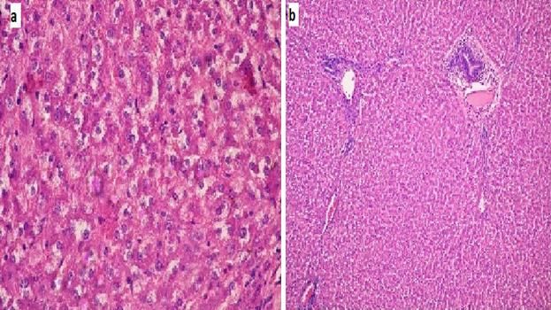
Fig. 5. a. Liver of rat treated with MXC for 6 months showed localized area of necrosis with
some apoptic changes in the hepatocytes, congestion, cellular infiltration of mononuclear
cells type. H & E; x25. b. Liver of rat treated with MXC for 6 months showed severe fatty
degeneration of the hepatocytes
patocytes with focal area of kupffer cell proliferation. H & E; x25
Fig. 6. a.. Liver hepatocytes of rat treated with MXC for 12 months’ showed apoptotic changes
including condensation, increase esinophillia of cytoplasm shrinkage of the nucleus
associated with area of cellular reaction. H & E; x25. b. Liver of rat treated with MXC for 12
months’ showed blood vessel filled with porteinous material and R.B.Cs. H & E; x25
31Elsharkawy et al.; ARJOCS, 3(1): 24-35, 2021; Article no.ARJOCS.415
Fig. 7. a. Liver of rat treated with MXC for 12 months showed newly formed bile ducteules
denotes neoplastic changes in the portal tract associated with cytoplasm coagulation. H & E;
x25. b. Liver of rat treated with MXC for 12 months showed large area of cellular infiltration in
the portal tract (lymphocyte type) associated with dissociation, disorganization of hepatocytes
along with some mitotic reactions. H & E; x25
Fig. 8. a. Liver of rat treated with mythoxychlor and camel milk showed nearly normal hepatic
cells and slightly congested central vein. H and E X 10. b. The liver of rats treated with camel
milk only showed normal hepatocytes, blood vessels and bile ducts.
consistence with the biochemical findings, protective role of CM. Furthermore, our findings
indicating that the decreased degeneration of indicated that MXC caused increased ROS
some hepatocytes and normal architecture of production, oxidative damage, and decreased
others. These effects demonstrated the hepato- antioxidant defense in the rat liver, which might
32Elsharkawy et al.; ARJOCS, 3(1): 24-35, 2021; Article no.ARJOCS.415
result in an oxidized state in the cells. It has been Taken together these nutrients enhance the
known that increased TBARS level and production of detoxifying molecules, absorption
decreased GSH concentration indicates an of antioxidant vitamins and activation of
increased generation of ROS, which cause lipid antioxidant enzymes which in turn activate the
peroxidation in the liver [39]. The mechanism of detoxification system and reduce the exerted
methoxychlor mediated oxidative stress is not oxidative stress.
very clear but it has been shown to be mediated
by the activation of microsomal monooxygenase, 5. CONCLUSION
which is involved in the conversion of
methoxychlor into its reactive metabolites [40]. In conclusion, camel milk may have a restoration
During this reaction the reactive metabolites, effect against MXC -induced liver damage and
possibly free radicals, bind covalently to may improve hepatic function parameters. Also,
microsomal components [8]. It has been shown camel milk has several antioxidant properties
that human cytochrome P-450 enzymes, could be efficient in the protection against the
responsible for the conversion of methoxychlor liver injury induced by MXC exposure in rats.
into its major metabolites and CYP1A2, have Therefore, camel milk may be recommended to
been shown to play a predominant role in this use against the hepatotoxic effects of MXC and
reaction [11]. It has been reported that ROS such further studies are needed toward other chemical
as H2O2 appears to be a key agent in the agents.
cytotoxic effects of the methoxychlor [41,42]. Our
results revealed that administration of camel milk COMPETING INTERESTS
in association with MXC slightly reduce TBARS
levels and elevate the level of GSH-Px, these Authors have declared that no competing
result were progressed in group treated with interests exist.
camel milk alone. That is due to the role of camel
milk in decreasing oxidative stress because it REFERENCES
contains high levels of antioxidants, vit. C, A and
E and is very rich in antioxidant minerals 1. Yousef MI. Aluminium-induced changes in
magnesium and zinc. Antioxidant vitamins are hemato-biochemical parameters, lipid
useful in reducing the oxidative stress. Vit. E and peroxidation and enzyme activities of male
magnesium have been suggested to enhance rabbits: protective role of ascorbic acid.
glutathione biosynthesis. Magnesium deficiency Toxicology. 2004;199(1):47-57.
has been associated with the production of 2. Darwish HA, Abd Raboh NR, Mahdy A.
reactive oxygen species [43]. Also, zinc is Camel’s milk alleviates alcohol-induced
essential for the activity of many enzymes in liver injury in rats. Food and Chemical
living organisms such as SOD and GPX. It has Toxicology. 2012;50(5):1377-1383.
been reported that zinc can prevent cell damage 3. MS Gorban A, Izzeldin OM. Fatty acids
through activation of the antioxidant system and lipids of camel milk and colostrum.
[44,45]. On the other hand, the protective International Journal of Food Sciences and
proteins (lactoferrin, lysozyme, and Nutrition. 2001;52(3):283-287.
immunoglobulins), and antioxidant enzymes 4. FitzGerald RJ, Meisel H. Milk protein-
(glutathione peroxidase and superoxide derived peptide inhibitors of angiotensin-I-
dismutase) play a crucial role in the cellular converting enzyme. British Journal of
oxidant–antioxidant balance. Furthermore, the Nutrition. 2000;84(S1):33-37.
bioactive peptides derived from Ca-M proteins 5. Korhonen H, Pihlanto A. Food-derived
during fermentation processes and hydrolysis bioactive peptides-opportunities for
reactions by proteolytic enzymes act protective designing future foods. Current
roles for body cells through the release of amino Pharmaceutical Design. 2003;9(16):1297-
acids such as proline [46]. In contrast with bovine 1308.
milk, Ca-M has lower antiallergic effects due to 6. Ibrahim ZS, Alkafafy M, Soliman MM,
the presence of immunoglobins and its protein Ahmed MM.. Molecular mechanism of
(free of β-lactoglobulin) profile. The short- and hepato-renal protection of camel milk
long-term regular consumption of Ca-M because against oxidative stress-perturbations.
of the suppression of oxidative/inflammation Journal of Camel Practice and Research.
stresses significantly improves diabetes and 2016;23(1):53-63.
hypertension in adults and the behavioral of 7. Mirmiran P, Ejtahed HS, Angoorani P,
children with autism spectrum disorder [47,46]. Eslami F, Azizi F. Camel milk has
33Elsharkawy et al.; ARJOCS, 3(1): 24-35, 2021; Article no.ARJOCS.415
beneficial effects on diabetes mellitus: A methoxychlor: The protective effect of
systematic review. International Journal of ascorbic acid. Journal of Advanced
Endocrinology and Metabolism. 2017; Veterinary Research. 2011;1(3):119-126.
15(2). 18. National Research Council, 2010. Guide
8. Bulger WH, Temple JE, Kupfer D. for the care and use of laboratory animals.
Covalent binding of [14C] methoxychlor National Academies Press.
metabolite (s) to rat liver microsomal 19. Murono EP, Derk RC, Akgul Y. In vivo
components. Toxicology and Applied exposure of young adult male rats to
Pharmacology. 1983;68(3):367-374. methoxychlor reduces serum testosterone
9. EL Nisr NA, El-Sharkawy EE, Abd Ellah levels and ex vivo Leydig cell testosterone
MR, Elsherif W, Kames GF, Sayed SM, formation and cholesterol side-chain
Wahba NM, Abdel-Hafeez MM, Aamer AA, cleavage activity. Reproductive Toxicology.
Abdel MFM. Ameliorative effect of propolis 2006;21(2):148-153.
against methoxychlor induced hepato renal 20. Althnaian T, Albokhadaim I, El-Bahr SM.
dysfunction. Basic Research Journal. Biochemical and histopathological study in
2013; 1:07-16. rats intoxicated with carbontetrachloride
10. Bulger WH, Kupfer DAVID. Characteristics and treated with camel milk. Springer Plus.
of monooxygenase-mediated covalent 2013;2(1):57.
binding of methoxychlor in human and rat 21. Doumas BT, Watson WA, Biggs HG.
liver microsomes. Drug Metabolism and Albumin standards and the measurement
Disposition. 1989;17(5):487-494. of serum albumin with bromcresol green.
11. Stresser DM, Kupfer D. Human Clinica Chimica Acta. 1971;31(1):87-96.
cytochrome P450–catalyzed conversion of 22. Rec GS. Determination of alkaline
the proestrogenic pesticide methoxychlor phosphatase. Journal of Clinical Chemistry
into an estrogen: Role of CYP2C19 & Clinical Biochemistry. 1972; 10:82.
and CYP1A2 in O-Demethylation. Drug 23. Rungby J, Ernst E. Experimentally induced
Metabolism and disposition. 1998;26(9): lipid peroxidation after exposure to
868-874. chromium, mercury or silver: interactions
12. Bondy SC, Naderi S. Contribution of with carbon tetrachloride. Pharmacology &
hepatic cytochrome P450 systems to the Toxicology. 1992;70(3):205-207.
generation of reactive oxygen species. 24. Sun Y, Elwell JH, Oberley LW. A
Biochemical Pharmacology. 1994;48(1): simultaneous visualization of the
155-159. antioxidant enzymes glutathione
13. Ochsendorf FR. Infections in the male peroxidase and catalase on
genital tract and reactive oxygen species. polyacrylamide gels. Free Radical
Human Reproduction Update. 1999;5(5): Research Communications. 1988;5(2):67-
399-420. 75.
14. Pelicano H, Feng L, Zhou Y, Carew JS, 25. Bradford MM. A rapid and sensitive
Hileman EO, Plunkett W, Keating MJ, method for the quantitation of microgram
Huang P. Inhibition of mitochondrial quantities of protein utilizing the principle of
respiration a novel strategy to enhance protein-dye binding. Analytical
drug-induced apoptosis in human leukemia Biochemistry. 1976;72(7):248-54.
cells by a reactive oxygen species- 26. Bancroft J, Stevens A, Turner D. Theory
mediated mechanism. Journal of Biological and practice of histological techniques 4th
Chemistry. 2003;278(39):37832-37839. Ed Churchill Living Stone, New York
15. Sujatha R, Chitra KC, Latchoumycandane, Edinburgh. Madrid, Sanfrancisco; 1996.
C, Mathur PP. Effect of lindane on 27. Nguyen NT, Braley S, Fleming NW,
testicular antioxidant system and Lambourne L, Rivers R, Wolfe BM.
steroidogenic enzymes in adult rats. Asian Comparison of postoperative hepatic
Journal of Andrology. 2001;3(2):135-138. function after laparoscopic versus open
16. Latchoumycandane C, Mathur PP. Effect gastric bypass. The American Journal of
of methoxychlor on the antioxidant system Surgery. 2003;186(1):40-44.
in mitochondrial and microsome-rich 28. Singhal RL, Merali Z. Biochemical toxicity
fractions of rat testis. Toxicology. 2002; of cadmium. In Cadmium toxicity. Marcel
176(1-2):67-75. Dekker New York. 1979;61-112.
17. Elsharkawy EE, Sharkawy AA. Evaluation 29. Salvatore F, Sacchetti L, Castaldo G.
of subacute toxicity induced by Multivariate discriminant analysis of
34Elsharkawy et al.; ARJOCS, 3(1): 24-35, 2021; Article no.ARJOCS.415
biochemical parameters for the 38. Al-Hashem F. Camel's milk protects
differentiation of clinically confounding liver against aluminum chloride-induced toxicity
diseases. Clinica Chimica Acta. 1997; in the liver and kidney of white albino rats.
257(1):41-58. American Journal of Biochemistry and
30. Hamad EM, Abdel-Rahim EA, Romeih EA. Biotechnology. 2009b;5(3):98-109.
Beneficial effect of camel milk on liver and 39. Nandi D, Patra RC, Swarup D. Effect of
kidneys function in diabetic Sprague- cysteine, methionine, ascorbic acid and
Dawley rats. International Journal of Dairy thiamine on arsenic-induced oxidative
Science. 2011;6(3):190-197. stress and biochemical alterations in rats.
31. Al-Fartosi KG, Majid A, Auda MA, Hussein Toxicology. 2005;211(1-2):26-35.
MH. The role of Camel’s milk against some 40. Miller KP, Gupta RK, Greenfeld CR, Babus
oxidant-antioxidant markers of male rats JK, Flaws JA. Methoxychlor directly affects
treated with CCl4. International Journal of ovarian antral follicle growth and atresia
Research of Pharmaceutical and through Bcl-2-and Bax-mediated
Biomedical Science. 2012;3(1):385-389. pathways. Toxicological Sciences. 2005;
32. Salwa MQ, Lina AK. Antigenotoxic and 88(1):213-221.
anticytotoxic effect of camel milk in mice 41. Gupta RK, Schuh RA, Fiskum G, Flaws
treated with cisplatin. Saudi Journal of JA. Methoxychlor causes mitochondrial
Biological Sciences. 2010;17(2):159-166. dysfunction and oxidative damage in the
33. Hamed H, Chaari F, Ghannoudi Z, ElFeki mouse ovary. Toxicology and Applied
A, Ellouz SC, Gargouri A. Beneficial effects Pharmacology. 2006;216(3):436-445.
of fermented camel milk by Lactococcus 42. Elsharkawy EE, Kames AO, Sayed SM,
lactis subsp cremoris on cardiotoxicity Nisr NA, Wahba NM, Elsherif WM, Nafady
induced by carbon tetrachloride in mice. AM, Abdel-Hafeez MM, Aamer AA. The
Biomedicine & Pharmacotherapy. 2018; ameliorative effect of propolis against
97:107-114. methoxychlor induced ovarian toxicity in
34. Zhu WW, Kong GQ, Ma MM, Li Y, Huang rat. Experimental and Toxicologic
X, Wang LP, Peng ZY, Zhang XH, Liu XY, Pathology. 2014;66(9-10):415-421.
Wang XZ. Camel milk ameliorates 43. Klevay LM. January. Advances in
inflammatory responses and oxidative cardiovascular-copper research. In First
stress and downregulates mitogen- International Biominerals Symposium:
activated protein kinase signaling Trace Elements in Nutrition, Health and
pathways in lipopolysaccharide-induced Disease, Montreal, Canada: Institut Rosell.
acute respiratory distress syndrome in rats. 2002;64-71.
Journal of Dairy Science. 2016;99(1):53- 44. Powell SR. The antioxidant properties of
56. zinc. The Journal of Nutrition. 2000;
35. Uversky VN, El-Fakharany EM, Abu-Serie 130(5):1447S-1454S.
MM, Almehdar HA, Redwan EM. Divergent 45. Sonia S, Fatima H, Tahar SM, Abdelhamid
anticancer activity of free and formulated K. Interrelationships between cadmium,
camel milk α-lactalbumin. Cancer zinc and antioxidants in the liver of the rat
Investigation. 2017;35(9):610-623. exposed orally to relatively high doses of
36. Palanivel MG, Rajkapoor B, Kumar RS, cadmium and zinc. Ecotoxicology and
Einstein JW, Kumar EP, Kumar MR, Environmental Safety. 2011;74(7):2099-
Kavitha K, Kumar MP, Jayakar B. 2104.
Hepatoprotective and antioxidant effect of 46. Izadi A, Khedmat L, Mojtahedi SY.
Pisonia aculeata L. against CCl4-induced Nutritional and therapeutic perspectives of
hepatic damage in rats. Scientia camel milk and its protein hydrolysates: A
Pharmaceutica. 2008;76(2):203-216. review on versatile biofunctional
37. Al-Hashem F, Mohammad D, Bashir N, properties. Journal of Functional Foods.
Mohammad A, Riyadh E, Mohammad K, 2019;60: 103441.
Al-Khateeb M. Camel's milk protects 47. Ismail T, Ahmad Z, Sestili P, Hussain M,
against cadmium chloride induced toxicity Akram K, Ismail A, Akhtar S. Camel's milk
in white albino rats. American Journal of concentrate inhibits streptozotocin induced
Pharmacology and Toxicology. 2009a; diabetes. Food Bioscience. 2018;26:
4(3):107-117. 73-79.
_____________________________________________________________________________________________________
© Copyright Global Press Hub. All rights reserved.
35You can also read