Correlation of dystonia severity and iron accumulation in Rett syndrome
←
→
Page content transcription
If your browser does not render page correctly, please read the page content below
www.nature.com/scientificreports
OPEN Correlation of dystonia severity
and iron accumulation in Rett
syndrome
Tz‑Yun Jan1,11, Lee‑Chin Wong2,11, Ming‑Tao Yang3,4,11, Chien‑Feng Judith Huang5,6,
Chia‑Jui Hsu7, Steven Shinn‑Forng Peng8*, Wen‑Yih Isaac Tseng1,5,8,9 & Wang‑Tso Lee1,10*
Individuals with Rett syndrome (RTT) commonly demonstrate Parkinsonian features and dystonia
at teen age; however, the pathological reason remains unclear. Abnormal iron accumulation in deep
gray matter were reported in some Parkinsonian-related disorders. In this study, we investigated the
iron accumulation in deep gray matter of RTT and its correlation with dystonia severity. We recruited
18 RTT-diagnosed participants with MECP2 mutations, from age 4 to 28, and 28 age-gender matched
controls and investigated the iron accumulation by susceptibility weighted image (SWI) in substantia
nigra (SN), globus pallidus (GP), putamen, caudate nucleus, and thalamus. Pearson’s correlation
was applied for the relation between iron accumulation and dystonia severity. In RTT, the severity of
dystonia scales showed significant increase in subjects older than 10 years, and the contrast ratios
of SWI also showed significant differences in putamen, caudate nucleus and the average values of
SN, putamen, and GP between RTT and controls. The age demonstrated moderate to high negative
correlations with contrast ratios. The dystonia scales were correlated with the average contrast ratio
of SN, putamen and GP, indicating iron accumulation in dopaminergic system and related grey matter.
As the first SWI study for RTT individuals, we found increased iron deposition in dopaminergic system
and related grey matter, which may partly explain the gradually increased dystonia.
Rett syndrome (RTT), a rare neurodevelopmental disease, is typically delineated by developmental variation in
cognition, hand use and motor functions. This disease involves approximately 1:9–10,000 female b irths1–3, most
of whom demonstrate a mutation in the MECP2 (methyl-CpG-binding protein 2) gene which discovered by
Amir et al.1,4–7. Although individuals with RTT exhibit relatively normal infant development8–11, they manifest
stereotypical hand movements and gait abnormalities with poor balance between ages 1 and 4 years12. Between
ages 2 and 10 years, symptoms stabilize after the period of regression. Although some individuals with RTT may
remain at this stage for the rest of their lives, others may deteriorate from the teen years or early twenties, show-
ing muscle weakness, rigidity, and spasticity and developing bradykinesia, scoliosis, and distal-limb distortion
due to a long period extrapyramidal involvement13,14. Parkinsonian features may also develop with dystonia
in the limbs and the head and neck. These symptoms are possibly correlated with nigrostriatal–dopaminergic
pathway dysfunction and abnormal MECP2 i nput15,16. The possibility of premature aging was also s uggested17.
However, the exact cause of degeneration and parkinsonian features in RTT is not clear. Abnormal iron
accumulation in deep brain nuclei was found in a heterogeneous group of neurodegenerative diseases with
brain iron accumulation (NBIA), characterized by extrapyramidal movements. Individuals with NBIA may
show approximately three times the normal deposition of iron in the globus pallidus (GP) and substantia nigra
(SN) but normal amounts in other brain regions18,19, indicating that accumulation of iron in deep brain nuclei
may associate to extrapyramidal symptoms. Iron may also accumulate in brain structure of healthy people due
1
Graduate Institute of Brain and Mind Sciences, College of Medicine, National Taiwan University, Taipei,
Taiwan. 2Department of Pediatrics, Cathay General Hospital, Taipei, Taiwan. 3Department of Pediatrics, Far
Eastern Memorial Hospital, New Taipei City, Taiwan. 4Department of Chemical Engineering and Materials Science,
Yuan Ze University, Taoyuan, Taiwan. 5Institute of Medical Device and Imaging, National Taiwan University
College of Medicine, Taipei, Taiwan. 6Institute of Biomedical Engineering, National Taiwan University College
of Medicine, Taipei, Taiwan. 7Department of Pediatrics, National Taiwan University Hospital SinChu Branch,
Taipei, Taiwan. 8Department of Medical Imaging, National Taiwan University Hospital and National Taiwan
University College of Medicine, Taipei, Taiwan. 9Molecular Imaging Center, National Taiwan University, Taipei,
Taiwan. 10Department of Pediatrics, National Taiwan University Hospital and National Taiwan University College
of Medicine, 8, Chung‑Shan South Road, Taipei 100, Taiwan. 11These authors contributed equally: Tz-Yun Jan,
Lee-Chin Wong and Ming-Tao Yang. *email: steven0131@mail2000.com.tw; wangtsolee@ntu.edu.tw
Scientific Reports | (2021) 11:838 | https://doi.org/10.1038/s41598-020-80723-1 1
Vol.:(0123456789)www.nature.com/scientificreports/
to aging. A postmortem study revealed that iron was accumulated at a higher level in extrapyramidal structures
than in the cortex20. Susceptibility weighted imaging (SWI) studies also revealed that mineralization of the deep
gray matter increased with age from infancy to late a dulthood21–23. Excessive iron deposits may induce oxidative
stress and damage mitochondrial function in neurons. Therefore, the over-accumulation of iron in deep brain
nuclei may cause irreversible cellular injury, leading to dystonia or parkinsonian features.
Recent studies investigating differences in iron deposition in Parkinson’s disease (PD) and atypical PD found
increased iron in the SN and caudate nucleus (CN) in PD and in the putamen and GP in atypical PD. Iron content
was also correlated with clinical severity24,25. Recently, the effect of extraordinary iron accumulation in other
neurological diseases mimicking atypical RTT has been deliberated. An individual with WDR45 mutation was
reported to have signal loss in SN and GP from T1 and T2* weighted imaging26. However, age-related changes in
iron content in RTT and its correlation with parkinsonian features and dystonia severity in typical RTT remain
unclear. Therefore, the primary purpose of this study was to depict iron deposition in the deep gray matter in
RTT and investigate its relation with dystonia severity. We used a ferromagnetic-sensitive technique, SWI, to
gain information about mineralization in the basal ganglia in individuals with RTT. We hypothesized that iron
accumulation in the deep gray matter may be related to dystonia severity in patients with RTT.
Method
Participants. A total of 18 female RTT patients with MECP2 mutations were recruited. All study partici-
pants were enrolled from the Joint Clinic for Rett syndrome in National Taiwan University Hospital and were
diagnosed to be RTT based on the revised diagnostic criteria27 by the major neurologist (Lee W.T.). We further
subdivided the patients with RTT into two age groups (age < 10 years and age > 10 years). Twenty-eight age- and
gender-matched healthy controls without neurologic diseases (e.g., psychiatric diagnoses, history of neurologi-
cal impairment, neuropsychiatric conditions, or genetic disorders) were also enrolled for comparison. Patients
with RTT were sedated with chloral hydrate before scanning to avoid motion artifacts. During the scanning
period, heart rate and pulse oximetry were continuously monitored. The study was approved by the Institutional
Review Board of the National Taiwan University Hospital, and informed consent was obtained from all partici-
pants or their legal guardians (201510011RINC). All methods were performed in accordance with the relevant
guidelines and regulations.
MRI protocol. In the present study, we utilized SWI to acquire information on abnormal metal concentra-
tions in the deep gray matter. The SWI method applies the principal of susceptibility difference, and tissues can
be distinguished by their contrast signals in sufficiently long echo times. Iron has paramagnetic properties and
increases local magnetic susceptibility when an external magnetic field is applied. Tissues containing iron will
strengthen and interfere with the local magnetic field. This characteristic can be applied to quantitatively deter-
mine the iron concentration in a tissue28,29. To generate images, SWI uses a three-dimensional gradient echo
sequence with a long echo time and flow compensation to acquire high-resolution magnitude imaging and phase
imaging. Phase imaging can reflect static magnetic field inhomogeneities and be utilized as a high-pass filter to
mask the original magnitude imaging by multiplication30.
All of the scans were acquired using a Siemens 3 T scanner (Tim Trio, Germany) with a 32-channel phased
array coil. The MR imaging protocol consisted of sagittal T1-weighted, axial fast spin-echo T2-weighted imag-
ing, and SWI with flow compensation, and the following sequences were acquired: T1 MPRAGE (slice thick-
ness = 1.0 mm, TR/TE = 2000/2.98 ms, flip angle = 9°, FOV read = 256 × 192 mm2, and 208 slices per slab), T2
(slice thickness = 2.5 mm, TR/TE = 9650/103 ms, FOV = 200 × 200 mm2, and 56 contiguous axial-weighted
images), and SWI (slice thickness = 2.5 mm, TR/TE = 28/20 ms, flip angle 15°, FOV = 200 × 200 m m2, and 56
contiguous axial-weighted images) with flow compensation (TR/TE = 28/20 ms, FOV = 230 × 230 mm2, voxel
size = 0.8 × 0.7 × 1.6 mm, flip angle 15°, and 72 slices per slab with 22.2% oversampling). Images were read on a
commercially available workstation of Siemens by the neuroradiologist (Peng, S. F.) for excluding the presence
of infarcts and hemorrhages.
Image analysis. Analysis of signal intensity on SWI was documented according to the anatomical struc-
tures of bilateral regions of interest (ROIs) (SN, CN, putamen, GP, and thalamus) on the slice, where they were
visualized best. Visual grading scale, a semi-quantitative evaluation was applied to score the signal intensity in
10 ROIs. Hypointensity was measured according to a relative grading scale (compared with the cerebrospinal
fluid, CSF) by visual discrimination, ranging from 0 to 3 points (0: SI similar to CSF intensity; 1: mild; 2: moder-
ate; 3: severe)31. Additionally, to compare the inhomogeneity, a quantitative evaluation, contrast ratio was used
to examine the image variation of 10 ROIs. Each ROI was measured bilaterally as signal intensity (SROI) and
calculated against the occipital white matter (SO) as a contrast ratio “CR = (SROI − SO)/SO”. The occipital white
matter of individuals with RTT is considered less variable, so it was chosen as a comparable signal baseline. To
investigate the effect of changes in the dopaminergic system on dystonia severity, the average value of all regions
in the dopaminergic system and related grey matter, including the SN, putamen and GP, was obtained as a new
variable. All images were analyzed independently by an experienced neuroradiologist (Peng, S. F.), who was
blind to clinical symptoms, following defined radiological protocols.
Behavioral measurements. Parents/caregivers were requested to complete a questionnaire to collect
clinical information, including age, gene mutations, motor function, and clinical features. The Rett Syndrome
Gross Motor Scale includes 15 gross motor items with levels from ability to sit to challenging mobility32. We
grouped items into four subgroups to present a general view of motor function. The hand function scale is uti-
lized to evaluate the level of hand–eye coordination. Considering the hand function of patients with RTT, the
Scientific Reports | (2021) 11:838 | https://doi.org/10.1038/s41598-020-80723-1 2
Vol:.(1234567890)www.nature.com/scientificreports/
scale was designed ranging from level 1 (“no observed hand function”) to level 8 (can orientate and determine
with reasonable accuracy the position and size of objects when the hand is approaching them)32. For evaluation
of clinical features, the Rett Syndrome Severity Scale and Rett Syndrome Behaviour Questionnaire were used for
research assessment. The descriptions include the severity level of symptoms, pathological behaviors and diverse
clinical features in RTT33–35. Lastly, severity scales of dystonia were measured by experienced neurologists. We
used the Fahn–Marsden scale to evaluate dystonia in 9 body areas with a weighting factor (eyes, mouth, speech
and swallowing, neck, bilateral arms, bilateral legs, trunk), which halves the contribution of dystonia in the eyes,
mouth and n eck36. Another scale, the Unified Dystonia Rating Scale, was also used to perform a more detailed
assessment of body areas (e.g., to separate the item ‘face’ into ‘eye and upper face’ and ‘lower face’) and to add
severity and duration ratings for a more comprehensive observation37.
Statistical analysis. Clinical features of the two age groups (age < 10 years and age > 10 years) were com-
pared by using a nonparametric Mann–Whitney U test. The nonparametric U test was also applied to assess
between-group differences in signal intensity on the visual grading scale. An independent t test was used to com-
pare contrast ratios between the RTT and control groups in 5 ROIs and the average value of 3 ROIs. Additionally,
a multiple linear regression model was used to explore the abnormal association between signal intensity and
age in the RTT group. We assigned group as a dummy variable (control = 0, RTT = 1) and used centering meth-
odology to reduce multicollinearity with the age variable and interaction term (age × group). A full regression
model containing both the main effects of age and group, as well as interaction effects, was represented by the
following formula:
Ycontrast ratio = Intercept + β1 group + β2 age_centered + β3 age_centered × group + e
Moreover, Pearson correlation analysis was used to investigate the relation between dystonia severity and
signal intensity for each ROI and average values of 3 ROIs. All statistical analyses were performed using IBM SPSS
Statistics 20 (SPSS Inc., Chicago, IL, USA) and adjusted for multiple comparison with a Benjamini–Hochberg
correction. Differences were considered statistically significant at P < 0.05.
Financial disclosure. There is no financial disclosure for all authors.
Results
Demographic and clinical characteristics. We recruited 18 female RTT patients with MECP2 muta-
tions (the RTT group) and 28 age- and gender-matched healthy controls (the control group); however, one indi-
vidual with RTT and two controls were excluded due to poor SWI imaging quality. The mean ages of the RTT
group (n = 17) and the control group (n = 26) were 15.30 ± 8.1 years and 16.21 ± 7.9 years, respectively (p = 0.812).
There was no significant difference in clinical features of speech, gross motor function, fine motor function, Rett
syndrome severity, and symptoms between the two age groups in patients with RTT, although the older patients
with RTT showed more impairment in each item compared with the control group (Table 1). Dystonia scales
showed significantly increased dystonia severity in patients older than 10 years in both the Unified Dystonia
Rating Scale (p = 0.010) and Fahn–Marsden scale (p = 0.007).
Differences in signal intensity in the deep gray matter. In the present study, a visual grading scale
and contrast ratio were applied to evaluate the abnormality of iron deposition in RTT. On the visual grading
scale, both controls and patients with RTT had lower hypointensity scores (0 or 1) for the CN, putamen, and
thalamus. Although there was no significant difference in visual grading scale between the RTT and control
groups (corrected p = 0.295), patients with RTT showed a more severe hypointensity (mild: 56%; moderate: 33%;
severe: 0.6%) than controls (mild: 82%; moderate: 11%) in GP. In contrast, the signal intensity in the SN differed
significantly between the RTT and control groups (p = 0.014) (Fig. 1). There was no significant difference in
hypointensity in the thalamus for both control and patient groups. In the contrast ratios, although mean values
in the RTT group were more negative than those in the controls in all regions, only the putamen (p < 0.001), CN
(p < 0.001), and average (p = 0.025) showed significantly lower values in RTT after correction (Table 2). The mean
value of GP was also lower in RTT although it did not reach statistic significance (p = 0.056).
The results indicated that visual grading scales may be less reliable than contrast ratios in evaluating the
change in SWI.
Age-related changes in iron accumulation were also investigated in the present study (Fig. 2). In healthy
controls, age demonstrated moderate to high negative correlations with contrast ratios in all regions. In con-
trast, in the RTT group, age only demonstrated a moderate to high negative correlation in the SN (R = − 0.759,
p < 0.001), putamen (R = − 0.536, p = 0.027), and GP (R = − 0.732, p = 0.001). In addition, the contrast ratios in
individuals with RTT were significantly lower than those in healthy controls with more negative slopes in the
SN and GP (Figs. 2, 3).
In a multiple regression model, age showed a significant effect on the change in iron deposition in 5 ROIs
(Table 3). However, the group effect played a more important role than age in the CN and putamen (Table 3).
Although the contrast ratio decreased significantly in individuals with RTT over the entire age range in GP
(standardized B of group difference = − 0.328, p = 0.006), the change was more related to age factor (standard-
ized B of age difference = − 0.547, p = 0.001) than group factor. In all multiple regression models, no interaction
was found between age and group.
Scientific Reports | (2021) 11:838 | https://doi.org/10.1038/s41598-020-80723-1 3
Vol.:(0123456789)www.nature.com/scientificreports/
RSGMS Fahn–
RSSS level Marsden
No Age Gene mutation Speech Sitting Standing Walking Challenge HF (score) RSBQ scale UDRS
1 4.2 MECP2 c.880C>T, p.R294X − + + + + 2 Moderate (9) 45 28 37.5
2 6.0 MECP2 c.392 C>A + + + + + 8 Mild (7) 30 28 42.5
3 6.2 MECP2 c.392 C>A + + + + + 8 Mild (3) 29 24.5 37.5
4 7.2 MECP2 c.502 C>T p.R168X − + + + − 3 Mild (6) 20 26.5 33.5
5 8.1 MECP2 c.502 C>T p.R168X − + + + − 1 Moderate (13) 38 30.5 65.5
6 8.9 MECP2 c.808 C>T p.R270X − − − − − 2 Severe (16) 44 53.5 54.5
7 9.1 MECP2 c.502 C>T p.R168X − + − − − 1 Moderate (10) 51 30.5 52.5
8 14.3 MECP2 c.1161 C>A − + − − − 1 Moderate (13) 34 82.5 72
9 14.2 MECP2 c.502 C>T p.R168X − + + + − 1 Mild (6) 31 53 66.5
10 15.6 MECP2 c.446C>A p.S149Y − + + + − 7 Moderate (8) 20 13 33.5
MECP2 856-859del4 AAAG del,
11 18.0 − + − − − 5 Severe (18) 56 86.5 75
frameshift (K286fs )
MECP2 frameshift insertion
12 20.8 − + + + − 2 Moderate 10) 70 65 66
38_39insTCCT (G13fs)
13 22.8 MECP2 c.763C>T p.R255X + + + + − 7 Moderate (10) 49 38.5 56
14 25.3 MECP2 c.763C>T p.R255X − − − − − 1 Moderate (11) 56 72.5 60.5
15 25.9 MECP2 c. 502 C>t, p.R168X − + + + − 2 Moderate (12) 39 59 71
16 26.9 MECP2 mutation (losing data) − + + + − 1 Moderate (12) 63 73 84.5
17 28.1 MECP2 c.763C>T p.R255X − + + + − 1 Moderate (11) 38 63 73
Age < 10 2(29%) 6(86%) 5(71%) 5(71%) 3(43%)
Age > 10 1(10%) 9(90%) 7(70%) 7(70%) 0
36.71 31.64
Age < 10 Mean (SD) 3.57 (2.52) 9.14 (4.37) 46.21 (11.60)
(10.88) (9.86)
45.6 60.60
Age > 10 Mean (SD) 2.8 (2.53) 11.1 (3.17) 65.80 (13.82)
(15.74) (21.82)
p value 0.232 0.315 0.230 0.010* 0.007**
Table 1. Demographic and clinical features of patients with Rett Syndrome. Age group < 10 years
(n = 7), > 10 years (n = 10); Speech +: single word, −: mute. MECP2 methyl-CpG-binding protein 2, RSGMS
Rett Syndrome Gross Motor Score, HF hand function scale, RSSS Rett Syndrome Severity Score, RSBQ Rett
Syndrome Behaviour Questionnaire, UDRS Unified Dystonia Rating Scale. *p < 0.05, **p < 0.001. Adjusted for
multiple comparison with Benjamini–Hochberg correction.
Figure 1. SWI hypointensity score in five deep gray matter. Stacked column chart descripted the percentage
of visual grading scale for hypointensity of RTT group and control group. Significant difference was noted in
substantia nigra (p = 0.014). Generally, hypointensity signal was higher in RTT group than the controls, except
the signal in thalamus. (CTL control group, RTTRett syndrome, SN substantia nigra, CN caudate nucleus, PU
putamen, GP globus pallidus, TH thalamus).
Correlation of clinical dystonia severity and signal intensity. Abnormal iron accumulation has
been found in patients with PD and WDR45 mutations. Because adult individuals with RTT also developed
more dystonia and parkinsonian features, we further investigated the correlation of iron accumulation and dys-
tonia severity (Table 4). Both the Fahn–Marsden scale and the Unified Dystonia Rating Scale had a high correla-
Scientific Reports | (2021) 11:838 | https://doi.org/10.1038/s41598-020-80723-1 4
Vol:.(1234567890)www.nature.com/scientificreports/
RTT CTL 95% CI
Deep gray matter regions Mean Std. deviation Mean Std. deviation t p value (2-tailed) Lower Upper
Substantia nigra (SN) − 0.189 0.093 − 0.165 0.094 − 0.803 0.427 − 0.082 0.036
Caudate nucleus (CN) − 0.090 0.041 − 0.021 0.068 − 3.725 < 0.001** − 0.106 − 0.031
Putamen − 0.063 0.043 0.022 0.074 − 4.307 < 0.001** − 0.125 − 0.045
Globus pallidus (GP) − 0.213 0.121 − 0.147 0.098 − 1.963 0.056 − 0.134 0.002
Thalamus − 0.022 0.049 0.004 0.071 − 1.330 0.191 − 0.066 0.014
Average value − 0.154 0.079 − 0.096 0.080 − 2.332 0.025* − 0.108 − 0.007
Table 2. Comparison of contrast ratio in five deep gray matter between patients with Rett syndrome and
controls. Average value: average values of SN, PU and GP. *p < 0.05, **p < 0.001. Adjusted for multiple
comparison with Benjamini–Hochberg correction.
Figure 2. Scatter plots of the estimated age-related regression between age and contrast ratio. Regression model
indicated the contrast ratio decreased significantly with age in both RTT and control groups. However, the
contrast ratios were lower in RTT. The green cross/bold line was the data for RTT, and the blue square/dash line
was the data for the controls.
tion (R = 0.84, p < 0.001) in the present study. The Fahn-Marsden scale showed a moderately negative correlation
with the contrast ratio in SN (R = − 0.518, p = 0.033), GP (R = − 0.514, p = 0.035), and the average value of 3 ROIs
(R = − 0.446, p = 0.003), indicating that the contrast ratio was associated with dystonia severity. In addition, the
Unified Dystonia Rating Scale showed a significant negative correlation with SN (R = − 0.529, p = 0.029). How-
ever, only the correlations between the Fahn–Marsden scales and the average values of the contrast ratios in the
SN, putamen, and GP remained significant after Benjamini–Hochberg correction.
Discussion
To our best knowledge, the present study is the first of its kind on age-related changes in SWI for RTT. We found
evidence of age-related changes in iron accumulation in the deep gray matter in both RTT and control groups.
Furthermore, iron deposition, revealed abnormally increased in the CN, putamen, and dopaminergic system
and related grey matter (SN, putamen, and GP) in RTT, which was correlated with dystonia severity.
Abnormalities in the deep gray matter of RTT have been widely reported. A postmortem study mentioned
that small cells, increased packing density, and underpigmentation in the zona compacta were shown in SN38,39.
Neuropathological and neurochemical studies also showed abnormalities in dopaminergic terminals and deficits
Scientific Reports | (2021) 11:838 | https://doi.org/10.1038/s41598-020-80723-1 5
Vol.:(0123456789)www.nature.com/scientificreports/
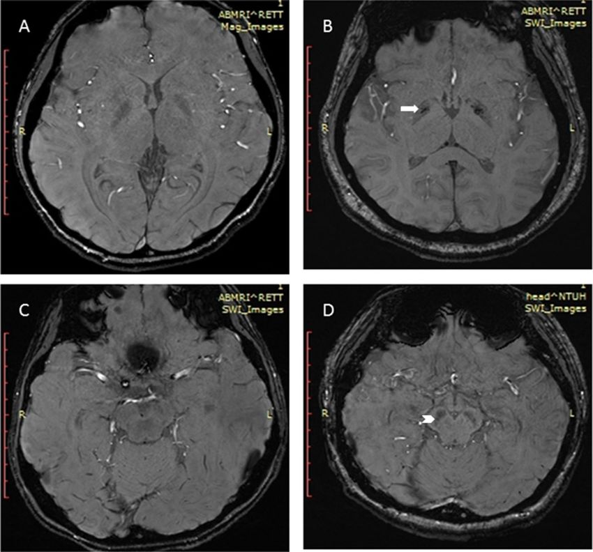
Figure 3. Susceptibility-weighted images in healthy control (A,C) and Rett syndrome patient (B,D) showing
the relative hypointensity in globus pallidus (B, arrow) and substantia nigra (D, arrowhead) in Rett syndrome.
in fluorodopa uptake in the CN and p utamen38,40. Moreover, structural alterations, including volume reductions,
15,41
were found in the CN and t halamus . Nevertheless, the association between these abnormalities and dystonia
severity in RTT remains unclear. In our study, we found significantly increased iron deposition in the SN, puta-
men, and GP, and the iron deposition was correlated with dystonia severity in patients with RTT. These regions
also showed more negative age-related iron changes in patients with RTT than in controls.
Dysfunctional iron regulation and abnormal iron aggregation may cause oxidative stress leading to cell
death23.The link between oxidative stress and RTT has been extensively investigated42–44. One of the functions
of the MECP2 gene might be protection of the CNS from hypoxia. Individuals with RTT are known to have
autonomic dysfunction, characterized by dysregulated respiration and reduced arterial o xygen45. This might lead
to hypoxic conditions and a systemic redox imbalance in the brain, leading to higher iron release46. Abnormal
iron release may cause excessive oxidative stress, eventually resulting in extrapyramidal dysfunction, such as
dystonia47. Along with age-related iron accumulation, dystonia may also worsen in individuals with RTT. The
present study showed that individuals with RTT older than 10 years were significantly deteriorated in dystonia
compared with those at a younger age, which may be related to iron accumulation in the dopaminergic system
and related grey matter. This result is coincident with previous studies showing that older girls with RTT had
more dystonia and gross motor d eterioration17,32.
In healthy people, iron is distributed in a regional, age-related pattern in the extrapyramidal system. A post-
mortem study discovered that non-heme iron in SN and GP increased rapidly from birth to the first two decades
of life; then, there was no further change in subsequent years, even though the variability of iron values in the SN
increased considerably with age. On the other hand, iron accumulated more slowly in the putamen and CN of
healthy people and was deposited maximally after middle a ge20,48. In the present study, individuals with RTT and
healthy subjects demonstrated a similar age-related change in signal intensity in the dopaminergic system and
related grey matter, but there was an increased rate of iron accumulation in individuals with RTT from a young
age, indicating exacerbation of aging in these patients. The regression model showed that the group difference
Scientific Reports | (2021) 11:838 | https://doi.org/10.1038/s41598-020-80723-1 6
Vol:.(1234567890)www.nature.com/scientificreports/
Standardized (B) t Sig R Adj. R2 F Sig
Substantia nigra
Intercept − 11.96 < 0.001** 0.697 0.446 12.272 < 0.001**
Group − 0.161 − 1.396 0.171
Age − 0.655 − 4.366 < 0.001**
Age × group − 0.048 − 0.318 0.752
Caudate nucleus
Intercept − 1.824 0.076 0.600 0.31 7.301 0.001**
Group − 0.514 − 4.004 < 0.001**
Age − 0.422 − 2.522 0.016*
Age × group 0.223 1.333 0.19
Putamen
Intercept 2.177 0.036* 0.699 0.449 12.387 < 0.001**
Group − 0.577 − 5.031 < 0.001**
Age − 0.49 − 3.273 0.002**
Age × group 0.125 0.837 0.407
Globus pallidus
Intercept − 9.032 < 0.001** 0.709 0.464 13.135 < 0.001**
Group − 0.328 − 2.902 0.006**
Age − 0.547 − 3.709 0.001**
Age × group − 0.14 − 0.951 0.348
Thalamus
Intercept 0.495 0.624 0.509 0.202 4.54 0.008**
Group − 0.224 − 1.624 0.112
Age − 0.554 − 3.074 0.004**
Age × group 0.161 0.892 0.378
Table 3. Multiple linear regression for contrast ratio of deep gray matter with age and group. Group was
calculated as Dummy variable: control = 0, RTT = 1. *p < 0.05, **p < 0.01.
Age Average SN CN PU GP TH
Fahn_Marsden
R 0.632** − 0.446* − 0.518* 0.137 − 0.274 − 0.514* − 0.496
p value 0.006 0.003 0.033 0.599 0.288 0.035 0.053
UDRS
R 0.686** − 0.465 − 0.529* 0.198 − 0.305 − 0.403 − 0.355
p value 0.002 0.060 0.029 0.447 0.235 0.109 0.162
Age
R – − 0.637** − 0.759** − 0.122 − .0536* − 0.737* − 0.404
p value – < 0.001 < 0.001 0.641 0.026 0.001 0.108
Table 4. The correlation between dystonia feature, age and contrast ratio of deep gray matter regions in
patients with RTT. CTL control, Average average values of SN, PU and GP, SN substantia nigra, CN caudate
nucleus, PU putamen, GP globus pallidus, TH thalamus, UDRS Unified Dystonia Rating Scale. *p < 0.05,
**p < 0.01. Adjusted for multiple comparison with Benjamini–Hochberg correction.
between RTT and controls influenced iron accumulation in the putamen and CN significantly more than age,
but the alterations in these regions were not correlated with dystonia severity and other clinical presentations,
possibly indicating a sign of early brain aging in RTT. Early aging had been observed in clinical characteristics
of middle-age RTT. Previous studies demonstrated premature neuromuscular aging in RTT, with loss of muscle
strength and muscle volume and a change in the texture of skin and h air14,17, compatible with the brain changes
in the present study.
In our study, dystonia severity in RTT was only correlated with iron accumulation in the dopaminergic system
and related grey matter (SN, putamen and GP). The damage caused by iron accumulation in the dopaminergic
system and related grey matter may lead to oxidative stress, cell death, and secondary defects in axonal transport,
leading to dystonia.
As this is the first investigation of iron accumulation in RTT, there were some limitations in our study. First,
methodological limitations lead to differences in the results for the visual grading scale and contrast ratio.
Scientific Reports | (2021) 11:838 | https://doi.org/10.1038/s41598-020-80723-1 7
Vol.:(0123456789)www.nature.com/scientificreports/
Although the visual grading scale may be influenced by the signal-to-noise ratio and lead to deviation in visual
judgment, it remains the simplest and the most immediate analytical tool for clinical experts. The small sample
size is the second limitation of the present study. The age range of the study also limited the interpretation of
iron deposition. Another limitation of this study is that we did not have sufficient parkinsonian information on
RTT. Therefore, a longitudinal follow-up study with an effective sample size and a more comprehensive clinical
investigation is desired in the future.
In conclusion, the present study found abnormal iron accumulation in the deep gray matter of individuals
with RTT, with exacerbated iron deposition in the putamen, CN, and dopaminergic system and related grey
matter (SN, putamen, and GP). Iron accumulation in the dopaminergic system and related grey matter was also
significantly correlated with dystonia severity in RTT. This result revealed a new perspective on how unusual iron
accumulation accelerates neuropathological damage and aging in the RTT brain. Further longitudinal studies
are mandatory for clarification of early aging and the change in iron accumulation in the brains of individuals
with RTT.
Data availability
Anonymized data will be shared by request from any qualified investigator.
Received: 31 January 2020; Accepted: 21 December 2020
References
1. Laurvick, C. L. et al. Rett syndrome in Australia: A review of the epidemiology. J. Pediatr. 148, 347–352 (2006).
2. Wong, V. C. N. & Li, A. S. Y. H. Rett syndrome: Prevalence among Chinese and a comparison of MECP2 mutations of classic Rett
syndrome with other neurodevelopmental disorders. J. Child. Neurol. 22, 1397–1400 (2007).
3. Bienvenu, T. et al. The incidence of Rett syndrome in France. Pediatr. Neurol. 34, 372–375 (2006).
4. Amir, R. E. et al. Rett syndrome is caused by mutations in X-linked MECP2, encoding methyl-CpG-binding protein 2. Nat. Genet.
23, 185–188 (1999).
5. Vacca, M. et al. Mutation analysis of the MECP2 gene in British and Italian Rett syndrome females. J. Mol. Med. 78, 648–655 (2000).
6. Bourdon, V. et al. A detailed analysis of the MECP2 gene: Prevalence of recurrent mutations and gross DNA rearrangements in
Rett syndrome patients. Hum. Genet. 108, 43–50 (2001).
7. Wong, L. C., Hung, P. L., Jan, T. Y., Lee, W. T. & Taiwan Rett Syndrome Association. Variations of stereotypies in individuals with
Rett syndrome: A nationwide cross-sectional study in Taiwan. Autism Res. 10, 1204–1214 (2017).
8. Chahrour, M. & Zoghbi, H. Y. The story of Rett syndrome: From clinic to neurobiology. Neuron 56, 422–437 (2007).
9. Naidu, S. Rett Syndrome: A disorder affecting early brain growth. Ann. Neurol. 42, 3–10 (1997).
10. Smeets, E. E. J., Pelc, K. & Dan, B. Rett syndrome. Mol. Syndrom. 2, 113–127 (2011).
11. Neul, J. L. The relationship of Rett syndrome and MECP2 disorders to autism. Dial. Clin. Neurosci. 14, 253–262 (2012).
12. Temudo, T. et al. Stereotypies in Rett syndrome: Analysis of 83 patients with and without detected MECP2 mutations. Neurology
68, 1183–1187 (2007).
13. Smeets, E. et al. Rett syndrome in adolescent and adult females: Clinical and molecular genetic findings. Am. J. Med. Genet. A
122A, 227–233 (2003).
14. Hagberg, B. Rett syndrome: Long-term clinical follow-up experience over four decades. J. Child. Neurol. 20, 722–727 (2005).
15. Dunn, H. G. et al. Rett syndrome: Investigation of nine patients, including PET scan. Can. J. Neurol. Sci. 29, 345–357 (2002).
16. Su, S. H., Kao, F. C., Huang, Y. B. & Liao, W. MeCP2 in the rostral striatum maintains local dopamine content critical for psycho-
motor control. J. Neurosci. 35, 6209–6220 (2015).
17. Roze, E. et al. Rett syndrome: An overlooked diagnosis in women with stereotypic hand movements, psychomotor retardation,
Parkinsonism, and dystonia?. Mov. Disord. 22, 387–389 (2007).
18. Delgado, R. F. et al. Missense PANK2 mutation without ‘“eye of the tiger”’ sign: MR findings in a large group of patients with
pantothenate kinase-associated neurodegeneration (PKAN). J. Mag. Reson. Imag. 35, 788–794 (2012).
19. Hogarth, P. Neurodegeneration with brain iron accumulation: Diagnosis and management. J. Mov. Disord. 8, 1–13 (2015).
20. Hallgren, B. & Sourander, P. The effect of age on the non-haemin iron in the human brain. J. Neurochem. 3, 41–51 (1958).
21. Xu, X., Wang, Q. & Zhang, M. Age, gender, and hemispheric differences in iron deposition in the human brain: An in vivo MRI
study. Neuroimage 40, 35–42 (2008).
22. Wang, D., Li, W. B., Wei, X. E., Li, Y. H. & Dai, Y. M. An investigation of age-related iron deposition using susceptibility weighted
imaging. PLoS One 7, 50706 (2012).
23. Hagemeier, J., Geurts, J. J. & Zivadinov, R. Brain iron accumulation in aging and neurodegenerative disorders. Expert Rev. Neuro-
therap. 12, 1467–1480 (2012).
24. Schneider, E. et al. Susceptibility-weighted MRI of extrapyramidal brain structures in Parkinsonian disorders. Medicine (Baltimore)
95, e3730 (2016).
25. Wang, Z., Luo, X. G. & Gao, C. Utility of susceptibility-weighted imaging in Parkinson’s disease and atypical Parkinsonian disorders.
Transl. Neurodegener. 5, 17 (2016).
26. Ohba, C. et al. De novo WDR45 mutation in a patient showing clinically Rett syndrome with childhood iron deposition in brain.
J. Hum. Genet. 59, 292–295 (2014).
27. Neul, J. L. et al. Rett syndrome: Revised diagnostic criteria and nomenclature. Ann. Neurol. 68, 944–950 (2010).
28. Haacke, E. M., Mittal, S., Wu, Z., Neelavalli, J. & Cheng, Y. C. Susceptibility-weighted imaging: Technical aspects and clinical
applications, part 1. Am. J. Neuroradiol. 30, 19–30 (2009).
29. Mittal, S., Wu, Z., Neelavalli, J. & Haacke, E. M. Susceptibility-weighted imaging: Technical aspects and clinical applications, part
2. Am. J. Neuroradiol. 30, 232–252 (2009).
30. Haacke, E. M., Xu, Y., Cheng, Y.-C.N. & Reichenbach, J. R. R. Susceptibility weighted imaging (SWI). Magn. Reson. Med. 52,
612–618 (2004).
31. Gupta, D., Saini, J., Kesavadas, C., Sarma, P. S. & Kishore, A. Utility of susceptibility-weighted MRI in differentiating Parkinson’s
disease and atypical parkinsonism. Neuroradiology 52, 1087–1094 (2010).
32. Downs, J. et al. Validating the Rett syndrome gross motor scale. PLoS One 11, e0147555 (2016).
33. Kaufmann, W. E. et al. Social impairments in Rett syndrome: characteristics and relationship with clinical severity. J. Intellect.
Disabil. Res. 56, 233–247 (2012).
34. Barnes, K. V. et al. Anxiety-like behavior in Rett syndrome: characteristics and assessment by anxiety scales. J. Neurodev. Disord.
7, 30 (2015).
Scientific Reports | (2021) 11:838 | https://doi.org/10.1038/s41598-020-80723-1 8
Vol:.(1234567890)www.nature.com/scientificreports/
35. Mount, R. H., Charman, T., Hastingsm R. P., Reilly, S. & Cass, H. The Rett Syndrome Behaviour Questionnaire (RSBQ): refining
the behavioural phenotype of Rett syndrome. J. Child Psychol. Psychiatry 43, 1099–1110 (2002).
36. Burke, R. E., Fahn, S., Marsden, C. D., Bressman, S. B., Moskowitz, C. & Friedman, J. Validity and reliability of a rating scale for
the primary torsion dystonias. J. Neurol. 35, 73–77 (1985).
37. Comella, C. L. et al. Rating scales for dystonia: a multicenter assessment. Mov. Disord. 18, 303–312 (2003).
38. Bauman, M. L., Kemper, T. L. & Arin, D. M. Pervasive neuroanatomic abnormalities of the brain in three cases of Rett’s syndrome.
Neurology 45, 1581–1586 (1995).
39. Jellinger, K., Armstrong, D., Zoghbi, H. Y. & Percy, A. K. Neuropathology of Rett syndrome. Acta Neuropathol. 76, 142–158 (1988).
40. Chiron, C. et al. Dopaminergic D2 receptor SPECT imaging in Rett syndrome: Increase of specific binding in striatum. J. Nucl.
Med. 34, 1717–1721 (1993).
41. Reiss, A. L. et al. Neuroanatomy of Rett syndrome: A volumetiric imaging study. Ann. Neurol. 34, 20 (1993).
42. Felice, C. D. et al. Systemic oxidative stress in classic Rett syndrome. Free Radic. Biol. Med. 47, 440–448 (2009).
43. Felice, C. D. et al. The role of oxidative stress in Rett syndrome: An overview. Ann. New York Acad. Sci. 1259, 121–135 (2012).
44. Filosa, S., Pecorelli, A., D’Esposito, M., Valacchi, G. & Hajek, J. Exploring the possible link between MeCP2 and oxidative stress
in Rett syndrome. Free Radic. Biol. Med. 88, 81–90 (2015).
45. Marcus, C. L. et al. Polysomnographic characteristics of patients with Rett syndrome. J. Pediatr. 125, 218–224 (1994).
46. Ciccoli, L. et al. Iron release in erythrocytes and plasma non protein-bound iron in hypoxic and non hypoxic newborns. Free Radic.
Res. 37, 51–58 (2003).
47. Chiueh, C. C. Iron overload, oxidative stress, and axonal dystrophy in brain disorders. Pediatr. Neurol. 25, 138–147 (2001).
48. Ramos, P. et al. Iron levels in the human brain: A post-mortem study of anatomical region differences and age-related changes. J.
Trace Elem. Med. Biol. 28, 13–17 (2014).
Acknowledgements
The study was supported by Far Eastern Memorial Hospital National Taiwan University Hospital Joint Research
Program (Grant nos.: 105-FTN27, 106-FTN24, and 107-FTN22) and Cathay General Hospital (Grant no:
CGH-MR-A10822).
Author contributions
T.Y.J. contributed analysis of the data and wrote the initial draft of the paper; L.C.W collected data and patient
evaluation; M.T.Y. contributed patient evaluation; C.F.J.H. executed MRI scanning; C.J.H. contributed patient
evaluation; S.S.F.P. contributed SWI evaluation and data analysis; W.Y.I.T. contributed data analysis; and W.T.L.
contributed the main idea, designed the study, and revised the paper.
Competing interests
The authors declare no competing interests.
Additional information
Correspondence and requests for materials should be addressed to S.S.-F.P. or W.-T.L.
Reprints and permissions information is available at www.nature.com/reprints.
Publisher’s note Springer Nature remains neutral with regard to jurisdictional claims in published maps and
institutional affiliations.
Open Access This article is licensed under a Creative Commons Attribution 4.0 International
License, which permits use, sharing, adaptation, distribution and reproduction in any medium or
format, as long as you give appropriate credit to the original author(s) and the source, provide a link to the
Creative Commons licence, and indicate if changes were made. The images or other third party material in this
article are included in the article’s Creative Commons licence, unless indicated otherwise in a credit line to the
material. If material is not included in the article’s Creative Commons licence and your intended use is not
permitted by statutory regulation or exceeds the permitted use, you will need to obtain permission directly from
the copyright holder. To view a copy of this licence, visit http://creativecommons.org/licenses/by/4.0/.
© The Author(s) 2021
Scientific Reports | (2021) 11:838 | https://doi.org/10.1038/s41598-020-80723-1 9
Vol.:(0123456789)You can also read