Comparing the Light Response of D-Glucose in Polyacrylamide Hydrogel and Water in NIR Spectral Region by Using an LED Based Portable Device
←
→
Page content transcription
If your browser does not render page correctly, please read the page content below
Comparing the Light Response of D-Glucose in Polyacrylamide Hydrogel and Water in NIR Spectral Region by Using an LED Based Portable Device Onur Burak Ozdemir Istanbul Teknik Universitesi Fen-Edebiyat Fakultesi Ali Gelir ( gelira@itu.edu.tr ) Istanbul Teknik Universitesi Fen-Edebiyat Fakultesi https://orcid.org/0000-0001-6534-2253 Sedat Ozdemir Halic Universitesi Omer F. Kadi Istanbul Teknik Universitesi Fen-Edebiyat Fakultesi Sinem N. Seyhan Istanbul Teknik Universitesi Fen-Edebiyat Fakultesi Kadir Berat Yildirim Istanbul Teknik Universitesi Fen-Edebiyat Fakultesi Research Article Keywords: Glucose, infrared absorption, polyacrylamide hydrogel, portable device Posted Date: January 19th, 2022 DOI: https://doi.org/10.21203/rs.3.rs-1155894/v1 License: This work is licensed under a Creative Commons Attribution 4.0 International License. Read Full License
Comparing the Light Response of D-Glucose in Polyacrylamide Hydrogel and Water
 in NIR Spectral Region by Using an LED Based Portable Device

Onur Burak Ozdemir1, Ali Gelir1*, Sedat Ozdemir2, Omer F. Kadi1, Sinem N. Seyhan1, Kadir
 B. Yildirim1

 1
 Istanbul Technical University, Physics Engineering Department, 34469, Maslak, Istanbul,
 Turkey.

 2
 Haliç University, Faculty of Medicine, 34445, Beyoglu / Istanbul

ABSTRACT

The spectral properties of the molecules depend on the matrix in which it is present. The
interaction of the molecule with the solute molecules affects the vibrational and rotational
modes of the molecule. In this study, an absorption-based system was designed to show how
the absorbance properties of glucose change in polyacrylamide hydrogel, and the measurements
were performed at different wavelengths; 960 nm, 1450 nm, 1550 nm, and 1950 nm. It was
observed that the system is sensitive to glucose at 1450 nm and 1950 nm wavelengths in
polyacrylamide hydrogel, whereas it is only sensitive at 1450 nm in water which is due to the
high absorbance of water at 1950 nm. In polyacrylamide hydrogel, water molecules mostly
gather around the polymer chains via electrostatic interactions and the absorbance of water
decreases which results in an increasing absorbance of glucose. According to the results, the
responsivity of the system at 960 nm and 1550 nm, which are the wavelengths commonly used
LED-based systems for measuring glucose in literature, is not high enough for reliable glucose
measurements when compared with 1450 nm and 1950 nm.

Keywords: Glucose, infrared absorption, polyacrylamide hydrogel, portable device
INTRODUCTION

Light response of the molecules directly depends on the properties of the matrix (environment)
in which the molecules are present. The rigidity, viscosity, polarity, temperature are some of
the important parameters which define the matrix. For example, the number of possible
vibrational states of a molecule increases with the increasing temperature or decreasing
viscosity (Lakowicz, 2006).Therefore, by controlling the parameters of the matrix, the light
response of the molecules can be changed and/or adjusted.

Blood measuring devices work with two different methods; these are photometric and
electrochemical methods. In the photometric method, sugar in the blood and a particular reagent
interact, and the new color formed is analyzed optically by the device (Demitri and Zoubir,
2017). In the electrochemical method, glucose interacts with the test strip and generates an
electric current, this current is measured, and the sugar value is calculated (Salacinski et al.,
2014).

Blood measurements have played an essential role in the treatment of diabetic patients until
now, but continuous measurements make this method painful and uncomfortable for patients
considering they need blood samples for measurements (Dalvi, 2013; Ahmad, Kamboh and
October, 2015; Javid, Faranak and Zakeri, 2018). Measurements from the blood became a
bigger problem in children with diabetes. Thus, more modern measurement methods have
become necessary for diabetic patients, enabling comfortable and frequent tests (Fei et al.,
2004; Formosa, 2013). In light of the same idea, optical systems that work without taking blood
samples from the patient have begun to be studied. Systems that measure optical absorbance,
reflectance, and measurements from intercellular fluid (reverse iontophoresis) can be given as
examples (Leboulanger, Guy and Delgado-Charro, 2004; Poddar et al., 2008; Zhang et al.,
2016). Among these systems, measurement by optical absorbance method was preferred in this
paper due to its low cost and easy application (Jung and Hwang, 2013; Xue et al., 2014; Ionescu
and Doctorala, 2019). Because it is optically based, it provides excellent ease of use. The low
production cost allows better access to people in need (Haxha and Jhoja, 2016). With the
increased awareness, the value and the number of optical-based systems are rising every day
(Gonzales, Mobashsher and Abbosh, 2019). Optical measurement systems are an active field
where research is continuing.

It is aimed in this paper to test and compare the wavelength of the infrared light to be used in
different matrices, which are aqueous and PAAm gel, and present it to the literature to enable
further advancement in the area of non-invasive glucose measurements. Therefore, the
wavelength at which the absorbance is highest for the glucose molecule will be determined in
the artificially designed system that may simulate the tissue. The experiment consists of three
stages; (i) preparation of samples in different concentrations, (ii) design and installation of the
experimental setup, and (iii) performing the measurements. After the experiment structure was
planned, a circuit was designed accordingly. Then a device was designed to hold the electronic
circuit and sample together and was printed using a 3D printer. The wavelengths selected in
this study were 960 nm, 1450 nm, 1550 nm, and 1950 nm according to the spectrum of the
glucose given in (Hotmartua et al., 2015). It was observed that the system is sensitive to glucose
at 1450 nm and 1950 nm wavelengths for PAAm gel, whereas it is only sensitive at 1450 nm
for water which is due to high absorbance values of free water. According to the results, the
responsivity of the system at 960 nm and 1550 nm, which are the wavelengths commonly used
in led-based systems for measuring glucose in literature, is not high enough for reliable glucose
measurements when compared with 1450 nm and 1950 nm.

MATERIALS and METHODS

Chemicals and Synthesis of PAAm Gels

The monomer (acrylamide, AAm), the initiator (ammonium persulfate, APS), and glucose were
supplied by Merck (Darmstadt, Germany). All chemicals were used as received. Distilled water
was used when preparing the glucose solutions and for polymerization. At five different
concentrations, the aqueous glucose solutions, 20, 40, 60, 80, 100 mg/ml, were prepared in the
polystyrene spectrophotometer cuvettes. The polymer concentration was adjusted to 4M and
was synthesized via free radical polymerization of AAm. The pre-polymer solutions were
prepared by including five different glucose concentrations, 20, 40, 60, 80, 100 mg/ml, and the
polymerization was performed in the presence of glucose in the heat bath. The reaction
temperature was adjusted to 60 oC, and the reaction took 30 minutes. Since the monomer
concentration is high, we obtain a physical gel at the end of the reaction. The polymer samples
also were prepared in the polystyrene spectrophotometer cuvettes. The SEM image of the
PAAm gel was taken in freezed dried condition by using Zeiss EVO LS 10 model SEM device.

Experimental Setup

The block diagram of the measurement setup was given in Figure 1. The light source and the
photodiode compartments are modular, and they can easily be changed depending on the
wavelength. The collimators were used to obtain an almost parallel light beam before and after
the sample. A transimpedance amplifier with an adjustable gain was used in the data acquisition
section, as seen in Fig. 2.

 Collimator

 IR Data Acq.
 Driver Sample Photodiode
 LED Stage

 Fig. 1. The block diagram of the experimental setup.

 Fig. 2. Circuit scheme of the transimpedance amplifier used in this study.

Due to changing light intensity of the LED and different photodiode responses, circuit gain has
been adjusted to the wavelength in which the measurements were performed. Therefore, to
obtain accurate results, the gain, which is –Rf, must be optimized. The photodiode was used in
the photovoltaic mode for the low noise response.

The operational amplifier was selected as TL081CP (ST Microelectronics, General purpose
JFET OPAMP), the output voltage is recorded on the computer by using a data logger (UNI-T
71C), and the system is powered by using a low-noise power source (Rigol-DP831A).

The properties of the LEDs and photodiodes used in this study were tabulated in Table 1.

Table 1. Devices used in this study.
 Device Manufacturer Model Wavelength Radiant Flux Active area R
 (nm) (mW) (mm2) (A/W)
 LED Vishay TSUS5202 950 15.0 - -
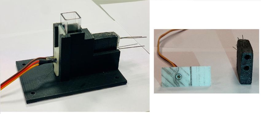
LED OSA Opto. OIS-150 1450P 1400-1500 1.20 - -
 LED OSA Opto. OIS-330 1550P 1500-1600 1.05/sr - -
 LED Thorlabs LED1900P 1800-2050 1.00 - -
 PD Vishay BPV23F 870-1050 - 4.4 0.6
 PD OSI Opto. FCI-InGaAs-1000 1100-1620 - 0.79 ~ 0.9
 PD Thorlabs FD05D 900 - 2600 - 0.20 1.3

The LED, PD, and sample holder were designed by SolidWorks 3D design program and printed
by FlashForge Inventor model 3D printer. The printed holders were shown in Fig. 3 and it is
clearly seen that the system is designed for 10 mm spectrometer cuvettes by which all
measurements were performed except the PAAm gel at 1950 nm. At this wavelength, the AAm
gel was sandwiched between two glass slides with a light path of 0.4 mm due to the very high
absorption in 10 mm cuvette which makes the system unresponsive.

Fig. 3. The holder designed in this study (left) and modular led and PD holders (right).

Measuring NIR Spectra

The NIR spectra of the glucose in water and PAAm gel were taken by using Shimadzu UV-
3600 Spectrometer to compare the results obtained by the LED-based system. The spectra of
the water samples were taken in quartz cuvettes with a 1 mm light path, and the PAAm gel
samples were sandwiched between two glass slides with a light path of 0.4 mm.

Theoretical Model

In this study, the glucose absorption is measured using Beer-Lambert Law which is given in
Eq. 1.

 = − (1)
here Io is the initial light intensity, I is the light intensity after passing the sample, , C and d are molar absorption coefficient, the concentration, and the sample's thickness, respectively. Data vout Acquisitio n Io I Photodiode d Fig. 4. Schematic representation of the measurement model. The output voltage of the amplifier, which was given in Fig. 2, depending on the photodiode current is given in Eq. 2. = − (2) where , , and are the output potential, the photodiode current, and the feedback resistor, respectively. As seen in this equation, the output potential is directly proportional to the photodiode current. The photocurrent, ipd, induced in the photodiode is proportional to the light intensity [20]. According to Eq. 2, measured potential (vout) depends on this photocurrent. Therefore, Eq. 1 can be written in terms of the potential given in Eq. 3. ( ) (0) = − (3) where (0) and ( ) are the output potential measured without and with glucose, respectively. By taking the logarithm of both sides of Eq. 3, the absorbance can be obtained as given in Figs. 8 and 10. (0) = 10 ( ( )) = (4) The molar absorption coefficient of the glucose for each wavelength was calculated by fitting the measured data to Beer-Lambert's law given in Eq. 4. RESULTS and DISCUSSION
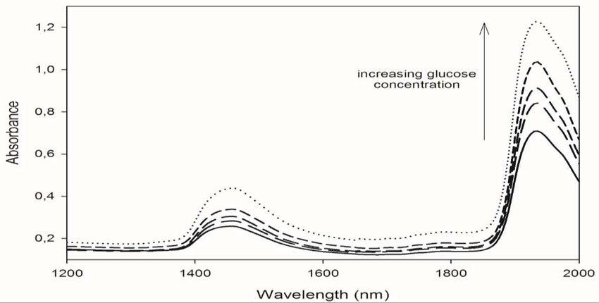
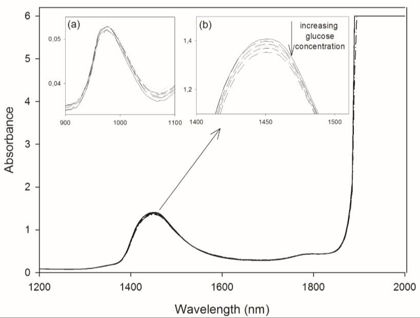
The glucose absorption measurements were performed at four different wavelengths, 960 nm,
1450 nm, 1550 nm, and 1900 nm, to carry out the most sensitive wavelength for detecting the
glucose in water and in PAAm gel. The wavelengths were selected based on the data given in
reference (Hotmartua et al., 2015) and the spectra given in Figs. 5 and 6. In fact, glucose has
higher absorbance at higher wavelengths, but the photodiode and light source for this region is
costly; therefore, it is not cost-effective for a wearable device.

 Fig. 5. NIR spectra of D-Glucose in water at different concentrations. The absorption at 960
 nm and 1450 nm were magnified as seen in inset figures (a) and (b), respectively.
When the NIR spectra given in Figs. 5 and 6 were compared, it is clearly seen that the overall
absorption in water is much higher than in PAAm gel and it saturates in water above 1900 nm.
This is due to the high absorption property of water in this wavelength region and the absorption
is dominated by water which decreases the response of the system against glucose. In PAAm
gel, the overall absorption is lower and the responsivity of the system against glucose is higher,
that is the change in the absorption is higher for changing concentration of the glucose (Fig. 6).
In addition, the absorption peak of glucose at 1950 nm is visible for glucose in PAAm gel which
is the most efficient wavelength for glucose measuring.
Fig. 6. NIR spectra of D-Glucose in PAAm gel at different concentrations.
Before starting the absorbance measurements for glucose, The aging of glucose in water was
studied first. For this purpose, five glucose samples with different concentrations, 20, 40, 60,
80, and 100 mg/mL were prepared, and the output potential for each sample was measured for
20 days at 23 oC. The slopes of the concentration-output voltage graph were given in Table 2.
It is seen from the values in this Table that the slope did not change considerably in the first ten
days, but it changes considerably at the end of the 20 days. Therefore, it was concluded that the
aqueous glucose solution should not be used after ten days.

 Table 2. The slope of the concentration-output voltage graph with respect to time.

 Time /
 (Day) ( . / )
 1 0.0003
 3 0.0004
 5 0.0003
 10 0.0002
 20 0.0019

The background-subtracted potential values, which were measured by using an LED-based
system for glucose in water, at 960 nm, 1450 nm, 1550 nm, and 1950 nm were given in Fig. 7
as a function of glucose concentration. The potential changes linearly with the concentration of
the glucose in water and the slopes were calculated as 0.0287 mV/mM, 0.0422 mV/mM, and
0.0071 mV/mM for 960 nm, 1450 nm, and 1550 nm, respectively. The highest slope was
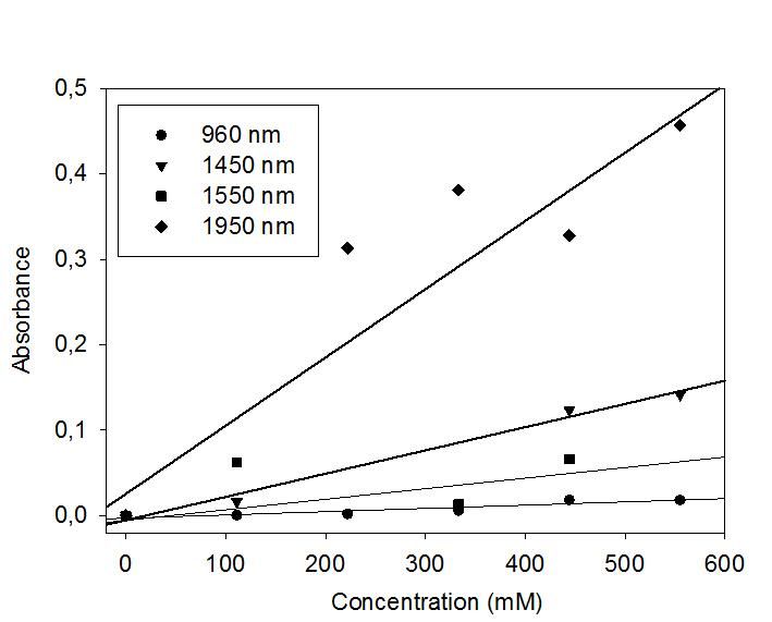
obtained for 1450 nm, and it is seen clearly from Fig. 7 that the system's response to the glucose
is high at this wavelength. It can be concluded that this wavelength is more suitable for
designing a system for measuring the glucose for wearable technology. This is also clearly seen
from the absorption spectrum of the glucose in water in the NIR region given in Fig. 5 where
the glucose absorbs the light more efficiently at 1450 nm. Additionally, the LED-based system
does not respond to the concentration changes of the glucose in water at 1950 nm due to the
high absorption of water at this wavelength which is in accordance with the results given in Fig.
5 where the absorption reached saturation above 1900 nm.

Fig. 7. The background-subtracted potential change as a function of glucose concentration in
 water at different wavelengths. Left: 960 nm, 1450 nm, 1550 nm, right: 1950 nm. The
 measurements were performed by using an LED-based system designed in this study.

The absorbances as a function of glucose concentration at 960 nm, 1450 nm, and 1550 nm were
given in Fig. 8. The absorbance changes linearly with the concentration and decreases with
increasing glucose concentration contrary to the expectation. However, this unexpected
behavior was also observed by different groups in literature and it was explained as the
waveguide effect of the glucose molecules in water (Javid, Faranak and Zakeri, 2018). That is,
when the concentration of the glucose increases in water, they are aligned to construct a
waveguide-like structure for electromagnetic waves and leads to an increase in the transmitted
light intensity through the sample.
Fig. 8. The absorbance of glucose in water as a function of glucose concentration at different
wavelengths. The measurements were performed by using an LED-based system designed in
 this study.

In water, since the absorbance dependence on the glucose concentration is completely different,
enhancement factors, , were calculated from the slopes of Fig. 8 instead of molar absorption
coefficients and they were found to be 5.22x10⁻³, 5.49x10⁻³, and 1.01x10⁻³ mM-1 for 960 nm,
1450 nm, and 1550 nm, respectively. The enhancement factor is higher at1450 nm than the
other wavelengths. When the absorption spectrum given in Fig. 5 is examined in detail, it is
seen that the absorption is higher at 1450 nm, which shows that the system designed in this
study gives results that are in accordance with the commercial spectrometer.

The potential changes, which were measured by using an LED-based system, as a function of
glucose concentration in PAAm gel were given in Fig. 9 at different wavelengths. The system's
responsivity to the glucose was found to be 0.0549 mV/mM, 0.1563 mV/mM, 0.0939, and
0.3242 mV/mM for 960 nm, 1450 nm, 1550 nm, and 1950 nm, respectively. This result clearly
shows that the system's responsivity to the glucose is highest at 1950 nm which couldn’t be
observed in water due to the high absorption.
Fig. 9. The background-subtracted potential change as a function of glucose concentration in
PAAm gel at different wavelengths. The measurements were performed by using LED based
 system designed in this study.

The absorbance variations as a function of glucose concentration in PAAm gel were given in
Fig. 10 at different wavelengths. Here, it is seen that the change at 1950 nm is more significant
which is in accordance with the spectra given in Fig. 6. When the absorbance values given in
Figs. 8 and 10 were compared, it is clearly seen that the glucose absorbs light more in PAAm
gel than in water.

The responsivities and calculated molar absorption coefficients were tabulated in Table 3. The
responsivity of the LED system is higher at all wavelengths in PAAm gel and it is highest at
1950 nm in gel. This is due to the high absorption of the free water when compared with gel.
In addition, the molar absorption coefficients calculated by using the LED based system and
the NIR Spectrometer are close to each other which shows that the LED based system designed
in this study works correctly and gives reliable results.
Fig. 10. The absorbance of glucose in PAAm gel as a function of glucose concentration. The SEM image of the PAAm gel was shown in Fig. 11 and it is clearly seen that it has a very porous structure. The glucose molecules can easily be trapped in the pores of the gel. It is well known in literature that PAAm gel is highly hydrophylic and the positively charged amine groups on the polymer chains interact with the water electrostatically which gathers the water molecules around the polymer chains (Yilmaz et al., 2009). Table 3. The responsivity (R) and the molar absorption coefficients () of glucose at different wavelengths. (NM: Couldn’t be measured) LED based system NIR Spectrometer Wavelength Rwater Rgel gel 10−3 gel 10−3 (nm) (mV/mM) (mV/mM) (c −1 −1 ) (c −1 −1 ) 960 0.0287 0.0549 1.74 ± 0.25 2.32 ± 0.37 1450 0.0422 0.1563 5.21 ± 0.62 5.88 ± 0.39 1550 0.0071 0.0939 3.57 ± 0.76 3.37 ± 0.50 1950 NM 0.3242 26.45 ± 2.16 27.73 ± 1.56
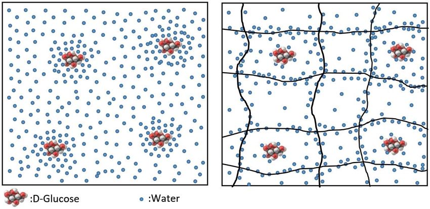
Fig. 11. SEM image of the PAAm gel. The pores are clearly visible in the gel.

Possible mechanism which affects the light response of the glucose in water and in PAAm gel
can be depicted as seen in Scheme 1. In the left scheme, the distribution of the glucose dissolved
in water was shown. Here, the water in the left scheme was named as free water to be able to
distinguish from the water in PAAm gel. In the left scheme, water molecules are distributed
almost homogeneously and the glucose molecules are mostly surrounded by water. During the
light absorption experiments performed in the free water system, while the most of the light is
absorbed by water molecules only small amount of light is absorbed by the glucose due to the
shielding effect of water. This description is consistent with the results given in Fig. 5 where
the absorption of water is very high when compared with glucose. In the right scheme, water
molecules are mostly gathered around the polymer chains due to the electrostatic interactions
between water and positively charged amine groups of polymer chains. In the pores of the gel,
since some part of the water molecules are gathered around the polymer chains, the number
density of the glucose molecules increases relatively when compared with water molecules.
During the light absorption experiments performed in the PAAm gel, the overall absorption
decreases since some part of the water molecules are shielded by the polymer chains and the
absorption by the glucose molecules increases due to the decreased number of water molecules
which shield the glucose molecules before. This description is also consistent with the results
given in Fig. 6 where the overall absorption decreased and the glucose absorption became more
visible.
Scheme 1. The schematic representation to describe the effect of the PAAm gel on the light
 response of glucose in free water (left) and in PAAm gel (right). Lines in right indicate the
 polymer chains.

Conclusions

It was concluded that the water plays a crucial role in the measurement of the glucose by using
an optical system based on absorption/transmission of light. Water absorbs light in NIR region
considerably and makes the optical system blind against the glucose as observed in the
experiments performed in this study. To be able to increase the responsivity of the optical
system against glucose, water molecules should be passivated. In this study, we showed that
the PAAm gel can be used for passivating the water molecules where the water molecules are
gathered around the polymer chains due to electrostatic interactions and polymer chains prevent
the water molecules to absorb light.

According to the results, it was concluded that the 1950 nm wavelength is the most efficient
wavelength. However, this wavelength cannot be used in water due to the very high absorption
of water at this wavelength and the light at 1450 nm wavelength should be used for the system
used for water.

It can be said that the system designed in this study is appropriate for detecting glucose.
Therefore, the results of this study will provide a basis for future works about wearable non-
invasive glucose measuring devices.
Acknowledgments. This work was supported by the Grant No FLO-2019-42073 from the
Scientific Research Projects of Istanbul Technical University. We thank to Mesut Balaban in
Physics of Istanbul Technical University for his valuable support in the NIR spectral
measurements.

References
Ahmad, M., Kamboh, A. and October, A. K. (2015) ‘Non-invasive blood glucose monitoring
using near-infrared spectroscopy | EDN’, (October 2013), pp. 1–7.

Dalvi, N. (2013) ‘Glucose Meter Reference Design’, p. 20.

Demitri, N. and Zoubir, A. M. (2017) ‘Measuring Blood Glucose Concentrations in
Photometric Glucometers Requiring Very Small Sample Volumes’, IEEE Transactions on
Biomedical Engineering, 64(1), pp. 28–39. doi: 10.1109/TBME.2016.2530021.

Fei, S. et al. (2004) ‘Near-infrared spectral methods for noninvasively measuring blood
glucose’, Optical Transmission, Switching, and Subsystems, 5281(May 2004), p. 595. doi:
10.1117/12.521999.

Formosa, N. (2013) ‘Blood glucose monitoring in children and adolescents with Type 1
Diabetes Mellitus’, Malta Medical Journal, 25(1), pp. 31–35.

Gonzales, W. V., Mobashsher, A. T. and Abbosh, A. (2019) The progress of glucose
monitoring—A review of invasive to minimally and non-invasive techniques, devices and
sensors, Sensors (Switzerland). doi: 10.3390/s19040800.

Haxha, S. and Jhoja, J. (2016) ‘Optical Based Noninvasive Glucose Monitoring Sensor
Prototype’, IEEE Photonics Journal, 8(6), pp. 1–11. doi: 10.1109/JPHOT.2016.2616491.

Hotmartua, R. et al. (2015) ‘Noninvasive blood glucose detection using near infrared sensor’,
Proceedings - 5th International Conference on Electrical Engineering and Informatics:
Bridging the Knowledge between Academic, Industry, and Community, ICEEI 2015, pp. 687–
692. doi: 10.1109/ICEEI.2015.7352586.

Ionescu, M. and Doctorala, S. (2019) ‘Measuring and detecting blood glucose by methods
non-invasive’, Proceedings of the 10th International Conference on Electronics, Computers
and Artificial Intelligence, ECAI 2018, (July). doi: 10.1109/ECAI.2018.8678943.

Javid, B., Faranak, F. G. and Zakeri, F. S. (2018) ‘Noninvasive optical diagnostic techniques
for mobile blood glucose and bilirubin monitoring’, Journal of Medical Signals and Sensors,
8(3), pp. 125–139. doi: 10.4103/jmss.JMSS-8-18.

Jung, Y. and Hwang, J. (2013) ‘Near-infrared studies of glucose and sucrose in aqueous
solutions: Water displacement effect and red shift in water absorption from water-solute
interaction’, Applied Spectroscopy, 67(2), pp. 171–180. doi: 10.1366/12-06635.

Lakowicz, J. R. (2006) ‘Principles of fluorescence spectroscopy’, Principles of Fluorescence
Spectroscopy, (January 2006), pp. 1–954. doi: 10.1007/978-0-387-46312-4.

Leboulanger, B., Guy, R. H. and Delgado-Charro, M. B. (2004) ‘Reverse iontophoresis for
non-invasive transdermal monitoring’, Physiological Measurement, 25(3). doi: 10.1088/0967-
3334/25/3/R01.

Poddar, R. et al. (2008) ‘Non-Invasive Glucose Monitoring Techniques: A review and current
trends’, (May 2014).

Salacinski, A. J. et al. (2014) ‘Validity and reliability of a glucometer against industry
reference standards’, Journal of Diabetes Science and Technology, 8(1), pp. 95–99. doi:
10.1177/1932296813514315.

Xue, J. et al. (2014) ‘Noninvasive measurement of glucose in artificial plasma with near-
infrared and Raman spectroscopy’, Applied spectroscopy, 68(4), pp. 428–433. doi:
10.1366/13-07250.

Yilmaz, Y. et al. (2009) ‘Elucidation of multiple-point interactions of pyranine fluoroprobe
during the gelation’, Spectrochimica Acta - Part A: Molecular and Biomolecular
Spectroscopy, 72(2), pp. 332–338. doi: 10.1016/j.saa.2008.09.012.

Zhang, Z. et al. (2016) ‘Estimation of glucose absorption spectrum at its optimum pathlength
for every wavelength over a wide range’, Spectroscopy Letters, 49(9), pp. 588–595. doi:
10.1080/00387010.2016.1231117.

Statements and Declarations
Funding. This work was supported by Istanbul Technical University (ITU) with grant number
FLO-2019-42073.
Competing Interests
The authors have no relevant financial or non-financial interests to disclose.
Author Contributions
All authors contributed to the study conception, design and analysis. The 3D design of the
system and the electronic control card design and production were performed by Ali Gelir, Onur
Burak Ozdemir, Omer F. Kadi, and Kadir B. Yildirim, polymer hydrogel synthesis and material
preparation were performed by Ali Gelir, Onur Burak Ozdemir and Sinem N. Seyhan,
absorption measurements were performed by Ali Gelir, Onur Burak Ozdemir and Sedat
Ozdemir. All authors read and approved the final manuscript.
Data availability. The datasets generated during the current study are available from the
corresponding author on reasonable request..
You can also read