Choroidal Thickness and Volume Modifications Induced by Aerobic Exercise in Healthy Young Adults
←
→
Page content transcription
If your browser does not render page correctly, please read the page content below
Research Article
Ophthalmic Res 2021;64:604–612 Received: July 2, 2020
Accepted: August 25, 2020
DOI: 10.1159/000511201 Published online: December 16, 2020
Choroidal Thickness and Volume
Modifications Induced by Aerobic
Exercise in Healthy Young Adults
Gema Insa-Sánchez a, b Lorena Fuentes-Broto a, b Alberto Cobos c
Elvira Orduna Hospital a, c, d Francisco Segura a, c, d Ana Sanchez-Cano a, c
Lorena Perdices a Isabel Pinilla a, e, f
aAragón Health Research Institute (IIS Aragón), Zaragoza, Spain; bDepartment of Pharmacology, Physiology
and Legal and Forensic Medicine, Universidad de Zaragoza, Zaragoza, Spain; cDepartment of Applied Physics,
Universidad de Zaragoza, Zaragoza, Spain; dDepartment of Ophthalmology. Miguel Servet University Hospital,
Zaragoza, Spain; eDepartment of Ophthalmology. Lozano Blesa University Hospital, Zaragoza, Spain; fDepartment
of Surgery, Gynecology and Obstetrics, Universidad de Zaragoza, Zaragoza, Spain
Keywords areas, and in some areas, the values were even smaller than
Exercise · Choroid · Optical coherence tomography · Eye the baseline measurements. The CV values showed changes
after exercise similar to those of thickness. The total CV re-
covery after exercise was related to sex and physical activity
Abstract level. Conclusion: Individuals with higher physical activity
Introduction: Our aim was to evaluate the changes in cho- habits had greater CV at rest than those with lower physical
roidal thickness (CT) and volume (CV) following aerobic activity levels. During exercise, healthy young people adjust
physical exercise in healthy young adults. Methods: This CT and CV. At 3 min post-exercise, CT and CV increase. Wom-
study included 72 eyes from healthy volunteers between 22 en and individuals with greater physical activity levels re-
and 37 years old. Using the International Physical Activity duce their total CV more than others during recovery.
Questionnaire, total physical activity was computed. Mea- © 2020 S. Karger AG, Basel
surements using an autorefractometer, ocular biometry, and
spectral-domain optical coherence tomography using the
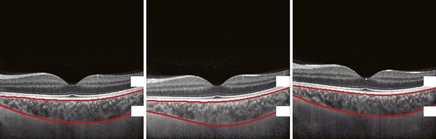
Enhanced Depth Imaging protocol were taken. OCT was per- Introduction
formed as a baseline measurement and after performing 10
min of dynamic physical exercise (3 and 10 min post-exer- During exercise, the increased O2 requirement of con-
cise). The choroidal layer was manually segmented, and the tracting muscles is satisfied by an increase in blood supply
CT and CV in different areas from the Early Treatment Dia- because the heart pumps more blood per minute due to
betic Retinopathy Study grid were obtained. Results: In circulatory adaptations and regulation of vessel diameter;
healthy adults, at 3 min post-exercise, CT was higher in the these adaptations enable a large part of the bloodstream
subfoveal, the 3-mm nasal, and the 6-mm superior areas. Be- to reach the muscles rather than the less active tissues.
tween 3 and 10 min post-exercise, the CT was reduced in all Exercise affects systemic haemodynamics, resulting in in-
karger@karger.com © 2021 S. Karger AG, Basel Correspondence to:
www.karger.com/ore Lorena Fuentes-Broto, lfuentes @ unizar.escreased blood flow, increased arterial systolic pressure, other external factors that affect vascular regulation: caf-
and increased heart frequency. feine causes the CT to decrease in the 4 h following inges-
Ocular blood flow is indispensable for adequate visual tion [28–31]; tobacco produces a decrease in CT follow-
performance. The choroid, a vascularized tissue between ing smoking [32–34], although no baseline changes were
the sclera and the retina, accounts for 85% of the ocular observed in healthy chronic smokers [35]; and alcohol
blood supply. In addition to blood supply, other choroi- increases CT between 1 h and 2 h after ingestion [36].
dal functions such as light absorption, thermoregulation The effects of exercise on the IOP, OPP, AL, corneal
via heat dissipation, and the role in the drainage of the thickness, depth of the anterior chamber, thickness of the
aqueous humour from the anterior chamber via the uveo- lens, and blood flow in the ophthalmic artery, the central
scleral pathway have been considered [1]. Traditionally retinal artery, the optic nerve head, the retina, and the
some authors have claimed that unlike the retina and an- choroid have been studied, but the effects of exercise on
terior uvea, the choroid has little or no self-regulation [2]. CT are still controversial [37]. Some authors demonstrate
However, other hypotheses have emerged in which they that exercise increases CT [38], but others did not see any
state that the choroid has the ability to regulate its choroi- changes [37, 39, 40], probably related to methodological
dal blood flow (ChBF) during changes in ocular perfusion differences or differences in the population.
pressure (OPP), having a greater ability to adapt to mean We hypothesized that, due to the great choroidal vas-
arterial pressure (MAP) than to intraocular pressure cularization, changes in the blood circulation induced by
(IOP) [3, 4]. exercise could generate changes at the choroidal vascular-
The introduction of optical coherence tomography ization. The aim of our study was to assess with SD-OCT
(OCT) has had an important effect on the development the CT changes after physical exercise. The normalization
of methods for studying the choroid [5, 6]. The latest gen- of a database of choroidal variations before and after ex-
erations of OCT and the introduction of new scanning ercise in healthy young people would be very useful, so
protocols and visualization have improved the possibility that their results could be extrapolated to help us under-
of studying the choroid. This is achievable due to the in- stand the influence of exercise in the eyes with patholo-
troduction of the enhanced depth imaging (EDI) tech- gies involving the choroid.
nique that inverts the retinal image and has less vitreous
resolution but better choroidal images [7]. Spectral-do-
main OCT (SD-OCT) with the EDI protocol allows visu- Materials and Methods
alization of the deeper layers of the retina [8].
Studies report values of choroidal thickness (CT) be- Participants
Seventy-two eyes from healthy individuals (32 men and 40
tween 270 and 350 µm [9] and average values of 354 µm women) with a mean age of 25 ± 5 years (range from 22 to 37 years)
[10] in healthy populations. CT may be modified by in- were recruited from among hospital workers and friends in the
ternal factors. CT has a negative correlation with age [9– Ophthalmology Department of the University Clinical Hospital
15] and axial length (AL) [10]. Another very important Lozano Blesa, Zaragoza, Spain. The inclusion criteria were age be-
tween 20 and 40, best corrected VA measured by Snellen charts of
factor to consider is the diurnal variation since the cho-
20/20, no history of ocular or systemic disease, and no treatments.
roid decreases in thickness as the hours of the day pass The exclusion criteria were having a refractive error of ≥±6.50 di-
[16–19]. On the other hand, there is a positive relation- optres (D) spherical or ≥±3 D cylindrical.
ship between best visual acuity (VA) and central subfo- This study was carried out in accordance with the tenets of the
veal thickness in patients older than 50 years [12, 20]. As 1964 Helsinki Declaration and its later amendments or compara-
ble ethical standards of Declaration of Helsinki (2013) and was
for the difference by sex, there is controversy, some stud-
approved by the Clinical Research Ethics Committee of Aragon
ies do not find differences, others, despite not being sig- (project licence PI19/352). Written informed consent was ob-
nificant, suggest that the thickness is greater in women tained from the subjects who participated in this study; confiden-
than in men, and others that choroid is thicker in men tiality and protection of data were ensured by applying interna-
than in women [13, 21–24]. In this aspect, it has to be tional recommendations and current Spanish legislation (Ley de
Investigacion Biomedica 14/2007 and LOPD).
considered that during the menstrual period a woman
Each subject underwent a preliminary ophthalmologic exami-
decreases her CT [25]. CT is also modified by the refrac- nation to exclude any pre-existing, undiagnosed ocular disease.
tive error [9, 10, 12, 26] and by many diseases that affect The ophthalmologic examination included refraction, an evalua-
the retina, such as age-related macular degeneration tion of intrinsic and extrinsic ocular motility, an anterior segment,
(AMD), central serous choroidopathy, macular hole, and and central ocular fundus study by biomicroscopy and SD-OCT.
pathological myopia [9, 27]. CT may also be modified by
Choroidal Modifications by Exercise Ophthalmic Res 2021;64:604–612 605
DOI: 10.1159/000511201Assessment of the Levels of Physical Activity Data obtained were statistically treated using a licensed version
Levels of physical activity in a typical week, according to inten- of SPSS 19.0 for Windows (SPSS, Chicago, IL, USA). Descriptive
sity, frequency, and duration, were estimated using the Interna- statistics for continuous variables included the mean and standard
tional Physical Activity Questionnaire [41]. One metabolic equiv- deviation. For each continuous variable, the Kolmogorov-Smirnov
alent (MET) is defined as the amount of oxygen consumed while test was applied to check normality.
sitting at rest (3.5 mL O2/kg/min). Moderate intensity refers to The CT measures were analysed at baseline and at 3 and 10 min
activities that cause small increases in breathing or heart rate. Vig- after exercise by the Friedman test, a repeated measure analysis, as
orous intensity refers to activities that require hard physical effort the data were not normally distributed. Post hoc analysis of the
and cause large increases in breathing or heart rate. MET-adjusted baseline, 3-min post-exercise, and 10-min post-exercise choroidal
minutes (MET-min) of physical activity in the last week were cal- values was performed by the Wilcoxon test. Adjustment for mul-
culated by assigning 4 METs to the time spent in moderate activi- tiple comparisons was performed using the Holm-Bonferroni
ties and 8 METs to the time spent in vigorous activities. method.
To determine the relationship of age, sex, AL, and physical ac-
Protocol for the Examinations and Exercise tivity levels with total CV, multivariate linear regression models
Subjects were instructed to refrain from ingesting alcohol and were analysed using forward stepwise selection. The assumptions
caffeine and from doing exercise in the 24 h prior to the examina- of linear regression (normality, linearity, and homoscedasticity of
tion. All subjects were examined under the same conditions with- residuals) were tested and examined through the scatter plots of
out mydriatics and by the same investigator. All examinations the residuals. The level of significance considered was p < 0.05.
were obtained during the same time period to avoid diurnal varia-
tions (2 p.m.–3 p.m.).
Ocular biometric measures were collected using the IOLMaster
biometer (IOLMaster; Carl Zeiss Meditec, Jena, Germany) to de- Results
termine the AL. The measurement was taken before exercise. Each
measurement was performed at least 3 times, and the mean was Seventy-two healthy eyes (from 32 men and 40 wom-
calculated. Refractometry (KR-7000P; Topcon Medical Systems en) with a mean age of 25 ± 5 years (ages 22–37 years)
Inc., Oakland, NJ, USA) was performed, and the measurement was were included in this study. The average ametropy was
taken before exercise. Each measurement was performed at least 3
times, and the mean was defined as the spherical equivalent. Fi- −1.82 ± 1.82 D (range: −6.25 to +0.75 D), and the AL was
nally, the CT was measured by SD-OCT with a Spectralis OCT 24.05 ± 1.25 mm (range: 20.60–26.01 mm). At baseline,
device (software version 5.6b; Heidelberg Engineering, Heidel- the temporal measurements were significantly higher
berg, Germany) with EDI, a fast macular protocol, and 25 frames. than the nasal measurements in both CT and CV, both in
The focus system was adjusted to obtain a higher quality image. the perifoveal and parafoveal rings (p < 0.001 in all com-
When taking the images, the Eye-Tracking® system was used to
minimize possible eye movements during the scans and to guar- parisons). However, between the superior and inferior ar-
antee good image quality, and subjects were warned to look at an eas, there were no significant differences in the CT or CV
internal fixation point. The highest quality scans obtained were of the parafoveal ring (p = 0.760 and p = 0.393, respec-
chosen for evaluation. Their quality line, rated in the OCT from 0 tively) or the perifoveal ring (p = 0.297 and p = 0.248, re-
to 40 dB (decibels), was always higher than 25 dB. spectively).
After this baseline measurement, volunteers with their refrac-
tive error corrected were required to perform 10 min of exercise. Table 1 shows the multiple linear regression with the
The exercise was going up and down stairs at a moderate pace. An explanatory variables sex, age, AL, and physical activity
examiner (GI-S) controlled the exercise level to assure that it was levels with the total CV at rest as the dependent variable.
similar in all participants. After exercising, 2 more OCT measure- The AL and physical activity level were the best explana-
ments, 3 and 10 min after completing the exercise, were taken fol- tory variables (adjusted R2 = 0.366, p < 0.001 for the mod-
lowing the same protocol as in the baseline measurement.
el). Sex and age were not found to be significant for ex-
Data Analysis plaining the total CV at baseline (p = 0.453 and 0.255,
Following each measurement session, the OCT images were respectively). The mean CV values were 8.25 ± 2.23 mm3
exported from the device. In each OCT profile, we could distin- and 8.80 ± 1.89 mm3 for men and women, respectively.
guish the retinal thickness, which was delimited by the internal For the β-coefficient of the significant explanatory vari-
limiting membrane and the Bruch membrane (BM), which served
as the outer limit. Choroidal sections were obtained with manual ables, for each 1 mm that AL increased, the total CV di-
segmentation, which exhibited a high degree of intraobserver and minished 0.94 mm3, and for each 1 MET-min/week that
interobserver repeatability [26, 42]. In each of the 25 frames, an physical activity level increased, the total CV increased
experienced, masked observer moved the BM line to the choroidal- 372 picolitres.
sclera interface and the internal limiting membrane line to the BM Participants were required to go up and down stairs at
location. After moving the lines, the OCT provided the CT and the
CV in the retinal map, specifically in each one of the 9 macular a moderate pace for 10 min. At 3 min post-exercise, the
areas of the Early Treatment Diabetic Retinopathy Study (ETDRS) CT values were higher than those at baseline, although
[43]. only significant differences were found in the subfoveal
606 Ophthalmic Res 2021;64:604–612 Insa-Sánchez et al.
DOI: 10.1159/000511201Table 1. Multiple linear regression models of total choroidal volume
Independent variable Unstandardized 95% confidence interval for β Standardized p value Model
β-coefficient β-coefficient summary
lower upper
Dependent variable: total choroidal volume (mm3) at baseline
(Constant) 30.56 22.59 38.54Table 2. Choroidal thickness of the 72 healthy eyes at baseline and 3 and 10 min post-exercise
Choroidal thickness, µm Baseline 3 min 10 min p value: baseline p value: baseline p value: 3 min
post-exercise post-exercise versus 3 min versus 10 min versus 10 min
post-exercise post-exercise post-exercise
Subfoveal 331±91* 341±95*,# 333±90# 0.016 0.777 0.001
Inner circle (ETDRS parafoveal region: 3 mm)
Superior 330±87 337±89# 329±84# 0.183 0.191 0.004
Temporal 328±80 333±81# 327±77# 0.130 0.604 0.003
Nasal 303±90* 310±95*,# 302±89# 0.019 0.830 0.001
Inferior 329±85$ 332±89# 325±83$,# 0.327 0.042 0.001
Outer circle (ETDRS perifoveal region: 6 mm)
Superior 320±79* 330±83*,# 322±78# 0.023 0.907 0.002
Temporal 309±72$ 308±68# 303±66$,# 0.355 0.027 0.002
Nasal 239±73 241±75# 237±73# 0.584 0.625 0.023
Inferior 315±73$ 316±74# 310±72$,# 0.886 0.048 0.016
ETDRS, Early Treatment Diabetic Retinopathy Study. * p < 0.05, baseline versus 3 min post-exercise. $ p < 0.05, baseline versus 10
min post-exercise. # p < 0.05, 3 min versus 10 min post-exercise (Wilcoxon test).
Table 3. Choroidal volume of the 72 healthy eyes at baseline and 3 and 10 min post-exercise
Choroidal volume, mm3 Baseline 3 min 10 min p value: baseline p value: baseline p value: 3 min
post-exercise post-exercise versus 3 min versus 10 min versus 10 min
post-exercise post-exercise post-exercise
Subfoveal 0.26±0.07* 0.27±0.07*,# 0.26±0.07# 0.019 0.621 0.003
Inner circle (ETDRS parafoveal region: 3 mm)
Superior 0.52±0.14 0.53±0.14# 0.52±0.13# 0.255 0.119 0.003
Temporal 0.51±0.12 0.52±0.13# 0.51±0.12# 0.107 0.882 0.002
Nasal 0.48±0.14* 0.49±0.15*,# 0.48±0.14# 0.043 0.954 0.010
Inferior 0.51±0.13 0.52±0.14# 0.51±0.13# 0.202 0.087 0.001
Outer circle (ETDRS perifoveal region: 6 mm)
Superior 1.70±0.42* 1.75±0.45*,# 1.70±0.41# 0.024 0.924 0.002
Temporal 1.64±0.38$ 1.63±0.36# 1.61±0.35$,# 0.295 0.024 0.005
Nasal 1.26±0.40 1.27±0.40 1.26±0.39 0.551 0.817 0.140
Inferior 1.66±0.39$ 1.68±0.39# 1.64±0.37$,# 0.729 0.032 0.004
Total volume 8.56±2.05 8.64±2.12# 8.49±1.99# 0.241 0.160 0.000
ETDRS, Early Treatment Diabetic Retinopathy Study. * p < 0.05, baseline versus 3 min post-exercise. $ p < 0.05, baseline versus 10
min post-exercise. # p < 0.05, 3 min versus 10 min post-exercise (Wilcoxon test).
plain the total CV changes between 3 min versus 10 min Discussion
post-exercise (adjusted R2 = 0.141, p = 0.003 for the mod-
el). This result suggests that women and individuals with Choroid is a vascularized layer, and vascular changes
higher physical activity habits had the greatest decreases have been described related not only to pathological con-
in total CV during recovery, that is, from 3 to 10 min post- ditions such as central serous chorioretinopathy with a
exercise. mean CT of 505 µm [5], polypoidal choroidal vasculopa-
thy with a mean CT of 438 µm, and exudative AMD with
608 Ophthalmic Res 2021;64:604–612 Insa-Sánchez et al.
DOI: 10.1159/000511201a CT
tolic and diastolic blood pressures, heart rate, mean ocular    with retinal diseases. These findings could be a recom-
perfusion pressure, and the mean blur rate [37], in which       mendation to avoid the pathological influence of external
these values were modified after exercise. However, not all     factors in pachychoroid diseases, AMD, or other vascular
authors found similar changes, as IOP [38], diastolic blood     retinal diseases where choroidal flow may be crucial.
pressure [39], AL, previous anterior chamber depth, lens            For example, in patients with primary open-angle
thickness, and vitreous length [40] values were not found       glaucoma and ocular hypertension, the baseline ChBF re-
to be different after exercise. Previous studies in a small     sponse to isometric exercise indicates less active regula-
series on changes in CT after physical exercise also showed     tory capacity [58]. We should also keep in mind that aer-
opposite results. Alwassia and colleagues, in a sample of       obic exercise decreases IOP while isometric exercise can
15 patients with a mean age of 61 years, concluded that         cause a temporary elevation in IOP that can aggravate the
there was no variation in CT after performing physical          problem. In pseudoexfoliative glaucoma, patients who
exercise [39]. This lack of variation in CT after exercise in   performed at least 30 min of exercise per week had less
the Alwassia study could depend on the age of the studied       progression in the visual field defect than those who did
participants, not only because of the presence of choroidal     not exercise [59].
diminution but also because of their cardiac condition.             When we talk about AMD, some studies in dry AMD
   However, the same conclusion was shown by Hong, in           said that there were no differences between the ChBF
a study of 25 subjects with a mean age of 25.4 years [40],      changes observed in control subjects and those observed
and by Kinoshita, in a study of 38 subjects with a mean of      in patients with AMD during the isometric exercise phase
39 years [37]. However, Sayin et al. [38] studied 19 sub-       and after exercise [48]. However, Pournaras CJ et al. [49]
jects with a mean age of 27 years and found similar results     found that in response to an acute, moderate increase in
to our study: a small time after finishing the exercise (3      OPP induced by isometric exercise, there were no chang-
min in our study and 5 min in Sayin’s work), they found         es in ChBF. In contrast, in patients with neovascular
a CT increase, and then there was a diminution after a          AMD, this flow increases, indicating an altered regulation
later timepoint (10 in our study and 15 in Sayin’s one). In     in response to the increase in OPP.
the study by Sayin et al. [38], the CT values 15 min post-          To prescribe exercise to patients with eye diseases,
exercise did not differ significantly from the baseline.        more research is needed to determine if they have altera-
   Our results provide new findings because when ana-           tions that do not allow them to recover properly after
lysing the data only 10 min after the end of the exercise,      acute exercise. On the other hand, chronic exercise, as in
the CT values were lower than those at baseline in some         other haemodynamic variables [4], could also produce
areas. Although we could differentiate between isometric        adaptations in the choroidal flow, which could be benefi-
activities (with continuous muscle contractions) and dy-        cial for these patients. Therefore, more studies are needed
namic activities (contraction and relaxation phases), the       to determine whether training improves their choroidal
practice of exercise involves an increase in heart rate as      condition. For all this, an exercise protocol during an
well as cardiac output and generates redistribution of          ophthalmologic examination could be useful to obtain an
blood flow.                                                     early diagnosis in ocular diseases because our results
   On the other hand, variations in blood flow, systolic        show that physical activity level and sex have an effect on
blood pressure, ventilation, and hormonal changes occur         total CV.
[55]. Also note that during isometric exercises, blood              The present study has several limitations that need to
pressure in the ophthalmic and brachial arteries in-            be addressed. The lack of objective ocular and systemic
creased, and the same occurred with choroidal vascular          measures is still a problem in choroidal measurements.
resistance [56].                                                Independent measurements have been made by 2 experi-
   Regarding the regulation of choroidal blood flow dur-        enced evaluators to minimize subjectivity. The intensity
ing isometric exercise at different levels of IOP, Popa-        of the exercise has not been controlled with an ergometer,
Cherecheanu said that the regulation of choroidal blood         but it must be considered that the objective is to carry out
flow during changes in ocular perfusion pressure is con-        these measurements in a simple way in the ophthalmo-
trolled by complex mechanisms in humans and has less            logic room. However, a step and a timer are easy to achieve
capacity to adapt to IOP elevation than to MAP elevation        in the consultation and allow controlling the amount of
[57]. Choroidal changes after exercise, smoking, caffeine       exercise performed. So, in this simple way, the measures
drinking, or other external factors need to be described        after exercise provide much more information than the
not only in healthy and young subjects but also in patients     simple basal measurement at rest. In this study, the ma-

610                  Ophthalmic Res 2021;64:604–612                                  Insa-Sánchez et al.
                     DOI: 10.1159/000511201
jority of examined eyes were myopic. This is the result of Aragón, project licence PI19/352) and with the 1964 Helsinki Dec-
the narrow age range of the sample (22–37 years), and laration and its later amendments or comparable ethical standards.
Written informed consent was obtained from all individual par-
that most of them were university students, but it is nec- ticipants included in the study.
essary to take this into account because myopia could in-
fluence the final CT differently than in emmetropic or
other ametropic subjects. Conflict of Interest Statement
We can conclude that in our sample of healthy young
adults, individuals with higher physical activity levels had The authors have no conflicts of interest to declare.
greater CV at rest than those with lower physical activity
levels. Moreover, the average values of CT and CV were
affected by the performance of acute exercise. Funding Sources
Within a few minutes of completing a dynamic physi-
The study was supported by the Instituto de Salud Carlos III
cal exercise, the CT and CV increased, and after a short
(Ocular Pathology National Net RETICS-Oftared RD16/0008);
period of time, they recovered, although some area values the Fondo Europeo de Desarrollo Regional (FEDER) funds: “Una
were even lower than the baseline measurements. Thus, manera de hacer Europa”; and the Government of Aragon (Group
we can conclude that healthy young people are able to B08_17R and Predoctoral Grant L. Perdices).
adjust CT during exercise. Additionally, women and in-
dividuals with greater physical activity levels reduce their
total CV more during recovery after exercise than others. Author Contributions
G.I.S. and I.P.L. conceived of and designed the experiments;
G.I.S. performed the experiments; G.I.S. and L.F.B. analysed the
Statement of Ethics data; all authors contributed to the interpretation of the data; the
first draft was written by G.I.S.; and all authors provided critical
All procedures performed in studies involving human partici- input and revisions. All authors read and approved the final man-
pants were in accordance with the ethical standards of the institu- uscript.
tional research committee (Clinical Research Ethics Committee of
References
1 Nickla DL, Wallman J. The multifunctional choroidal morphologic features and vascula- grafía de coherencia óptica con imagen de
choroid. Prog Retin Eye Res. 2010 Mar;29(2): ture in healthy eyes using spectral-domain profundidad mejorada. Revista Mexicana de
144–68. optical coherence tomography. Ophthalmol- Oftalmología. 2017 Jan 1;91(1):2–8.
2 Delaey C, Van De Voorde J. Regulatory ogy. 2013 Sep;120(9):1901–8. 14 Akhtar Z, Rishi P, Srikanth R, Rishi E, Bhende
mechanisms in the retinal and choroidal cir- 8 Huang W, Wang W, Zhou M, Chen S, Gao X, M, Raman R. Choroidal thickness in normal
culation. Ophthalmic Res. 2000 Nov-Dec; Fan Q, et al. Peripapillary choroidal thickness Indian subjects using Swept source optical co-
32(6):249–56. in healthy Chinese subjects. BMC Ophthal- herence tomography. PLoS One. 2018; 13(5):
3 Polska E, Simader C, Weigert G, Doelemeyer mol. 2013 Jun 10;13:23. e0197457.
A, Kolodjaschna J, Scharmann O, et al. Regu- 9 Shin JW, Shin YU, Cho HY, Lee BR. Measure- 15 Bhayana AA, Kumar V, Tayade A, Chandra
lation of choroidal blood flow during com- ment of choroidal thickness in normal eyes M, Chandra P, Kumar A. Choroidal thickness
bined changes in intraocular pressure and ar- using 3D OCT-1000 spectral domain optical in normal Indian eyes using swept-source op-
terial blood pressure. Invest Ophthalmol Vis coherence tomography. Korean J Ophthal- tical coherence tomography. Indian J Oph-
Sci. 2007 Aug;48(8):3768–74. mol. 2012 Aug;26(4):255–9. thalmol. 2019 Feb;67(2):252–5.
4 Ong SR, Crowston JG, Loprinzi PD, Ramulu 10 Ikuno Y, Kawaguchi K, Nouchi T, Yasuno Y. 16 Chakraborty R, Read SA, Collins MJ. Diurnal
PY. Physical activity, visual impairment, and Choroidal thickness in healthy Japanese sub- variations in axial length, choroidal thickness,
eye disease. Eye. 2018 Aug;32(8):1296–303. jects. Invest Ophthalmol Vis Sci. 2010 Apr; intraocular pressure, and ocular biometrics.
5 Imamura Y, Fujiwara T, Margolis R, Spaide 51(4):2173–6. Invest Ophthalmol Vis Sci. 2011 Jul 11;52(8):
RF. Enhanced depth imaging optical coher- 11 Margolis R, Spaide RF. A pilot study of en- 5121–9.
ence tomography of the choroid in central se- hanced depth imaging optical coherence to- 17 Tan CS, Ouyang Y, Ruiz H, Sadda SR. Diurnal
rous chorioretinopathy. Retina. 2009 Nov- mography of the choroid in normal eyes. Am variation of choroidal thickness in normal,
Dec;29(10):1469–73. J Ophthalmol. 2009 May;147(5):811–5. healthy subjects measured by spectral domain
6 Barteselli G, Chhablani J, El-Emam S, Wang 12 Ding X, Li J, Zeng J, Ma W, Liu R, Li T, et al. optical coherence tomography. Invest Oph-
H, Chuang J, Kozak I, et al. Choroidal volume Choroidal thickness in healthy Chinese sub- thalmol Vis Sci. 2012 Jan 25;53(1):261–6.
variations with age, axial length, and sex in jects. Invest Ophthalmol Vis Sci. 2011 Dec 20; 18 Read SA, Pieterse EC, Alonso-Caneiro D,
healthy subjects: a three-dimensional analy- 52(13):9555–60. Bormann R, Hong S, Lo CH, et al. Daily
sis. Ophthalmology. 2012 Dec;119(12):2572– 13 Sardi Correa C, Acosta Cadavid C, Rodríguez morning light therapy is associated with an
8. Gómez AM, Mejía Estrada ME, Vásquez increase in choroidal thickness in healthy
7 Branchini LA, Adhi M, Regatieri CV, Nanda- Trespalacios EM. Grosor coroideo central en young adults. Sci Rep. 2018 May 29; 8(1):
kumar N, Liu JJ, Laver N, et al. Analysis of sujetos hispanos sanos medido por tomo- 8200.
Choroidal Modifications by Exercise Ophthalmic Res 2021;64:604–612 611
DOI: 10.1159/00051120119 Uyar E, Dogan U, Ulas F, Celebi S. Effect of 33 Zengin MO, Cinar E, Kucukerdonmez C. The 46 Cicek A, Duru N, Duru Z, Altunel O, Hashas
fasting on choroidal thickness and its diurnal effect of nicotine on choroidal thickness. Br J ASK, Arifoglu HB, et al. The assessment of
variation. Curr Eye Res. 2019 Jul; 44(7): 695– Ophthalmol. 2014 Feb;98(2):233–7. choroidal thickness with spectral-domain op-
700. 34 Wei X, Kumar S, Ding J, Khandelwal N, Agar- tical coherence tomography during Valsalva
20 Shao L, Xu L, Wei WB, Chen CX, Du KF, Li wal M, Agrawal R. Choroidal structural maneuver. Int Ophthalmol. 2017 Aug; 37(4):
XP, et al. Visual acuity and subfoveal choroi- changes in smokers measured using choroidal 843–8.
dal thickness: the Beijing Eye Study. Am J vascularity index. Invest Ophthalmol Vis Sci. 47 Kara N, Demircan A, Karatas G, Ozgurhan
Ophthalmol. 2014 Oct;158(4):702–e1. 2019 Apr 1;60(5):1316–20. EB, Tatar G, Karakucuk Y, et al. Effects of two
21 Ruiz-Medrano J, Flores-Moreno I, Peña-Gar- 35 Teberik K. The effect of smoking on macular, commonly used mydriatics on choroidal
cía P, Montero JA, Duker JS, Ruiz-Moreno choroidal, and retina nerve fiber layer thick- thickness: direct and crossover effects. J Ocul
JM. Macular choroidal thickness profile in a ness. Turk J Ophthalmol. 2019 Feb 28; 49(1): Pharmacol Ther. 2014 May;30(4):366–70.
healthy population measured by swept-source 20–4. 48 Park KA, Oh SY. Choroidal thickness in
optical coherence tomography. Invest Oph- 36 Kang HM, Woo YJ, Koh HJ, Lee CS, Lee SC. healthy children. Retina. 2013 Oct; 33(9):
thalmol Vis Sci. 2014 May 20;55(6):3532–42. The effect of consumption of ethanol on sub- 1971–6.
22 Abbey AM, Kuriyan AE, Modi YS, Thorell foveal choroidal thickness in acute phase. Br J 49 Li XQ, Larsen M, Munch IC. Subfoveal cho-
MR, Nunes RP, Goldhardt R, et al. Optical co- Ophthalmol. 2016 Mar;100(3):383–8. roidal thickness in relation to sex and axial
herence tomography measurements of cho- 37 Kinoshita T, Mori J, Okuda N, Imaizumi H, length in 93 Danish university students. In-
roidal thickness in healthy eyes: correlation Iwasaki M, Shimizu M, et al. Effects of exer- vest Ophthalmol Vis Sci. 2011 Oct 31;52(11):
with age and axial length. Ophthalmic Surg cise on the structure and circulation of cho- 8438–41.
Lasers Imaging Retina. 2015 Jan;46(1):18–24. roid in normal eyes. PLoS One. 2016; 11(12): 50 Gale J, Wells AP, Wilson G. Effects of exercise
23 Ooto S, Hangai M, Yoshimura N. Effects of e0168336. on ocular physiology and disease. Surv Oph-
sex and age on the normal retinal and choroi- 38 Sayin N, Kara N, Pekel G, Altinkaynak H. thalmol. 2009 May–Jun;54(3):349–55.
dal structures on optical coherence tomogra- Choroidal thickness changes after dynamic 51 Castro EF, Mostarda CT, Rodrigues B,
phy. Curr Eye Res. 2015 Feb;40(2):213–25. exercise as measured by spectral-domain op- Moraes-Silva IC, Feriani DJ, De Angelis K, et
24 Tuncer I, Karahan E, Zengin MO, Atalay E, tical coherence tomography. Indian J Oph- al. Exercise training prevents increased intra-
Polat N. Choroidal thickness in relation to thalmol. 2015 May;63(5):445–50. ocular pressure and sympathetic vascular
sex, age, refractive error, and axial length in 39 Alwassia AA, Adhi M, Zhang JY, Regatieri modulation in an experimental model of met-
healthy Turkish subjects. Int Ophthalmol. CV, Al-Quthami A, Salem D, et al. Exercise- abolic syndrome. Braz J Med Biol Res. 2015
2015 Jun;35(3):403–10. induced acute changes in systolic blood pres- Apr;48(4):332–8.
25 Ulas F, Dogan U, Duran B, Keles A, Agca S, sure do not alter choroidal thickness as mea- 52 Miles L. Physical activity and health. Nutr
Celebi S. Choroidal thickness changes during sured by a portable spectral-domain optical Bull. 2007;32(4):314–63.
the menstrual cycle. Curr Eye Res. 2013 Nov; coherence tomography device. Retina. 2013 53 Hirata M, Tsujikawa A, Matsumoto A, Hangai
38(11):1172–81. Jan;33(1):160–5. M, Ooto S, Yamashiro K, et al. Macular cho-
26 Sanchez-Cano A, Orduna E, Segura F, Lopez 40 Hong J, Zhang H, Kuo DS, Wang H, Huo Y, roidal thickness and volume in normal sub-
C, Cuenca N, Abecia E, et al. Choroidal thick- Yang D, et al. The short-term effects of exer- jects measured by swept-source optical coher-
ness and volume in healthy young white cise on intraocular pressure, choroidal thick- ence tomography. Invest Ophthalmol Vis Sci.
adults and the relationships between them ness and axial length. PLoS One. 2014; 9(8): 2011 Jul 1;52(8):4971–8.
and axial length, ammetropy and sex. Am J e104294. 54 Read SA, Collins MJ. The short-term influ-
Ophthalmol. 2014 Sep;158(3):574–e1. 41 Craig CL, Marshall AL, Sjöström M, Bauman ence of exercise on axial length and intraocu-
27 Esmaeelpour M, Kajic V, Zabihian B, Othara AE, Booth ML, Ainsworth BE, et al. Interna- lar pressure. Eye. 2011 Jun;25(6):767–74.
R, Ansari-Shahrezaei S, Kellner L, et al. Cho- tional physical activity questionnaire: 55 Green DJ, Hopman MT, Padilla J, Laughlin
roidal Haller’s and Sattler’s layer thickness 12-country reliability and validity. Med Sci MH, Thijssen DH. Vascular adaptation to ex-
measurement using 3-dimensional 1060-nm Sports Exerc. 2003 Aug;35(8):1381–95. ercise in humans: role of hemodynamic stim-
optical coherence tomography. PLoS One. 42 Chhablani J, Barteselli G, Wang H, El-Emam uli. Physiol Rev. 2017 Apr;97(2):495–528.
2014;9(6):e99690. S, Kozak I, Doede AL, et al. Repeatability and 56 Riva CE, Titze P, Hero M, Movaffaghy A,
28 Vural AD, Kara N, Sayin N, Pirhan D, Ersan reproducibility of manual choroidal volume Petrig BL. Choroidal blood flow during iso-
HB. Choroidal thickness changes after a sin- measurements using enhanced depth imag- metric exercises. Invest Ophthalmol Vis Sci.
gle administration of coffee in healthy sub- ing optical coherence tomography. Invest 1997 Oct;38(11):2338–43.
jects. Retina. 2014 Jun;34(6):1223–8. Ophthalmol Vis Sci. 2012 Apr 24;53(4):2274– 57 Popa-Cherecheanu A, Schmidl D, Werkmeis-
29 Zengin MO, Cinar E, Karahan E, Tuncer I, 80. ter RM, Chua J, Garhöfer G, Schmetterer L.
Kucukerdonmez C. The effect of caffeine on 43 Photocoagulation for diabetic macular ede- Regulation of choroidal blood flow during
choroidal thickness in young healthy subjects. ma. Early treatment diabetic retinopathy isometric exercise at different levels of intra-
Cutan Ocul Toxicol. 2015;34(2):112–6. study report number 1. Early treatment dia- ocular pressure. Invest Ophthalmol Vis Sci.
30 Altinkaynak H, Ceylan E, Kartal B, Keleş S, betic retinopathy study research group. 2019 Jan 2;60(1):176–82.
Ekinci M, Olcaysu OO. Measurement of cho- Arch Ophthalmol. 1985 Dec; 103(12): 1796– 58 Portmann N, Gugleta K, Kochkorov A, Pol-
roidal thickness following caffeine intake in 806. unina A, Flammer J, Orgul S. Choroidal blood
healthy subjects. Curr Eye Res. 2016 May; 44 Chung SE, Kang SW, Lee JH, Kim YT. Cho- flow response to isometric exercise in glau-
41(5):708–14. roidal thickness in polypoidal choroidal vas- coma patients and patients with ocular hyper-
31 Dervisogullari MS, Totan Y, Yuce A, Kulak culopathy and exudative age-related macular tension. Invest Ophthalmol Vis Sci. 2011 Sep
AE. Acute effects of caffeine on choroidal degeneration. Ophthalmology. 2011 May; 9;52(10):7068–73.
thickness and ocular pulse amplitude. Cutan 118(5):840–5. 59 Perez CI, Singh K, Lin S. Relationship of life-
Ocul Toxicol. 2016 Dec;35(4):281–6. 45 Li X, Wang W, Chen S, Huang W, Liu Y, style, exercise, and nutrition with glaucoma.
32 Sizmaz S, Küçükerdönmez C, Pinarci EY, Wang J, et al. Effects of Valsalva maneuver on Curr Opin Ophthalmol. 2019 Mar; 30(2): 82–
Karalezli A, Canan H, Yilmaz G. The effect of anterior chamber parameters and choroidal 8.
smoking on choroidal thickness measured by thickness in healthy Chinese: an AS-OCT and
optical coherence tomography. Br J Ophthal- SS-OCT study. Invest Ophthalmol Vis Sci.
mol. 2013 May;97(5):601–4. 2016 Jul 1;57(9):OCT189–95.
612 Ophthalmic Res 2021;64:604–612 Insa-Sánchez et al.
DOI: 10.1159/000511201You can also read