Catching Nucleosome by Its Decorated Tails Determines Its Functional States
←
→
Page content transcription
If your browser does not render page correctly, please read the page content below
REVIEW
                                                                                                                                              published: 14 July 2022
                                                                                                                                     doi: 10.3389/fgene.2022.903923

                                              Catching Nucleosome by Its
                                              Decorated Tails Determines Its
                                              Functional States
                                              Parveen Sehrawat†, Rahul Shobhawat† and Ashutosh Kumar *
                                              Department of Biosciences and Bioengineering, Indian Institute of Technology Bombay, Mumbai, India

                                              The fundamental packaging unit of chromatin, i.e., nucleosome, consists of ~147 bp of DNA
                                              wrapped around a histone octamer composed of the core histones, H2A, H2B, H3, and H4, in
                                              two copies each. DNA packaged in nucleosomes must be accessible to various machineries,
                                              including replication, transcription, and DNA damage repair, implicating the dynamic nature of
                                              chromatin even in its compact state. As the tails protrude out of the nucleosome, they are easily
                                              accessible to various chromatin-modifying machineries and undergo post-translational
                                              modifications (PTMs), thus playing a critical role in epigenetic regulation. PTMs can regulate
                            Edited by:        chromatin states via charge modulation on histones, affecting interaction with various
                     Dileep Vasudevan,
  Institute of Life Sciences (ILS), India     chromatin-associated proteins (CAPs) and DNA. With technological advancement, the list
                         Reviewed by:         of PTMs is ever-growing along with their writers, readers, and erasers, expanding the
                Sivaraman Padavattan,         complexity of an already intricate epigenetic field. In this review, we discuss how some of
National Institute of Mental Health and
    Neurosciences (NIMHANS), India
                                              the specific PTMs on flexible histone tails affect the nucleosomal structure and regulate the
                     Lars Nordenskiöld,       accessibility of chromatin from a mechanistic standpoint and provide structural insights into
  Nanyang Technological University,           some newly identified PTM–reader interaction.
                             Singapore
                     Alexey V. Onufriev,      Keywords: nucleosome, PTMs, histone tails, acetylation, acylation, methylation
          Virginia Tech, United States
                   *Correspondence:
                      Ashutosh Kumar          1 INTRODUCTION
                   ashutoshk@iitb.ac.in
     †
     These authors have contributed
                                              In the eukaryote’s nucleus, DNA is packaged into the macromolecular “beads on a string”-like
   equally to this work and share first        structure called chromatin using highly basic histone proteins. A nucleosome is the basic and
                           authorship         efficient unit of this organization in which 145–147 bp of DNA are wrapped around a histone
                                              octamer (two molecules of each histone H2A, H2B, H3, and H4). Two pairs of H3–H4 dimer form a
                     Specialty section:       tetramer stabilized by a characteristic hydrophobic four-helix bundle structure between H3 and H3ʹ,
          This article was submitted to       and then two dimers of H2A–H2B interact with H3–H4 tetramer on each side through a second
         Epigenomics and Epigenetics,         homologous hydrophobic four-helix bundle structure between H2B and H4, forming a globular
                a section of the journal      octamer from which disordered tails protrude out. Through extensive hydrogen-bonding and
                   Frontiers in Genetics
                                              electrostatic interactions, histones coordinate with DNA via conserved histone fold domains,
            Received: 24 March 2022           resulting in the bending of negatively charged DNA over a positively charged octamer surface.
             Accepted: 07 June 2022
                                              This bent conformation of DNA brings the phosphate backbone of the two strands closer, and this
             Published: 14 July 2022
                                              energetically constrained conformation is maintained by neutralizing negative charges by positively
                             Citation:        charged lysine and arginine side chains (Luger et al., 1997).
       Sehrawat P, Shobhawat R and
                                                   These strong and extensive interactions render the nucleosome a stable disc that can sterically
Kumar A (2022) Catching Nucleosome
 by Its Decorated Tails Determines Its
                                              inhibit the binding of chromatin-associated proteins (CAPs). Virtually all eukaryotic organisms use
                     Functional States.       the inhibitory nature of this packaging to regulate access to DNA. However, the information encoded
             Front. Genet. 13:903923.         inside the DNA must be retrieved at appropriate times. Although DNA is very tightly compacted, it
     doi: 10.3389/fgene.2022.903923           still remains accessible to many enzyme machineries that replicate it, repair it, and use it to produce RNA

Frontiers in Genetics | www.frontiersin.org                                         1                                          July 2022 | Volume 13 | Article 903923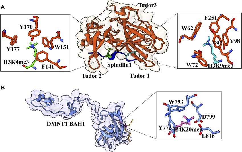
Sehrawat et al.                                                                                          Decorated Tail Recognition by Specific Reader

molecules and proteins. For doing this, chromatin and nucleosomes            pioneering work of Vincent Allfrey in the 1960s and subsequent
must be inherently dynamic and highly malleable. Numerous                    studies revealed that these tails are subjected to many post-
biochemical and structural studies established the dynamic nature            translation modifications like acetylation, methylation, and
of the nucleosome in terms of its conformation and composition. The          phosphorylation, thus acting as a hub of chromatin signaling
disordered N-terminal tail of histones have an affinity to DNA,               (Millán-Zambrano et al., 2022). Covalent modifications of
forming a dynamic complex with DNA termed as “fuzzy                          histone tails can alter the chromatin structure via cis-effects or
conformational ensembles,” which regulate the chromatin                      trans-effects. Cis-effects are employed by changing the
structure and dynamics (Ghoneim, Fuchs, and Musselman 2021;                  biophysical properties of modified histone chains, like altering
Peng et al., 2021; Shukla, Agarwal, and Kumar 2022). Polach and              the electrostatic charge or structure of the tail, which in turn
Widom, (1995) demonstrated the phenomenon of intrinsic structural            affects internucleosomal contacts. For example, histone
dynamics of nucleosome known as “DNA breathing,” i.e., partially             acetylation on lysine residue exerts its effect by neutralizing
unwrapping and rewrapping of DNA spontaneously. This dynamic                 the positive charge of histone tails. Charge-neutralized tails
unwrapping/rewrapping phenomenon is exploited by several DNA-                generate a localized decondensation of the chromatin fiber,
binding proteins like transcription factors in a tunable and analogous       resulting in better availability of DNA double helix to the
fashion. Using FRET experiments, the Langowski group showed that             transcription machinery. Acetylation at H4K16 inhibits the
disassembly of nucleosome is initiated by DNA breathing resulting in         packaging of a nucleosomal array in a compact 30-nm
a dynamic “octasome,” which opens on a 50 µs time scale at an angle          chromatin fiber in vitro and further abolishes cross-fiber
of ≈20°. This results in disruption of dimer tetramer interface with         interactions (Michael et al., 2006). In fact, out of four
H2A–H2B dimer evicting first followed by H3–H4 tetramer removal               acetylations possible in the H4 tail, K16 acetylation is unique
(Böhm et al., 2011; Gansen et al., 2018).                                    as only this modification reduces the cation-induced folding of
    In addition to DNA breathing, cells have also evolved various            the 12-mer nucleosome array implicating cis-effect of acetyl mark
other mechanisms to make nucleosomal DNA more accessible:                    (Allahverdi et al., 2011). Multiscale computational studies
histones posttranslational modifications, histone chaperones,                 supported by NMR experiments revealed that acetylated
histone variants, and chromatin remodelers. These regulatory                 H4 tails lose local contacts and reduced tail availability for
mechanisms control the genome function without changing the                  forming critical internucleosomal interactions resulting in the
nucleotide sequence, also referred to as “epigenetic” marks.                 unfolding of chromatin fiber (Collepardo-Guevara et al., 2015;
Histone post-translational modification is the process of covalently          Bascom and Schlick 2018). Similarly, phosphorylation adds a net
attaching adducts like methyl group, acetyl group, phosphate group,          negative charge generating “charge patches,” which result in
and ubiquitin group. These modifications present on free N-terminal           alteration of nucleosome packaging (Dou and Gorovsky 2000).
tails or inside the histone fold domain affect the structure and             Bulky groups, such as ADP-ribose and ubiquitin, also affect the
dynamics of nucleosomes locally and chromatin globally and                   arrangements of the histone tails and open up nucleosome arrays.
provide the binding platform for different groups of proteins like               Histone modifications also act via trans-effects, where
transcription factors, chromatin remodelers, histone-/DNA-                   modification-binding partners are recruited to the chromatin.
modifying enzymes, and chaperones, especially the charge-altering            This is similar to “reading” a specific covalent histone mark by
PTMs inside the globular histone octamer core can modify the                 modification reader proteins. For example, the acetylation mark
electrostatic interaction of histone–histone or histone–DNA,                 is read by proteins having “bromodomains” (Jacobson et al.,
thereby altering the structure and dynamics of nucleosomes                   2000). Similarly, methylated lysine or arginine residues are read
(Fenley et al., 2018). For instance, phosphorylation in combination          by chromodomains or similar domains (e.g., MBT and Tudor) to
with acetylation inside the nucleosomal DNA entry–exit site                  facilitate the modulation of chromatin (Maurer-Stroh et al.,
modulates DNA accessibility by transcription complexes (Brehove              2003). Acetylation, methylation, and phosphorylation were the
et al., 2015). Misregulation of these modifications can cause many            initially detected and extensively studied modifications. Recent
diseases like cancer; therefore, regulating this epigenetic mark is          advancement in the high-sensitive mass spectrometry technique
necessary for proper functioning. In this review, we have                    has played a pivotal role in revealing a wide array of new
discussed the role of four PTMs (acetylation, acylation,                     modifications, including ubiquitylation, SUMOylation, ADP-
serotonylation, and methylation) present on flexible and                      ribosylation, a dozen of various acyl groups, serotonylation,
intrinsically disordered histone tails in regulating chromatin               and lactylation (Zhao and Garcia 2015). Based on diversity
accessibility and function. Several excellent reviews on other               and biological specificity of distinct modifications, Strahl and
modifications like phosphorylation (Sawicka and Seiser 2014;                  Allis, (2000) proposed the “histone code” hypothesis, which states
Treviño, Wang, and Walker 2015), SUMOylation (Ryu and                        that “multiple post-translational modifications form a specific
Hochstrasser 2021), and ubiquitination (Mattiroli and Penengo                pattern either in combination or sequential fashion on same or
2021) are good read to get a better understanding.                           different histone tail, to perform a specific downstream function.”
                                                                             The key players involved in defining the histone code are the
                                                                             enzymes or proteins that write, read, and then erase these marks
2 HISTONE TAILS AND PTMS                                                     in a specific sequence or modification. This fine-tuned action is
                                                                             critical for regulating most nuclear processes, including
The nucleosome is a globular structure, but the unstructured                 replication, recombination, DNA damage and repair,
N-terminal tail of each histone protrudes out from its core. The             transcription, and differentiation.

Frontiers in Genetics | www.frontiersin.org                              2                                      July 2022 | Volume 13 | Article 903923
Sehrawat et al.                                                                                       Decorated Tail Recognition by Specific Reader

    The crosstalk and specific recognition of histone PTM by its            turnover of this modification is controlled by two groups of
cognate reader define the temporal and spatial modulation of the            enzymes: histone acetyltransferases (HATs) and histone
genome. After the initial discovery of the bromodomain as an               deacetylases (HDACs) (discussed in detail in Marmorstein and
acetylation mark reader and chromodomain as a methylation                  Zhou, (2014) and Xia et al. (2020)). This mark is found on all
mark reader, several epigenetic studies have identified a diverse           histone tails H2A (K5 and K9), H2B (K5, K12, K15, K16, K20, and
repertoire of “readers” regulating the dynamic nature of the               K120), H3 (K4, K9, K14, K18, K23, K27, K36, and K56), and H4
chromatin landscape. For example, RAG2 protein of RAG1/                    (K5, K8, K12, K16, K20, and K91) (Musselman et al., 2012).
2 V(D)J recombinase reads H3K4me3 modification and                          Although acetylation was discovered about 60 years ago, the first
induces V(D)J recombination at the T- and B-cell receptor                  reader of acetylated lysine, a bromodomain, was discovered only
gene locus. Mutations in the reader motif of RAG2 impair                   in 1999 (Dhalluin et al., 1999). Till now, three types of protein
V(D)J recombination and can result in immunodeficiency                      domains able to “read” acetyllysine marks have been identified:
syndromes (Matthews et al., 2007). Many chromatin-                         bromodomains, DPF domain, and YEATS domain (Figure 1).
associating multi-subunit enzymatic complexes contain a set of             Here, we will discuss these newly identified reader proteins of
multiple readers within one or different subunits, and these               acetylation mark.
readers having specificities for different marks can be in close
proximities. These complexes can be “writers” or “erasers”
[histone acetyltransferases and histone deacetylases] that can
                                                                           3.1 Recognition of H3K14Ac by the
redefine the epigenetic landscape by adding or removing                     Bromodomain Module of RSC Chromatin
modifications at different sites or chromatin remodelers that can           Remodeler
alter the structure and dynamics of chromatin. Combinatorial               Bromodomains are evolutionarily conserved domains that act as
readout of multivalent histone PTMs on the same tail or at a               histone lysine acetylation readers. In humans, 61 bromodomains
different tail can provide a lock and key type mechanism to carry          in 46 different proteins have been identified, and these proteins
out a specific biological function at targeted genomic loci. Owing to       are part of transcription-regulating complexes, chromatin
their fundamental role, any misreading of these epigenetic                 remodelers, and PTM writers. Based on the structure and
modifications has been shown to contribute to many human                    sequence, bromodomains are divided into eight subfamilies
diseases, including cancer and developmental and autoimmune                (I–VIII) (Filippakopoulos et al., 2012). Even with little
disorders (Chi, Allis, and Wang 2010; Shen and Laird 2013).                sequence homology, all bromodomains have a conserved
    In some cases of acute myeloid leukemia, the reader module of          structural fold consisting of four α-helices (αZ, αA, αB, and
H3K9 trimethylation (PHD motif) is found to be fused with                  αC) (Figure 1A). The two highly variable loops, ZA and BC,
nuclear pore protein (NUP98). This fusion protein remains                  joining these helices, form a deep hydrophobic acetyllysine
bound to H3K9me3, interfering with the removal of this                     binding pocket (Sanchez and Zhou 2009).
modification and the addition of H3K27me3, thereby affecting                    One of the yeast chromatin remodelers, the RSC complex,
the normal differentiation of progenitor and hematopoietic cells           consists of seven bromodomains. Acetylation of histone H3 lysine
(Wang et al., 2009). In fact, misinterpretation of acetyl marks by         at the 14th position enhanced RSC binding to nucleosomes and
their respective reader domains has been implicated in uterine,            augmented the RSC remodeling activity (Duan and Smerdon
bladder, cervical, and other tumors (Zhao et al. 2021). Targeting          2014; Lorch, Maier-Davis, and Kornberg 2018). Recently, Chen
one of the bromodomain protein families BET (bromodomain                   et al. (2020) showed that out of seven bromodomains present in
and extraterminal domain) by small molecules resulted in the               RSC (one in the Sth1 subunit and two each in Rsc1, Rsc2, and
reversal of a cancer cell phenotype in the patient-derived NUT             Rsc4 subunits), the C-terminal bromodomain of Sth1 is the
midline carcinoma cell line (Filippakopoulos et al., 2010).                primary domain responsible for recognizing H3K14Ac. ITC
Understanding the basic aspects of epigenetic control and the              experiments using H3K14Ac containing H36–21 peptide
genesis of epimutation-induced human disorders requires an                 revealed that Rsc1 and Rsc2 have no significant interaction,
understanding of the molecular mechanism and functional                    while Rsc4 (dissociation constant of 263 μM) has a 16-fold
importance of PTM–reader interactions. In the following                    weaker interaction than Sth1 bromodomain (dissociation
sections, we discuss the molecular mechanism of PTM readout                constant of 16 μM). Further ITC results with an array of
by different reader modules and the functional significance of              histone peptides containing different acetylation sites
these newly identified PTM–reader interactions.                             demonstrated that the Sth1 bromodomain could also strongly
                                                                           bind to H3K20Ac with similar KD as that of H3K14Ac. Sequence
                                                                           analysis of these peptides revealed a conserved feature in both
3 ACETYLATION                                                              H3K14Ac and H4K20Ac peptides: the following two residues
                                                                           after lysine are hydrophobic, and the third one is a conserved
Acetylation of the lysine residue at the ε-amino group was the first        arginine (K(Ac)ΦΦR motif, where Φ represents any hydrophobic
PTM discovered in thymus histones by Philips in 1961 (Allfrey              amino acid). Mutation at the +1 and +2 position with neutral or
et al., 1964). The negative charge on the acetyl group neutralizes         polar amino acid and mutation of +3 arginine abolish the
the positive charge of the lysine side chain, thereby altering the         interaction between the peptide and Sth1 bromodomain.
electrostatic properties of histone proteins. This modification is              Like other bromodomain-containing proteins, Sth1 has a
generally correlated with a transcriptionally active state, and the        hydrophobic pocket formed by four amphipathic α-helices in

Frontiers in Genetics | www.frontiersin.org                            3                                     July 2022 | Volume 13 | Article 903923
Sehrawat et al.                                                                                                                Decorated Tail Recognition by Specific Reader

  FIGURE 1 | Structures of acetyllysine reader modules (A). Bromodomain of YEATS Sth1 (RSC remodeler): four helices are shown in green (PDB: 6KMB) (B).
  YEATS domain of AF9 protein (PDB: 4TMP) (C). DPF domain of MOZ protein: antiparallel β-sheets are shown in green, and zinc ions are shown in red color (PDB: 4LJN).

  FIGURE 2 | Readout of acetyllysine by different readers. (A) Left: overall structure of Sth1BD (green ribbons) with H3K14ac6–21 (yellow color, K14 shown as red
  sticks). Right: close-up view of H3K14ac-binding sites of Sth1. H3K14Ac is shown in red color, and Sth1BD residues are shown in green color. Residues in the blue circle
  interact with the aliphatic side chain of K14, while residues in the yellow circle interact with the methyl group of acetyl mark. Hydrogen bonds are shown as blue dashed
  line (PDB ID: 6KMJ). (B) Left: overall structure of the AF9 YEATS domain (purple color) with H3K9Ac3–10 (orange–red color, K9 shown in yellow color). K9ac (yellow
  color) can be seen inserted into the narrow end-open pocket. Right: close-up view of interacting residues of H3K9Ac (yellow color) and AF9 residues (pink color). A serine
  (S58)-lined aromatic cage (F28, H56, F59, Y78, and F81) is formed in which the acetylated lysine snugly fits (PDB ID: 4TMP). (C). Overall structure of H3K14Ac3–15 (stick
  model in cyan color) with MOZDPF (pink surface). K14ac (red color) can be seen inserted in the “dead end” pocket of MOZ protein (PDB ID: 4LLB).

which K14Ac is inserted. In addition to hydrophobic contacts,                              group of acetyl mark with V1339, I1277, and F1278, and the
interactions between the aliphatic side chain of K14 with three                            hydrogen bond between the carbonyl oxygen of the acetyl group
aromatic amino acids (Y1287, Y1332, and Y1290), the methyl                                 and N1333 of Sth1 are responsible for strong affinity (Figure 2A).

Frontiers in Genetics | www.frontiersin.org                                            4                                               July 2022 | Volume 13 | Article 903923
Sehrawat et al.                                                                                             Decorated Tail Recognition by Specific Reader

The other three residues of the K(Ac)ΦΦR motif also make                      pocket are involved in multiple sets of CH–π interactions, which
extensive hydrogen bonds and hydrophobic contacts with the                    collectively contribute to the stable binding (Figure 2B). Key residues
Sth1 bromodomain, explaining the specific recognition of this                  involved in the generation of the aromatic cage are highly conserved
motif by Sth1BD. Critical residues of hydrophobic pockets                     among different YEATS domain-containing proteins from yeast to
(Y1332 and N1333) are not present in Rsc1 and Rsc2 subunit                    humans. The interaction of the YEATS domain and H3 tail is also
bromodomains which explains their insignificant interaction                    highly dependent on amino acids flanking the K9, especially arginine,
with the H3K14Ac peptide (Chen et al., 2020).                                 at the eighth position, as mutation at this site resulted in a 200-fold
   More recently, the C. elegans homolog of Sth1, SMARCA4, was also           binding decline (Li et al., 2014).
shown to be highly selective for H3K14Ac and showed a similar                     AF9 is subunit of a large protein complex, Super Elongation
binding affinity. The hydrogen-bonding and hydrophobic interactions            Complex (SEC), which has been shown to mediate enhanced
are very well conserved in C. elegans SMARCA4 bromodomain and                 transcription of several loci in MLL-rearranged leukemias and
H37–20K14Ac-modified peptide complex. In this case also, the                   developmental genes by releasing paused Pol II (Smith, Lin, and
K(Ac)ΦΦR motif is involved in the extensive electrostatic,                    Shilatifard 2011). ChIP-seq and CoIP experiments suggested that
hydrophobic, and hydrogen-bonding interactions that ensure                    the YEATS domain (N-terminal part of AF9) is critical for the
specific and robust binding between SMARCA4 and H3K14Ac-                       recruitment of AF9 at the H3K9ac mark around the transcription
containing peptides (Enríquez et al., 2021).                                  start sites and C-terminal of AF9 is required for the interaction
   The RSC complex is the most abundant and well-                             with other proteins of SEC complex in vivo. One of the critical
characterized chromatin remodeler of the SWI/SNF family,                      interacting      partners        of     AF9      is    DOT1L,        an
comprising about 17 subunits in Saccharomyces cerevisiae. Like                H3K79 methyltransferase, and H3K79me3 mark is associated
other remodelers, using its main catalytic subunit Sth1, the RSC              with active transcription. Several in vivo experiments revealed
complex catalyzes the ATP hydrolysis reaction and uses this                   that AF9 is required for DOT1L recruitment at targeted genes and
energy to evict or side the histone octamer to expose DNA-                    subsequent deposition of H3K79me3 to promote active
binding sites on chromatin. At the H3K14Ac-enriched                           transcription (Li et al., 2014).
transcription start sites (TSSs), the RSC complex is recruited,
which generates a nucleosome-free region enabling RNA Pol II to
initiate transcription (Carey, Li, and Workman 2006; Lorch et al.,            3.3 Recognition of H3K14ac by the DPF
2011). Also, H3K14Ac is found at UV-irradiated DNA sites,                     Domain
which recruit the RSC complex to facilitate DNA repair by                     The double PHD finger(DPF) domain, a subgroup of PHD (plant
chromatin remodeling (Yu et al., 2005; Duan and Smerdon 2014).                homeodomain) fingers, is a tandem of PHD fingers with a face-to-
                                                                              back orientation where two domains form a single structure. This
                                                                              domain has been found in two protein families, histone
3.2 Recognition of H3K9ac by the YEATS                                        acetyltransferase MYST family proteins (MOZ or KAT6A and
Domain                                                                        MORF or KAT6B) and subfamilies of SWI/SNF chromatin
A study published in 2014 showed YEATS domain as a novel reader               remodeler (BAF and PBAF complex). The DPF domain from all
of histone acetylation marks. It is an evolutionarily conserved protein       these proteins is homologous, and all the key residues are conserved,
module from yeast to humans and is named after its founding                   forming a highly similar secondary structure consisting of two
domain-containing proteins, Yaf9, ENL, AF9, Taf14, and Sas5                   antiparallel β-sheets followed by a C-terminal α-helix which is
(Masson et al., 2003). Three YEATS domain-containing proteins                 coordinated by two zinc atoms via Cys4-His-Cys3 motif in a
in S. cerevisiae and four proteins in humans are associated with              cross-brace topology (Figure 1C). Two PHD fingers are linked
transcription-regulating      complexes,        chromatin-remodeling          with one another in a face-to-back orientation mediated by the
complexes, and HAT complexes (Schulze et al., 2009). ITC and                  interaction between glutamic acid and arginine in the α-helix of the
pull-down assay revealed that binding of the YEATS domain of                  first PHD finger. Although the PHD finger was originally recognized
AF9 protein to histone H3 tail is acetylation dependent, and the              as a methylation mark reader, the DPF domain of DPF3b was shown
AF9 YEATS domain binds strongly to H3K9Ac (KD of 3.7 μM) as                   to bind H3K14 acetylation mark. The structural aspects of
well as to H3K27Ac (KD of 7.0 μM) and H3K18Ac (KD of 11.0 μM),                acetyllysine–H3K14Ac interaction are discussed in the next section.
however, to a lesser extent (Li et al., 2014). The crystal structure of
YEATS domain with different acetylated histone peptides uncovered
a unique serine-lined aromatic sandwich pocket for specific                    4 ACYLATIONS
acetyllysine readout. The AF9 YEATS domain adopts an
immunoglobin fold in which eight antiparallel β strands form a                The latest advancements in mass spectrometry revealed a wide
two-layer β sandwich, and H3K9Ac long side chain is inserted into a           array of acylation marks in histones apart from classical
serine-lined aromatic cage formed in the cleft of loops L4 and L6             acetylation modification. These acyl marks include
(Figures 1B, 2B). In the AF9 YEATS–H3K9Ac complex, the YEATS                  butyrylation (Kbu) (Chen et al., 2007), propionylation (Kpr)
domain uses strands β2 and β7 and loops L1, L4, L6, and L8 to form            (Chen et al., 2007), crotonylation (Kcr) (Tan et al., 2011),
extensive hydrogen bonds and hydrophobic interactions with the T3-            succinylation (Ksucc) (Xie et al., 2012), malonylation (Kma)
S10 segment of H3. In addition to hydrogen and hydrophobic                    (Xie et al., 2012), 2-hydroxyisobutyrylation (Khib) (Dai et al.,
interactions, multiple aromatic residues in the acetyllysine binding          2014), β-hydroxybutyrylation (Kbhb) (Xie et al., 2016),

Frontiers in Genetics | www.frontiersin.org                               5                                        July 2022 | Volume 13 | Article 903923
Sehrawat et al.                                                                                          Decorated Tail Recognition by Specific Reader

  FIGURE 3 | Chemical structure of different types of acylations.

benzoylation (Kbz) (Huang et al., 2018), lactylation (Kla) (Zhang            where 49 bromodomains were assayed for their binding affinity to
et al., 2019), glutarylation (Kglu) (Bao et al., 2019), and                  different acyl-modified H3 peptides revealed that only
isobutyrylation (Kibu) (Zhu et al., 2021) (Figure 3). These                  bromodomains having larger binding pockets such as
modifications are derived from their respective acyl-CoAs, a                  CECR2 and BRD9 were able to bind long-chain Kbu
product of different metabolic pathways. Therefore, these                    modification, and the second bromodomain of TAF1 was able
specific marks can identify the metabolic state of the cell and               to interact with Kcr, albeit with reduced affinity compared with
regulate chromatin dynamics and gene expression according to                 Kac (Flynn et al., 2015). All these studies implied that
the need of the cell (Nitsch, Shahidian, and Schneider 2021). Also,          bromodomains could read a few acyl marks, but these
various new studies suggest that these different acylation marks             interactions are not strong and significant compared to acetyl
are important for eliciting specific epigenetic responses (Dutta,             modification.
Abmayr, and Workman 2016).
   Till date, no specific or selective writer, reader, or eraser for
non-acetyl acylation modification has been identified so far. A                4.2 Recognition of H3K9acyl/H3K18acyl/
recent study with high-throughput profiling of an acyl-CoA/                   H3K27cr by the YEATS Domain
protein using CoA/AcetylTraNsferase Interaction Profiling                     In the crystal structure of the AF9 YEATS domain–H3K9Ac
(CANTIP) revealed only known acetyl mark-interacting                         complex, a clear open space at the end of the aromatic sandwich
proteins (Levy et al., 2020). In fact, p300 lysine                           cage led to the hypothesis that this open space can accommodate
acetyltransferase (also known as KAT3B) has been shown to                    a large chain of bulkier acyl marks (Li et al., 2014) (Figure 1B).
be able to catalyze the transfer of all types of different acyl groups       Further calorimetric titrations and NMR 2D 15N-1H
(Nitsch, Shahidian, and Schneider 2021).                                     heteronuclear single quantum coherence (HSQC) spectra
                                                                             revealed that, indeed, the AF9 YEATS domain could bind to
                                                                             the H3 tail peptides, which has crotonylation (cr), propionylation
4.1 Recognition of Acyl Marks by                                             (pr), butyrylation (bu), and formylation (fo) modifications at K9,
Bromodomains                                                                 K18, and K27 positions with no significant binding for the
Given that bromodomain is a major protein module that reads                  H3K14 site (Li et al., 2016; Zhang et al., 2016). An increase in
lysine acetylation, an initial study found out that the                      the hydrocarbon chain beyond the acetyl group resulted in 2.4-,
bromodomain of bromodomain-containing protein (BRD4)                         1.9-, and 1.4-fold binding enhancement for Kcr-, Kpr-, and Kbu-
was able to bind Kbu and Kpr but with very less affinity than                 modified peptides, respectively. Similar studies with Taf14 and
Kac (Vollmuth and Geyer 2010). A more comprehensive study                    YEATS2 (subunit of ATAC histone acetyltransferase complex)

Frontiers in Genetics | www.frontiersin.org                              6                                      July 2022 | Volume 13 | Article 903923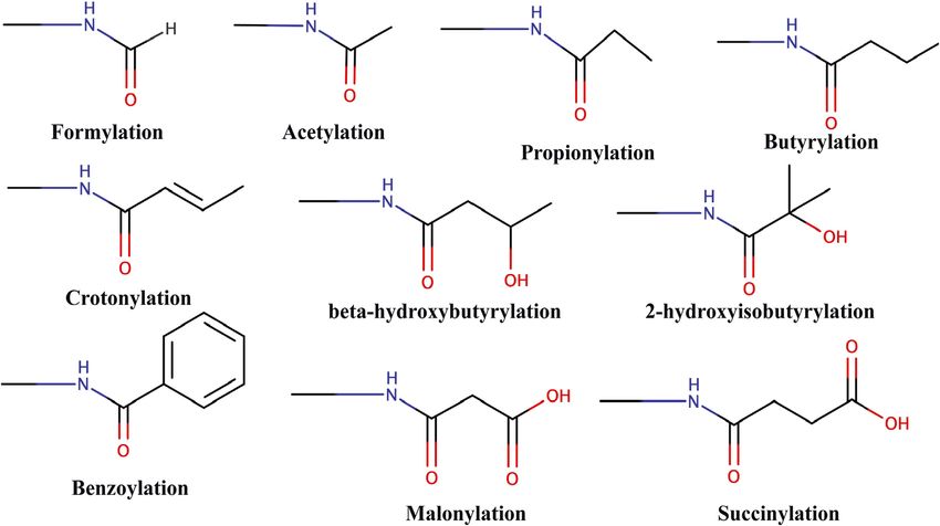
Sehrawat et al.                                                                                                        Decorated Tail Recognition by Specific Reader

  FIGURE 4 | Readout of acyl marks by YEATS and DPF domains. (A) Longer chain of the crotonyl group (blue) can be accommodated in the Taf14 YEATS domain
  (magenta). (PDB ID: 5D7E and 5IOK). (B) Superimposition of residues involved in interaction among YEATS domains of different proteins: YEATS2 YEATS
  domain–H3K27cr (cyan; PDB ID: 5IQL), Taf14 YEATS domain–H3K9cr (yellow; 5IOK), and AF9 YEATS domain–H3K18cr (purple; 5HJD). (C) Opposite orientation of
  H3 peptides across Taf14 (PDB ID: 5IOK) and YEATS2 (PDB ID: 5IQL) proteins. In the Taf14–H3K9cr5–13complex, R8 (cyan) interacts with aspartate at 104th
  position. In YEATS2–H3K27cr24–31 complex, R26 (cyan) is facing away from the YEATS domain but proline at 30th position (magenta) makes contacts with the
  hydrophobic pocket. (D) Top: overall structure of the DPF domain of MORF protein with H3K14bu1–16 (wheat) and H3K14cr1–19 (cyan). Bottom: close-up view of amino
  acids involved in interaction between crotonylated lysine (yellow) and MORFDPF protein (magenta) (PDB ID: 6OIE and 5B76).

established that the YEATS domain is a Kac reader module with                         neutral asparagine residue, which does not recognize “R(−1).” While
the highest affinity to crotonylation mark (Andrews et al., 2016;                      Taf14 and AF9 YEATS domain prefer H3K9cr and H3 N-terminal
D.; Zhao et al., 2016). The crystal structure of all three proteins in                residues “K4-Q5-T6-A7-R8” have extensive interactions with loops
complex with H3 tail peptide having crotonylation mark showed                         L6 and L8 surface residues, YEATS2 binds H3K27cr more strongly
a highly similar structure where the extended side chain of Kcr fits                   and oppositely oriented C-terminal residues “S28-A29-P30-A31” fits
comfortably into the narrow end-open pocket of the YEATS                              nicely on the surface of L6 and L8 loops (Figure 4C). In the crystal
domain (Figure 4A).                                                                   structure of YEATS2, the YEATS domain in complex with H3K27cr
   Two conserved aromatic residues in different YEATS domains                         revealed a hydrophobic pocket in YEATS2 in which H3P30 fits
(F62 and W81 for Taf14, F59 and Y78 for AF9, and Y268 and                             snugly and facilitates the correct positioning of H3K27cr, explaining
W282 for YEATS2) make a sandwich arrangement with the                                 the site specificity of YEATS2 (D. Zhao et al., 2016).
planar crotonylamide group, which crosses the β-sandwich                                  A recent study showed the role of histone crotonylation and
cage at a 90° angle in a corkscrew-like manner (Figure 4B).                           Taf14 in the yeast metabolic cycle. In the yeast metabolic cycle,
This arrangement favors a novel “aromatic–π–aromatic” stacking                        acetylation increases in high oxygen consumption state, followed
(also called “π–π–π” stacking) (Klein et al., 2018). Additionally,                    by generation of crotonylation intermediates (Gowans et al.,
extensive hydrophobic interactions, the amide−π interactions, the                     2019). As the cells shift to a low oxygen consumption state,
CH–π interactions, and electrostatic interactions (mainly of Cα                       acetylation mark is replaced by crotonylation marks and in this
and Cβ of the alkene moiety with the carbonyl oxygen of Q79)                          LOC state H3K9cr and Taf14 repress the pro-growth genes,
between the side chain of crotonylated lysine and pocket residues                     contrary to earlier studies showing their role in gene
significantly contribute to specific recognition of Kcr by the                          expression (Sabari et al., 2015).
YEATS domain (Krone et al., 2020).
   Flanking residues in all three H3K9, H3K18, and H3K27 are
conserved, sharing a common motif “A(−2)R(−1)KS(+1).” An acidic                       4.3 Recognition of H3K14acyl by the DPF
aspartate residue of Taf14 and AF9 YEATS domains forms charge-                        Domain
stabilized hydrogen-bonding interaction with (n–1) arginine of                        Pull-down assays and ITC experiments using an array of
H3K9cr peptide. Interestingly, in the YEATS domain of                                 H3K14 bearing different acylations revealed that the DPF
YEATS2 protein, this acidic aspartate residue is replaced by the                      domain of DPF2 and MOZ HAT displays more affinity for

Frontiers in Genetics | www.frontiersin.org                                       7                                            July 2022 | Volume 13 | Article 903923
Sehrawat et al. Decorated Tail Recognition by Specific Reader
H3K14cr, H3K14bu, and H3K14pr than H3K14Ac with proteins via the transamidation reaction (Hummerich et al.,
crotonylation being the most favored (Xiong et al., 2016). On 2012).
a similar line, a combination of fluorescence spectroscopy, NMR, It was previously known that TGMs could modify histones
and histone peptide pull-down assay established the specificity of in vitro and do so very fast compared to some of the known native
the DPF domain of MORF HAT for H3K14cr and H3K14bu substrates (Abad, and Franco 1996). But recently, using the bio-
(Klein et al., 2017). In all the crystal structures solved for the DPF orthogonal metabolic-labeling approach, Farrelly et al. (2019)
domain in complex with H3K14cr or H3K14bu, H3 tail peptide showed that TGM2 can catalyze serotonylation of glutamine at
has extensive contacts with double PHD finger of the DPF the fifth position of histone H3 trimethylated lysine 4
module with segments H34–11 and H317–25 (in case of full (H3K4me3)-marked nucleosomes, resulting in the presence of
length taken) adopting α-helical conformation (Xiong et al., combinatorial H3K4me3Q5ser in vivo.
2016; Klein et al., 2017, 2019). The overall structural analysis
revealed binding of the DPF module to acyllysine in a ping-pong- 5.1 Recognition of H3Q5ser by WDR5
like manner with three characteristic interactions (Klein et al., In a pull-down assay, WDR5 was captured using H3 peptide with
2019). H3K4me3Q5ser dual marks as the bait, suggesting that
The first PHD domain of the DPF module forms a unique WDR5 could be a potential reader of this modification
zinc-finger domain in which a hydrophobic pocket is formed at (Farrelly et al., 2019). WDR5 is a core subunit of a histone
the β-sheet-2 surface. In the MORF (and MOZ) proteins, the methyltransferase enzyme; the MLL (mixed-lineage leukemia)
hydrophobic pocket is formed by the amino acid residues complex is responsible for trimethylation of H3K4. Further
I228–C230 of β-1, N235–G237 of β-2, and amino acid pull-down assays and ITC experiment established that
residues involved in zinc ion coordination S210 (S217), F211 serotonylated H3Q5 enhances the binding of WDR5 by at
(F218), L242 (L249), W257 (W264), C259 (C266), I260 (I267), least two-fold than that of unmodified H3, and
and E261 (Figure 4D). The planar crotonylation group of H3K4 trimethylation mark has no significant effect on this
H3K14 is inserted snugly into this hydrophobic reader pocket binding, also supported by the observation that there was no
and stably positioned with the help of four water-mediated electron density for the trimethyl group of K4 in the crystal
hydrogen-bonding interactions and four pairs of hydrophobic structure of WDR5–H3K4me3Q5ser complex (Jie et al., 2022)
contacts. The structural and sequence alignment analysis of DPF (Figure 5B).
domains showed that glycine residue G237 of β-strand-2 is a The crystal structure of WDR5–H3Q5ser complex revealed
critical component of the hydrophobic pocket due to its free side that the serotonyl group is placed in a shallow hydrophobic
chain. In classical PHD fingers, this glycine residue is replaced by surface pocket of WDR5. The WDR5–Q5ser interaction is
bulky amino acids like phenylalanine or tyrosine, which fill the stabilized via a network of hydrogen bonds (one between OH
pocket and block large chain acylation mark insertion. One of the group of serotonin with amide group N130 residue of WDR5 and
phenylalanines (F211 in MOZ and F218 in MORF) is in close another between amide group of serotonin with WDR5 D172 side
proximity to the inserted crotonyl group and forms a π−π chain) and van der Waals contacts (between hydrophobic moiety
interaction between the C=C double bond of the crotonyl of serotonyl group and aromatic side chains of Y131, F149, and
group and the aromatic ring of phenylalanine. This additional Y191 of WDR5) (Figure 5A). Additionally, R2 of H3 peptide also
interaction is responsible for selectivity and strengthens the participates in this complex formation as it is anchored into a
interaction between DPF domain and H3K14cr (Klein et al., negatively charged central channel and interacts with WDR5’s
2019). Additionally, H3 residues R2 and K4 are inserted into two F133 and F263 through cation–π interactions (Jie et al., 2022).
“acidic” pockets formed at the surface of β-1 sheet of the second In neuroblastoma cells, upon recognition of H3Q5ser
PHD domain and held by hydrogen bonding and electrostatic modification, WDR5 is recruited to promoter regions of
interactions. oncogenic genes GPX1, C-MYC, and PDCD6 that can promote
tumor formation (Jie et al., 2022). Knockdown studies implied
that serotonylation of H3Q5 is not dependent on H3K4me3;
5 SEROTONYLATION instead, there was a decrease in the level of H3K4me3 upon
WDR5 knockout, also seen in the case of TGM2 knockdown,
Serotonylation is the attachment of the serotonin molecule to the which may be due to less recruitment of MLL1 complex.
glutamine residue of proteins. Serotonin [or 5-HT (hydroxy H3K4me3Q5ser displays a ubiquitous pattern of tissue
tryptamine)] is a monoamine with an abundant presence and expression in mammals, with enrichment observed in the
diverse functions varying from neurotransmitter to hormone brain and gut, two organ systems responsible for the bulk of
release and gastrointestinal motility. Additionally, serotonin 5-HT production. Genome-wide analyses of human serotonergic
has been shown to have the ability to covalently modify neurons, developing mouse brain, and cultured serotonergic cells
several proteins, including RacI, small guanosine indicate that H3K4me3Q5ser nucleosomes are enriched in
triphosphatase, and fibronectin and thereby regulate their euchromatin, are sensitive to cellular differentiation, and
functions (Walther et al., 2003; Watts, Priestley, and correlate with permissive gene expression—phenomena that
Thompson 2009). The tissue transglutaminase 2 (TGM2) are linked to the enhanced function of TFIID (Farrelly et al.,
enzyme is responsible for conjugating serotonin to cytosolic 2019).
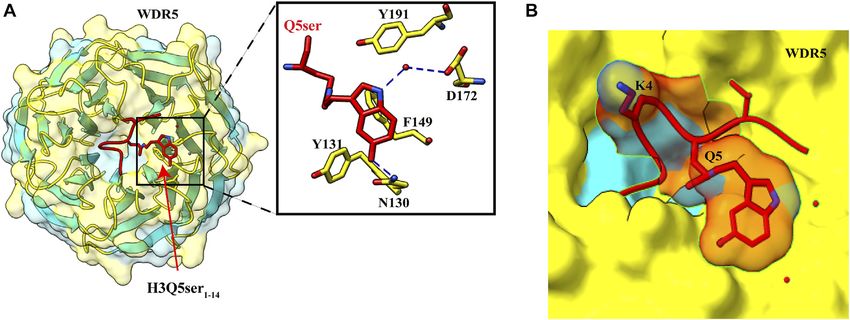
Frontiers in Genetics | www.frontiersin.org 8 July 2022 | Volume 13 | Article 903923Sehrawat et al. Decorated Tail Recognition by Specific Reader
FIGURE 5 | Recognition of H3Q5ser by WDR5. (A) Left: overall structure of WDR522–334 in complex with H3Q5ser1–14 peptide (red). Right: enlarged view showing
residues of WDR522–334 (yellow) interacting with Q5ser (red). Hydrogen bonds are shown with blue dashed lines. (B) Lysine at fourth position is protruding away from the
WDR5, resulting in no effect of trimethylation of K4 on the Q5ser–WDR5 interaction (PDB ID: 7CFQ).
6 METHYLATION were discovered that interact specifically with the methylated
histone. These “reader” proteins contain methyl-lysine-binding
Histone methylation and its importance in transcription were motifs, including PHD, chromo, Tudor, PWWP, WD40, BAH,
first observed in the 1960s. There are three lysine methylation ADD, ankyrin repeat, and MBT domains (Figure 6). These
states: -mono, -di, and -tri (me1, me2, and me3); since readers can distinguish target methyllysine based on their
methylation does not change histone’s charge configuration, methylation state and surrounding amino acid sequence
the primary function of these methylations is to interact with (Musselman et al., 2012).
effector molecules that specifically recognize these modifications.
Generally, all other histone modifications are specific for the 6.1 Royal Superfamily
active or repressed state, while in methylated chromatin it In this superfamily, domains are structurally related and have β-
depends on its methylation state and the modification barrel topology. It is believed that they come from a common
position. For example, H3K4, H3K36, and ancestor, which has the conserved binding ability with the
H3K79 methylations are considered to mark active methylated substrate. All the family members consist of a
transcription (Heintzman et al., 2007), whereas H3K9, H3K27, slightly curved β-barrel with three β-strands followed by a
and H4K20 methylations are associated with silenced chromatin short 310 helix, and different members are distinguished based
states (Bernstein et al., 2005; Barski et al., 2007). on additional strands or helices. This family includes MBT,
Methyllysine-specific readers have a peculiar characteristic: Tudor, chromodomain, and PWWP (Yap and Zhou 2010).
they recognize histone modification by an aromatic cage that
comprises two to four aromatic amino acids. These aromatic 6.1.1 Recognition of H4K20me1/me2 by the MBT
amino acids in some complexes are perpendicular to each other, Domain of L3MBTL1
which helps encircle the entire lysine methylation. The Isothermal titration calorimetry (ITC) assay using different
compartment of the aromatic cage defines whether mono-, di-, methylation states of H4K20 peptide revealed that the MBT
or tri-methylation state interacts with it. Therefore a small domain of human L3MBTL1 displays more affinity toward
compartment limits its interaction with a higher methylation mono- and dimethylation states and does not bind to
state because of steric hindrance, while a large compartment unmodified and trimethylated histone peptide. However, the
favors the interaction with a higher methylation state. Interaction binding is relatively of low affinity (KD = 5–40 μM) and
in the compartment between the methylammonium group and promiscuous (Min et al., 2007). The crystal structure of
the aromatic cage is stabilized by cation–π interactions and the L3MBT1 with three MBT domains bound to histone peptide
hydrophobic and van der Waal interactions. Amino acids of 11 residues (H4 residue 15–25 with H4K20me2) shows the
surrounding methyllysine play a vital role in the reader’s similar structure of all MBT domains assembled in a triangular
specificity for a particular methylated lysine. Some readers shape. The structure is consistent with previously known
show very low specificity, while others are specific for a structures of the MBT domain. It consists of four β-strands
specific methylated state. Beyond caging of the methyllysine, that form β-barrel and other extended arms of helices and a
the mechanism of recognition of surrounding residues varies shorter strand (Wang et al., 2003). L3MBTL1 has three repeats of
among readers. A number of evolutionarily conserved domains the MBT domain; however, only the second MBT domain binds
Frontiers in Genetics | www.frontiersin.org 9 July 2022 | Volume 13 | Article 903923Sehrawat et al. Decorated Tail Recognition by Specific Reader
FIGURE 6 | Structural features of domains capable to “read” methyl marks. (A) Second MBT domain of human KIAA1617: β-strands in light green and α-helix in
pink (PDB ID: 1WJR). (B) PWWP domain of Pdp1 (PDB ID: 2L89). (C) Chromodomain of MPP8 (PDB ID: 3QO2). (D) Tudor domain of human PHF20 (PDB ID: 3SD4). (E)
WD40 repeats of MYC (PDB ID: 6U8O). (F) Ankyrin repeats of human liver-type glutaminase (PDB ID: 5U0K). (G) Bromo-adjacent homology domain of human
polybromo-1(PDB ID: 6OXB). (H) PHD of human BPTF protein (PDB ID: 2F6N).
to methyllysine (Santiveri et al., 2008; Eryilmaz et al., 2009; Li that H4K20me1 nucleosomal arrays were less compacted than
et al., 2007; Min et al., 2007). Superimposition of all three MBT H4K20me0 and H4K20me3 nucleosomal arrays, and the H4 tail
domains revealed that the shorter side chain of cysteine 363 in the was more dynamic in K20 mono-methylation. This study
aromatic cage of the second MBT was replaced by bulky amino suggests that mono-methylation of H4K20 facilitates the
acids in MBT1 and MBT3. Thus, steric hindrance prevents the opening and accessibility of chromatin (Shoaib et al., 2021).
binding of MBT1 and MBT3 with methylated lysine. It also
preferentially reads mono- and dimethylation by the cavity 6.1.2 PWWP Domain
insertion mode, and specificity toward a lower methylation PWWP domain was first identified in the WHSC1 protein that
state is because of the aspartate residue present in the contains the 100–130 amino acid structural motif (Stec et al.,
aromatic cage. Typical interactions in the aromatic cage are 1998). It also has a conserved Pro-Trp-Trp-Pro motif, and it
cation–π between aromatic residues and positively charged consists of five β-strand barrels packed against the helical bundle.
methylammonium of methyllysine. Additionally, aspartate Despite the sequence conservation in different proteins, some
binds to methylammonium via hydrogen bonding in the MBT variation in the PWWP motif can occur. For example,
domain. methyltransferase DNMT3a/b has SWWP (Qiu et al., 2002),
In contrast, the flanking residues of the peptide substrate show and hepatoma-derived growth factor (HDGF) has a PHWP
little interaction with the protein. Only two water-mediated motif instead of a PWWP motif (Sue et al., 2004). It was
hydrogen bonds are present, first between the backbone initially identified as the DNA-binding protein; however, its
carbonyl group of H18 and Y386 of the protein and a second similarity to the Tudor and chromodomain suggests that it
between the carbonyl NH group of K20 and N358 of the protein. might have the ability to bind methylated lysine. DNMT3a
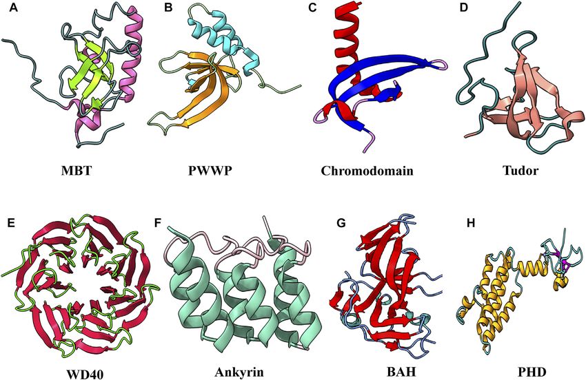
H4K20 methylation was previously linked to chromatin protein responsible for DNA methylation contains the PWWP
compaction. However, H4K20me1 was found in the actively domain. This domain is also known to interact with methylated
transcribing genes, contradicting the previously suggested role histone tails, which led to assumptions that it might have dual
of methylation in chromatin compaction. A recent study revealed binding to histone tails and the dsDNA.
Frontiers in Genetics | www.frontiersin.org 10 July 2022 | Volume 13 | Article 903923Sehrawat et al. Decorated Tail Recognition by Specific Reader
Pdp1 protein, which contains PWWP domains, binds to The noncanonical chromodomain proteins are based on the
methylated lysine, and dsDNA was seen by fluorescence chromo ATPase/helicase-DNA-binding (CHD) protein. They
polarization assay (FPA) (Qiu et al., 2012). The binding contain two chromodomains, both at the N-terminal, for
studies showed that PWWP domains of Pdp1 bind to the example, SNF2-type helicase, which is involved in chromatin
H3K20 trimethylation. After that, many other PWWP remodeling. CHD7 specifically recognizes H3K1me1 as the
domains were shown to exhibit the binding with methylated enhancer for the gene.
lysine. Except for Pdp1, all the proteins containing PWWP
domains bind specifically to the H3K36 methylation, 6.1.4 Recognition of H3K4me3K9me3 Bivalent Mark
suggesting its role as the H3K36 methylation sensor (Vezzoli by the Tudor Domain of Spindlin1
et al., 2010; van Nuland et al., 2013; Wang et al., 2020). As it binds Tudor domains are structurally diverse and mediate
to the trimethylation state of lysine, it suggests that the binding protein–protein interactions. Tudor domains interact with all
cavity of the PWWP domain is wider to accommodate the bulkier methylation states. This domain consists of approximately
me3 group than the MBT domain, which can only interact with 60 amino acids of four or five β-strands which form a β-barrel
mono- and dimethylated states. Therefore, this domain shows structure followed by one or two helices (Selenko et al., 2001).
less specificity for the degree of methylation state. Tudor domain-containing protein interacts with the H3K4me3
Structural analysis of Pdp1 revealed that the aromatic cage is (Wang et al., 2011; Yang et al., 2012), H3K9me2 (Arita et al.,
formed by Y63, W66, and F94. Cation–π interactions are used by 2012), H3K36me3 (Cai et al., 2013), and H4K20me3 (Hirano
the Pdp1 PWWP domain to recognize the trimethylated lysine at et al., 2012). Almost 30 known proteins have this Tudor domain,
20th position, and two residues (D97 and N99) from the loop including JMJD2, 53BP1, SGF29, Spindlin1, UHRF1, PHF1,
between β3 and β4 form an extensive network of hydrogen bonds OHF19, LBR, and TDRD3 (which recognizes methylated
with the histone H4 tail residues (R19, K20, and V21) (Qiu et al., arginine residues). Proteins in this family are involved in
2012). Y63, W66, and F94 amino acid side chains are various biochemical processes like DNA methylation,
perpendicularly oriented, forming an aromatic cage nonhomologous end joining, DNA damage and repair,
accommodating the trimethylammonium group. Mutations of transcription activation and repression, and rRNA expression.
the residues that compose the aromatic cage abolish methylated To meet the growing demand for ribosomes in rapidly
histone peptide binding. growing cells, more copies of rRNA are produced at a greater
transcription rate. The repressive histone methylation marks
6.1.3 Recognition of H3K9me3 Marks by present on H3K9/K27 and H4K20 in the heterochromatin
HP1 Chromodomain region are linked to rRNA transcription suppression. Because
The chromatin organization modifier domain (chromodomain) it has been established that H3K4 methylation is required for
is the smallest member of this superfamily. The structural active gene expression, the cell must establish H3K4 methylation
motif is based on the HP1 fold, consisting of three curved and H3K9 demethylation to convert the suppressed rRNA
antiparallel β-sheets followed by the α-helix (Ball et al., 1997). expression to active expression. Although H3K4 and
There are approximately 55 proteins identified which contain H3K9 trimethylation are mutually exclusive, bivalent
chromodomain. These chromodomain-containing proteins H3K4me3 and H3K9me3 have been documented in specific
were associated with chromatin silencing. These proteins cell types (Mikkelsen et al., 2007; Bilodeau et al., 2009; Rugg-
are divided into two groups: canonical (based on Gunn et al., 2010; Matsumura et al., 2015). It was previously
HP1 structure) group includes polycomb proteins Cbx1-9, reported that KDM4A and KDM4C recognize the H3K4me3 via
CMT1-3, and CYD; and noncanonical, including CHD1-8, their Tudor domain and help in the demethylation of H3K9me3.
RBBP1, and HRP1. This study suggests that these methylation marks can coexist on
HP1 and polycomb proteins recognize H3K9me3 (Jacobs and the same H3 N-terminal tail and functionally crosstalk (Huang
Khorasanizadeh 2002) and H3K27me3 (Min, Zhang, and Xu et al., 2006; Yamamoto, and Fujimori 2016). It was also previously
2003), respectively, through their ARKS/T motif. The histone tail known that euchromatin rRNA genes contain bivalent mark
inserted between two strands forms the complete β-barrel in both H3K4me3K9me3 (Murayama et al., 2008). So for rRNA
proteins. This insertion of the histone tail is stabilized by the synthesis, the bivalent mark of H3K4 and
electrostatic interactions and the hydrogen bond between the H3K9 trimethylation is needed.
backbone. This interaction involves seven amino acids preceding The Tudor domain is found in Spindlin1, a protein that aids in
methyllysine and one following amino acid. This recognition rRNA expression. Splindin1 creates a complex with C11orf84 that
method prefers the recognition of trimethylation over mono- and recognizes the bivalent mark on the histone H3 tail. Tudor
dimethylation. Recent cryo-EM structure of H3K9me3 di- 2 domain residues F141, W151, Y170, and Y177 form an
nucleosome with HP1α, HP1β, and HP1γ revealed how aromatic pocket for trimethylated K4, whereas Tudor
heterochromatin is organized. In this structure, di- 1 domain residues W62, W72, Y91, and Y98, as well as Tudor
nucleosomes trimethylated at K9 are bridged by two 3 domain residue F251, recognize trimethylated K9 (Figure 7A)
symmetric molecules of HP1. Linker DNA between the (Du et al., 2021). In addition to cation–π interactions formed by
nucleosome is not interacting with the HP1, which leaves dual methylated lysine, the N-terminal amino group of
linker DNA to interact with ACF (ATP-utilizing chromatin H3A1 forms a hydrogen bond with the side-chain carboxylate
assembly and remodeling factor) (Machida et al., 2018). group of D189, and guanidino moiety of H3R2 is ion-paired with
Frontiers in Genetics | www.frontiersin.org 11 July 2022 | Volume 13 | Article 903923Sehrawat et al. Decorated Tail Recognition by Specific Reader
FIGURE 7 | Interaction details of Spindlin1 and DNMT1 with trimethylated histone peptides. (A) Structure of Spindlin1 with H3K4me3K9me3 peptide; Spindlin1
shown in orange, H3K4me3 in green and H3K9me3 in cyan. Key residues involved in the interactions are depicted as a ball‑stick model shown in an enlarged view (PDB
ID: 7CNA). (B) Structure of DNMT1 BAH1 with H4K20me3 peptide; DNMT1 BAH1 is shown in light blue and H4K20me3 in pink (PDB ID: 7LMK).
the side-chain carboxylate group of D184 (Du et al., 2021). This H3K4me2 and me3. However, the crystal structure of
bivalent recognition further helps in the dislocation of HP1 from WDR5 with the unmodified, mono-, di-, and trimethylated
the rRNA chromatin, which relaxes the chromatin and helps in H3K4 reveals that WDR5 interacts with H3R2 and acts as an
the recruitment of RNA polymerase I, which leads to the arginine reader. WRD5 interacts with lysine only through E332,
expression of rRNA. Spindlin1 Tudor 3 domain is responsible present at the protein’s surface. WRD5 anchor as the arginine
for the binding to C11orf84, while the other two Tudor domains pocket binds to the unmodified and demethylated arginine
act in concert to recognize a noncanonical bivalent histone mark residue (Dharmarajan et al., 2012). It was proposed to act as a
H3K4me3K9me3. histone modification intermediate that binds to arginine and
presents lysine for methylation.
6.2 The WD40 Repeats
WD40 repeats are present in proteins showing very diverse
protein–protein interactions. WD repeats usually have 6.3 Recognition of H3K9me1/me2 by
40–60 amino acids and conserved tryptophan–aspartate (WD) Ankyrin Repeat Domain of G9a
residue. This motif can be found in each blade of a WDR domain. It is a ~33 amino acid repeat, and each repeat consists of
The structural plasticity of WDR domains allows them to keep helix–loop–helix structure with a β-hairpin/loop region; and in
their β-propeller fold even after deletion of WD repeats, which most proteins, 4–7 repeats are present, which stack onto each
can range from five to eight but are generally seven (Böcskei, and other to form the right-handed solenoid-like structure that helps
Polgár 1998; Juhász et al., 2005). Each of these repeats folds into a in the protein–protein interaction (Sedgwick and Smerdon 1999).
four-stranded β-sheet, and these propellers are large, usually A short-distance interaction between inter α-helices helps form
containing ~300 amino acids. the solenoid, forming a typical globular protein shape. The
SET1 methyltransferase (catalyze methylation of H3K4) structure of the AR domain is stabilized through inter- and
subunit WDR5 contains seven WD repeats to form the β- intra-helical hydrophobic and hydrogen bonding via polar
propeller structure. SET1 needs WDR5 for its assembly and residues at the N-terminal. Hydrophobic interactions between
activity. First, it was found that WDR5 binds to P5 and H7 form the L-shape of the domain, and hydrogen bond
Frontiers in Genetics | www.frontiersin.org 12 July 2022 | Volume 13 | Article 903923You can also read