To Better Generate Organoids, What Can We Learn From Teratomas?
←
→
Page content transcription
If your browser does not render page correctly, please read the page content below
REVIEW
                                                                                                                                            published: 16 July 2021
                                                                                                                                    doi: 10.3389/fcell.2021.700482

                                            To Better Generate Organoids, What
                                            Can We Learn From Teratomas?
                                            Hongyu Li, Lixiong Gao, Jinlin Du, Tianju Ma, Zi Ye and Zhaohui Li*
                                            Department of Ophthalmology, The Chinese People’s Liberation Army General Hospital, Beijing, China

                                            The genomic profile of animal models is not completely matched with the genomic
                                            profile of humans, and 2D cultures do not represent the cellular heterogeneity and
                                            tissue architecture found in tissues of their origin. Derived from 3D culture systems,
                                            organoids establish a crucial bridge between 2D cell cultures and in vivo animal models.
                                            Organoids have wide and promising applications in developmental research, disease
                                            modeling, drug screening, precision therapy, and regenerative medicine. However,
                                            current organoids represent only single or partial components of a tissue, which
                                            lack blood vessels, native microenvironment, communication with near tissues, and a
                                            continuous dorsal-ventral axis within 3D culture systems. Although efforts have been
                                            made to solve these problems, unfortunately, there is no ideal method. Teratoma, which
                          Edited by:
                                            has been frequently studied in pathological conditions, was recently discovered as a
                    Yangming Wang,          new in vivo model for developmental studies. In contrast to organoids, teratomas have
              Peking University, China      vascularized 3D structures and regions of complex tissue-like organization. Studies have
                      Reviewed by:          demonstrated that teratomas can be used to mimic multilineage human development,
                      Ryuji Morizane,
      Brigham and Women’s Hospital          enrich specific somatic progenitor/stem cells, and even generate brain organoids. These
         and Harvard Medical School,        results provide unique opportunities to promote our understanding of the vascularization
                       United States
                              Yan Liu,
                                            and maturation of organoids. In this review, we first summarize the basic characteristics,
     Nanjing Medical University, China      applications, and limitations of both organoids and teratomas and further discuss the
                *Correspondence:            possibility that in vivo teratoma systems can be used to promote the vascularization and
                          Zhaohui Li        maturation of organoids within an in vitro 3D culture system.
           zhaohuili650@hotmail.com
                                            Keywords: teratoma, organoid, 3D culture, ESC, IPSC
                    Specialty section:
         This article was submitted to
                  Stem Cell Research,      INTRODUCTION
                a section of the journal
  Frontiers in Cell and Developmental      The progress of clinical medicine cannot be separated from research on disease pathogenesis and
                                Biology
                                           drug screening. Restricted to ethical requirements, in vivo animal models and two-dimensional
             Received: 26 April 2021       (2D) cell cultures have frequently been used to study human development and diseases in the last
            Accepted: 21 June 2021
                                           few decades. However, both models fail to mimic the complex architectures, cell-cell interactions,
            Published: 16 July 2021
                                           unique microenvironment and organ-level functionality of the human body (Kim et al., 2020).
                              Citation:    Since the isolation of human embryonic stem cells (ESCs) and the reprogramming of human
   Li H, Gao L, Du J, Ma T, Ye Z and
                                           somatic cells into induced pluripotent stem cells (iPSCs) succeeded, these pluripotent stem cells
       Li Z (2021) To Better Generate
Organoids, What Can We Learn From
                                           (PSCs) have become a source for cell replacement therapy and a new model to simulate human
                           Teratomas?      development (Hoffman and Carpenter, 2005). By exploring the self-organizing features of PSCs in
      Front. Cell Dev. Biol. 9:700482.     a 3D culture environment, stem cell studies have improved our understanding of key aspects of
     doi: 10.3389/fcell.2021.700482        organogenesis (Garreta et al., 2020). These advances have led to the generation of new cell culture

Frontiers in Cell and Developmental Biology | www.frontiersin.org                1                                             July 2021 | Volume 9 | Article 700482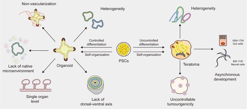
Li et al.                                                                                                              To Better Generate Organoids

systems named organoids that could be used to produce organ-               of 3D culture. In this review, we substantially summarize the
like tissues. The first organoid was successfully generated from           general characteristics, applications and limitations of current
mouse ESCs and formed a fully stratified neural retinal tissue             organoids and teratomas, and then focus on the progress and
architecture in a spatiotemporal manner mimicking in vivo                  potential of teratoma systems as an in vivo tool for better
development (Eiraku et al., 2011). Then, a large number of                 generating organoids.
culturing systems were introduced to generate a variety of tissue-
specific organoids, including the brain, lung, intestine, liver,
pancreas, and kidney (Lancaster et al., 2013; Watson et al.,               ORGANOIDS: A BETTER IN VITRO
2014; Dye et al., 2015; Broutier et al., 2016; Takasato et al.,            MODEL
2016). Organoids have compositions and architectures similar
to living organs, which provides valuable information about                The 3D cultures have advantages over 2D cultures and animal
the mechanisms underlying human development and organ                      models. When placed flat in a Petri dish, cells cannot behave as
regeneration. However, there are some limitations and challenges           usual. Although animal models may be useful tools, they also have
in current organoids, including the low reproducibility, the               limitations because there are large differences between model
lack of blood vessels and native microenvironment, which                   animals and humans. Therefore, it is usually difficult to translate
limits their applications in disease modeling and drug screening           the results from animal models into physiological understanding
(Fatehullah et al., 2016; Kaushik et al., 2018; Rossi et al., 2018).       of the human body with accuracy. The term “organoid” is
The development of the organoid system is still in its infancy             defined as a complex 3D structure that develops from stem
in comparison to 2D cultures and animal models. Further                    cells or organ-specific progenitors and displays architectures and
study is instrumental and necessary to better generate and                 functionalities similar to the architectures and functionalities of
apply organoids.                                                           living organs (Ashok et al., 2020). Organoid systems are changing
    In fact, there is a significant difference between in vivo             the way scientists model organ development and expanding
and in vitro microenvironment. The microenvironment of 3D                  basic biological research and medical research into a more
cultures is provided mainly by a solid extracellular matrix                physiologically meaningful human environment.
(ECM), which supports cell growth and promotes cell adhesion
(Badekila et al., 2021). However, their complexity and variability         Generating Organoids
in composition makes it more difficult to control the cultural             Self-Organization of Organoids
microenvironment. Moreover, the characteristics of cell-to-cell            The development of organoids usually involves the self-
adhesion and the other 3D properties are more sufficiently                 organization of a comparatively homogeneous cell population.
showed in in vivo than in vitro conditions (Aleckovic and Simón,           Self-organization is the spontaneous formation of ordered
2008). And in vivo microenvironment is relatively stable and               patterns and structures from specific elements or individuals. The
could not impede the infiltration of drugs comparing to a natural          basic processes of self-organization involve the following three
ECM with complex compositions.                                             processes: self-assembly, self-patterning, and self-morphogenesis
    Once PSCs grow in in vivo environment, teratomas would                 (Sasai, 2013). Self-assembly refers to the process of autonomously
be formed. As a new in vivo model of human multilineage                    forming patterns by selectively gathering cells or rearranging the
development, teratomas have similarities and differences                   relative positions of cells (Whitesides and Grzybowski, 2002).
with organoids (McDonald et al., 2020). Both organoid and                  Only through the self-assembly of one or several types of cells
teratoma are derived from PSCs. Organoid formation involves                can highly ordered structures be formed (Dobrescu and Purcarea,
gradually controlled differentiation of PSCs and subsequent                2011). Self-patterning is the spatial and temporal control of cell
self-organization into tissue-specific organ-like structures.              states to acquire heterogeneous properties in a region-specific
Teratoma formation involves uncontrolled differentiation and               manner (Sasai, 2013). Self-patterning starts with a symmetry
self-organization into various somatic tissues from all three germ         disrupting event (Turner et al., 2016), which is affected by a
layers. Teratoma systems provide an in vivo tool that enables              variety of mechanisms, including reaction diffusion, asymmetric
more physiologically relevant experiments to be performed,                 cell division, and stability of regulatory networks (Fleury
which cannot be reproduced in animals or in vitro cultures.                and Watanabe, 2004; Sasai, 2013; Green and Sharpe, 2015).
Recently, several studies showed that single specific lineage              Self-morphogenesis, driven by internal organization mechanics
cell types, such as skeletal muscle stem cells, neural stem cells,         and occurring automatically without external forces or spatial
and hematopoietic stem cells, have already been enriched from              constraints, is the most crucial step that determines the final
mouse PSCs through teratoma formation (Chan et al., 2018;                  organoid formation (Sasai, 2013). Self-morphogenesis should
Philipp et al., 2018; Kim et al., 2019). These enriched cells could        include complex control of directionally internal forces and
be cultured in vitro, and even specific vascularized tissues could         dynamic control of the cooperative response to mechanical
be isolated to generate organoids (Lee et al., 2020). Moreover,            force, as well as dynamic changes in tissue viscosity and
the variations of cell types in teratomas could be an advantage            stiffness (Lecaudey et al., 2008; Sasai, 2013). Successful formation
to generate complex tissues, which provides a possibility to               of organoids depends on each described process, and three
investigate human development at a multi-organ level. All these            main features need to be carefully considered: the physical
results indicated that an in vivo teratoma system could provide            characteristics of the culture environment, the requirement
a powerful platform to improve the method and technology                   for endogenous and exogenous signals, and the initial origin

Frontiers in Cell and Developmental Biology | www.frontiersin.org      2                                        July 2021 | Volume 9 | Article 700482
Li et al. To Better Generate Organoids
of culturing cell types (Rossi et al., 2018). The choice made lineage. Organoids derived from ASCs were first generated in the
for each of these features could affect the characteristics of small intestine (Sato et al., 2009). ASC-derived organoids contain
the final organoid. only the polarized epithelial components of organs and lack
the stroma, nerves, and vasculature (Bartfeld et al., 2015; Huch
The 3D Culture Microenvironment et al., 2015). The ASC-derived organoids have lower complexity
The microenvironment of 3D cultures is provided mainly by and predictability than PSC-derived organoids. The fundamental
a solid extracellular matrix (ECM), which supports cell growth differences between PSC- and ASC-derived organoids make
and promotes cell adhesion and is the most common way to them complementary model systems. In addition, intestinal and
enhance the 3D properties of organoids (Badekila et al., 2021). pancreatic organoids could be derived from dissociated fetal cells
Matrigel, an animal-derived hydrogen from mouse sarcoma, or tissues (Fordham et al., 2013; Greggio et al., 2013).
is the initial natural ECM used in 3D cultures. Matrigel
or another natural matrix (collagen I) is a complex mix of Applications of Current Organoids
ECM components and growth factors, making cell growth and The ability to grow near-physiological, self-renewing organoids
differentiation very efficient. However, their complexity and provides us with a fabulous model system for basic research
variability in composition makes it more difficult to control and transplantation application. Organoids are genetically stable,
the cultural microenvironment and may reduce organoid-to- contain a variety of differentiated cell types, and produce
organoid reproducibility (Badekila et al., 2021). A hydrogel with specialized cell types that cannot be cultured in vitro (Schutgens
chemical composition has recently been introduced to substitute and Clevers, 2020). Moreover, the self-organization of tissues and
for the undefined natural matrix (Gjorevski et al., 2016; Lindborg the spatiotemporal regulation of cells in organoids reproduce the
et al., 2016). Although hydrogels could control the mechanical development of human bodies. Following the establishment of
and biochemical characteristics of the culture microenvironment, human stem cell-derived organoids, various human diseases have
they need to be customized according to the specific requirements been studied in vitro. Organoid systems have opened remarkable
of different organoids because of their inherent low biological opportunities in human development and disease modeling,
activity (Blondel and Lutolf, 2019). All these protocols derived for drug screening and precision therapy, and regenerative medicine
different specific organoids depend largely on experience (Rossi (Fatehullah et al., 2016).
et al., 2018; Ashok et al., 2020). It is relatively difficult to evaluate
the advantages and disadvantages of each protocol. Development and Disease Modeling
As organoids retain the basic characteristics of their initial
Endogenous and Exogenous Signals developmental stage, the detailed process of early embryonic
The derivation of organoids relies partially/exclusively on development could be observed in a dish with the systematic
endogenous or exogenous signals (Rossi et al., 2018). For induction of cell differentiation (Rookmaaker et al., 2015). For
example, mESC-derived optic cup organoids are generated under example, by inducing PSCs to differentiate into specific tissues,
low growth factor conditions that support the formation of the involvement of FGF, BMP and Wnt signaling pathways in
neuroepithelia, and then spatially separated domains of the regulating the development of the brain, retina, stomach and
neural retina and retinal pigmented epithelium are driven pancreas has been clarified (Nakano et al., 2012; Greggio et al.,
by self-organized mechanisms (Eiraku et al., 2011). The 2013; Lancaster et al., 2013; McCracken et al., 2014). Additionally,
morphogenesis of neural retinal organoids proceeds without a large number of disease models have been established with3D
additional exogenous signals. However, in fact, most protocols cultures, including infectious, genetic and tumor diseases, which
for organoids require the addition of specific exogenous signals, provides a theoretical basis for reproducing the pathological
as the starting cell population does not include all the necessary features of human diseases (Qian et al., 2016; Dutta et al.,
components to self-organize properly (McCracken et al., 2014; 2017; Artegiani and Clevers, 2018; Drost and Clevers, 2018;
Takasato et al., 2014). The decision on which specific signals to Bresnahan et al., 2020; Campaner et al., 2020). For instance,
use and when to apply them is generally obtained by the current Helicobacter pylori can be injected into stomach organoids to
understanding of relevant in vivo developmental mechanisms. study the mechanisms of gastritis and the bacterial contribution
to carcinogenesis (Salama et al., 2013). Patient-derived tissues or
Starting Cell Type of Organoids specific genetic mutations could be used to produce organoids
The characteristics of the final organoid also depend on the for genetic or tumor disease modeling (Cristobal et al., 2017;
starting cell types. Organoids can be produced from PSCs, Finnberg et al., 2017; Cruz and Freedman, 2018; Berkers
ASCs and fetal progenitor cells (Nakano et al., 2012; Fordham et al., 2019; Kampmann, 2020; Teriyapirom et al., 2021).
et al., 2013; Broutier et al., 2017). PSC-derived organoids have The combination of the CRISPR-Cas9 system and organoid
a complex composition containing epithelial, mesenchymal and technology would constitute a successful approach to investigate
endothelial cells. For instance, major cell populations of kidney the underlying mechanisms and signaling pathways of genetic
organoids are tubular epithelia, mesenchyme and podocyte cells and cancer diseases through gene mutation, fusion and repair
(Wu et al., 2018). However, not all differentiation processes of (Hendriks et al., 2020). A number of mutational combinations
PSCs are efficient, and sometimes unexpected cell types may resulting from gene knockout based on the CRISPR-Cas9
occur. Wu et al. (2018) found that in their kidney organoids, system and gene insertion on transposons represent the diverse
10–20% of the cells are non-renal cells, including a neuronal mutational patterns of brain cancer organoids (Bian et al., 2018).
Frontiers in Cell and Developmental Biology | www.frontiersin.org 3 July 2021 | Volume 9 | Article 700482Li et al. To Better Generate Organoids
Similar applications were also found in intestine, liver and Limitations of Current Organoids
kidney organoids (Freedman et al., 2015; Drost et al., 2017; As a new model that could substitute for in vivo research,
Artegiani et al., 2019). In summary, human organoids enable organoids are growing faster in popularity than 2D cell
us to investigate in depth the processes that control embryonic culture and animal models because of their extreme fidelity.
development, present lineage characteristics, and mimic the The application mentioned above depends on the repeatable
occurrence and progression of diseases. formation of organoids that are highly similar to living organoids
in terms of architecture and function (Rossi et al., 2018).
Drug Screening and Precision Therapy Although the field of organoids is advancing at an impressive rate,
There are often limitations, such as unpredictable results, time- there are some limitations and challenges that need to be resolved
consuming tests, and individual differences in newly developed (Figures 1A–E).
drugs for human diseases (Eglen and Randle, 2015; Martin,
2015; Sampaziotis et al., 2015). The opinion that organoids could Heterogeneity
mimic the pathological process of human disease has provided a One of the major limitations of human organoids is
feasible tool for drug screening and precision therapy (Artegiani heterogeneity. Many protocols have been described to
and Clevers, 2018). For example, models of colon cancers and generate organoids, but there is still a lack of a widely accepted
cystic fibrosis could be replicated through a 3D culture system, standardized solution. For example, intestinal organoids vary
and then corresponding biobanks could be established for drug from different laboratories, where derivation from ASCs is
testing (van de Wetering et al., 2015; Dekkers et al., 2016). exclusively composed of epithelial tissues, yet derivation from
A colon cancer biobank was used to screen 83 drugs currently PSCs may generate mesenchymal tissues (Sato et al., 2011; Spence
applied as cancer treatment drugs in the clinic and confirmed et al., 2011). Velasco et al. (2019) analyzed 166,242 cells isolated
the association of known genetic drugs (Bartfeld and Clevers, from 21 brain organoids using single-cell RNA sequencing and
2017). Similarly, organoids derived from cancer patients could found that 95% of organoids produced almost indistinguishable
be used to determine the ideal treatment for a particular patient cell types. Brain organoids generated by four distinct protocols
because they preserve the genetic heterogeneity of the primary showed significant differences in overall external morphology
tumor (Huang et al., 2015; Nielsen et al., 2016). High-throughput and size (Velasco et al., 2019). Due to the lack of a reliable way to
screening paradigms are another advance in organoid-based synchronize the size, shape and survival of organoids, research
drug screening, which was recently demonstrated in the kidney design and data analysis become complicated, resulting in high
(Czerniecki et al., 2018). The generation of organoids based variability of organoid phenotypes (Kaushik et al., 2018). Efforts
on specific diseases or even specific individuals is expected to should be made to develop clear guidelines for evaluating the
become a powerful tool for precision therapy (Eglen and Randle, quality and effectiveness of organoid systems. Although chemical
2015; Walsh et al., 2016; Pasteuning-Vuhman et al., 2020). hydrogel scaffolds and single-cell sequencing techniques are
helpful to reduce the heterogeneity of organoids to some extent,
Regenerative Medicine generating a purified organoid with high tissue consistency
Human organoids have also become a hopeful source of remains an unsolved problem (Wang et al., 2017; Collin et al.,
transplantable tissues and functional cell types in regenerative 2019). A new standardized culture technique is needed to
medicine. Proof-of-concept studies have already proven the improve organoid-to-organoid reproducibility.
transplantation of human organoids into animals (Takebe
et al., 2015; van de Wetering et al., 2015). When intestinal Non-vascularization
organoids were transplanted into mice, colonic mucosa injuries Another indispensable limitation is the absence of blood vessels,
regenerated (Cruz-Acuña et al., 2017). Organoids of neural which impedes the maturation and function of organoids.
retinas derived from mouse PSCs could be transplanted into Indeed, as the size increases to a specific level, nutrients and
a mouse with retinal degeneration (RD), which produced oxygen cannot fully reach the central part of the organoids,
mature photoreceptor cells and constituted synaptic connections eventually resulting in cell death and growth arrest (Lancaster
with the host cells (Assawachananont et al., 2014). Similar and Knoblich, 2014a,b). Vascular systems are specifically required
results existed in primate and rat models of RD (Shirai for nutrient, oxygen and waste exchange, sometimes for signal
et al., 2016). In our previous study, we successfully used transmission (Daniel and Cleaver, 2019). Moreover, blood
cell surface markers (C-Kit+ /SSEA4− ) to effectively eliminate vessels surrounding stem cells serve as a microenvironment
tumorigenic embryonic cells and enriched retinal progenitor cells for maintaining homeostasis, which plays a vital role in the
from human ESC-derived retinal organoids, which significantly differentiation and self-renewal of stem cells during embryonic
improved vision and preserved retinal structure following development (Otsuki and Brand, 2017). The lack of vascular
subretinal transplantation into RD models of rats and mice circulation systems can cause hypoxia and may accelerate
(Zou et al., 2019). In addition, transplantation trials of kidney necrosis during organoid culture, thus hindering the normal
and liver organoids have also been successfully implemented development and migration of cells (Fantin et al., 2013). To
in animal models (Taguchi et al., 2014; Huch et al., 2015). solve these limitations, studies have tried to produce vascularized
Although these results are preliminary, it is promising that organoids by co-culturing organoids with vascular endothelial
cells and tissues derived from organoids might be a source for cells or by combining organ-on-chip techniques with planting
clinical transplantation. vascular endothelial cells (Takebe et al., 2013; Vatine et al., 2019).
Frontiers in Cell and Developmental Biology | www.frontiersin.org 4 July 2021 | Volume 9 | Article 700482Li et al. To Better Generate Organoids
FIGURE 1 | Limitations of organoid and teratoma. (A) Due to the lack of a reliable way to synchronize the size, shape and survival of organoids, the organoids vary
among different laboratories. (B) The absence of blood vessels in organoids impedes their maturation and function. (C) Organoids lack native microenvironment
which impede their applications in disease modeling and drug screening. (D) Most organoids are based on the single organ level and merely imitate a small part of
the human body, not the entire part. (E) The absence of a continuous anterior-posterior or dorsal-ventral axis that guides and supports proper organoid directionality,
especially for cerebral organoids. (F) Cell proliferation and differentiation are affected and controlled by cell lineages, host types and graft sites, resulting in the
heterogeneity of teratomas. (G) The development of teratomas is asynchronous. In the 10-week teratoma, neural cells were highly similar to prefrontal cortex cells at
human gestational weeks 16–17, while gut cells were most similar to human gut cells at gestational weeks 8–11. (H) The tumorigenicity of PSCs (pluripotent stem
cells) is uncontrollable, leading to the uncontrollable tumorigenicity of teratomas.
Yet most of these blood vessels are arranged in a disorderly with immune cells (Nozaki et al., 2016; Finnberg et al., 2017).
manner and cannot replace the functionalities of living vessels Yet these immune cell populations are less mature, or could
(Vatine et al., 2019; Shi et al., 2020). Transplanting organoids only be maintained for a short period of time that is not
into immunodeficient mice is a simple way to generate blood conducive to investigating the long-term response of immune
vessels (Mansour et al., 2018). However, it is difficult to systems to chronic diseases or new drugs (Drost and Clevers,
estimate the source of vascular endothelial cells because whether 2018). Besides, most of human organoids do not robustly retain
organoids contain host- or graft-derived blood vessels relies the complex full diversity and physical architecture of the
on the location of transplantation (Daniel and Cleaver, 2019). native microenvironment (Neal et al., 2018). Controlling the
Currently, a functional vascular-like system has been established culture environments by engineering approaches may supply
in human brain organoids, by expressing ETS variant 2 (ETV2) repeatable experimental conditions and results (Hagiwara and
that contributed to forming a complex vascular-like network Koh, 2020). In addition, it is possible to quantitatively change the
in cortical organoids (Cakir et al., 2019). Another stable environmental condition by controlling the microenvironment
and reproducible method is needed to establish vascularized surrounding the cells (Rossi et al., 2018; Hagiwara and Koh,
organoids of the others. As vascular endothelial cells display 2020). However, it still remains a difficult obstacle to overcome.
stark heterogeneity based on their local environment, there is a
long way to go before achieving the vascularization of organoids Other Limitations
(Daniel and Cleaver, 2019). Other than the limitations described above, human organoids
still face many challenges. First, current organoids merely
Lack of Native Microenvironment imitate a small part of the human body, not the entire
The third limitation of organoids is the lack of native part, because only organoids of a single organ origin have
microenvironment that precludes studies about interaction of been established (Fatehullah et al., 2016). Human organoids
stem cells with their niches or immune cells, etc., particularly in still lack communication with near organs or tissues and are
adult stem cell-derived organoids (Fatehullah et al., 2016; Drost limited to studying the reproduction of organ-specific or tissue-
and Clevers, 2018; Ashok et al., 2020; Kim et al., 2020). The specific microphysiology (Kim et al., 2020). A multichannel
modeling of cell-to-cell communication with immune systems 3D microfluidic system or a chamber device, which enables
and the development of vascular networks in organoid systems the individual cultivation of different organoids, may facilitate
are essential to be addressed. Significant progress has recently organoid-to-organoid communication in the future (Zhang et al.,
been achieved with co-culture systems, such as tumor organoids 2009; Achberger et al., 2019; Koike et al., 2019). However, there is
Frontiers in Cell and Developmental Biology | www.frontiersin.org 5 July 2021 | Volume 9 | Article 700482Li et al. To Better Generate Organoids
a long way to go before implementing the human multiorganoid immunodeficient mice, which is the most essential approach to
model in vitro. Additionally, the lack of a dorsal-ventral axis is assess the differentiation potential of cells (Lensch et al., 2007).
another issue which affects the proper directionality of cerebral Both kinds of teratomas are composed of various somatic tissues
organoids (Ashok et al., 2020). Although some efforts are in arranged in a random manner and can form in humans and
progress to improve these limitations, current organoids still animals (rats and mice) (Bulic-Jakus et al., 2016). Histologically,
lack functions, such as light responsiveness of retinal organoids teratomas contain highly organized structures on behalf of all
(Lancaster et al., 2013), and filtering blood of kidney organoids three embryonic germ layers: nerve and epidermis tissue from
(Homan et al., 2019). ectoderm, bone and muscle tissue from mesoderm, bronchus
All of the abovementioned methods to solve these limitations and gut tissues from endoderm (Thomson et al., 1998). In
rely heavily on an in vitro environment that provides essential this section, we systematically expound the characteristics of
materials for cell growth through the addition of growth factors. experimental teratomas.
The in vitro environment is unable to provide blood vessels, Experimental teratomas are generated by transplanting
intercellular junctions and specific factors, which develop in normal PSCs to an ectopic site (Hultman et al., 2014). The
in vivo conditions. A new technique developed under in vivo in vivo environment of teratoma system could provide necessary
conditions might be an opportunity to address these challenges. growth factors and critical signaling pathways, to promote cell
Human PSC-derived teratomas have the same cellular origin growth and differentiation. It also has a more sufficient cell-to-
as human organoids and a similar self-organizing pattern. cell adhesion and 3D properties than in vitro microenvironment,
Researchers indicated that, as teratomas could generate a which is relatively stable and could not impede the infiltration of
wide array of cell types and grow to a large size because drugs comparing to a natural ECM with complex compositions
of their vascularization, they could be used as a model for (Aleckovic and Simón, 2008). Yet it is worth noting that the
multilineage human development (McDonald et al., 2020). In in vivo environment might modify tumorigenesis of stem cells,
view of the rapid technical development in this field, we believe through specific epigenetic pathways (Berger et al., 2009) or
that teratomas are in a time of extraordinary opportunity to by eliciting an immune response (Bulic-Jakus et al., 2016). The
better generate organoids (Suzuki et al., 2013; Lee et al., 2020; environmental cues within the embryo influence the proliferation
McDonald et al., 2020). and differentiation of hESC after transplantation (Cunningham
et al., 2012). The normal PSC spontaneously organizes when
PSCs are injected into immunodeficient mice by subcutaneous,
TERATOMAS: A EMERGING IN VIVO intramuscular, or intratesticular routes (Hentze et al., 2009).
MODEL Teratomas are strongly dependent on the site of engraftment,
and the simplest and most effective method is subcutaneous
In recent years, studies of human embryonic development have implantation. Generally, at least 103 PSCs must be transplanted
been limited by a scarcity of relevant biological material and to guarantee the formation of teratomas (Thomson et al., 1998;
key ethical constraints. Although organoids have solved these Heins et al., 2004). However, no evidence has indicated that the
limitations to a certain extent, they can only be studied in quantity of implanted cells has an effect on teratoma formation.
an in vitro setting. Thus, there has been a push to establish In addition, teratomas are generally surrounded by a capsule that
in vivo models specific to human development. Teratomas are prevents integration of stem cells into host tissues and limits cell-
derived from PSCs and formed under in vivo conditions. As cell interactions. Cells with teratomas are exactly mature, easy
teratomas contain tissues from all three embryonic germ layers, to remove and do not invade adjacent tissues (Gertow et al.,
they have become an emerging in vivo model in stem cell 2007). Although the tissue arrangement in teratomas seems to be
research (Thomson et al., 1998). In this section, we mainly discuss chaotic, their proliferation and differentiation are not completely
the general characteristics of teratomas and their applications random but are affected and controlled by many factors, such as
and limitations. ESC lineages, host types and graft sites (Aleckovic and Simón,
2008). Since human ESC lineages are derived from individual
General Characteristics of Teratomas embryos, they may have different differentiation potency, even
The term “teratoma” is derived from the Greek words “teras” in monozygotic twin ESC lines (Lauss et al., 2005). Studies
meaning monster and a suffix denoting a tumor (Damjanov have demonstrated that the level of mRNA expression differs
and Andrews, 2007). Teratomas are deemed tumors due to their among different human ESC lineages (Skottman et al., 2005;
progressive, uncoordinated and unregulated growth. The term McDonald et al., 2020). The structure of teratomas derived from
has been used to describe both benign and malignant tumors different hESC lineages may also exhibit differences to a certain
composed of multiple tissues foreign to the anatomic site from extent. Previous studies have shown that the differentiation of
which they originate. Currently, researchers use the term as ESCs is affected by neighboring cells and signaling molecules
simply “teratoma” for benign tumors and “teratocarcinoma” as from the microenvironment (Blum and Benvenisty, 2007). For
malignant tumors. Natural teratomas in humans are benign germ instance, ESCs injected subcutaneously have a higher incidence
cell tumors that occur mainly in gonads, such as ovaries and testes of teratomas, teratomas in the knee grow slower and smaller
(Damjanov and Solter, 1974). A teratoma could occasionally (Wakitani et al., 2003), and teratomas in the liver grow faster
be found in the retroperitoneum and anterior mediastinum. and contain many fluid-filled cavities (Cooke et al., 2006).
Experimental teratomas are generated by transplanting PSCs into Current research on the detailed characteristics of differentiation
Frontiers in Cell and Developmental Biology | www.frontiersin.org 6 July 2021 | Volume 9 | Article 700482Li et al. To Better Generate Organoids
events after human PSC xenotransplantation is still limited associated with at least one mouse fetal cell type (McDonald
(Hultman et al., 2014). However, most of the human PSC et al., 2020). McDonald et al. (2020) also used the CRISPR-Cas9
xenotransplantation events focus mainly on basic morphological system to establish a single-cell genetic knockout screen on 24
visualization and lack a consensus standard protocol to generate main lineage-specific genes and to assay the functions of multiple
experimental teratomas, which requires further exploration lineage genes that are critical to human development, proving
(Gropp et al., 2012). that teratomas were able to act as new models for studying
multilineage human development, pantissue functional genetic
Applications of Teratomas screening, and tissue engineering (Aleckovic and Simón, 2008).
Teratomas provide a new model for stem cell research. Moreover, disease models could be successfully established
The in vivo growth environment indicates that the teratoma by teratomas to investigate pathogenic genes and regulatory
system can highly reproduce the differentiation of PSCs and mechanisms (Tzukerman et al., 2003; McDonald et al., 2020).
embryonic development in the early stage under physiological In McDonald’s study, three rare neurodevelopmental diseases,
conditions. In addition to being used for pluripotency testing, the including Rett, L1, and Pitt-Hopkins syndromes, were built
teratoma system has also shown potential applications in human combining genetic knockout systems and teratoma formation
development modeling, disease modeling, and tissue engineering. (McDonald et al., 2020). Additionally, teratoma formation could
also be applied in cancer research (Hultman et al., 2014). When
Tumorigenicity and Pluripotent Assay ovarian tumor cells were transplanted into mature teratomas of
Experimental teratomas were primitively a tumor test before immunodeficient mice, tumor cells invaded into surrounding
they were used to prove the pluripotency of stem cells (Peterson normal differentiated tissues and led to the growth of new
et al., 2011). As human stem cell therapies have been available blood vessels (Tzukerman et al., 2003). Therefore, teratomas
for diseases and injuries, such as diabetes, macular degeneration, might serve as an adequate experimental model to study and
cancer, and spinal cord injury in recent years, tumorigenicity manipulate the local microenvironment in the growth of tumor
assays are especially important (Yamanaka, 2020). Only non- cells, thereby contributing to the progress of cancer research. In
tumorigenic stem cells can be safely injected into the human general, the teratoma system provides a new platform to deepen
body for treatment. Due to the low immunity of immunodeficient our understanding of human development and better establish
animals, the short lifespan of rodents, or the requirement of disease models (Menendez et al., 2006).
specific factors to support tumor growth, teratoma assays may
underestimate tumorigenic potential, which impedes long-term Tissue Engineering
monitoring of transplanted stem cells (Cunningham et al., 2012). Furthermore, the complex organ-like structures of teratomas may
Another important application of teratomas is to test whether provide a new approach to study tissue engineering (Aleckovic
cells are pluripotent (Knoepfler, 2009; Ben-David and Benvenisty, and Simón, 2008). Current tissue engineering attempts to
2011; Cunningham et al., 2012; Buta et al., 2013). Pluripotent imitate the in vivo environment to produce a more realistic
assays are indispensable for identifying the multidirectional in vitro cell differentiation model (Zheng et al., 2018). However,
differentiation potential of PSCs, and experimental teratomas are due to the multidirectional differentiation potential of stem
the most common and simple method to verify the pluripotency cells, it is difficult to control their arrangement into a high-
of isolated ESCs and reprogrammed iPSCs. However, there is order structure composed of multiple cell types (Hoffman and
insufficient proof to fully demonstrate the composition and Carpenter, 2005). Unlike in vitro models, experimental teratomas
benignancy of experimental teratomas (Hultman et al., 2014). To could differentiate into multiple lineage tissues in vivo, which
better detect tumorigenicity and pluripotency with pertinence, reproduces the differentiation process of living cells in high
the standardization of teratoma assays should be an essential goal fidelity (Buta et al., 2013). Finding the possible process and
that has not yet been achieved (Reubinoff et al., 2000). mechanism of the complex organ-like structure in the teratoma
can increase the knowledge of tissue growth and may be helpful to
Development and Disease Modeling generate functional tissues. Overall, experimental teratomas may
Teratomas could provide information about the molecular not only represent proof of pluripotency but can also be used
pathways and mechanisms in early embryonic development in several areas, such as embryogenesis, cancer research, tissue
by patterning developmental processes (Aleckovic and Simón, engineering and regeneration medicine.
2008). A “developmental gradient” will form within the teratoma
during its development: undifferentiated stem cells in the center, Limitations of Current Teratomas
followed by tissues in the early developmental stage and tissues Although the teratoma system has demonstrated its potential as
in the late developmental stage in the outermost layers (Wernig a new in vivo model, there are some limitations (Figures 1F–H).
et al., 2007). This different developmental gradient might be One of the important limitations is heterogeneity (Velasco et al.,
useful to assess the gradual differentiation process and related 2019). Previous study demonstrated that tissue arrangement
molecular mechanisms of stem cells. Recently, McDonald et al. in teratomas and their proliferation and differentiation were
(2020) performed single-cell RNA sequencing of 179,632 cells affected and controlled by stem cell types, nature of the PSC
across 23 teratomas from three human ESC lines and one human line, nature of the host and graft sites (Aleckovic and Simón,
iPSC line and found that teratomas reproducibly contained 2008). All these factors, especially different stem cell lines, would
approximately 20 cell types. Every teratoma cell type is highly affect the structures and components of the final teratoma. To
Frontiers in Cell and Developmental Biology | www.frontiersin.org 7 July 2021 | Volume 9 | Article 700482Li et al. To Better Generate Organoids
control heterogeneity, the standardization of generation methods could also be used to enrich specific cell lineages (Lee et al., 2020).
is important (Müller et al., 2010). The most effective way is to Moreover, teratomas spontaneously develop blood vessels with a
transplant the same PSC line in the same site from the same short duration of neoplasia, which could promote vascularization
host type. Additionally, genetic engineering technology is also and shorten the cultivation time of organoids (Stachelscheid et al.,
a useful method to minimize the variations of teratoma. For 2013; Lee et al., 2020; McDonald et al., 2020).
instance, through miRNA-based molecular sculpting, teratomas
could be engineered toward a desired lineage to apply for
studying developmental biology and human disease (McDonald Inducing the Formation of
et al., 2020). Moreover, desired cell types could be enriched Embryoid-Body-Like Aggregates
in teratomas by genetic engineering to investigate tissues of Embryoid bodies (EBs) usually refer to the 3D structure
our interest (Suzuki et al., 2013; Lee et al., 2020). Another spontaneously formed during the suspension culture of PSCs
limitation of teratomas is their complicated somatic tissues (Itskovitz-Eldor et al., 2000). Similar embryoid-body-like
arranged in a semirandom manner. Indeed, the tumorigenicity aggregates are also formed in the early developmental stage of
of PSCs is uncontrollable (Singh et al., 2016). A previous study 3D culture, which requires the involvement of specific inducible
showed that the genetic and epigenetic differences between ESCs factors (Watanabe et al., 2005; Eiraku et al., 2008; Sasai, 2013).
and iPSCs have an effect on their tumorigenicity (Ben-David The formation of embryoid-body-like aggregates is the first step
and Benvenisty, 2011). When transplanting human fetal grafts in organoid formation. However, partially reprogrammed iPSCs
into severe combined immunodeficient mice, undifferentiated sometimes fail to differentiate in vitro, resulting in the inability
tumors could be generated (Shih et al., 2007). Autologous iPSCs of an embryoid-body-like aggregate to form (Lee et al., 2020),
that present no immune barrier would increase the chance possibly because the isolated iPSCs retain some somatic memory,
of generating a teratoma (Kawamata et al., 2015). Sometimes at least at low passages (Kim et al., 2011; Ohi et al., 2011). The
teratomas even contain organotypic tissues (e.g., hair, teeth, other reason for the failure of an embryoid-body-like aggregate to
limbs), resulting in difficulty in studying the properties of the form is that the established iPSC lines may contain aberrant traits,
tissues of interest (Bulic-Jakus et al., 2016). The third limitation which may be caused by the stoichiometry of reprogramming
is the asynchronous development of teratomas. A study indicated factors and culture conditions (Newman and Cooper, 2010;
that in the 10-week teratoma, neural cells were highly similar to Carey et al., 2011). Moreover, appropriate differentiation
prefrontal cortex cells at human gestational weeks 16–17, while protocols should be applied to induce in vitro differentiation of
gut cells were most similar to human gut cells at gestational weeks PSCs, depending on the intrinsic trait of differentiation potency
8–11 (McDonald et al., 2020). The impact of this asynchronous of each cell line, especially in iPSCs (Liang and Zhang, 2013).
development was two-pronged. The upside is that tissues in the Induced PSCs have differential identities distinct from ESCs,
late stage of human development could be generated through attributing to the different epigenetic regulatory mechanisms
short-term culture of the tissues isolated from teratomas. The which play important roles in shaping the cellular identity (Bock
downside is that this asynchronous manner can become an et al., 2011). The different characteristics of iPSCs have been
obstacle to capture specific mature cell types developed in a suggested to simply reflect the polymorphism of pluripotency
highly ordered microenvironment. Therefore, it is urgent to find that can be observed in variable ESC lines (Guenther et al., 2010;
a unique dissociation protocol that could capture cell types as Newman and Cooper, 2010). A more effective method is required
specific as possible. In addition, teratomas were generally induced to ensure that PSCs can form embryoid-body-like aggregates to
in mice and rats that had a relatively short lifespan, which promote organoid formation.
impeded the long-term observation of diseases that may persist Unlike the formation of EBs, teratomas are relatively easy to
in humans for several years (Cunningham et al., 2012). Further implement (Lee et al., 2009). Histologically, EBs have a much
studies on teratoma systems are significantly needed to overcome lower level of differentiation than teratomas. The central area
these limitations. of EBs may be necrotic (Stachelscheid et al., 2013). However,
teratomas are completely differentiated and composed mainly
of neuroectodermal and mesenchymal tissues (McDonald et al.,
LESSONS FORM TERAOMTAS TO 2020). Differentiation through teratoma formation could be
BETTER GENERATE ORGANOIDS universally applicable because not only naive PSCs but also
partially reprogrammed cells could form teratomas (Hong et al.,
The use of organoid platforms has led to advancements in 2016; Kim et al., 2017). Lee et al. (2020) showed that neural
in vitro organogenesis and disease modeling. However, the differentiation through teratoma formation presented similar
variations between organoids and living organs seriously hinder results irrespective of the cell type used. Embryoid-body-like
the applications of organoids. Teratoma systems provide a more aggregates that are difficult to form in vitro could be induced by
advanced in vivo tool that enables more physiologically relevant in vivo teratoma formation and then through suspension culture
experiments to be performed, which cannot be reproduced in or other in vitro culture methods to promote organoid formation.
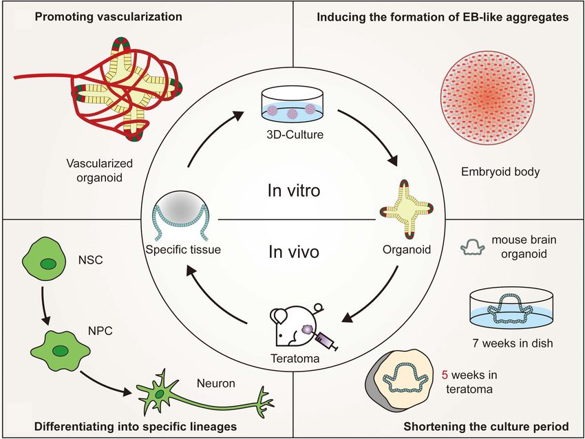
animals or in vitro cultures (Figure 2). Teratomas provide a Moreover, PSC-derived tissues that have developed to a certain
microenvironment that is more suitable for stem cell growth. The stage in vitro could be transplanted subcutaneously into mice
teratoma system facilitates the formation of embryoid-body-like and differentiated according to teratoma development patterns,
aggregates from stem cells with poor ability to differentiate and or related growth factors can be added to promote organoid
Frontiers in Cell and Developmental Biology | www.frontiersin.org 8 July 2021 | Volume 9 | Article 700482Li et al. To Better Generate Organoids
FIGURE 2 | Advantages of in vivo teratoma systems to better generate organoids. (A) Vascularized tissues could be isolated from in vivo teratomas by genetic
engineering and then cultured in vitro to generate vascularized organoids. (B) Pluripotent stem cells (PSCs), showing poor embryoid-body (EB)-forming ability in vitro,
are usually defective in the initial stage of differentiation. However, the PSCs could form teratomas in vivo to generate embryoid-body-like aggregates to promote
organoid formation. (C) By combining teratoma systems with genetic engineering technology, teratoma systems could be used to enrich specific lineage cells with
more accuracy to better generate organoids and improve the reproducibility of organoids. (D) Mouse embryonic stem cell-derived brain organoids, which are
produced in 7 weeks by in vivo 3D culture, could be generated in 5 weeks by in vitro culturing nerve tissues isolated from teratomas (NSC, neural stem cell; NPC,
neural progenitor cell).
formation. Due to the limited number of correlative studies, more Hong et al., 2016; Chan et al., 2018). As early as 2013, Suzuki
studies focusing on this aspect can be carried out in the future. et al. (2013) found that hematopoietic stem niche-like cells
were present in iPSC-derived teratomas and even migrated
Differentiating Into Specific Lineages into the bone marrow of mice. When these cells were injected
Under 3D culture conditions, PSCs first differentiated into intravenously into irradiated recipients, lymphoid and myeloid
progenitor cells and subsequently formed tissue-specific cells could be reconstituted (Amabile et al., 2013). Moreover,
organoids (Foley, 2017). However, these organoids commonly injecting Olig2-GFP transgenic ESCs into immunodeficient mice
include multiple lineage cell types (Lancaster and Knoblich, could also generate neural stem cells that then differentiated
2014a). For instance, single-cell sequencing results showed into terminal neuronal and glial cells (Hong et al., 2016). Neural
that kidney organoids contained non-renal cells, including stem cells could also be obtained in the primary culture of
three neuronal clusters and one muscle cluster, and that retinal human infantile teratomas (Kim et al., 2019). In addition,
organoids contained fibroblast cells and vascular cells (Wu Chan et al. (2018) showed that myogenic progenitors could
et al., 2018; Wang et al., 2021). If progenitors from a single be produced from mouse PSCs without genetic modification
lineage could be enriched in vitro, the production of unexpected through teratomas. Only 40,000 myogenic progenitor cells
organoid cells could be reduced, and the target organoid would transplanted into diseased muscles of NSG-mdx(4Cv) mice
be relatively easy to obtain. In recent years, several studies regenerated 80% of the total muscle volume, which matured into
have successfully enriched single lineage progenitor/stem cells functional muscle cells in vivo and was able to improve force
through a teratoma formation system (Amabile et al., 2013; generation (Chan et al., 2018).
Frontiers in Cell and Developmental Biology | www.frontiersin.org 9 July 2021 | Volume 9 | Article 700482Li et al. To Better Generate Organoids
However, the semirandom differentiation trait of teratomas could be cultured in vitro to generate vascularized organoids.
makes it difficult to distinguish various tissues, as different However, due to the small number and disordered arrangement
cell lines might be found in the same spatial region (Bulic- of blood vessels in teratomas, it is relatively difficult to generate
Jakus et al., 2016). McDonald and his colleagues applied genetic vascularized organoids, such as the brain and kidney that
engineering techniques to solve this issue at the single-cell level, require a plentiful blood supply (Gerecht-Nir et al., 2004).
which they called the miRNA circuit technique (McDonald et al., Recently, Wimmer et al. (2019) reported the development of
2020). Neural lineage cells, including early neurons, neuronal self-organizing 3D blood vessel organoids from PSCs, which
progenitors and Schwann cells, could be enriched by regulating produced a stable perfused vascular tree after transplantation
tissue-specific miRNA-124. Furthermore, clumps of enriched under the renal capsule of mice. Blood vessel organoids could
neural lineage cells through teratoma formation might be be transplanted into the site of teratoma formation or co-
cultured in vitro to generate brain organoids, which had a better cultured with isolated specific vascularized tissues to increase
consistency in terms of cellular arrangements and tissue elements the number of blood vessels. Moreover, organoids could be
compared to traditional 3D culture systems (Lee et al., 2020). transplanted into mice to promote organoid vascularization
Normally, PSC-derived teratomas are well-differentiated (Lensch according to the development pattern of teratoma vascular
et al., 2007). By combining genetic engineering technology, a systems. Hematopoietic stem cells or vascular endothelial cells
teratoma system could be used to enrich cells from specific could be coinjected into mice to promote vascular formation
lineages with more accuracy (McDonald et al., 2020), which is (Philipp et al., 2018; Wimmer et al., 2019). In brief, the vascular
a good example of reducing heterogeneity and improving the system in teratomas provides a reliable platform for organoid
reproducibility of current organoids. vascularization (Gerecht-Nir et al., 2004).
Promoting Vascularization Shortening the Culture Period
Vasculogenesis refers to the process by which capillaries sprout The culture time of organoids varies according to different
from existing blood vessels and invade avascular tissues (Risau, organ types (Spence et al., 2011; Nakano et al., 2012; Lancaster
1997). On the 18th day of human embryo development, et al., 2013; van den Berg et al., 2018). In general, the more
endothelial cells can be differentiated from the endoderm and precise and more complex the tissue, the longer it takes to
subsequently form vascular networks that invade embryonic grow. With increasing culture time, the center of the medium
tissues (Risau and Flamme, 1995). These vascular networks gradually lacked the exchange of nutrients and oxygen (Lancaster
eventually differentiate into arteries, veins, and lymphatic tissues and Knoblich, 2014a,b), resulting in retinal organoids dying in
(Semenza, 2007). The development of human organs and blood 126 days (Nakano et al., 2012). Brain organoids also start to
vessels is synchronous (Ruhrberg et al., 2002). Vascular systems shrink after 6 months (Lancaster et al., 2013). The maximum
provide nutrients and oxygen to promote maturation and diameter of organoids is only 4 mm (Lancaster et al., 2013).
maintain the normal function of human organs. However, in Current organoids are similar to fetal tissues in the early
the 3D culture environment, blood vessels cannot follow the stage of development (Fatehullah et al., 2016; Ashok et al.,
formation of organoids (Eiraku et al., 2008; Nakano et al., 2020). To obtain mature organoids, one method is to promote
2012), which limits both the size enlargement and functional vascularization (Rossi et al., 2018), and the other is to shorten
maturity of organoids (Kim et al., 2020). Although several the cultivation time, which theoretically might speed up the
technologies, such as co-culture, 3D printing, and bioreactors differentiation of PSCs before nutrients run out in a limited space,
have already been adopted to promote the vasculogenesis of prolonging the lifespan of organoids (Lee et al., 2020).
organoids, the volume of organoids is still much smaller than the One advantage of using the teratoma system to generate
volume of embryonic organs (Takebe et al., 2013; Phelan et al., organoids is that using the teratoma system can shorten
2019; Brassard et al., 2020). In vitro-generated blood vessels are the time of 3D culture. Generally, when 106 undifferentiated
arranged in a disorderly manner, and there is a lack of blood diploid mouse ESCs are inoculated into immunodeficient mice,
supply to verify their functions to transport oxygen and nutrients teratomas arise in a high proportion of recipients within 3–
(Ashok et al., 2020). 6 weeks (Gertow et al., 2004; Cooke et al., 2006; Valbuena et al.,
However, human PSC-derived teratomas offer a complex 2006; Hentze et al., 2009; Prokhorova et al., 2009). An mESC-
3D microenvironment, with corresponding tissue patterning, derived brain organoid was generated in 5 weeks by in vitro
extracellular matrices and epigenetic imprinting, to support culturing nerve tissues isolated from a teratoma (Lee et al., 2020).
vessel development (Gertow et al., 2004; Damjanov and Andrews, Compared with traditional 3D culture, the cultivation time of
2016). Little is known about the formation and maintenance mouse brain organoids was reduced by nearly 2 weeks (Eiraku
of blood vessels in teratomas. Some studies have suggested that et al., 2008). In the traditional 3D culture microenvironment,
the hematopoietic cell phenotype may reside in bone marrow- PSCs are involved in stepwise differentiation and subsequent
like structures, liver cells and mesenchymal stroma of teratomas self-organization, mimicking tissue formation and organogenesis
(Amabile et al., 2013; Dekkers et al., 2016). Vascularized tissues in mammalian embryonic development (Morgani et al., 2017).
could be isolated from in vivo teratomas by genetic engineering These processes could be partially generalized in teratoma
or obtained from in vitro teratomas cultured in a 3D perfusion formation, although they involve uncontrolled differentiation
bioreactor (Stachelscheid et al., 2013; Lee et al., 2020; McDonald and self-organization (Singh et al., 2016). The characteristic of
et al., 2020). Then, these isolated specific vascularized tissues uncontrolled differentiation may be related to the asynchronous
Frontiers in Cell and Developmental Biology | www.frontiersin.org 10 July 2021 | Volume 9 | Article 700482You can also read


