Case report: an unusual unilateral pterygium - a secondary pterygium caused by parasitosis in the scleral fistula
←
→
Page content transcription
If your browser does not render page correctly, please read the page content below
Zeng et al. BMC Ophthalmology       (2021) 21:323
https://doi.org/10.1186/s12886-021-02083-2

 CASE REPORT                                                                                                                                      Open Access

Case report: an unusual unilateral
pterygium — a secondary pterygium
caused by parasitosis in the scleral fistula
Wenjie Zeng1,2, Zhaoyi Pan1,2, Jun Wang1,2, Xianghui Deng1,2 and Wenmin Jiang1,2*

  Abstract
  Background: Ocular parasitosis can cause eye damage, which contribute to eye symptoms such as burning, itching
  and even blindness. It is uncommon to see the parasitosis lying in the sclera layer, neither it causing pterygium.
  Here, we present an unusual case of a secondary pterygium caused by intrascleral worm.
  Case presentation: A 52-year-old women complained about discomfort in right eye for 6 years. Slit-lamp examination
  indicated a thickened triangular layers of conjunctiva extending from the nasal edge to the cornea. The diagnosis was
  pterygium in the right eye. To our surprise, after scleral of nasal side exposed, we could see a tiny fistula right in the
  sclera which lied right under the pterygium, with an alive and motile worm inside. An intrascleral fistula was noted.
  Then the worm was removed by forceps from the fistula, which was creamy white, thread-like and 1 cm long.
  Discussion and conclusions: As far as we known, it is the first case of an intrascleral worm hidden beneath the
  conjunctiva which caused the secondary pterygium. It is hard to know the etiology of the secondary pterygium which
  caused by parasitosis in the scleral fistula untill excision surgery. It is hard to imagine the worm was living in the sclera
  of the patient for a long-time.
  Keywords: Scleral fistula, Ocular parasitosis, Secondary pterygium, Unilateral pterygium

Background                                                                           contacting animals that carrying worm eggs [1, 5]. Due to
Most ocular parasites infections can result from parasitic                           the blood-eye barrier and immunosuppressive micro-
migration through bloodstream or adjacent tissues in the                             environment, worms can remain in the eye for a long time
host, fewer from inoculation in the eye [1]. It can cause                            without being killed by ocular immune system.
eye damage by disrupting normal structure mechanically,                                Pterygium—a conjunctival fibrovascular degeneration
secreting toxic metabolites and inducing immune or aller-                            disease—may be caused by ultraviolet, increasing age,
gic reactions [2, 3]. Ocular parasites—involving three spe-                          male sex, outdoor occupation, alcohol use, heredity et al.
cies: worms, protozoa, and arthropods—are an alive                                   [6, 7]. It is a chronic inflammation process [8]. Long-
organism living in the host eye that acquire some of its                             term parasites infection may relate to chronic inflamma-
nutritional requirements through intimate contact with                               tion [9], Studies have suggested that mites may play a
the host [4]. Common routes of worm infection include                                role in the development of pterygium [10, 11]. The fol-
ingesting unclean water, soil, undercooked food and                                  lowing is a report of a rare case of intrascleral worm hid-
                                                                                     den beneath a fleshy pterygium for a time till pterygium
* Correspondence: wenminjiang@csu.edu.cn                                             excision surgery.
1
 Department of Ophthalmology, The Second Xiangya Hospital, Central South
University, Changsha, People’s Republic of China
2
 Hunan Clinical Research Center of Ophthalmic Disease, Changsha, People’s
Republic of China

                                      © The Author(s). 2021 Open Access This article is licensed under a Creative Commons Attribution 4.0 International License,
                                      which permits use, sharing, adaptation, distribution and reproduction in any medium or format, as long as you give
                                      appropriate credit to the original author(s) and the source, provide a link to the Creative Commons licence, and indicate if
                                      changes were made. The images or other third party material in this article are included in the article's Creative Commons
                                      licence, unless indicated otherwise in a credit line to the material. If material is not included in the article's Creative Commons
                                      licence and your intended use is not permitted by statutory regulation or exceeds the permitted use, you will need to obtain
                                      permission directly from the copyright holder. To view a copy of this licence, visit http://creativecommons.org/licenses/by/4.0/.
                                      The Creative Commons Public Domain Dedication waiver (http://creativecommons.org/publicdomain/zero/1.0/) applies to the
                                      data made available in this article, unless otherwise stated in a credit line to the data.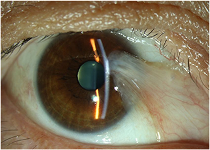
Zeng et al. BMC Ophthalmology (2021) 21:323 Page 2 of 3
eosinophils and basophils were increased; the ratio of
each were 8.50 and 1.50%. After the above examina-
tions, we diagnosed the patient with pterygium in the
right eye and decided to undergo excision surgery
next day.
After surgical excision of the pterygium, with the
scleral of nasal side exposed, we could see a tiny fis-
tula right in the sclera which lied right under the pte-
rygium (Fig. 2a). We used a toothed forceps to clip
the raised spot; creepily an alive and motile worm
was pulled out from the fistula with a little bleeding
(Fig. 2b). After clipping out the worm, we examined
the bare sclera carefully, we found that the fistula was
in one of the layer of the sclera with the worm living
Fig. 1 Preoperative image of the patient’s right eye. Slit-lamp in. The worm (Fig. 2c) was creamy white, thread-like,
examination showing a fleshy nasal pterygium in right eye,
1 cm long, and round with bilateral symmetry. Then
extending at 3 o’clock and 2 mm away from the limbus, covering
part of corneal pupillary area we underwent the corneal limbal stem cell autograft
for patient to prevent the pterygium from recurring.
After surgery, we send the excised pterygium and the
worm for pathological examination at our hospital.
Unfortunately, the report only showed a few dissocia-
tive keratins under the microscope, which may be ex-
Case presentation cluded from the worm (Fig. 3). Postoperatively, the
A 52-year-old women was referred to our ophthal- patient received sodium hyaluronate eye drops 4
mology department with discomfort in right eye for 6 times per day, deproteinized calfblood extract eye gel
years and no other symptoms. Her best-corrected vis- 1 times per day, levofloxacin eye drops 3 times per
ual acuity was 20/20 OD (+ 1.0 Diopters Sphere) and day for right eye about 1 week. 10 days after the oper-
20/20 OS (− 0.5 Diopters Sphere), with intraocular ation, the patient re-examined at our ophthalmology
pressure of 17 and 18 mmHg respectively. In slit-lamp clinic, which showed the surgical incision recovering
examination, the patient’s right eye showed a fleshy well (Fig. 4a and b).
nasal triangular membrane (Fig. 1), extending at 3
o’clock and 2 mm away from the limbus, covering Discussion and conclusions
part of corneal pupillary area. The anterior chamber Cases of intrascleral worms have been rarely reported,
depth was normal in both eyes. In blood routine, especially those hidden beneath the pterygium. Most
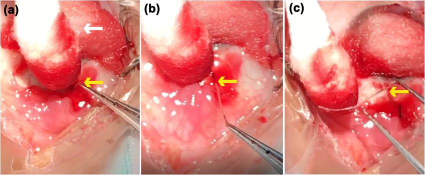
Fig. 2 The worm exposing process during the surgery. The patient’s cornea was covered with the cotton ball (white arrow); the nasal raised
spot (yellow arrow) was exposed 3 mm away from the limbus after excising the pterygium (a). The alive and motile worm (yellow arrow) was
being pulled out from the fistula inside the sclera by toothed forceps (b). The worm (yellow arrow) was completely pulled out from the fistula
with a little bleeding, which was creamy white, thread-like, 1 cm long (c)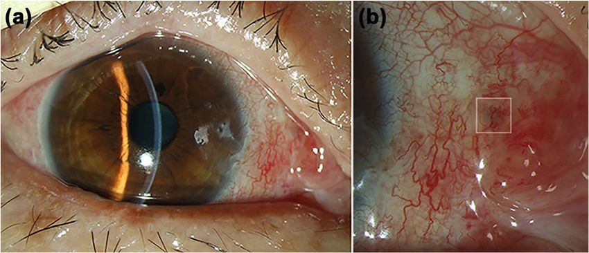
Zeng et al. BMC Ophthalmology       (2021) 21:323                                                                                         Page 3 of 3

                                                                       pterygium till excision surgery. It also gives us a hint
                                                                       that monocular pterygium maybe secondary caused by
                                                                       parasite infection.
                                                                       Acknowledgements
                                                                       Not applicable.

                                                                       Authors’ contributions
                                                                       WJZ drafted the manuscript and collected patient information, ZYP followed
                                                                       the patient, JW and XHD edited the photos, WMJ critically revised the
                                                                       manuscript for intellectual content. All authors read and approved the final
                                                                       manuscript.

                                                                       Funding
                                                                       Not applicable.

                                                                       Availability of data and materials
                                                                       All data generated or analyzed during this study are included in this
                                                                       published article.
 Fig. 3 Pathologic finding. Pathological examination of the excised
 pterygium and the worm showing a few dissociative keratins under      Declarations
 the microscope, which may be excluded from the worm
                                                                       Ethics approval and consent to participate
                                                                       This report has been performed in accordance with the Declaration of
                                                                       Helsinki. As this is a single case report excluding the data that can identify
ocular worms can infect the conjunctiva, eyelid, anter-                the patient, no ethical approval was required by the review board of the
ior chamber, orbit and retina [12], but rarely sclera,                 Second Xiangya Hospital, Central South University.
which may due to its dense and crisscrossed fibrosis,
                                                                       Consent for publication
preventing most worms from penetrating through the                     Written informed consent was obtained from the patient for publication of
ocular surface.                                                        this case report and any accompanying images.
   The origin of the parasitosis was hard to know, we could
                                                                       Competing interests
only surmise it comes from conjunctival vessels, migrating to          The authors declare that they have no competing interests.
the surface of the scleral, and caused a fistula. Pterygium is a
chronic inflammation process [8], and long-term mite infec-            Received: 4 November 2020 Accepted: 26 August 2021
tion can cause ocular chronic inflammation, which may play
a role in the development of pterygium. The pterygium re-              References
currence rate was higher in patients with mite infection,              1. Padhi TR, Das S, Sharma S, et al. Ocular parasitoses: A comprehensive
                                                                           review. Surv Ophthalmol. 2017;62:161–89.
which may relate to chronic inflammation mediated by T                 2. Rathinam SR, Annamalai R, Biswas J. Intraocular parasitic infections. Ocul
helper cell 1 7[11]. The secretions and metabolites of demo-               Immunol Inflamm. 2011;19:327–36.
dex may produce more inflammatory factors and vascular                 3. Nimir AR, Saliem A, Ibrahim IA. Ophthalmic parasitosis: a review article.
                                                                           Interdiscip Perspect Infect Dis. 2012;2012:587402.
endothelial growth factor in the cornea, which stimulate the           4. Rathinam SR, Ashok KA. Ocular manifestations of systemic disease: ocular
surrounding conjunctival fibrous tissue and blood vessels to               parasitosis. Curr Opin Ophthalmol. 2010;21:478–84.
proliferate, thus promoting pterygium course [10].                     5. Otranto D, Eberhard ML. Zoonotic helminths affecting the human eye.
                                                                           Parasit Vectors. 2011;4:41.
   To the best of our knowledge, this is the first case                6. Rezvan F, Khabazkhoob M, Hooshmand E, et al. Prevalence and risk factors
of an intrascleral worm hidden beneath a secondary                         of pterygium: a systematic review and meta-analysis. Surv Ophthalmol.
                                                                           2018;63:719–35.
                                                                       7. Chu WK, Choi HL, Bhat AK, et al. Pterygium: new insights. Eye (Lond). 2020;
                                                                           34:1047–50.
                                                                       8. Hill JC, Maske R. Pathogenesis of pterygium. Eye (Lond). 1989;3(Pt 2):218–26.
                                                                       9. McSorley HJ, Maizels RM. Helminth infections and host immune regulation.
                                                                           Clin Microbiol Rev. 2012;25:585–608.
                                                                       10. Tarkowski W, Moneta-Wielgos J, Mlocicki D. Do Demodex mites play a role
                                                                           in pterygium development? Med Hypotheses. 2017;98:6–10.
                                                                       11. Huang Y, He H, Sheha H, et al. Ocular demodicosis as a risk factor of
                                                                           pterygium recurrence. Ophthalmology. 2013;120:1341–7.
                                                                       12. McBurney-Lin S, Khorram D, Gee S, et al. A new worm infiltrating the
                                                                           human cornea: a report of three cases. Am J Ophthalmol Case Rep. 2018;9:
                                                                           124–30.

 Fig. 4 Postoperative image of the patient’s right eye. The incision
 recovered well after excising the pterygium and removing the worm     Publisher’s Note
 (a). Magnified image shows that the fistula, where the worm living    Springer Nature remains neutral with regard to jurisdictional claims in
                                                                       published maps and institutional affiliations.
 inside, turned into a black spot (white square) during the healing
 process (b)
You can also read