Eagle syndrome with hidden stylocarotid syndrome examined using dynamic ultrasonography: illustrative case
←
→
Page content transcription
If your browser does not render page correctly, please read the page content below
J Neurosurg Case Lessons 1(26):CASE21286, 2021
DOI: 10.3171/CASE21286

Eagle syndrome with hidden stylocarotid syndrome examined using dynamic
ultrasonography: illustrative case
Yukiko Tanaka, MD,1,2 Hidenori Anami, MD,1,2 Hiroyuki Kurihara, MD,1,2 Satoru Miyao, MD,1,2 Hidetoshi Nakamoto, MD,1,2
Yuichi Kubota, MD, DMedSci,1,2 and Takakazu Kawamata, MD, PhD2
1
 Department of Neurosurgery and Stroke Center, TMG Asaka Medical Center, Saitama, Japan; and 2Department of Neurosurgery, Tokyo Women’s Medical University,
Tokyo, Japan

    BACKGROUND Eagle syndrome, or elongated styloid process syndrome, is a rare cause of cerebral infarction. When the styloid process is elongated
    but the internal carotid artery (ICA) is morphologically normal on three-dimensional computed tomography angiography (3D-CTA), determining the
    causal relationship between elongation and cerebral infarction is difficult.
    OBSERVATIONS The patient was a 27-year-old man who experienced two left cerebral infarctions in 3 months. On 3D-CTA, the styloid process was
    elongated, but the structure of the ICA was normal. When the patient’s neck was rotated leftward, the peak systolic velocity and pulsatility index
    increased (shown via dynamic subtraction ultrasonography) and ICA stenosis was evident (shown via subtraction angiography). The styloid process
    was removed, and the cerebral infarction did not recur in the 2 years after surgery.
    LESSONS This is the first report to document that indirect compression of ICA by the styloid process can cause Eagle syndrome. The blood flow
    changes of the ICA on dynamic ultrasonography revealed morphological changes that were hidden on 3D-CTA or nondynamic subtraction angiography.

    https://thejns.org/doi/abs/10.3171/CASE21286
    KEYWORDS Eagle syndrome; elongated styloid process; stroke; dynamic ultrasonography; surgery

    Eagle syndrome was first reported by W.W. Eagle in 1937,1 who attrib-              Illustrative Case
uted its symptoms to elongation of the styloid process or calcification of                 The patient was a 27-year-old man with no particular medical history.
the stylohyoid ligament. The styloid process arises from the base of the              He experienced a sudden onset of dysarthria and was diagnosed with
temporal bone and connects to the stylohyoid ligament. It normally ranges             left cerebral infarction at a different hospital. Cerebral vascular imaging
in length from 2.5 to 4 cm2–4 and is considered elongated in most reports             showed no occlusion or stenosis. Transthoracic and transesophageal
when more than 3 cm long.4,5 The incidence of styloid process elongation              echocardiography results and the levels of several autoantibodies were
is 4% to 33.4%;6–8 however, only a few cases become symptomatic.                      normal. The patient’s neurological status returned to normal, and he
    There are two clinical presentations of Eagle syndrome. One is                    was discharged after receiving single antiplatelet therapy.
characterized by throat pain, facial pain, dysphagia, or pharyngeal                       He was referred to our hospital 3 months later because of tran-
foreign body sensation. The other, termed “stylocarotid syndrome,”                    sient right hemiparesis. On physical examination, his consciousness
is characterized by transient ischemic attack or stroke due to steno-                 level was Glasgow Coma Scale E4V5M6, and he complained of
sis, occlusion, or dissection.9–11                                                    mild left motor weakness. The manual muscle test score for the left
    We report a case in which dynamic ultrasonography revealed                        extremities was 4 out of 5; he was otherwise neurologically normal.
internal carotid artery (ICA) stenosis associated with elongation of                      Head magnetic resonance imaging (MRI) showed small cerebral
the styloid process.                                                                  infarctions in the left parietal lobe (Fig. 1A). No major artery stenosis

ABBREVIATIONS 3D-CTA = three-dimensional computed tomography angiography; DSA = digital subtraction angiography; ICA = internal carotid artery
MRI = magnetic resonance imaging; PI = pulsatility index; PSV = peak systolic velocity.
INCLUDE WHEN CITING Published June 28, 2021; DOI: 10.3171/CASE21286.
SUBMITTED May 7, 2021. ACCEPTED May 18, 2021.
© 2021 The authors, CC BY-NC-ND 4.0 (http://creativecommons.org/licenses/by-nc-nd/4.0/).

                                                                                                            J Neurosurg Case Lessons | Vol 1 | Issue 26 | June 28, 2021 | 1

                                                                                                                   Unauthenticated | Downloaded 12/24/21 04:36 AM UTC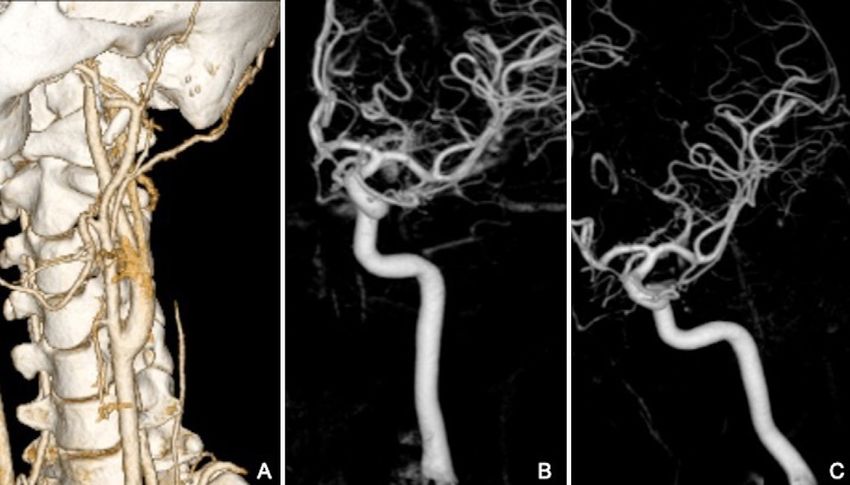
increased when the head was rotated to the left but not in other
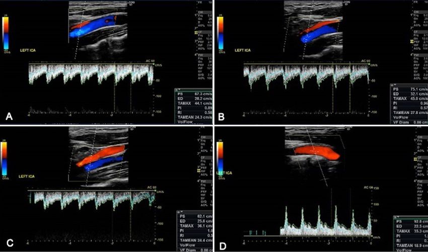
directions (Fig. 2). Digital subtraction angiography (DSA) revealed
focal left ICA stenosis at the crossing point of the styloid process
upon head rotation to the left (Fig. 3).
We thought that intermittent ICA stenosis caused blood flow turbu-
lence, which resulted in cerebral infarction. We then surgically removed
a portion of the left stylomastoid process (up to 3.5 cm long) using a
transcervical approach (Figs. 4 and 5A). After surgery, the patient did
not complain of any neurological symptoms. Postoperative DSA sho-
wed no morphological changes upon head movement (Fig. 5B). No
recurrence of stroke had occurred 2 years after surgery.
FIG. 1. A: Diffusion-weighted MRI showing a cerebral infarction in the Discussion
left hemisphere. B: 3D-CTA showing elongation of the left styloid pro- Observations
cess (red arrows). When an elongated styloid process accompanies a stroke, direct
compression of the ICA by the process usually causes stenosis,
occlusion, dissection, or other morphological blood vessel–related
was observed on MR angiography. Dual antiplatelet therapy was initi- abnormality, making it easy to determine the causal relationship
ated, and the symptoms gradually resolved. Laboratory data showed between elongated styloid process and a stroke. However, in our
no coagulation disorders. No abnormalities were detected on trans- case, no obvious morphological abnormalities of the ICA were
thoracic or transesophageal echocardiography. Three-dimensional found other than elongation of the styloid process.
computed tomography angiography (3D-CTA) revealed an elongated Anatomically, the styloid process is a columnar bone arising
left styloid process (Fig. 1B); however, no occlusion of the intracranial from the bottom of the temporal bone. It runs anteromedially and
or extracranial arteries was observed. The elongated process was inferiorly between the internal and external carotid arteries, con-
approximately 5 cm long and ran close to the left ICA. nects to the stylohyoid ligament, and is continuous with the hyoid
No obvious structural or blood flow abnormalities were noted on bone. Cervical rotation can cause the styloid process or the stylo-
ultrasonography. Dynamic ultrasonography with head rotation sho- hyoid ligament to compress the ICA, even indirectly. In particular,
wed that the peak systolic velocity (PSV) and pulsatility index (PI) ipsilateral cervical rotation tends to increase ICA compression due
FIG. 2. Cervical ultrasonography showing no obvious blood flow differences in the head in the neutral (A), extension (B), and flexion (C) positions. Flexing
the head downward and to the left caused the PSV and PI to increase relative to the other positions (D).
2 | J Neurosurg Case Lessons | Vol 1 | Issue 26 | June 28, 2021
Unauthenticated | Downloaded 12/24/21 04:36 AM UTC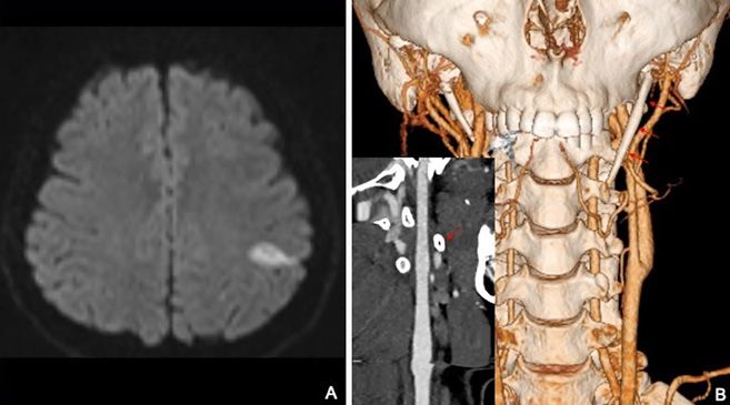
In the present case, the relationship between elongated styloid
                                                                                  process and stroke was not clear because of the absence of mor-
                                                                                  phological changes in the ICA. Dynamic ultrasonography with cervi-
                                                                                  cal rotation showed that the PSV and PI increased only when the
                                                                                  patient rotated his head leftward. The elevation of PSV usually means
                                                                                  stenosis of blood vessels, and PI increases also imply peripheral vas-
                                                                                  cular resistance increases. These hemodynamic changes made us
                                                                                  think that the indirect compression of the ICA by the elongated styloid
                                                                                  process caused strokes.
                                                                                      In patients with a cerebral infarction who have an elongated styloid pro-
                                                                                  cess, the possibility of developing stylocarotid syndrome should be consid-
  FIG. 3. DSA showing that the ICA (red arrows) is bent when the                  ered even if continuous compression of the blood vessels is absent.
  patient’s neck is flexed in the left-downward direction (A) compared             Ayyildiz et al. emphasized the importance of not only the length of the sty-
  with when it is in the neutral position (B).                                    loid process but also its medial angulation.13 They indicated that the devia-
                                                                                  tions of the styloid process and its length as well as the surrounding
                                                                                  structures may cause Eagle syndrome. In previous reports, changes in
                                                                                  blood flow were observed only in patients with obvious morphological
                                                                                  changes in the ICA. Our report is the first to document the occurrence of
                                                                                  stylocarotid syndrome due to indirect compression by the styloid process,
                                                                                  and morphological change in the ICA was first evident when the patient
                                                                                  rotated his head leftward.
                                                                                      Styloidectomy is the gold standard treatment for Eagle syndrome. Although
                                                                                  a transcervical approach has been used,14–18 a transoral approach is usually
                                                                                  chosen. Because it is difficult to expose the deeper part of the styloid process,
                                                                                  the transcervical approach is the more suitable option for stylocarotid syn-
                                                                                  drome. In some cases, carotid artery stenting was performed.10,19 Miyata
                                                                                  et al.,10 for example, used this procedure along with styloidectomy for treat-
  FIG. 4. Removal of the styloid process, which measured 3.5 cm.                  ment of stent compression by the styloid process due to cervical rotation.

                                                                                  Lessons
to the spatial positioning relationship between the styloid process and               In conclusion, we encountered a case of repeated cerebral infarc-
the carotid artery. Li et al. reported a case of transient ischemic attack in     tion associated with Eagle syndrome. Our case shows that indirect
which transcranial Doppler imaging combined with color Doppler flow                compression of the ICA by an elongated styloid process can cause
imaging showed a marked decrease in blood flow in the middle cerebral              stylocarotid syndrome. Dynamic ultrasonography with cervical rotation
artery,12 thereby revealing the usefulness of simultaneous assessment             centered on ipsilateral head movement may be useful for diagnosis
of intracranial and extracranial hemodynamics.                                    of hidden stylocarotid syndrome.

                        FIG. 5. 3D-CTA confirms the removal of the left styloid process (A). After the surgery, DSA with the head in
                        the neutral position (B) and left-downward position (C) shows no morphological changes.

                                                                                                        J Neurosurg Case Lessons | Vol 1 | Issue 26 | June 28, 2021 | 3

                                                                                                               Unauthenticated | Downloaded 12/24/21 04:36 AM UTC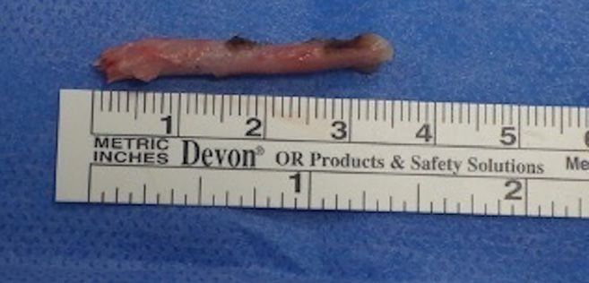
Acknowledgments                                                               14. Pigache P, Fontaine C, Ferri J, Raoul G. Transcervical styloidec-
   We would like to thank Editage for English language editing.                   tomy in Eagle’s syndrome. Eur Ann Otorhinolaryngol Head Neck
                                                                                  Dis. 2018;135(6):433–436.
References                                                                    15. Badhey A, Jategaonkar A, Anglin Kovacs AJ, et al. Eagle syn-
 1. Eagle WW. Elongated styloid processes: report of two cases. Arch              drome: a comprehensive review. Clin Neurol Neurosurg.
    Otolaryngol. 1937;25(5):584–587.                                              2017;159:34–38.
 2. Monsour PA, Young WG. Variability of the styloid process and stylo-       16. Gallaway E, Bayoumi S, Hammond D, Halsnad M. Case report: an
    hyoid ligament in panoramic radiographs. Oral Surg Oral Med Oral              atypical presentation of Eagle syndrome. J Surg Case Rep.
    Pathol. 1986;61(5):522–526.                                                   2017;2017(8):rjx152.
 3. Langlais RP, Miles DA, Van Dis ML. Elongated and mineralized sty-         17. Liu Y, Yang H, Cui X. A case of a very elongated styloid process.
    lohyoid ligament complex: a proposed classification and report of a            Clin Med Insights Ear Nose Throat. 2017;10:1179550617728899.
    case of Eagle’s syndrome. Oral Surg Oral Med Oral Pathol.                 18. Ceylan A, K€oybaşioglu A, Çelenk F, et al. Surgical treatment of
    1986;61(5):527–532.                                                           elongated styloid process: experience of 61 cases. Skull Base.
 4. Kaufman SM, Elzay RP, Irish EF. Styloid process variation. Radio-             2008;18(5):289–295.
    logic and clinical study. Arch Otolaryngol. 1970;91(5):460–463.           19. Todo T, Alexander M, Stokol C, et al. Eagle syndrome revisited:
 5. Koshy J, Narayan M, Narayanan S, et al. Elongated styloid process:            cerebrovascular complications. Ann Vasc Surg. 2012;26(5):
    a study. J Pharm Bioallied Sci. 2015;7(5):S131–S133.                          729.e1–729.e5.
 6. Eagle WW. Elongated styloid process: further observations and a
    new syndrome. Arch Otolaryngol. 1948;47(5):630–640.                       Disclosures
 7. H€arm€a R. Stylalgia: clinical experiences of 52 cases. Acta Otolar-      The authors report no conflict of interest concerning the materials
    yngol. 1966;63(suppl 224):149.                                            or methods used in this study or the findings specified in this
 8. Bruno G, De Stefani A, Balasso P, et al. Elongated styloid process:       paper.
    an epidemiological study on digital panoramic radiographs. J Clin
    Exp Dent. 2017;9(12):e1446–e1452.                                         Author Contributions
 9. Chuang WC, Short JH, McKinney AM, et al. Reversible left hemi-            Conception and design: Anami, Miyao. Acquisition of data: Anami,
    spheric ischemia secondary to carotid compression in Eagle syn-           Tanaka, Miyao. Analysis and interpretation of data: Miyao, Kubota.
    drome: surgical and CT angiographic correlation. AJNR Am J                Drafting the article: Miyao. Critically revising the article: Miyao.
    Neuroradiol. 2007;28(1):143–145.                                          Reviewed submitted version of manuscript: Miyao, Nakamoto.
10. Miyata H, Nakahara I, Ohta T, et al. A case of internal carotid artery    Administrative/technical/material support: Kurihara, Kubota. Study
     dissection caused by an elongated styloid process: successful treat-     supervision: Kubota, Kawamata.
     ment with carotid artery stenting and partial resection of the styloid
     process. Surg Cereb Stroke. 2016;44(2):145–150.
11. Smoot TW, Taha A, Tarlov N, Riebe B. Eagle syndrome: a case               Supplemental Information
    report of stylocarotid syndrome with internal carotid artery dissec-      Previous Presentations
    tion. Interv Neuroradiol. 2017;23(4):433–436.                             Portions of this work were presented at Stroke2020, Annual Meeting of
12. Li Z, Hua Y, Yang J, Li J. Ultrasound evaluation of transient ische-      the Japanese Society on Surgery for Cerebral Stroke, Yokohama,
     mic attack caused by styloid process elongation: a case report.          Japan, March 26, 2020.
     Front Neurol. 2019;10:26.
13. Ayyildiz VA, Senel FA, Dursun A, Ozturk K. Morphometric examination of    Correspondence
     the styloid process by 3D-CT in patients with Eagle syndrome. Eur Arch   Hidenori Anami: TMG Asaka Medical Center, Saitama, Japan.
     Otorhinolaryngol. 2019;276(12):3453–3459.                                hidenori272@gmail.com.

4 | J Neurosurg Case Lessons | Vol 1 | Issue 26 | June 28, 2021

                                                                                                     Unauthenticated | Downloaded 12/24/21 04:36 AM UTC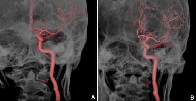
You can also read