Case Report: Acute Intracardiac Thrombosis in Children With Coronavirus Disease 2019 - (COVID-19)
←
→
Page content transcription
If your browser does not render page correctly, please read the page content below
CASE REPORT
                                                                                                                                                     published: 25 June 2021
                                                                                                                                             doi: 10.3389/fped.2021.656720

                                                Case Report: Acute Intracardiac
                                                Thrombosis in Children With
                                                Coronavirus Disease 2019
                                                (COVID-19)
                                                Hamid Bigdelian 1,2 , Mohsen Sedighi 3*, Mohammad Reza Sabri 2 , Bahar Dehghan 2 ,
                                                Chehreh Mahdavi 2 , Alireza Ahmadi 2 , Mehdi Ghaderian 2 , Hamid Rahimi 2 ,
                                                Atefeh Sadeghizadeh 4 , Monirsadat Emadoleslami 4 , Seyed Nasser Mostafavi 4 ,
                                                Rana Saleh 4 , Niloofar Javadi 5 , Maryam Derakhshan 6 , Zahra Pourmoghaddas 4 and
                                                Shima Sarfarazi Moghadam 2
                                                1
                                                  Department of Cardiovascular Surgery, School of Medicine, Isfahan University of Medical Science, Isfahan, Iran, 2 Pediatric
                                                Cardiovascular Research Center, Cardiovascular Research Institute, Isfahan University of Medical Sciences, Isfahan, Iran,
                                                3
                                                  Department of Anesthesiology and Pain Medicine, Pain Research Center, Iran University of Medical Sciences, Tehran, Iran,
                                                4
                                                  Department of Pediatrics, School of Medicine, Isfahan University of Medical Sciences, Isfahan, Iran, 5 Student Research
                           Edited by:
                                                Committee, School of Medicine, Isfahan University of Medical Sciences, Isfahan, Iran, 6 Department of Pathology, School of
                        Matthias Sigler,
                                                Medicine, Isfahan University of Medical Science, Isfahan, Iran
     University of Göttingen, Germany

                        Reviewed by:
                       Kei Yamamoto,            We herein describe a case series of children with SARS-CoV-2 infection (COVID-19)
 National Center for Global Health and
                                                complicated with acute intracardiac thrombosis. The diagnosis of COVID-19 was
                      Medicine, Japan
              Sachin Gajanan Damke,             confirmed through the reverse transcription-polymerase chain reaction (RT-PCR).
      Datta Meghe Institute of Medical          Transthoracic echocardiography of patients revealed large intracardiac mobile masses
                        Sciences, India
                                                resected successfully via cardiac surgery. The underlying mechanisms of this thrombus
                  *Correspondence:
                      Mohsen Sedighi
                                                in the COVID-19 infection may be attributed to the hypercoagulation and inflammatory
                Sedighi.mo@iums.ac.ir           state of the disease incurred by the SARS-CoV-2 virus.
                                                Keywords: COVID-19, acute thrombosis, children, cardiac surgery, case series
                    Specialty section:
          This article was submitted to
                   Pediatric Cardiology,
                a section of the journal        INTRODUCTION
                  Frontiers in Pediatrics
                                                Coronavirus disease 2019(COVID-19) caused by a severe acute respiratory syndrome coronavirus
            Received: 16 March 2021
             Accepted: 25 May 2021
                                                2 (SARS-CoV-2) was reported for the first time in Wuhan, Hubei, China in December 2019
            Published: 25 June 2021             and has spread rapidly throughout the world (1). According to the daily report of the World
                                                Health Organization (WHO), the epidemic of COVID-19 so far registered 2,751,166 cases and
                             Citation:
   Bigdelian H, Sedighi M, Sabri MR,
                                                76,936 deaths in Iran (2). However, epidemiological and clinical patterns of the COVID-19
  Dehghan B, Mahdavi C, Ahmadi A,               remain largely unclear, particularly among pediatric patients. Children with COVID-19 have their
              Ghaderian M, Rahimi H,            specific clinical features and therapeutic responses but clinical manifestations of this disease in
  Sadeghizadeh A, Emadoleslami M,               children might be less severe (3). Although COVID-19 is recognized as an acute respiratory
    Mostafavi SN, Saleh R, Javadi N,            tract infection, accumulating clinical data indicates cardiovascular complications of the disease,
   Derakhshan M, Pourmoghaddas Z
                                                including myocarditis, arrhythmias, cardiogenic shock, and acute myocardial injury (4). More
   and Sarfarazi Moghadam S (2021)
     Case Report: Acute Intracardiac
                                                importantly, cardiovascular thrombosis due to COVID-19-induced hypercoagulopathy is a serious
          Thrombosis in Children With           and life-threatening complication that required emergency intervention in some cases (5, 6).
           Coronavirus Disease 2019             To our knowledge, hypercoagulopathy associated with COVID-19 in children has been rarely
(COVID-19). Front. Pediatr. 9:656720.           reported. Here, we report three COVID-19 children with acute intracardiac thrombosis treated
     doi: 10.3389/fped.2021.656720              with cardiac surgery.

Frontiers in Pediatrics | www.frontiersin.org                                          1                                               June 2021 | Volume 9 | Article 656720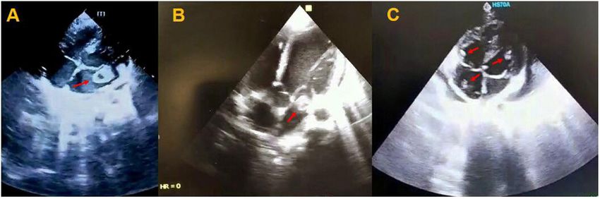
Bigdelian et al. Acute Intracardiac Thrombosis in COVID-19 Children
TABLE 1 | Laboratory test results of patients at the time of admission. CASE 2
Variables Reference range Case 1 Case 2 Case 3 An 7-year-old girl was referred to the Emergency Department
of Pediatric Medical Center with fever (38.2◦ C), tachypnea
RBC count (×10*12/L) 4.5–6.5 3.75 3.89 3.79
(36 times per minute), lower extremities rashes, abdominal
Hematocrit (%) 41–51 32.9 34.2 33.9
pain, and bilious vomiting. She had no significant past
Hemoglobin (g/dl) 13–17 11.8 11 11.5
medical history, but she had close contact with a COVID-19
WBC (×10*9/L) 4.5–11 16.3 14.2 12.3
patient. In physical examination, tachycardia, and low blood
Lymphocyte count (%) 20–40 10 15 27
pressure were observed, and thus, she was transferred to the
Platelet count (×10*9/L) 150–450 316 219 252
PICU for respiratory support by continuous positive airway
ESR (mm/h) 0–20 56 80 22
pressure (CPAP) along with pharmacotherapy. Leukocytosis
CRP (mg/L) Up to 6 29 38 11
with increased CRP and erythrocyte sedimentation rate (ESR)
PT (s) 11–13 13 14.1 14
were found in blood test (Table 1) and nasopharyngeal swab
PTT (s) 26–45 38 38 34
analysis by RT-PCR confirmed COVID-19. According to the
INR 0.9–1.2 1 1.2 1.2
cardiorespiratory and gastrointestinal symptoms and laboratory
D-dimer Up to 500 490 530 2,000
results, early diagnosis of COVID-19-induced multisystem
Blood culture (3×) ± – – – inflammatory syndrome in children (MIS-C) was made and
RT-PCR SARS-CoV-2 ± + + + pharmaceutical treatment, including hydroxychloroquine (5
RBC, red blood cell; WBC, white blood cell; ESR, erythrocyte sedimentation rate; CRP, C- mg/kg/dose), lopinavir/ritonavir (230 mg/m2 /dose), intravenous
reactive protein; PT, prothrombin time; PTT, partial thromboplastin time; INR, international immunoglobulin (IVIG /2 gr/kg), methylprednisolone (30
normalized ratio. mg/kg), and enoxaparin (2 mg/kg per 12 h) was started. Pediatric
cardiology consultation was requested because of cardiomegaly
and pulmonary edema in her chest X-ray and transthoracic
CASE 1 echocardiography showed LVEF of 69% with better LV systolic
function, minimal pericardial effusion, mild TR, trivial PI, and
An 11-year-old girl was admitted to the Emergency Department a large mobile mass in the LA (Figure 1B). Consequently, she
of Pediatric Medical Center with fever (39◦ C), coughing, underwent cardiac surgery on the 6th day of hospitalization and
tachypnea (38 times per minute), low oxygen saturation (93% in intraoperative observations revealed multiple large thrombi in
room air), and decreased level of consciousness (LOC). Glasgow LA and right atrium (RA) with necrosis of posterior leaflet of
Coma Scale (GCS) was 13 and lower extremities rashes were the mitral valve. Intracardiac thrombi were removed completely
found in her physical examination. Also, results of the blood test and the mitral valve was repaired. Second echocardiography after
on admission time revealed leukocytosis and increased level of surgery showed normal heart function and she was discharged
C-reactive protein (CRP) (Table 1). The patient was transferred from hospital after 2 weeks in good health condition with
to the pediatric intensive care unit (PICU) and nasopharyngeal negative COVID-19 test. In the histopathological assessment,
swab and stool sample analysis by reverse transcription- fibrin clot, fibrinoid necrosis, and surface thrombus with dense
polymerase chain reaction (RT-PCR) confirmed COVID-19 neutrophilic infiltration were found in the necrotic tissue of
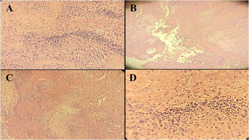
infection and, therefore, drug treatment for the COVID-19 was posterior leaflet of the mitral valve (Figures 2A–D).
started according to the Iranian pediatric protocol, including
hydroxychloroquine (5 mg/kg/dose), lopinavir /ritonavir (230 CASE 3
mg/m2 /dose), ceftriaxone (75 mg/kg/dose), and vancomycin (10
mg/kg/dose) (7). Because of persistent fever and tachycardia, An 8-year-old girl was referred to the Emergency Department
pediatric cardiology consultation was requested and done on of Pediatric Medical Center with fever (38.2◦ C), tachycardia (PR
the 3rd day of hospitalization showing left ventricular ejection 135), and tachypnea (40 times per min). Past medical history of
fraction (LVEF) of 64%, mild mitral regurgitation (MR), trivial the patient revealed that she had undergone orthopedic surgery a
tricuspid regurgitation (TR), trivial pulmonary insufficiency (PI), couple of weeks ago because of car accident injures. In her blood
and a large mobile mass in the left atrium (LA) with attachment test, leukocytosis, increased CRP, and a remarkable rise in D-
to the posterior leaflet of the mitral valve (Figure 1A). Hence, dimer were found and RT.PCR test for COVID-19 was positive
she became a candidate for emergency surgical intervention and (Table 1). Primary diagnosis of pulmonary thromboembolism
cardiac surgery was performed on the 5th day. Intraoperative (PTE) was made and pharmaceutical treatment, including
observations revealed a large thrombus in the LA with necrosis hydroxychloroquine (5 mg/kg/dose), methylprednisolone (30
of the posterior leaflet of the mitral valve. Left atrium mg/kg), enoxaparin (2 mg/kg per 12 h), lopinavir /ritonavir (230
thrombus was resected completely and the mitral valve was mg/m2 /dose) was started. On the 2nd day of hospitalization,
repaired. Her clinical conditions improved following surgery echocardiography was performed showing LVEF of 68%, mild
and the results of postoperative echocardiography showed a LA and LV enlargement, trivial TR, trivial PI, and multiple
normal heart function. She was discharged in good health homogenous masses in the LV and right side of the heart
condition after 10 days of hospitalization with negative RT-PCR as well as clot strands in the main pulmonary artery (MPA)
for COVID-19. (Figure 1C). Therefore, she underwent emergency cardiac
Frontiers in Pediatrics | www.frontiersin.org 2 June 2021 | Volume 9 | Article 656720Bigdelian et al. Acute Intracardiac Thrombosis in COVID-19 Children
FIGURE 1 | Transthoracic echocardiogram of patients showing a mobile mass in the left atrium with attachment to the posterior leaflet of the mitral valve (A), a large
mobile mass in the left atrium (B), and multiple mobile homogenous masses in the right side of the heart and left ventricle (C).
surgery on the 3rd day and multiple thrombi were resected from
the cardiac chambers with the repair of the tricuspid valve. Also,
blood clots were removed successfully from the MPA through
thrombectomy. She was admitted to the PICU and pediatric
ward and was discharged from the hospital after 2 weeks. Second
echocardiography indicated normal cardiovascular function and
she was discharged from the hospital in good health condition
after 15 days with a negative test for COVID-19.
Pathological assessment of specimens in three children
showed no fungal and bacterial infection and also SARS-CoV-2
RNA was not detected in the thrombi.
DISCUSSION FIGURE 2 | Histopathological manifestations (40x resolution) of the posterior
leaflet of mitral valve tissue in case 2 showing fibrinoid necrosis and cellular
Among the COVID-19 patients, the notifiable cases are adults debris (A), fibrinoid necrosis without evidence of bacterial colonization (B),
complicated with fever, dry cough, fatigue, and worsening dense neutrophilic infiltration with cellular debris (C), and necrotic area with
dyspnea, whereas pediatric cases are less reported in the literature neutrophilic debris induced by SARS-CoV-2 (D).
and clinical profiles of those children have not been well-defined
(8). A prior study in our country reported that fever is more
prevalent in COVID-19 children aged ≥5 years compared to
the patients agedBigdelian et al. Acute Intracardiac Thrombosis in COVID-19 Children
of lysed clots lodging in pulmonary arteries and consequent ETHICS STATEMENT
PTE (14).
To sum up, there is an increasing concern about This study was reviewed and approved by the Medical Research
hypercoagulopathy state in COVID-19 patients, especially Ethics Committee at Isfahan University of Medical Science,
children. Therefore, conservative treatment with anticoagulation Isfahan, Iran. Written informed consent was obtained from the
along with vigilant workup is advised in all COVID-19 patients patient’s next-of-kin for the publication of this research.
to prevent subsequent hypercoagulable states.
AUTHOR CONTRIBUTIONS
DATA AVAILABILITY STATEMENT
All authors contributed to the analysis, interpretation of data,
The original contributions generated for the study are included wrote the manuscript, approved the final version of the
in the article/supplementary material, further inquiries can be manuscript, and agreed to be accountable for all aspects of
directed to the corresponding author/s. the work.
REFERENCES 10. Klok FA, Kruip MJHA, van der Meer NJM, Arbous MS, Gommers
DAMPJ, Kant KM, et al. Incidence of thrombotic complications in
1. Guo T, Fan Y, Chen M, Wu X, Zhang L, He T. Cardiovascular implications of critically ill ICU patients with COVID-19. Thromb. Res. (2020) 191:145–
fatal outcomes of patients with coronavirus disease 2019 (COVID-19). JAMA 7. doi: 10.1016/j.thromres.2020.04.013
Cardiol. (2020) 5:811–8. doi: 10.1001/jamacardio.2020.1017 11. Maier CL, Truong AD, Auld SC, Polly DM, Tanksley C-L, Duncan A. COVID-
2. World Health Organization. Covid-19 (2021). Available online at: https:// 19 associate hyperviscosity: a link between inflammation and thrombophilia?
covid19.who.int (accessed October 5, 2021). Lancet. (2020) 395:1758–9. doi: 10.1016/S0140-6736(20)31209-5
3. Mahmoudi S, Mehdizadeh M, Shervin Badv R, Navaeian A, Pourakbari B, 12. Hékimian G, Kerneis M, Zeitouni M, Cohen-Aubart F, Chommeloux J,
Rostamyan M, et al. The coronavirus disease 2019 (COVID-19) in children: Bréchot N, et al. COVID-19 acute myocarditis and multisystem inflammatory
a study in an Iranian children’s referral hospital. Infect Drug Resist. (2020) syndrome in adult intensive and cardiac care units. Chest. (2021) 159:657–
13:2649–55. doi: 10.2147/IDR.S259064 62. doi: 10.1016/j.chest.2020.08.2099
4. Bandyopadhyay D, Akhtar T, Hajra A, Gupta M, Das A, Chakraborty S, et al. 13. Colafrancesco S, Scrivo R, Barbati C, Conti F, Priori R. Targeting
COVID 19 pandemic: cardiovascular complications and future implications. the immune system for pulmonary inflammation and cardiovascular
Am J Cardiovasc Drugs. (2020) 20:311–24. doi: 10.1007/s40256-020-00420-2 complications in COVID-19 patients. Front Immunol. (2020) 11:1439.
5. Tang N, Bai H, Chen X, Gong J, Li D, Sun Z. Anticoagulant treatment doi: 10.3389/fimmu.2020.01439
is associated with decreased mortality in severe coronavirus disease 14. Hussain N, Shattuck PE, Senussi MH, Velasquez Kho E, Mohammedabdul
2019 patients with coagulopathy. J Thromb Haemost. (2020) 18:1094– M, Sanghavi DK, et al. Large right atrial thrombus associated with central
9. doi: 10.1111/jth.14817 venous catheter requiring open heart surgery. Case Report Med. (2012)
6. Panigada M, Bottino N, Tagliabue P, Grasselli G, Novembrino C, 2012:501303. doi: 10.1155/2012/501303
Chantarangkul V, et al. Hypercoagulability of COVID-19 patients in intensive
care unit. A report of thromboelastography findings and other parameters of Conflict of Interest: The authors declare that the research was conducted in the
hemostasis. J Thromb Haemost. (2020) 18:1738–42. doi: 10.1111/jth.14850 absence of any commercial or financial relationships that could be construed as a
7. Karimi A, Rafiei Tabatabaei S, Rajabnejad M, Pourmoghaddas Z, potential conflict of interest.
Rahimi H, Armin S, et al. An algorithmic approach to diagnosis
and treatment of coronavirus disease 2019 (COVID-19) in children: Copyright © 2021 Bigdelian, Sedighi, Sabri, Dehghan, Mahdavi, Ahmadi,
Iranian expert’s consensus statement. Arch Pediatr Infect Dis. (2020) Ghaderian, Rahimi, Sadeghizadeh, Emadoleslami, Mostafavi, Saleh, Javadi,
8:e102400. doi: 10.5812/pedinfect.102400 Derakhshan, Pourmoghaddas and Sarfarazi Moghadam. This is an open-access
8. Jiehao C, Jin X, Daojiong L, Zhi Y, Lei X, Zhenghai Q, et al. A case series of article distributed under the terms of the Creative Commons Attribution License (CC
children with 2019 novel coronavirus infection: clinical and epidemiological BY). The use, distribution or reproduction in other forums is permitted, provided
features. Clin Infect Dis. (2020) 71:1547–51. doi: 10.1093/cid/ciaa198 the original author(s) and the copyright owner(s) are credited and that the original
9. McFadyen JD, Stevens H, Peter K. The emerging threat of (micro)thrombosis publication in this journal is cited, in accordance with accepted academic practice.
in COVID-19 and its therapeutic implications. Circ Res. (2020) 127:571– No use, distribution or reproduction is permitted which does not comply with these
87. doi: 10.1161/CIRCRESAHA.120.317447 terms.
Frontiers in Pediatrics | www.frontiersin.org 4 June 2021 | Volume 9 | Article 656720You can also read