Cap'n'collar differentiates the mandible from the maxilla in the beetle Tribolium castaneum - EvoDevo
←
→
Page content transcription
If your browser does not render page correctly, please read the page content below
Coulcher and Telford EvoDevo 2012, 3:25
http://www.evodevojournal.com/content/3/1/25

 RESEARCH                                                                                                                                      Open Access

Cap’n’collar differentiates the mandible from the
maxilla in the beetle Tribolium castaneum
Joshua F Coulcher and Maximilian J Telford*

  Abstract
  Background: The biting mandible of the arthropods is thought to have evolved in the ancestor of the insects,
  crustaceans and myriapods: the Mandibulata. A unique origin suggests a common set of developmental genes will
  be required to pattern the mandible in different arthropods. To date we have functional studies on patterning of
  the mandibular segment of Drosophila melanogaster showing in particular the effects of the gene cap’n’collar (cnc),
  however, the dipteran head is far from representative of insects or of more distantly related mandibulates;
  Drosophila does not even possess a mandibular appendage. To study the development of a more representative
  insect mandible, we chose the red flour beetle Tribolium castaneum and investigated the function of the Tribolium
  orthologs of cap’n’collar (Tc-cnc) and the Hox gene Deformed (Tc-Dfd). In order to determine the function of Tc-cnc
  and Tc-Dfd, transcripts were knocked down by maternal RNA interference (RNAi). The effects of gene knockdown
  were examined in the developing embryos and larvae. The effect of Tc-cnc and Tc-Dfd knockdown on the
  expression of other genes was determined by using in situ hybridization on Tribolium embryos.
  Results: Our analyses show that Tc-cnc is required for specification of the identity of the mandibular segment of
  Tribolium and differentiates the mandible from maxillary identity. Loss of Tc-cnc function results in a transformation
  of the mandible to maxillary identity as well as deletion of the labrum. Tc-Dfd and the Tribolium homolog of
  proboscipedia (Tc-mxp = maxillopedia), Hox genes that are required to pattern the maxillary appendage, are
  expressed in a maxilla-like manner in the transformed mandible. Tribolium homologs of paired (Tc-prd) and
  Distal-less (Tc-Dll) that are expressed in the endites and telopodites of embryonic appendages are also expressed in
  a maxilla-like manner in the transformed mandible.
  We also show that Tc-Dfd is required to activate the collar of Tc-cnc expression in the mandibular segment but not
  the cap expression in the labrum. Tc-Dfd is also required for the activation of Tc-prd in the endites of the mandible
  and maxillary appendages.
  Conclusions: Tc-cnc is necessary for patterning the mandibular segment of Tribolium. Together, Tc-cnc and Tc-Dfd
  cooperate to specify mandibular identity, as in Drosophila. Expression patterns of the homologs of cnc and Dfd are
  conserved in mandibulate arthropods suggesting that the mandible specifying function of cnc is likely to be
  conserved across the mandibulate arthropods.
  Keywords: Beetle, cap’n’collar, Deformed, Endite, Labrum, Mandible, Maxilla, RNAi, Tribolium

* Correspondence: m.telford@ucl.ac.uk
Department of Genetics, Environment and Evolution, University College
London, Darwin Building, Gower Street, London WC1E 6BT, UK

                                       © 2012 Coulcher and Telford; licensee BioMed Central Ltd. This is an Open Access article distributed under the terms of the
                                       Creative Commons Attribution License (http://creativecommons.org/licenses/by/2.0), which permits unrestricted use,
                                       distribution, and reproduction in any medium, provided the original work is properly cited.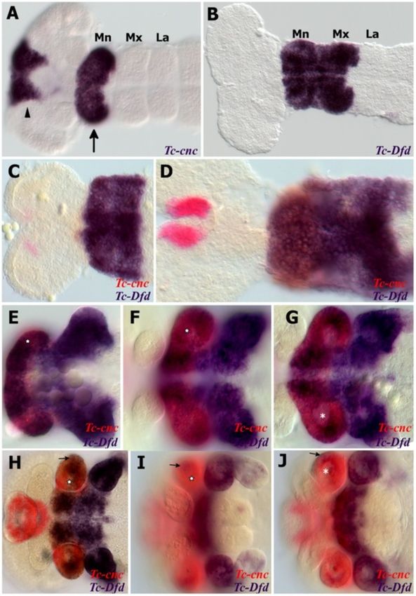
Coulcher and Telford EvoDevo 2012, 3:25                                                                            Page 2 of 16
http://www.evodevojournal.com/content/3/1/25

Background                                                     structure between mandibulate arthropods. We might
The arthropod mandible is an appendage adapted for bit-        therefore expect significant similarities in the embryonic
ing and chewing and is present in three arthropod groups,      patterning of the mandible between diverse mandibulate
the insects and crustaceans (collectively the Pancrustacea)    taxa. Finding the identity of the genes that pattern the
and the myriapods (millipedes and centipedes). The man-        mandible and showing how they function in diverse
dibulate arthropods, commonly grouped together in the          arthropod taxa could support the view that the mandible
monophyletic Mandibulata, constitute the majority of ani-      is homologous across the Mandibulata and, through com-
mals both in terms of numbers of species and biomass on        parisons with non-mandibulate sister groups, could give
this planet. The mandible is therefore an evolutionary         an insight into how the mandible evolved from a primitive
novelty of particular interest.                                arthropod limb.
  There are many different types of mandible, but the             We have undertaken a functional study of some of the
characteristic that most mandibles share, and which dif-       genes that pattern the mandible in a model organism
ferentiates it from other arthropod appendages, is the         with a typical insect mandible to compare its develop-
presence of a functional biting edge made up of the inci-      ment with the development of mandibles in other taxa.
sor and molar processes. This gnathal edge is widely           We chose to study the red flour beetle Tribolium casta-
considered to be a homologous structure within the             neum that, unlike Drosophila melanogaster, has a canon-
Mandibulata [1-3].                                             ical mandible in which the gnathal edge is made up of
  Other arthropod groups, the chelicerates and trilo-          the incisor and molar processes.
bites, do not have mandibles and instead have a walking
leg on the homologous segment to the mandibular seg-           Mandibular segment patterning in Drosophila
ment [4,5]. An unsegmented appendage, or lobopod, is           The majority of research into the function of genes pat-
present in closely related outgroups to the arthropods,        terning arthropod gnathal appendages has focused on
such as the onychophorans and tardigrades [6].                 insects with very derived mouthparts, in particular the
  An alternative phylogenetic hypothesis to the mono-          involuted larval head and non-biting adult proboscis of
phyletic Mandibulata is the Myriochelata hypothesis,           the dipteran D. melanogaster [18-25] and the stylet of
which groups the myriapods with the chelicerates.              the hemipteran Oncopeltus fasciatus [26,27].
Accepting this hypothesis would suggest that the man-             Although developing Drosophila embryos possess
dible evolved independently in the Myriapoda and Pan-          gnathal lobes (structures from which the gnathal appen-
crustacea or that it has reverted to a walking leg in the      dages are formed in other less derived insects [28]), fol-
Chelicerata [7]. While still controversial, recent molecu-     lowing head involution, Drosophila larvae do not have
lar phylogenies including evidence from unique micro-          any gnathal appendages [28-31] and both larval and
RNAs favour Mandibulata over Myriochelata. This                adult Drosophila lack an appendage on the mandibular
phylogeny is also strongly supported on morphological          segment.
grounds [8-11].                                                   In Drosophila, the gene Deformed (Dfd) is required for
                                                               the specification of both mandibular and maxillary iden-
Mandible evolution                                             tities [23-25,32,33]. Dfd does not differentiate the mandibu-
The mandible is serially homologous with other arthropod       lar segment from the maxillary segment; for this function
post-antennal appendages all of which are thought to have      another gene, cap’n’collar (cnc), is required [22-24]. cnc is
evolved from a segmented biramous limb. The archetypal         a basic leucine zipper family gene (bZIP) that is expressed
biramous limb consists of a protopodite (the base of the       in an anterior ‘cap’ domain in the labrum and a posterior
limb) to which are attached two branches: the telopodite       ‘collar’ domain in the mandibular segment and is necessary
(or palp) and an exopodite [12-14]. Structures called end-     for the development of both labral and mandibular derived
ites, often involved in food processing, are also present on   structures. It is likely that cnc achieves its mandible pattern-
the protopodite. The gnathal edge of the mandible is           ing function in part indirectly by repressing the maxilla
thought to have evolved from the proximal most endite          patterning function of Dfd: Dfd expression is repressed by
on the protopodite of this ancestral biramous limb [2,11].     cnc in the anterior of the mandibular gnathal lobe and the
The mandible is thought to be a gnathobasic structure          activity of the Dfd protein is also repressed by cnc in the
and this interpretation is supported by expression data:       mandibular segment. cnc null mutants lose both labral and
the distal limb expression domain of Distal-less (Dll) is      mandibular segment derived structures and have a duplica-
missing from the embryonic mandibular limb bud in              tion of maxillary structures [22-24,34].
diverse mandibulate arthropods [15-17].
   All arthropod mandibles appear to be gnathobasic and        Previous work in Tribolium
are restricted to a monophyletic group implying that the       In Tribolium, Brown et al. have demonstrated that the
mandible has a unique origin and is a homologous               homolog of Dfd, Tc-Dfd, is necessary for patterning the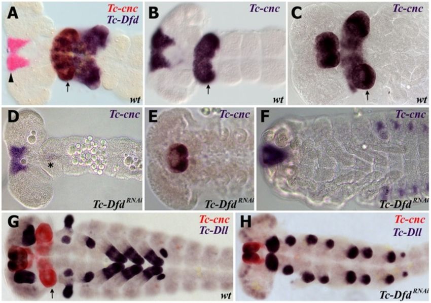
Coulcher and Telford EvoDevo 2012, 3:25                                                                           Page 3 of 16
http://www.evodevojournal.com/content/3/1/25

mandibular and maxillary segments and that Tc-Dfd ex-           GGATCGTCACAGTGTTGGTG-30). Accession numbers
pression is progressively downregulated in the mandibular       are as follows: Tc-cnc (GenBank: NM_001170642.1),
limb buds as in Drosophila [35,36]. In Tc-Dfd mutants           Tc-Dfd (GenBank: NM_001039421), Tc-mxp (GenBank:
there is a homeotic transformation of the mandible to an        NM_001114335), Tc-Dll (GenBank: NM_001039439),
antenna and a loss of the maxillary endites. Dfd, although      Tc-prd (GenBank: NM_001077622).
required for mandible development, does not differentiate
the mandibular segment from the maxillary segment in
                                                                Parental RNAi
Drosophila or Tribolium. The role of Tc-cnc in Tribolium
                                                                Parental RNAi was performed as previously described
is not known, however, it is expressed in a very similar pat-
                                                                [41]: 0.25 to 0.4 μl of Tc-cnc dsRNA (dissolved in dis-
tern to that seen in Drosophila [37] and this is also true of
                                                                tilled water at a concentration of 0.36 to 3 μg/μl) was
cnc in other mandibulate arthropods [38-40] suggesting it
                                                                injected into female pupae. Then, 633 bp of Tc-cnc
may have a conserved function. Embryonic expression in
                                                                dsRNA (positions 1,389 to 2,021, including part of the
non-mandibulate arthropods is not known.
                                                                bZIP domain which starts at position 1,932) was
                                                                injected. Embryos were either fixed 24 to 48 h after egg
Experimental outline
                                                                laying or left to develop into first instar larvae for cuticle
With the ultimate aim of understanding the origin of the
                                                                preparation. In total, 1,736 female beetle pupae were
mandible, we were interested in the role that Tc-cnc
                                                                injected for collecting embryos for in situ hybridization.
might play in patterning the mandibular segment of Tri-
                                                                   In order to characterize the Tc-cnc phenotype, 218 fe-
bolium castaneum, a mandible-bearing insect. In order
                                                                male pupae were injected with 1 to 2 μg/μl dsRNA and
to test its function in Tribolium, Tc-cnc was knocked
                                                                the cuticles of first instar larvae were analyzed. Of these
down using parental RNA interference (RNAi) by inject-
                                                                218 injected pupae, 195 successfully eclosed. At 20 days
ing double-stranded RNA (dsRNA) into female Tribo-
                                                                after injection a further 117 beetles (60%) had died.
lium pupae [41]. The knockdown phenotype was
                                                                Parental injection of Tc-cnc dsRNA resulted in the mor-
detected both in embryos and in the first instar larvae of
                                                                tality of a much greater number of injected females
offspring of injected parents. The effect of Tc-cnc knock-
                                                                compared to the numbers killed in other RNAi experi-
down on downstream genes was studied by in situ
                                                                ments, in which typically 10% of injected female beetles
hybridization in Tribolium embryos.
                                                                die by day 20 (data not shown). The higher mortality
                                                                rate may be a consequence of the effects of Tc-cnc
Methods
                                                                knockdown. Only one phenotype was detected in first
Tribolium castaneum culture
                                                                instar larvae: transformation of the mandible to maxil-
Wild-type T. castaneum (San Bernardino strain) were
                                                                lary identity and loss of the labrum.
kindly provided by Dr Gregor Bucher (Department of De-
                                                                   In order to obtain partial phenotypes (incomplete
velopmental Biology, Georg-August-University Göttingen,
                                                                transformations of the mandible to maxillary identity)
Göttingen, Germany) and raised at 32°C in organic whole-
                                                                we tried injecting lower concentrations of Tc-cnc dsRNA
meal flour supplemented with 5% brewer’s yeast.
                                                                (360 to 750 ng/μl). However, only wild-type larvae or
                                                                those with fully transformed mandibles were obtained,
Cloning of Tribolium orthologs
                                                                and no partial phenotypes were observed. Similar rates
Tc-cnc, Tc-Dfd, Maxillopedia the Tribolium ortholog of Pro-
                                                                of mortality were observed even at lower concentrations.
boscipedia (Pb) (Tc-mxp), the Tribolium ortholog of paired
                                                                   To obtain Tc-DfdRNAi embryos, 1,142 bp (positions
(Tc-prd) and Tc-Dll were amplified from mixed stage cDNA
                                                                491 to 1,632) Tc-Dfd dsRNA was injected into female
by polymerase chain reaction (PCR) amplification using the
                                                                pupae and embryos were fixed for in situ hybridization.
following primers: Tc-cnc, a 2,612 bp clone for hapten-
                                                                The Tc-DfdRNAi phenotype was confirmed by comparing
labelled RNA probe synthesis (forward: 50-GCAACAGTG
                                                                cuticle preparations of first instar larvae to previously
GGCCCTATTTA-30 and reverse: 50-GTGGTGGCTCCT
                                                                described phenotypes [36,42].
TGTGTTCT-30). Tc-cnc, a 633 bp clone for dsRNA synthesis
(forward: 50-GATTACAGCTATACGAGTCGG-30 and re-
verse: 50-GTCAGCCAGACTCAAAATCTG-30). Tc-Dfd                     Cuticle preparation
(forward: 50-CCAAGTGAGGAGTACAACCAG-30 and                       Cuticles from first instar larvae were prepared in
reverse: 50-TACAAGGCCGTGAGTCCGTAA-30), Tc-mxp                   Hoyer’s medium and lactic acid as previously described
(forward: 50-ATAGCTGCTTCGCTAGACCTTA-30 and                      [43]. The cuticle preparations were observed using dif-
reverse: 50-TCGCAGGTGGGGTCATTAT-30), Tc-Dll (for-               ferential interference contrast (DIC) and confocal fluor-
ward: 50-CAGCAGGTGCTCAATGTGTT-30 and reverse:                   escent microscopy (larval cuticle autofluoresces at
50-ATTAAACAGCTGGCCACACC-30), Tc-prd (forward:                   visible wavelengths). Cuticle preparations were observed
50-ATGCACAGACATTGCTTTGG-30 and reverse: 50-                     using confocal microscopy with an excitation frequency
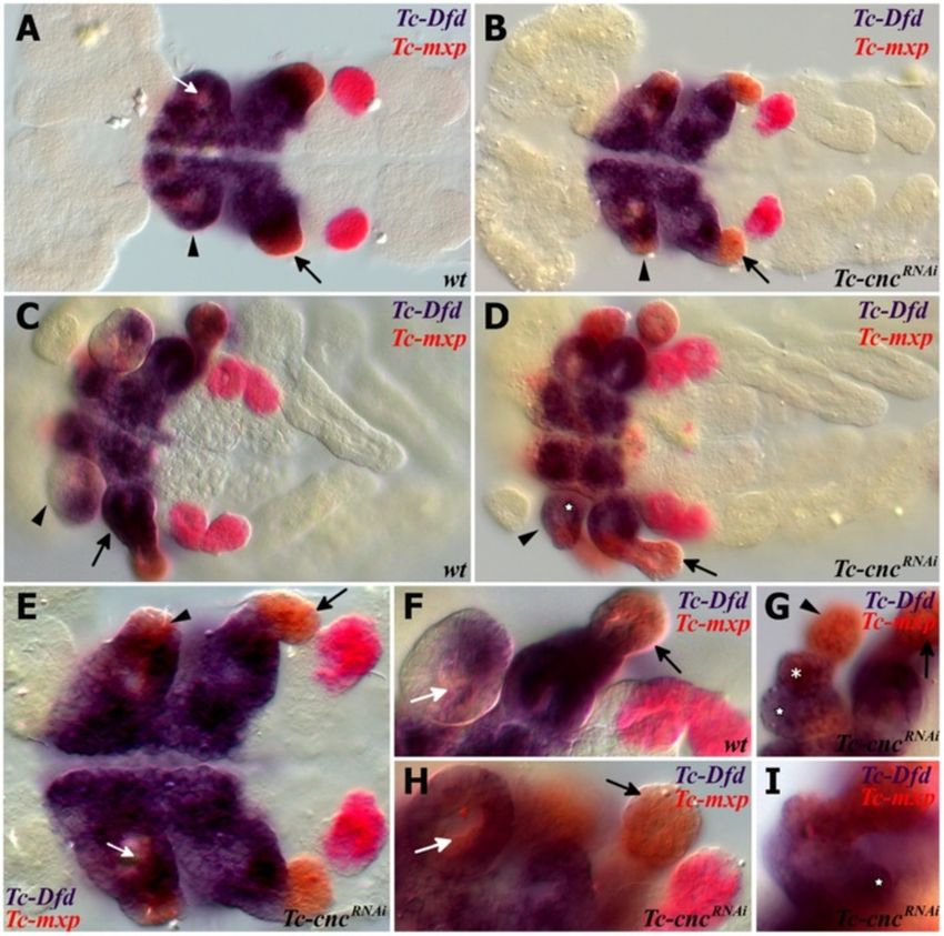
Coulcher and Telford EvoDevo 2012, 3:25                                                                                                 Page 4 of 16
http://www.evodevojournal.com/content/3/1/25

of 488 nm using an upright Leica TCS SPE confocal                           Whole mount in situ hybridization
microscope (Leica microsystems, Wetzlar, Germany).                          Embryos were fixed in 9% formaldehyde. Both single stain-
Images were obtained and edited using Leica applica-                        ings (nitro blue tetrazolium/5-bromo-4-chloro-3-indolyl
tion suite advanced fluorescence software, LAS-AF                           phosphate (NBT/BCIP)) and double stainings (NBT/BCIP
(Leica microsystems, Wetzlar, Germany).                                     and FastRed) were performed as previously described [44].

 Figure 1 Expression of Tc-Dfd and Tc-cnc in the mandibular and maxillary segments. All views are ventral with anterior to the left unless
 otherwise indicated. Gene expression was determined by in situ hybridization. (A) Expression of Tc-cnc in a germ band extending stage embryo.
 There is an anterior cap domain of Tc-cnc in the labrum (arrowhead). The posterior collar domain is present in the mandibular segment (arrow).
 (B) Tc-Dfd expression in a germ band extending embryo as limb buds are just about to form. Expression is present throughout the mandibular
 and maxillary segments. (C-J) Expression of Tc-Dfd (blue) and Tc-cnc (red) in wild-type embryos. Coexpression of Tc-Dfd and Tc-cnc is brown. (C)
 Early germ band extending embryo. (D) Germ band extending stage embryo prior to limb bud formation. (E) Germ band extending embryo. (F)
 Late germ band extending embryo. (G) Same embryo as (F), but a lower plane of focus that shows the reduction of Tc-cnc expression in the
 mesoderm (asterisk). (H) Germ band retracting embryo. (I) Embryo undergoing dorsal closure with the gnathal appendages moving towards the
 ventral midline. (J) Same embryo as (I), but a lower plane of focus that shows the reduction of Tc-cnc expression in the mesoderm (asterisk). (C,D)
 Prior to limb bud formation, Tc-Dfd expression is continuous throughout the mandibular segment. (E,F) As soon as the endites start to form, Tc-
 Dfd expression retracts from the developing mandibular endites (star) whilst Tc-cnc expression is maintained throughout the mandibular
 appendage. (G-J) By late embryogenesis, faint Tc-Dfd expression is only present in the lateral part of the mandibular limb bud (arrow), and
 missing from the ventral-medial region (star). Tc-Dfd expression is still strongly maintained in the maxillary limb bud. Mandibular (Mn), maxillary
 (Mx) and labial (La) segments.
Coulcher and Telford EvoDevo 2012, 3:25 Page 5 of 16
http://www.evodevojournal.com/content/3/1/25
Some modifications, for example in the frequency and of the mandibular limb bud becomes the outer lobe
duration of washes, were incorporated from alternative of the mandible and develops into the future
in situ hybridization protocols [45]. incisor process. Tc-Dfd is not present in this most
Stained embryos were dissected from their yolk and distal region, which is more clearly noticeable in
mounted in glycerol. Embryos (and cuticle preparations) lateral orientations of dissected Tribolium embryos
were observed using differential interference contrast (see Figure 2C).
(DIC) microscopy with an Imager M1 microscope (Carl We found that Tc-prd, in addition to its function as a
Zeiss Ltd., Cambridge, UK). Images were taken with secondary pair-rule gene [47], is expressed in the pre-
Axiocam HRC (Carl Zeiss Ltd., Cambridge, UK) and dicted location of the developing endites of the embry-
processed using Axiovision product suite software re- onic mandibular, maxillary and labial limb buds (see
lease 4.8.2 (Carl Zeiss Ltd., Cambridge, UK). Images Figure 2B) [48]. We therefore used Tc-prd expression as
were edited with GIMP (release 2.6.10.) [46]. a marker for endite development. Tc-prd expression
reveals that the ventral-medial region of the mandibular
Scanning electron microscopy limb bud, where Tc-Dfd expression is lost, encompasses
Embryos were fixed as described for the whole mount in the mandibular endite and the immediate surrounding
situ hybridization protocol. Fixed embryos were rinsed tissue. Tc-Dfd expression is retained in the lateral part of
in ethanol and immersed in hexamethyldisilazane the mandibular limb bud, but fades throughout embryo-
(HMDS), air dried and sputter coated with gold. Images genesis (Figure 2D). Tc-Dfd expression is absent (or con-
were taken in a JEOL JSM-5410LV scanning microscope siderably weaker) in the distal part of the maxillary palps
(JEOL Ltd., Tokyo, Japan) at a magnification of 100 to throughout embryogenesis (see arrow in Figure 2E).
350 fold and processed with DigitalMicrograph (Gatan
Inc., Pleasanton, California, USA).
Tc-cnc RNAi phenotype
In order to test the role Tc-cnc might play in patterning
Results
the mandibular segment, the gene was knocked down in
Tc-cnc expression
developing embryos by injecting Tc-cnc dsRNA into fe-
Tc-cnc is expressed in two distinct domains, an anterior
male pupae. The knockdown phenotype was determined
cap that includes the developing labrum and around the
in the offspring of injected parents using cuticle prepara-
stomodeum and a posterior collar domain in the man-
tions of their first instar larvae (see Figure 3).
dibular segment (see Figure 1A) [37]. Tc-cnc expression
Injection of Tc-cnc dsRNA produces phenotypes that
remains constant in these two domains from their first
relate to both the cap domain and the collar domain of
appearance during germ band elongation and through
Tc-cnc expression. The effect in the collar domain is the
late embryogenesis (see Figure 1D,F,H) and is expressed
homeotic transformation of the mandibular appendage
in regions of the mandibular limb bud where Tc-Dfd ex-
into a maxillary identity showing that the posterior col-
pression becomes repressed (see star in Figure 1E-I). In
lar domain of Tc-cnc expression differentiates the man-
the mandibular limb bud, Tc-cnc is expressed predomin-
dible from the maxillary appendage. This is shown in
antly in the ectoderm, with weaker expression (or no
Figure 3D,F, where Tc-cncRNAi larvae can be seen to pos-
discernable expression) in the mesoderm of the limb
sess an additional pair of maxillae. The mandibular
bud (see asterisk in Figure 1G,J).
appendages are transformed into a maxillary identity, in
possession of a maxillary palp, and maxillary endites
Tc-Dfd expression retracts from the developing mandible
(which in wild-type first instar Tribolium larvae are
Tc-Dfd is expressed throughout the mandibular and
fused to form the ventral branch; see Figure 3A,C).
maxillary segments in the early developing Tribolium
Knockdown of the cap domain results in a dramatic de-
embryo (see Figure 1B). As the mandibular limb buds
letion of the labrum showing Tc-cnc is necessary to pat-
start to form, Tc-Dfd expression progressively retracts
tern this structure (see Figure 4B). There are also
from the ventral-proximal region of the mandibular limb
abdominal defects visible in some embryos, although it
bud (see Figure 1E-J). Tc-Dfd continues to retract from
is possible that this aspect of the phenotype was an
this ventral-proximal region (star in Figure 1E).
artifact of the cuticle preparation procedure.
In the developing maxillae, Tc-Dfd expression is con-
tinually expressed in the protopodite (see Figure 1E-J).
Mandibular Tc-Dfd is increasingly repressed until only Tc-cnc represses Tc-Dll and modifies Tc-prd expression in
weak expression remains on the lateral side of the man- the Mandibular segment
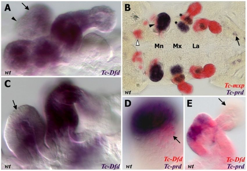
dible (see Figure 1H,I and Figure 2A,C,D). To investigate the transformed mandibular appendage in
The mandibular limb bud has two lobes, the inner Tc-cnc knockdown embryos, the expression patterns of
and the outer (see Figure 2A). The distal-most part the homeobox genes Tc-prd and Tc-Dll were studied as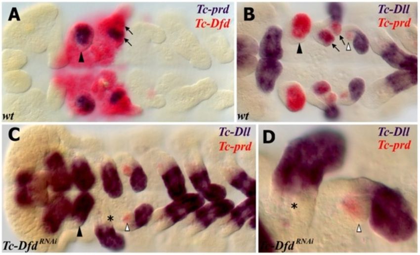
Coulcher and Telford EvoDevo 2012, 3:25                                                                                                     Page 6 of 16
http://www.evodevojournal.com/content/3/1/25

 Figure 2 Expression of Tc-Dfd, Tc-mxp and Tc-prd in dissected embryonic mandibles and maxillae. All views are ventral with anterior to
 the left unless otherwise indicated. Gene expression was determined by in situ hybridization. (A,C) Lateral view of the mandibular and maxillary
 appendages showing Tc-Dfd expression (blue) in a germ band fully retracted stage embryo. (A) Tc-Dfd expression is repressed from the
 developing mandibular endite, which consists of an inner lobe (arrowhead) and an outer lobe (arrow). (B) Embryo stained with Tc-mxp (red) and
 Tc-prd (blue). Tc-mxp is expressed in the maxillary and labial palps and the distal protopodite of both appendages. In the maxilla, protopodite
 expression relates to the position of the developing galea endite lobe (asterisk), which is marked by the distal domain of Tc-prd expression. Tc-
 mxp is expressed in the mesoderm of the mandibular limb bud (arrowhead). The intercalary domain of Tc-mxp expression is also visible (white
 arrowhead). Mesodermal expression of Tc-prd is present in the telopodites of post-antennal appendages but clearly visible in the developing leg
 appendages (arrow). (C) Tc-Dfd expression is missing from the outer lobe of the mandible (arrow). (D,E) Tc-Dfd expression (red) and Tc-prd
 expression (blue) in a dissected mandible and maxilla of a post germ band retracted stage embryo undergoing dorsal closure. Distal is top. (D)
 Lateral view of a dissected mandible. Tc-Dfd expression remains on the lateral side of the mandible (arrow). (E) Dissected maxilla, lateral is to the
 right. Tc-Dfd expression is throughout the protopodite and at the base of the palp. The distal part of the palp is lacking or has weak Tc-Dfd
 expression (arrow).

genetic markers of the developing endites and telopo-                         expression in the palp and in a proximal endite that
dites respectively (see Figure 5).                                            appears on the transformed mandible.
  In wild-type embryos, Tc-prd is expressed in the devel-                       The transformed mandibular appendage develops
oping endites of all three pairs of gnathal appendages                        more slowly than the adjacent true maxillary appendages
(mandibles, maxillae and labia; see Figure 2B and                             at several stages of embryogenesis resembling the maxil-
Figure 5A,C). There are two distinct domains of Tc-prd                        lary appendage of an earlier stage (see Figure 5E). By late
expression in the maxilla, which we assume correspond                         embryogenesis, there is no evident morphological differ-
to the developing lacinia and galea. There is a single do-                    ence between the maxillae and the ectopic maxillary
main of Tc-prd in the labial appendage and a larger sin-                      appendages on the mandibular segment.
gle domain of expression in the mandibular appendage.                           Asymmetry of different appendages in Tc-cnc RNAi
  Tc-Dll is expressed in the distal part of all appendages of                 embryos is often evident in germ band extending stage
wild-type Tribolium embryos except the mandible. In the                       embryos and occurs left or right at random (see
developing maxilla, there are two domains of Tc-Dll ex-                       Figure 5E). This does not appear to be an artifact of the
pression, a distal domain in the developing palp and a                        RNAi procedure or the in situ hybridization process as
proximal domain in the lacinia endite. Tc-cnc RNAi results                    appendages other than the mandible can be affected and
in homeotic transformation of the mandibular appendage                        parental RNAi experiments of other genes in Tribolium
into maxillary identity. The solitary domain of Tc-prd ex-                    have not yielded a similar result (data not shown). Instead
pression in the mandible is transformed into two domains                      this may be related to a loss of the role that cnc has been
of Tc-prd expression that relate to the maxillary endites                     shown to have in Drosophila in protecting the embryo
(see Figure 5B,D-F). Tc-Dll is de-repressed resulting in                      from oxidative stress [49].
Coulcher and Telford EvoDevo 2012, 3:25 Page 7 of 16
http://www.evodevojournal.com/content/3/1/25
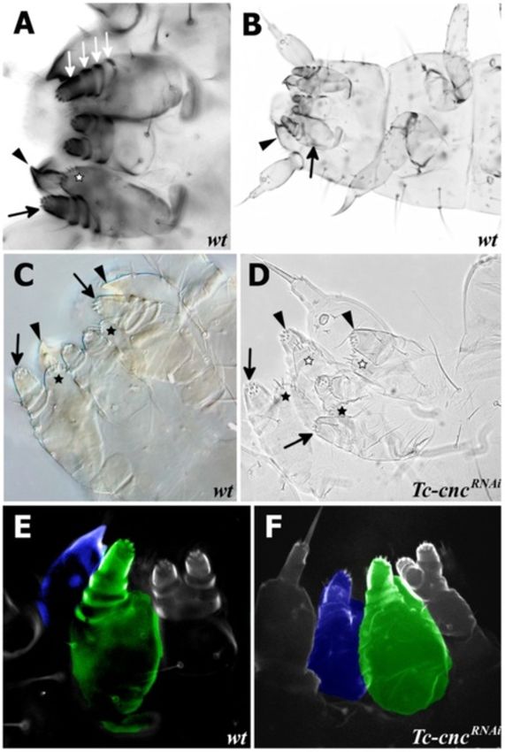
Figure 4 Tc-cncRNAi results in deletion of the Labrum. Scanning
electron micrographs (SEMs) of wild-type and Tc-cncRNA embryos
shows the deletion of the Labrum in Tc-cncRNAi embryos. All views
are ventral with anterior to the left. (A) SEM of a wild-type embryo
at fully extended germ band stage. The labral buds are clearly visible
at the anterior of the embryo (arrow). The mandible is indicated
(arrowhead). (B) SEM of Tc-cncRNAi embryo at germ band extending
stage. The labral buds are missing (arrow). The mandible is
transformed into maxillary identity (arrowhead).
in an additive fashion. Tc-Dfd is expressed in the proximal
part of the maxilla (the protopodite), and Tc-mxp is
expressed in the palp and is excluded from the proximal
part of the protopodite, although it is expressed in the dis-
tal protopodite and galea endite (see Figure 2B). Tc-Dfd
patterns the protopodite: the proximal part of the append-
age including the endite [36]. Tc-mxp patterns the telopo-
dite (the palp) and mutants of Tc-mxp possess legs instead
Figure 3 Tc-cncRNAi results in transformation of the mandible
of palps in both the maxillary and labial segments. These
into maxillary identity. Mandible (arrowhead), maxillary palp
(arrow) and maxillary ventral branch (star) are indicated on cuticle transformed appendages are attached to a protopodite that
preparations of wild-type and Tc-cncRNATribolium first instar larvae. is unaffected by the loss of Tc-mxp [50,51].
(A) Cuticle preparations of gnathal appendages visualized by As Tc-cnc RNAi results in a homeotic transformation
fluorescence microscopy. The maxillary appendages have a palp of the mandible into a maxilla, we predicted that both
with four segments (white arrows) attached to a protopodite with
the Hox genes responsible for patterning the maxillary
the maxillary endites (lacinia and galea) that, in first instar larvae, are
fused to form the ventral branch (star). (B) Cuticle preparation of a appendage will be expressed in the maxillary pattern in
first instar Tribolium larva. (C) Cuticle preparation of the larval gnathal the homeotically transformed appendage. It was found
appendages of a wild-type Tribolium larva visualized by DIC that this is indeed the case (see Figure 6).
microscopy. (D) Cuticle preparation of the gnathal appendages of a In wild-type embryos, Tc-Dfd expression retracts from
Tc-cncRNAi larva. Knockdown of Tc-cnc results in transformation of the
the mandibular limb bud (see Figure 6C,F). In Tc-cncRNAi
mandibular appendages into maxillary appendages (arrowheads).
The ventral branch is visible on the transformed appendages (white embryos, the mandible is transformed to maxillary identity
stars). The maxillary appendage is indicated with arrows (palp) and and Tc-Dfd expression is retained in the protopodite of
black stars (ventral branch). (E) Cuticle preparation of wild-type this transformed appendage (see Figure 6D,G-I).
Tribolium larva visualized by confocal microscopy. The mandibular Tc-mxp is expressed in the maxillary and labial palps in
appendage is highlighted in blue; the maxillary appendage is
wild-type embryos (see Figure 6A,C,F). In the maxillae, Tc-
highlighted in green. (F) Cuticle preparation of a Tc-cncRNAi larva
visualized by confocal microscopy. The transformed mandibular mxp is expressed in the distal part of the protopodite, in-
appendage is highlighted in blue and clearly resembles the maxillary cluding the galea endite. In the transformed mandibular
appendage (highlighted in green). appendage of Tc-cncRNAi embryos, Tc-mxp is expressed in
the ectoderm of the ectopic palp as it is in the maxillary
palp and also includes expression in the galea endite and
Tc-Dfd and Tc-mxp are expressed in a maxilla-like manner distal protopodite (see Figure 6B,D,E,G,I).
in the transformed mandibular limb bud of Tc-cncRNAi Tc-mxp is expressed in the mesoderm of the mandibu-
embryos lar appendages of wild-type embryos (white arrow in
The Hox genes Tc-Dfd and maxillopedia (Tc-mxp), the Figure 6A,F) [51]. Interestingly, this mesodermal expres-
Tribolium ortholog of pb, pattern the maxillary appendage sion of Tc-mxp is seen in Tc-cnc knockdown embryos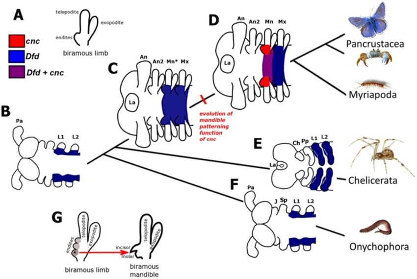
Coulcher and Telford EvoDevo 2012, 3:25                                                                                                       Page 8 of 16
http://www.evodevojournal.com/content/3/1/25

 Figure 5 Homeotic transformation of the mandibular appendage to maxillary identity in Tc-cnc knockdown embryos as revealed by
 the expression of markers for telopodites (Tc-Dll) and endites (Tc-prd). Gene expression was determined by in situ hybridization. All views
 are ventral with anterior to the left. (A-E) The mandibular segment appendage (arrowhead), lacinia (star), galea (asterisk) and telopodite (arrow)
 are indicated. (A,B) Expression of Tc-Dll (blue) and Tc-prd (red). (A) wild-type embryo. Tc-Dll is expressed in the maxillary lacinia endite lobe (star)
 and telopodite (arrow). Tc-prd is expressed in the endites of the mandible, maxilla and labial appendages. (B) Tc-cncRNAi embryo. Tc-Dll and Tc-prd
 are expressed in transformed mandible appendages in the same manner as in the maxillae. The labral domain of Tc-Dll is also missing at the
 anterior of the embryo. (C-E) Expression of Tc-Dll (red) and Tc-prd (blue) in germ band extending embryos earlier than those shown in A,B. (C)
 wild-type embryo. (D,E) Tc-cncRNAi embryos. (E) Tc-cncRNAi embryo. The telopodites and endites of some appendages are larger (white
 arrowheads) than the corresponding appendage on the other side of the same segment. There is an asymmetry between the different
 transformed mandibular appendages. The transformed mandibles resemble maxillae at an earlier stage of development and so have delayed
 development relative to the maxillary appendages. Mandibular (Mn), maxillary (Mx) and labial (La) segments shown.

(white arrow in Figure 6E,H). This suggests that there is                      Tc-Dfd activates Tc-prd expression in the mandible and
cnc independent regulation of Tc-mxp in the mandibular                         maxillary segments
limb bud. Tc-cnc is expressed in the ectoderm of the                           Brown et al. have shown that Tc-Dfd is required to pat-
mandibular limb bud, and expression is weaker (or ab-                          tern the mandible and the proximal part of the maxillary
sent) in the mesoderm.                                                         appendages In Tc-Dfd mutants, the mandible is trans-
                                                                               formed to antennal identity and the maxillae lose the
                                                                               endites whilst retaining the palp [36].
Tc-Dfd activates the posterior ‘collar’ domain of Tc-cnc in                      In order to further investigate the role of Tc-Dfd in pat-
the mandibular segment                                                         terning the gnathal appendages, we studied Tc-prd expres-
Experiments performed on Drosophila have shown that                            sion in Tc-DfdRNAi knockdown embryos. In Tc-DfdRNAi
Dfd does not activate cnc expression [52]. In order to in-                     knockdown embryos, Tc-prd expression is lacking in both
vestigate whether Tc-Dfd has any role in regulating Tc-                        the transformed mandible (ectopic antennae) and the
cnc expression in Tribolium, we knocked down Tc-Dfd                            affected maxillary appendages (see Figure 8C,D). Tc-prd is
by parental RNAi and detected Tc-cnc expression via in                         still expressed in the developing labial endite. This result
situ hybridization.                                                            shows that Tc-Dfd is necessary for the activation of Tc-prd
   Surprisingly, we found that in Tc-DfdRNAi embryos the                       expression in the mandibular and maxillary segments and
posterior collar domain of Tc-cnc expression is com-                           is further evidence that Tc-Dfd is required for develop-
pletely missing from all stages of embryo investigated,                        ment of the endites on these segments.
from germ band extending embryos through to stages
where embryos are undergoing dorsal closure (Figure 7).                        Discussion
The anterior cap domain of expression is unaffected.                           The role of Tc-cnc in patterning the mandible of Tribolium
This shows that, unlike in Drosophila, Tc-Dfd is neces-                        We sought to understand mandible patterning in a
sary for the activation of the posterior domain of Tc-cnc                      model arthropod that has a mandible with primitive
in the mandibular segment of Tribolium.                                        characteristics. Our results show that Tc-cnc is required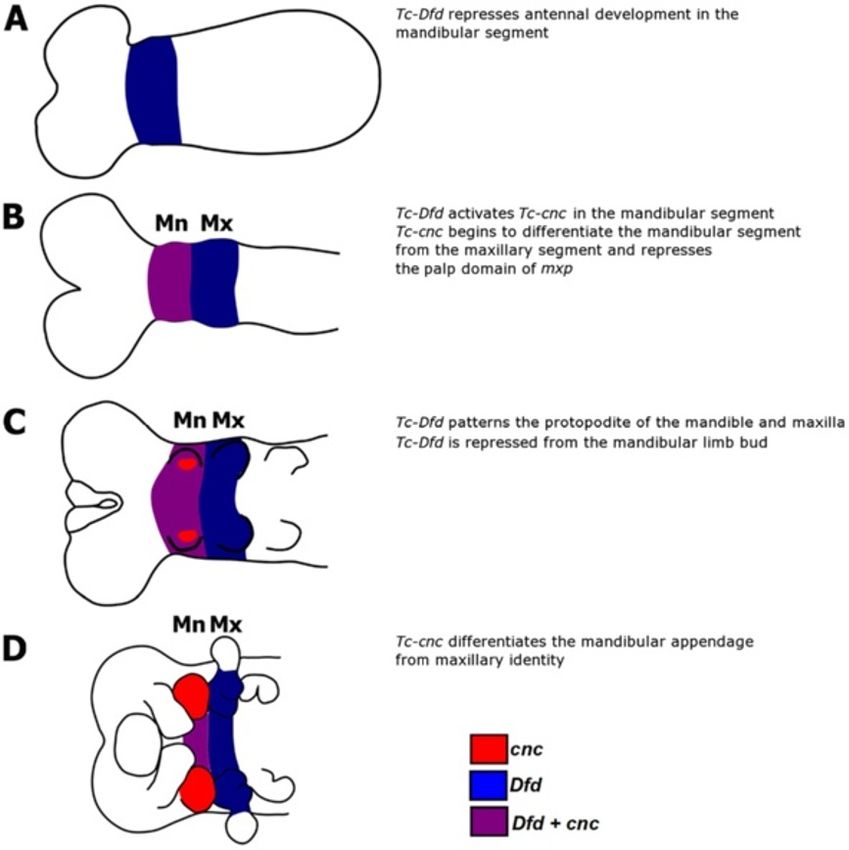
Coulcher and Telford EvoDevo 2012, 3:25                                                                                              Page 9 of 16
http://www.evodevojournal.com/content/3/1/25

 Figure 6 Expression of the Hox genes Tc-Dfd and Tc-mxp in wild-type and Tc-cncRNAi embryos. Knockdown of Tc-cnc by RNAi results in
 transformation of the mandibular appendage to maxillary identity and the expression of Hox genes in a similar manner to that seen in the
 maxilla. All views are ventral with anterior to the left. Expression of Tc-Dfd (blue) and Tc-mxp (red) was determined by in situ hybridization.
 Mandibular segment is indicated with an arrowhead, maxillary segment with a black arrow. Mesodermal expression of Tc-mxp is indicated with a
 white arrow. (A,C,F) Wild-type Tribolium embryos. (B,D,E,G-I) Tc-cncRNAi embryos. (A) Wild type germ band extending embryo. Tc-mxp is
 expressed in the developing maxillary and labial palps and the mesoderm in the mandibular segment (white arrow). (B) Tc-cncRNAi germ band
 extending embryo: Tc-mxp expression is present in the transformed mandibular appendage (arrowhead) in a telopodite domain consistent with
 the transformation of the mandible to maxillary identity. (C) Wild-type germ band retracting stage embryo. Tc-Dfd expression has retracted from
 the majority of the mandibular appendage. (D) Tc-cncRNAi embryo at a similar stage to C. Tc-Dfd expression is present in the transformed
 mandibular protopodite (star). Tc-mxp is expressed in the transformed mandibular appendage palp. (E) Higher magnification of the earlier germ
 band extending stage Tc-cncRNAi embryo shown in B. (F) Higher magnification of the gnathal appendages of a germ band retracting stage at a
 similar stage to C. (G, H, I) Higher magnification of the gnathal appendages of germ band retracting stage Tc-cncRNAi embryos. (G) Tc-Dfd is
 expressed throughout the transformed mandibular appendage, in the lacinia endite (star) and galea endite (asterisk). Tc-mxp is expressed in the
 palp (arrowhead) as well as the galea endite in a manner that is identical to the maxilla (arrow). (H) The mesodermal expression domain of Tc-
 mxp (white arrow) is observed in the transformed mandibular appendage. (I) Tc-Dfd is expressed throughout the maxilla, the rounded kink at the
 base of the maxilla is indicated (star).

for specification of the identity of the mandibular seg-                      The Hox genes Tc-mxp and Tc-Dfd are required to pat-
ment of Tribolium and differentiates the mandible from                     tern the maxillary appendage and do so in an additive man-
a maxilla.                                                                 ner, Tc-Dfd patterns the base of the appendage and Tc-mxp
  Knockdown of Tc-cnc transcripts by parental RNAi                         patterns the palp [36,51]. We show that in Tc-cnc knock-
results in a homeotic transformation of the mandible                       down embryos, Tc-Dfd and Tc-mxp are expressed in a max-
into maxillary identity in Tribolium embryos and first                     illa like pattern in the transformed mandibular appendage.
instar larvae. The homeotic transformation is also evi-                       We show that the ‘collar’ domain of Tc-cnc in the man-
dent in the changed expression of the genes Tc-Dll and                     dibular segment is activated by Tc-Dfd in Tribolium. The
Tc-prd (markers for the developing telopodite and endite                   mandibular segment collar domain of cnc is not activated
of the maxilla) in knockdown embryos.                                      or regulated by Dfd or by any other Hox gene in
Coulcher and Telford EvoDevo 2012, 3:25                                                                                                  Page 10 of 16
http://www.evodevojournal.com/content/3/1/25

 Figure 7 Tc-Dfd activates the posterior collar domain of Tc-cnc in the mandibular segment. Gene expression was determined by in situ
 hybridization. (2A-C) Tc-cnc expression in wild-type embryos. Throughout embryogenesis, Tc-cnc expression consists of an anterior cap domain in
 the labrum (arrowhead) and a collar domain (arrow) in the mandibular segment. (A) Tc-cnc (red) and Tc-Dfd (blue) expression in a germ band
 extending embryo. (B) Tc-cnc expression (blue) in a germ band extending embryo at a similar but slightly earlier stage to (A). (C) Tc-cnc
 expression (blue) in later stage embryo prior to dorsal closure. (D-F) Tc-cnc expression in Tc-DfdRNAi embryos. In all stages, from germ band
 extending (D), germ band retracted (E) and during dorsal closure (F), the posterior domain of Tc-cnc is missing in the mandibular segment, whilst
 the anterior domain of Tc-cnc is expressed as normal showing that Tc-cnc is activated by Tc-Dfd in the mandibular segment. There is a faint stripe
 of Tc-cnc in the mandibular segment of (D) (asterisk), this may be due to partial knockdown effects.( G) Expression of Tc-cnc (red) and Tc-Dll (blue)
 in wild-type germ band retracting embryo.( H) Expression of Tc-cnc (red) and Tc-Dll (blue) in a Tc-DfdRNAi germ band extending embryo. The
 posterior domain of Tc-cnc is missing..

Drosophila [52]. We also show that Tc-Dfd is necessary for                   mandibular and maxillary segments and in how these
the expression of Tc-prd in both the mandible and the                        segments are patterned. In both insects Dfd and cnc are
maxilla.                                                                     both required to pattern the mandibular segment. cnc is
  Based upon the results of this and previous studies we                     required for the patterning of labral derived structures
present a model for the roles of these genes in mandible                     and the differentiation of the mandible from maxillary
patterning in Tribolium (see Figure 9).                                      identity. cnc represses Dll expression in the mandibular
                                                                             segment.
The role of Tc-cnc in patterning the labrum of Tribolium                       The Hox genes Dfd and pb/Tc-mxp are also expressed
The deletion of the labrum in Tc-cncRNAi embryos is con-                     in similar proximal and distal domains respectively in
sistent with the loss of the cap domain of Tc-cnc expression                 the maxillary segment limb bud or gnathal lobe as are
in the labrum. The labrum is a structure of considerable                     prd and Dll. Dfd patterns proximal structures that are
interest as it is shared by all extant groups of euarthropods                derived from the maxillary lobe or limb buds. In both
whilst its evolution and development remain controversial.                   species, Dfd activates the proximal domain of Dll
The labrum has appendage-like characteristics and may                        [24,25,36]. Tc-Dfd activates the maxillary prd domain in
have evolved from a fused pair of appendages, for example                    both Drosophila, and also, as we have shown in this
from structures homologous to the anterior antennae of                       study, in Tribolium [55,56].
lobopods [53]. However, unlike all other paired arthropod                      There are nevertheless differences in the patterning of
appendages, the labrum is not associated with a segment                      the mandibular and maxillary segments between Tribo-
and may have a different origin [54].                                        lium and Drosophila. In Drosophila, loss of cnc function
                                                                             does not result in a full homeotic transformation of
Comparisons with Drosophila                                                  the mandibular gnathal lobe to maxillary identity, rather,
There are many similarities between Tribolium and                            the mandibular gnathal lobe is transformed into just the
Drosophila in the expression patterns of genes in the                        proximal part of the maxillary gnathal lobe [24]. This is
Coulcher and Telford EvoDevo 2012, 3:25 Page 11 of 16
http://www.evodevojournal.com/content/3/1/25
Figure 8 Tc-Dfd knockdown results in the loss of Tc-prd expression in the embryonic mandibular and maxillary segments. Tc-Dfd
patterns the endites of the mandibular and maxillary segments. The mandibular segment appendage is marked with an arrowhead. The maxillary
endites are marked with arrows in wild-type embryos (A,B). The labial endites are marked with white arrowheads. (A) Expression of Tc-prd (blue)
and Tc-Dfd (red) in a wild-type germ band extending embryo. Tc-prd is expressed in the developing endites of the mandible and maxilla. Tc-Dfd
is expressed in the mandibular and maxillary segments. (B) Expression of Tc-prd (red) and Tc-Dll (blue) in a wild-type germ band extending
embryo. Tc-prd is expressed in the mandible, maxillary and labial appendages. Tc-Dll is expressed in the lacinea endite lobe. There is no Tc-Dll
expression in the mandible (arrowhead). (C,D) Expression of Tc-prd (red) and Tc-Dll (blue) in a Tc-DfdRNAi germ band extending embryo. (C) The
mandible has been transformed into an ectopic antenna, which expresses Tc-Dll (arrowhead) and lacks Tc-prd expression. There is no endite and
no Tc-prd expression (asterisk) in the maxilla. The labial appendage has an endite (white arrowhead) marked with Tc-prd expression. (D)
Enlargement of the maxilla and labial appendage shown in (C).
in contrast to Tribolium where loss of Tc-cnc function mandibular segment, indicating that some mandibular
results in a complete transformation of mandible to max- expression of Dfd is not affected by the presence of cnc
illary identity. [23,24]. Dfd has also been shown to repress pb in the
In addition to the activation of Tc-cnc in the mandibu- ectoderm of the mandibular segment in Drosophila [61].
lar segment by Tc-Dfd, another difference between Dros- As the dynamics of Tc-Dfd expression in Tribolium re-
ophila and Tribolium is the regulation of collier (col) by semble the dynamics of Dfd expression in Drosophila,
cnc. The anterior mandibular expression of cnc is up- with initial coexpression followed by subsequent repres-
stream of col in Drosophila and both genes are required sion of Tc-Dfd in a part of the mandibular segment, it
to pattern the hypopharyngeal lobes [57-59]. In Tribo- seems likely that a similar situation is occurring in
lium, which does not have hypopharyngeal lobes, it has Tribolium.
been recently shown that Tc-cnc is not activated by Tc- We have shown that Tc-cnc is necessary for both the
col [60]. repression of Tc-Dfd expression in the mandibular limb
bud and the repression of the ectodermal palp domain
The role of cnc as a repressor of maxilla patterning Hox of Tc-mxp in the developing mandibular limb bud. How-
genes ever, further research is needed to determine whether
While we have shown that Tc-cnc patterns the mandible Tc-cnc has a direct functional role in the repression of
and differentiates the mandible from a maxilla, the pre- these Hox genes.
cise role that it has in patterning the mandibular seg-
ment is not clear. The many similarities in the The possible role of Tc-cnc as a direct activator of
patterning function of cnc in Tribolium and Drosophila mandible patterning genes
suggest that the molecular functions of Cnc protein In Drosophila, several lines of evidence suggest that Cnc
revealed by experiments in Drosophila may be similar in functions as an activator, activating mandibular segment
Tribolium. specific patterning genes and thereby indirectly repres-
Research in Drosophila has demonstrated the role of sing Hox genes [23]. cnc also patterns some mandibular
cnc as a repressor of Hox gene function in the mandibu- segment derived structures independently of Dfd. Ec-
lar segment [23,24]. cnc has been shown to repress Dfd topic activation of cnc in Drosophila embryos results in
transcription and Dfd protein activity in the anterior ectopic hypopharyngeal lobe derived structures [23]. Al-
mandibular segment in Drosophila [23,24]. There is co- though the hypopharyngeal lobes have been thought to
expression of cnc and Dfd in the posterior of the derive from the intercalary segment, it has recently beenCoulcher and Telford EvoDevo 2012, 3:25 Page 12 of 16
http://www.evodevojournal.com/content/3/1/25
Figure 9 A model of Tc-cnc and Tc-Dfd mandibular and maxillary patterning functions in Tribolium castaneum. Tc-Dfd patterns both the
mandible and maxillary segments. Tc-Dfd patterns the protopodites of these appendages. Note that the anterior ‘cap’ domain of Tc-cnc has been
omitted from this scheme for clarity. Tc-Dfd expression is shown in blue, Tc-cnc expression is shown in red. Tc-cnc and Tc-Dfd expression is shown
in purple. (A) Tc-Dfd is expressed in the mandibular and maxillary segments and patterns these segments. In the mandibular segment, Tc-Dfd
represses antennal development. Tc-Dfd patterns the maxillary segment in conjunction with Tc-mxp. (B) Tc-Dfd activates Tc-cnc expression and
together Tc-cnc and Tc-Dfd cooperate to pattern the mandibular segment. (C) Tc-Dfd patterns the protopodite of the mandibular and maxillary
appendages in germ band extending embryos, but is repressed from the mandibular limb bud as it develops. (D) In germ band retracting stage
embryos, Tc-cnc has differentiated the mandibular appendage from maxillary identity.
shown that they are in fact derived from the mandibular patterning of the maxilla may also be conserved. Dfd is
segment [37]. This result indicates that cnc is in fact ne- expressed in the mandible and maxilla bearing segments
cessary and sufficient to pattern some mandibular seg- in the majority of mandibulates and expression is stron-
ment derived structures suggesting that Tc-cnc may ger in the protopodite than in the palps of maxillary
directly activate mandible patterning genes in Tribolium. appendages [36,39,62-66]. There is loss of Dfd expres-
sion in the mandibular limb bud across mandibulates, as
Conserved expression of cnc, Dfd and pb in mandibulate in Tribolium and Drosophila [24,35,65,66]. Expression of
arthropods pb is conserved in the telopodites of these maxillary
Comparison of the expression of cnc homologs in man- appendages [39,65,67].
dibulates suggests that both functions of the labral pat- In an onychophoran, the closest extant outgroup to
terning anterior ‘cap’ domain and the mandible the Arthropoda, a homolog of Dfd is expressed in the
patterning posterior ‘collar’ domain are conserved in proximal region of each walking limb bud [68] suggest-
mandibulate arthropods. Species that have been studied ing that Dfd expression in the base of the mandibular
in addition to Drosophila and Tribolium include the and maxillary limbs may be the primitive condition in
cricket Acheta domestica, the milkweed bug Oncopeltus the Arthropoda.
fasciatus, and the firebrat Thermobia domestica [39,40].
Outside insects, only one species has been studied to cnc and the evolution of the mandible from a maxilla-like
date, the myriapod Glomeris marginata, which also precursor
shows expression in a cap and a mandibular collar [38]. The manner in which cnc differentiates the mandible
The expression patterns of orthologs of Dfd and pb from maxillary identity may ultimately provide clues
are also conserved in other mandibulates suggesting that about how the mandible has evolved from a maxilla-likeCoulcher and Telford EvoDevo 2012, 3:25 Page 13 of 16 http://www.evodevojournal.com/content/3/1/25 precursor in the stem lineage of mandibulate lineage leading to the mandibulate arthropods, cnc arthropods. acquired a new role patterning the mandibular segment: A study of the fossil record shows that the mandible has differentiating the mandibular endite and protopodite evolved from a particular type of jointed appendage, the from those of the maxilla resulting in the mandibular biramous limb (see Figure 10A). In the ancestor to the gnathal edge (see Figure 10D). arthropods, the primitive post-antennal limbs were similar The mandible has probably evolved from a biramous in structure [12]. As stem lineage arthropods diverged maxilla-like precursor by modification of the most prox- during the Cambrian, post-antennal biramous limbs imal endite to form the characteristic mandibular diverged from the primitive biramous limb structure. The gnathal edge whilst, at least primitively, retaining both likely precursor to the mandible was a maxilla-like ap- the telopodite palp and the exopodite (see Figure 10G). pendage, with numerous well-defined endites similar to those present on other post-antennal segments (see Figure 10G). Such a maxilla-like second post-antennal The role of cnc homologs in chelicerates and limb is present in numerous ‘crustaceamorph’ stem lineage onychophorans mandibulate arthropods like Martinssonia elongata and To test the idea that the function of cnc evolved to pat- the Phospatocopida [2,11,69,70]. tern the mandible in the lineage leading to the mandibu- We hypothesize that, in the stem lineage to the man- lates, it is necessary to study cnc homologs in outgroups dibulate arthropods, Dfd patterned the base of the an- to the Mandibulata with the prediction that it does not cestral monopodial limb (see Figure 10B) and the have a comparable role in patterning the segment hom- protopodite of the primitive biramous gnathal appen- ologous to the mandibular segment (the first leg seg- dages (see Figure 10C). At some point in the stem- ment in chelicerates). Figure 10 Hypothetical evolution of the mandible patterning function of cnc in embryos in the stem lineage of the mandibulate arthropods. (A) The post-antennal limbs of stem-lineage mandibulates are serially homologous biramous limbs with multiple endites, represented here as a single lobe for clarity, on the medial part of the protopodite. (B) Hypothetical expression of Dfd in a lobopod, the ancestor to all arthropods (and closely related taxa such as the Onychophora) based on expression of Dfd in an onychophoran [68]. Here, Dfd is expressed proximally in monopodial limbs with the anterior limit at the segment homologous to the first leg segment (L1) of chelicerates and onychophorans. (C) Hypothetical expression of Dfd in a hypothetical non-mandibulate ancestor to Mandibulata. (D) Expression of cnc and Dfd in a hypothetical ancestor to the mandibulate arthropods (Pancrustacea and Myriapoda). The mandibular segment identity is specified by cnc. We hypothesize that the mandible patterning function of cnc evolved in the stem lineage of the mandibulate arthropods. The mandibular and maxillary segments of mandibulates are homologous to the first and second leg segments of chelicerates and onychophorans. (E) Expression of Dfd in chelicerates based upon Dfd expression in spiders. There are two homologs of Dfd in spiders, both of which are expressed in the L1 to L4 segments. (F) Expression of Dfd in an onychophoran. (G) The mandibular gnathal edge, consisting of an incisor and molar, most likely evolved from the proximal endite (star) on the primitive biramous limb present in species such as Martinssonia. The other more distal endites were lost at some point. Labels are: antenna (An), first leg (L1), jaws (J), labrum (La), mandible (Mn), maxilla (Mx), maxilla-like mandible precursor (Mn*), primary antenna (Pa), second antenna (An2), second leg (L2), slime papilla (Sp).
Coulcher and Telford EvoDevo 2012, 3:25                                                                                         Page 14 of 16
http://www.evodevojournal.com/content/3/1/25

  The homologous segment to the mandibular segment in          differentiates the mandible from the maxilla in these
the chelicerates and the onychophorans is the first leg seg-   species and that cnc evolved a mandible patterning func-
ment and homologs of Dfd are expressed in this segment         tion in the lineage leading to the mandibulates and pos-
(see Figure 10E,F) [4,5,68]. In these groups there is no ob-   sibly acts in conjunction with Dfd to achieve this.
vious differentiation between the first leg appendage and        To show that cnc has a conserved role in patterning
the second leg appendage (maxilla homolog). It is there-       the mandible across Mandibulata requires study of the
fore not obvious what role a ‘collar’ domain of cnc would      function of cnc, or at the very least additional expression
perform in chelicerates or onychophorans.                      data, in more representatives of the mandibulate arthro-
  Although the expression of cnc is not known in non-          pods. In particular, expression data are lacking from any
mandibulate arthropods, expression of chelicerate anter-       crustacean species.
ior Hox genes such as Dfd and pb are different in several
respects to the conserved expression of these genes in         Competing interests
mandibulate arthropods [62]. This suggests that the con-       The authors declare that they have no competing interests.
served expression of Hox genes in the mouthparts of the
mandibulate arthropods is a synapomorphy for the Man-          Authors’ contributions
                                                               JFC and MJT conceived and designed the study. JFC collected the data and
dibulata [5,71,72].                                            JFC and MJT analyzed the results. JFC and MJT drafted the manuscript and
  The closest related outgroup of the Mandibulata in           approved the final manuscript for submission.
which a cnc homolog has been investigated is the nema-
tode Caenorhabditis elegans. The C. elegans cnc homo-          Acknowledgements
log, Skn1, has been shown to have developmental role in        The authors would like to thank Gregor Bucher (Göttingen) for providing a
                                                               culture of Tribolium castaneum beetles. This work was supported by funding
patterning mesoderm and endoderm derived structures            from the Biotechnology and Biological Sciences Research Council (BBSRC).
[73,74]. One important, non-developmental role of cnc
(and its homologs across Bilateria) that has been studied      Received: 25 June 2012 Accepted: 23 August 2012
                                                               Published: 1 November 2012
in some detail is its role in xenobiotic and oxidative
stress responses [49,75-77]. This role has been discov-
                                                               References
ered in diverse organisms and is likely to be present both     1. Edgecombe GD, Richter S, Wilson GDF: The mandibular gnathal edges:
in mandibulates and in closely related outgroups to the            homologous structures throughout Mandibulata? Afr Invertebr 2003,
Mandibulata such as the chelicerates.                              44:115–135.
                                                               2. Edgecombe GD: Arthropod phylogeny: an overview from the
                                                                   perspectives of morphology, molecular data and the fossil record.
Conclusions                                                        Arthropod Struct Dev 2010, 39:74–87.
Our study is the first functional investigation of some of     3. Kraus O: "Myriapoda" and the ancestry of the Hexapoda. Ann Soc Entomol
                                                                   (N S) 2001, 37:105–127.
the genes necessary specifically to pattern the mandible       4. Telford MJ, Thomas RH: Expression of homeobox genes shows chelicerate
of an arthropod species with a canonical mandible in               arthropods retain their deutocerebral segment. Proc Natl Acad Sci USA
which the gnathal edge is made up of the incisor and               1998, 95:10671–10675.
                                                               5. Damen WGM, Hausdorf M, Seyfarth EA, Tautz D: A conserved mode of
molar processes.                                                   head segmentation in arthropods revealed by the expression pattern of
  Using parental RNAi to knockdown gene transcripts                Hox genes in a spider. Proc Natl Acad Sci USA 1998, 95:10665–10670.
in Tribolium, we show that Tc-cnc is required for specifi-     6. Janssen R, Eriksson BJ, Budd GE, Akam M, Prpic NM: Gene expression
                                                                   patterns in an onychophoran reveal that regionalization predates limb
cation of the identity of the mandibular appendage and             segmentation in pan-arthropods. Evol Dev 2010, 12:363–372.
differentiates it from maxillary identity. Analysis of gene    7. Mayer G, Whitington PM: Velvet worm development links myriapods with
expression by in situ hybridization shows that Tc-cnc is           chelicerates. Proc Biol Sci 2009, 276:3571–3579.
                                                               8. Regier JC, Shultz JW, Ganley AR, Hussey A, Shi D, Ball B, Zwick A, Stajich JE,
required for the repression of the maxillary expression            Cummings MP, Martin JW, Cunningham CW: Resolving arthropod
domains of the Hox genes Tc-mxp and Tc-Dfd, which                  phylogeny: exploring phylogenetic signal within 41 kb of protein-coding
pattern the maxilla. We also show that Tc-cnc is neces-            nuclear gene sequence. Syst Biol 2008, 57:920–938.
                                                               9. Rota-Stabelli O, Telford MJ: A multi criterion approach for the selection of
sary for the formation of the labrum. The mandible dif-            optimal outgroups in phylogeny: recovering some support for
ferentiating function of Tc-cnc is similar to the role of          Mandibulata over Myriochelata using mitogenomics. Mol Phylogenet Evol
cnc in Drosophila in patterning the mandibular segment;            2008, 48:103–111.
                                                               10. Regier JC, Shultz JW, Zwick A, Hussey A, Ball B, Wetzer R, Martin JW,
in both beetle and fly, cnc and Dfd cooperate to specify           Cunningham CW: Arthropod relationships revealed by phylogenomic
mandibular identity. One significant difference is that            analysis of nuclear protein-coding sequences. Nature 2010, 463:1079–1083.
Tc-cnc is activated by Tc-Dfd in the mandibular segment        11. Rota-Stabelli O, Campbell L, Brinkmann H, Edgecombe GD, Longhorn SJ,
                                                                   Peterson KJ, Pisani D, Philippe H, Telford MJ: A congruent solution to
in Tribolium whereas cnc is activated independently of             arthropod phylogeny: phylogenomics, microRNAs and morphology
Dfd in Drosophila.                                                 support monophyletic Mandibulata. Proc Biol Sci 2011, 278:298–306.
  Similar expression patterns of cnc, Dfd and pb homo-         12. Boxshall GA: The evolution of arthropod limbs. Biol Rev Camb Philos Soc
                                                                   2004, 79:253–300.
logs in other mandibulate arthropods suggests that the         13. Chen JY: The sudden appearance of diverse animal body plans during
functions of these genes are conserved, that cnc also              the Cambrian explosion. Int J Dev Biol 2009, 53:733–751.
You can also read