Application story - Stress levels of zoo animals are kept to a minimum with FLIR thermal imaging cameras - viZaar
←
→
Page content transcription
If your browser does not render page correctly, please read the page content below
application story
Stress levels of zoo animals are kept to a
minimum with FLIR thermal imaging cameras
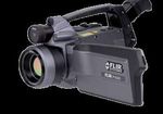
The FLIR P620 thermal imaging
FLIR thermal imaging camera used to inspect health issues in camera is accurate and completely
safe for use with zoo animals.
animals at Artis Royal Zoo Amsterdam
Zoo keepers all over the world are faced with the problem of determining whether an
animal should be treated or anesthetized or not. Many zoo animals are very sensitive to
emotional stress and to the physical side-effects of anesthetics, so in some cases veterinary
treatment can do more harm than good. To help them with this the keepers of the Artis
Royal Zoo in Amsterdam, the Netherlands, can call upon the help of veterinarian assistant
Daphne Valk and her FLIR P620 thermal imaging camera.
“It can provide important information that can help the vet to determine whether
treatment is necessary”, explains Valk. “This might not sound spectacular, but for keepers
The elephants at Artis are trained to show their feet to their
and veterinarians this is extremely important. You want to avoid treatment and handling keepers for a regular thermal imaging inspection.
unless it is really necessary, but in some situations waiting can be fatal. A thermal imaging
camera can help determine whether treatment is necessary. And the great thing about
thermal imaging technology is that it a non-invasive method, so the stress level of the
animal is kept to a minimum.”
The thermal imaging camera used by Valk is can use the thermal images to find
the FLIR P620. This highly sensitive thermal a multitude of health issues in animals
imaging camera contains an uncooled by looking for anomalies in the thermal
microbolometer detector that produces patterns. Generally speaking inflammations
thermal images at a resolution of 640 x 480 and injuries will show up as warmer and
pixels and a thermal sensitivity of 40 mK. scar tissue and nerve damage will be visible
Experienced veterinary thermographers as colder areas in the thermal image.
It is normal for the cuticles to show up as warm, but the area
around it is warm as well, indicating a possible infection.
www.flir.comapplication story
bull showed unusual
behavior, acting
grumpy and eating
less, so the keepers
suspected an infection.
But how were they to
find out? I was called
in with the thermal
imaging camera and
the keepers had the
The highly accurate FLIR P620 is a great for veterinary elephant open his
inspections according to veterinarian assistant Daphne Valk. mouth and move aside
the trunk. We train
FLIR P620 has proven its worth
them to do that on
In the short time the FLIR P620 thermal
command for exactly
imaging camera has been in operation it
this type of situation.
has certainly proven its worth, according
The thermal image
to Valk. “We started using it on elephants,
showed no signs of
like I had seen it demonstrated in the
an infection. So in this
States, taking thermal images of elephant
case the use of thermal
feet to see if they had infections or other
imaging prevented the
abnormalities.”
unnecessary use of
antibiotics.”
But thermal imaging cameras can do more
than just looking at elephant feet, as one
But the thermal
case with an elephant tusk showed. “One
imaging camera is used
of our elephants had an accident with one
more and more with
of his tusks, which caused part of the tusk
other animals as well.
to break off. The tusk stub featured a tare
“In one case a Scimitar
that continued to deep inside the jaw. The
Oryx had a swollen jaw. This Scimitar Oryx has a swollen jaw. The thermal image shows that the swollen area
The keepers noticed is not significantly warmer, so no drugs were administered. Less than 2 days later the
swelling was gone.
the swelling but they
were unsure about the
cause. It could be that it was just some looking at it with the thermal imaging
cud – food regurgitated by a ruminant to camera and urged the veterinarian to cut
chew on it a second time – that was stuck even deeper. I felt a little worried, for I didn’t
there, but it could also be an inflammation. want to cause unnecessary damage to the
The thermal image showed no signs of zebra, but the thermal image turned out
inflammation, since the area showed up as to be correct. In the end we found the
having a regular temperature. Secure in the infection and managed to remove it. If we
knowledge that it was probably just cud wouldn’t have had the thermal imaging
we took no further action and over time feedback we would never have dared to go
the swelling subsided. In this case the use in that deep.”
of the thermal imaging camera saved the
Scimitar Oryx from unnecessary sedation.” Thermal imaging succeeds where other
methods fail
Providing crucial feedback In some cases the thermal imaging camera
In another case the thermal imaging provides solutions when all other diagnostic
camera provided crucial feedback for the methods failed. “We had a camel that was
operating veterinarian. “We had a zebra limping and we just couldn’t find out what
with a limp and an inspection with the was causing it. Physical inspections and
Thermal imaging can be used to accurately track the recu- thermal imaging camera sowed it had an X-rays didn’t bring us any closer to finding
peration process of animals recovering from an inflamma- infected hoof. When the vet started to the cause, so I took out my thermal imaging
tion or injury. This thermal image shows an elephant foot
after treatment. The heat pattern is returning to normal,
remove the infected tissue I continued camera and I immediately spotted a warm
indicating that the treatment was successful.area in one of the toes. It turned out to be
an infection, which can be easily treated
with antibiotics. But you only use antibiotics
on ruminants if you are absolutely sure you
need to, because it can seriously damage
the good bacteria in their digestive system.”
The news of the thermal imaging camera
even spread to other zoos as well. “In one
case I was called to investigate a toucan in
another zoo. This toucan had a swelling next This thermal image shows a zebra hoof. The warm fluid run-
ning down the hoof is pus discharge caused by an infection
to its eye, but toucans are very sensitive to that was cut open by a veterinarian. The thermal imaging
stress so investigating it in a conventional camera provided crucial feedback during the operation.
manner might do more harm than good.
iguanas are very sensitive creatures you
To make sure that the animal is not put
don’t just administer antibiotics if it’s not
under any unnecessary stress I was called
absolutely necessary. But using thermal
in to investigate. The thermal image clearly
imaging proved to be a challenge, since
showed that the swelling was warm,
the iguana is kept in a warm room and
so I concluded that it was probably an
its body temperature is almost the same
infection and that further treatment was
as the room temperature. So I placed a
needed. The animal was anesthetized and
cold stone underneath the iguana’s claw,
further investigation showed that the initial
cooling down the claw slightly. The swollen the past we’ve had situations when we
assessment was correct.”
toe turned out to be warmer than the rest suspected there had been a fight but we had
of the claw, so we decided to administer no idea of how serious the injuries were, for
Thermography on cold blooded animals:
the antibiotics.” it is difficult to see if an animal is hurt if the
it is possible
wounds are hidden by the fur. Luckily we
An often made assumption is that veterinary
Another useful application for the thermal have not had such a situation since we’ve
thermography with cold-blooded animals
imaging camera in the Artis zoo Valk has acquired the FLIR P620 thermal imaging
is not feasible, but Valk had a case that
discovered is to determine whether animals camera, so I have not tested this theory, but
proved this assumption to be wrong. “We
have fought during the night. “This one I suspect that differences between wounds
had a green iguana with a swollen toe and
time the keepers noticed strange behavior and the surrounding skin and fur in both
we suspected it to be inflamed. But since
with our sea lions and they wondered
whether they might have fought during
the night, when no keepers were present
to notice it. So we had them leave the
basin in the morning, before the sun
came up, to prevent influence from direct
sunlight. I recorded some thermal images
of each animal and we found that although
there were no visible injuries there were
many warmer areas visible in the thermal
images. The fighting hadn’t resulted in any
visible wounds, but the invisible bruises
were made visible by the thermal imaging
camera. This was useful information for the
keepers who could take into account in the
way they treated the animals.”
Finding wounds through the fur
Valk suspects that this application might
turn out to be very useful if land dwelling
animals with fur, such as the chimpanzees,
The top image shows the toe of a camel with a hot area will start fighting. “It is not always easy to This thermal image shows the claw of a green iguana with
indicating an infected toe. The lower thermal image shows prevent fighting between zoo animals. In a suspicious swelling. The swollen toe is much warmer than
the feet of that camel after two weeks of treatment with the other toes, indicating an infection.
antibiotics.application story
temperature and in emissivity will make the of the swelling. An important characteristic
wounds stand out clearly on the thermal of this reaction to the antigen is also
image.” an increase in temperature, similar to an
inflammation, so I hope to be able to use
Another new application Valk is planning the thermal imaging camera to determine
to test as soon as the situation arises from a distance whether there is a swelling
is confirming the obligatory tuberculosis or not, so we can avoid putting the animals
check. “If an animal is moved to another through unnecessary stress and sedation.”
zoo we have to make sure that the animal
is not infected with tuberculosis, so each FLIR thermal imaging camera: a good
animal has to undergo a tuberculin skin investment Thermal imaging helps to determine the skin temperature
test. To do that we shave off a part of the All in all Valk is very pleased with the of this hippopotamus with a very serious rash from a safe
distance.
fur to reveal the skin, we then inject them decision to acquire the FLIR P620 thermal
with tuberculin antigens and after three imaging camera. “It really has been a good
days we check whether the antigens have investment. It has helped to find health individual animal. Sometimes an animal is
caused a swelling. If there is a swelling issues in while preventing unnecessary warm or cold in certain places that might
of a certain size in the location where surgery and minimizing stress. But I seem to indicate a health issue, while it
the antigens were administered then the would like to stress that with veterinary is just part of the normal pattern for that
animal is suspected of having tuberculosis thermography it is of vital importance to animal. I have been working with some our
and it cannot be transported.” keep critically assessing the validity of the animals for years and I still don’t have the
results. Not every anomaly in the thermal feeling that I know exactly what the normal
But this process can be very difficult with pattern is an infection or a bruise. You thermal patterns are for our individual
some animals. “Many zoo animals cannot have to understand the circumstances that animals.”
be kept separated for three days in a row, influence the thermal camera readings,
so we have to let them join the rest of like raindrops, mud, sunshine, sweat, thick Certification
the group or herd. But that makes it very fur or a breeze coming in from an opened Valk therefore hopes that in the near
difficult to determine whether there is window. All of these external influences future a certification for veterinary
enough cause to capture an sedate the – also called artifacts –can create thermal thermographers will be developed and
animal again in order to determine the size anomalies that have nothing to do with enforced by the European Union. “Currently
the health of the animal.” anyone can pick up a camera and call
himself a thermography consultant, but it
Valk therefore tries to eliminate these takes more than a thermal imaging camera
artifacts as much as possible. “We only alone.”
inspect the animals within their enclosure
in the morning before they head outside FLIR cooperates with the Infrared Training
with all the doors and windows shut tight, Center (ITC) to provide training courses and
to minimize the effect of sunlight and air reliable thermography certificates in three
convection on the thermal pattern. If the different levels. “I applaud the efforts taken
animal is dirty we use a hose to clean the by FLIR to promote reliable training and
animal and then wait for a couple of hours certification and I hope that in the future a
until the animal is dry. And we also use certification system for veterinarians will be
standardized viewing angles to enable given an official status.”
comparison between results from different
inspections.”
Individual differences
But even when these precautions are
taken, Valk is very careful to draw any
conclusions. “Not only do you have to For more information about thermal imaging
know about the physics of thermography, cameras or about this application,
please contact:
you also have to know a lot about the
animal you are dealing with. And I don’t FLIR Commercial Systems B.V.
The thermal image of this Tapir shows that the abscess
below the jaw is warmer than the surrounding tissue, which mean just the anatomy of that species; Charles Petitweg 21
warrants further investigation. I’m talking about the particulars of that 4847 NW Breda - Netherlands
Phone : +31 (0) 765 79 41 94
Fax : +31 (0) 765 79 41 99
T820171 {EN_uk}_A
e-mail : flir@flir.com
www.flir.comYou can also read


