Application of Intelligent Computer-Assisted Taylor 3D External Fixation in the Treatment of Tibiofibular Fracture: Retrospective Case Study ...
←
→
Page content transcription
If your browser does not render page correctly, please read the page content below
JMIR MEDICAL INFORMATICS                                                                                                              Sheng et al

     Original Paper

     Application of Intelligent Computer-Assisted Taylor 3D External
     Fixation in the Treatment of Tibiofibular Fracture: Retrospective
     Case Study

     Hongfeng Sheng1, MM; Weixing Xu1, MD; Bin Xu1, MM; Hongpu Song1, MM; Di Lu1, MM; Weiguo Ding1, MM;
     Henry Mildredl2, PhD
     1
      Department of Orthopaedics, Tongde Hospital of Zhejiang Province, Hangzhou, China
     2
      Federal Institute for Drugs and Medical Devices, Medical Devices Division, Bonn, Germany

     Corresponding Author:
     Hongpu Song, MM
     Department of Orthopaedics
     Tongde Hospital of Zhejiang Province
     234 Gu-cui Road
     Hangzhou, 310012
     China
     Phone: 86 0571 89972000
     Email: hongpusongzj@yeah.net

     Abstract
     Background: With the development of modern society, severe and complex tibial fractures caused by high-energy injuries such
     as traffic accidents have gradually increased. At present, the commonly used methods for the treatment of tibial fractures include
     plate fixation, intramedullary nail fixation, and external fixation. Most of these fractures are open wounds with severe soft tissue
     injury and wound contamination, and some involve bone defects, which makes internal fixation treatment difficult.
     Objective: This study aims to explore the use of intelligent computer-assisted Taylor 3D external fixation for the treatment of
     tibiofibular fractures.
     Methods: In total, 70 patients were included and divided into the Taylor 3D external fixation (TSF) group (28 patients with
     severe tibial fractures treated with TSF) and the internal fixation group (42 patients with complicated tibiofibular fractures treated
     by internal fixation). After the treatment, the follow-up evaluation of TSF for the treatment of tibiofibular fractures noted the
     incidence of complications, as well as the efficacy and occurrence of internal fixation for the treatment of tibial fractures in our
     hospital.
     Results: The results showed that TSF was superior to orthopedics in the treatment of tibiofibular fractures in terms of efficacy
     and complications.
     Conclusions: TSF for the treatment of tibiofibular fractures is more effective than internal fixation and the incidence of
     complications is low. This is a new technology for the treatment of tibiofibular fractures that is worthy of clinical promotion.

     (JMIR Med Inform 2021;9(5):e21455) doi: 10.2196/21455

     KEYWORDS
     intelligent computer-assisted instruction; Taylor three-dimensional external fixation; tibial fracture; internal fixation; external
     fixation

                                                                             Most of these fractures are open wounds with severe soft tissue
     Introduction                                                            injury and wound contamination, and some involve bone defects,
     With the development of modern society, severe and complex              which makes internal fixation treatment difficult. Potential
     tibial fractures caused by high-energy injuries such as traffic         complications include postoperative wound infection, chronic
     accidents have gradually increased. At present, the commonly            osteomyelitis, delayed fracture healing, and fracture nonunion.
     used methods for the treatment of tibial fractures include plate        The incidence of malunion healing is high, often resulting in
     fixation, intramedullary nail fixation, and external fixation.          treatment failure [1]. External fixation technology is a good

     https://medinform.jmir.org/2021/5/e21455                                                         JMIR Med Inform 2021 | vol. 9 | iss. 5 | e21455 | p. 1
                                                                                                                (page number not for citation purposes)
XSL• FO
RenderX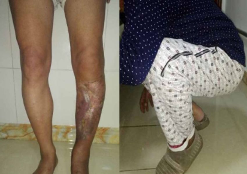
JMIR MEDICAL INFORMATICS                                                                                                                Sheng et al

     method for the treatment of such fractures. External fixation           of transverse shape fracture line, 6 cases of oblique shape, 3
     can reduce the damage to soft tissue, and reduce the risk of            cases of spiral shape, 5 cases of comminuted fracture, and 4
     postoperative wound infection, osteomyelitis, delayed fracture          cases of multiple fractures. According to the location of the
     healing, and fracture nonunion. While complications occur,              fracture, there were 7 cases in the proximal one-third of the
     fracture fixation can be performed in the early stages after an         bone, 5 cases in the middle one-third, 11 cases in the middle
     injury, which provides a better prognosis for soft tissue repair,       and distal junctions, and 5 cases in the distal one-third.
     limb care, and early functional exercise [2]. However, in the           Compartment syndrome occurred and 4 cases underwent open
     past, external fixation stents for the treatment of complicated         decompression.
     tibiofibular fractures have had poor stability for fracture fixation.
                                                                             The internal fixation group included 42 patients with severe
     They can only be used as a temporary fixation method. Most
                                                                             complicated tibiofibular fractures treated by internal fixation
     of the latter require secondary surgery to replace internal
                                                                             from January 2011 to March 2017, including 33 males and 9
     fixation, which makes the treatment period prolonged and
                                                                             females aged 17 to 70 years (mean 40.3 years old). There were
     significantly increases the cost of fracture treatment.
                                                                             26 cases of traffic injuries, 10 cases of heavy bruises, 6 cases
     Additionally, the fracture healing time is prolonged.
                                                                             of high fall injuries, 22 cases of open fractures (according to
     Based on the Ilizarov circular fixator (ICF), a previous study          Gustilo classification: 12 cases of type II, 10 cases of type III),
     [3] applied the Stewart platform and Charles theory to the field        20 cases of closed fractures (according to Tscherne
     of orthopedics, and combined these with computer software to            classification: 13 cases of grade 2, 7 cases of grade 3).
     invent the Taylor 3D space frame. Taylor 3D external fixation           According to fracture line classification, there were 16 cases of
     (TSF) is a good complement to the deficiencies of the ICF for           transverse fracture, 9 cases of oblique, 6 cases of spiral, 7 cases
     multidimensional planar fractures and deformity correction.             of comminuted, and 4 cases of multiple fractures. According
     TSF has the following advantages in the treatment of tibial             to the location of fracture, there were 11 cases in the proximal
     fractures: TSF is quick and easy to learn; accurate closed              one-third, 9 cases in the middle one-third, 14 cases in the middle
     reduction of fractures can be achieved with computer software           and distal junctions, and 8 cases in the distal one-third.
     assistance during or after surgery; TSF is a better option for          Compartment syndrome was treated with incision
     ensuring fixation stability of fractures; and the external fixator      decompression in 6 cases. There were 18 cases treated with
     can be used as a long-term fixation method. The stent is                steel plates and 24 cases treated with intramedullary nails.
     maintained during the entire process of fracture healing. The
     needle is fixed during the installation process. It does not cause
                                                                             Inclusion Criteria and Exclusion Criteria
     secondary damage to the local soft tissue. The risk of                  Inclusion criteria were the following: (1) high-intensity injury
     postoperative infection is low, and the rate of fracture nonunion       resulting in severe soft tissue injury or open severe complex
     is low. Fracture surgery can be performed soon after the injury         tibiofibular fracture, Gustilo type II-III or Tscherne grade 2-3
     to achieve early functional exercise. Postoperative bone defects        and (2) follow-up time ≥6 months. Exclusion criteria were the
     can be repaired by adjusting the external frame. There are many         following: (1) Simple low-energy tibiofibular fracture, Gustilo
     reports on the use of TSF in the treatment of limb deformities,         type I or Tscherne grade 1. (2) Total tibial plateau, pilon
     although there is little literature on the use of TSF in the            fracture, and other cumulative articular surface fracture patients.
     treatment of tibial fractures.                                          (3) Follow-up time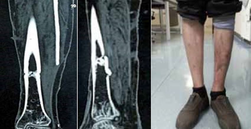
JMIR MEDICAL INFORMATICS                                                                                                                Sheng et al

     The Taylor external fixator installation procedure is the               again until ideal. Postoperative nail dressing and regular dressing
     following: after the affected limb is sterilized, the C-arm             care were provided. Knee and ankle joint functional exercise
     machine monitors the axial traction of the distal end of the            began the first day after surgery, with gradual weight-bearing
     affected limb, roughly resets the fracture end displacement             2 to 3 weeks after surgery, and a monthly review following
     (shortening, angulation, and rotational displacement), and              filming; the external fixator was dismantled after the fracture
     initially restores the length of the tibia. Cantering on the fracture   healed.
     line, a TSF ring is inserted into the distal and proximal fractures
                                                                             When evaluating fracture reduction and fixation, the internal
     (if the fracture segment is ≥3 cm, ensure that each fracture
                                                                             fixation group had an improved positive lateral radiograph.
     segment is fitted with one ring, because the TSF ring and the
                                                                             After the operation, the affected limb promoted blood return.
     tibia are placed ≥2 cm from the fracture line). When the
                                                                             On the second day, active and passive functional exercises of
     anatomical axis is vertical, at least 2 full needles or olive needles
                                                                             the knee and ankle joints were increased. Regular incision
     are inserted into the safety channel of each level of the tibia,
                                                                             dressing was provided (if the wound dressing had exudation,
     and connected with the TSF ring. If the stability is poor, a half
                                                                             the dressing was changed), and the wound was not bandaged
     needle can be implanted to increase stability and, according to
                                                                             after exudation. The incision healing was complete 10 to 14
     the adjacent TSF ring, install 6 adjustable connecting rods.
                                                                             days after surgery. If the wound became infected, the secretion
     Under the perspective of the C-arm machine, the fracture
                                                                             was assessed in the laboratory for bacteria, and an antibiotic
     displacement parameters were preliminarily calculated and the
                                                                             was intravenously provided according to the result; the drug
     fracture was repaired by adjusting the 6 connecting rods. If the
                                                                             was changed frequently, and if necessary, vacuum suction
     fracture is difficult to reset and the local soft tissue can be
                                                                             treatment was used. The patient avoided weight-bearing
     finitely cut at the fracture end, a comminuted fracture can be
                                                                             activities for 6 weeks after surgery, and then gradually added
     transformed into a relatively simple fracture by temporary
                                                                             weight with the help of progressive ablation. In this study, the
     fixation with Kirschner wire or by using plate fixation to
                                                                             x-ray films were reviewed in January, February, March, and
     maximize the recovery of the bone shaft. The tubular
                                                                             June. One year later, fracture healing was judged according to
     morphology was further reset after surgery with computer
                                                                             the films. After the fracture healed, the internal fixation was
     software.
                                                                             removed.
     Internal Fixation Group
                                                                             Observation Indicators
     The anesthesia method and the surgical position of the internal
                                                                             The main follow-up details recorded were the patient's surgical
     fixation group are the same as in the TSF group. Open fracture
                                                                             preparation time, operation time, fracture healing time, total
     emergency (6 to 8 hours) wound debridement is performed,
                                                                             weight-bearing time, length of hospital stays and expenses,
     followed by temporary external fixation of the fractured
                                                                             postoperative complications, as well as other indicators.
     unilateral outer frame; if there is a small wound surface and
     contamination is not serious, the wound can be closed in one            Statistical Methods
     stage; if the wound is large and contamination is heavy, VSD            We used SPSS Statistics software (Version 21.0; SPSS Inc) for
     negative pressure is used and a second-stage skin graft or flap         the following statistical analysis: the categorical variable data
     transfer is used to close the wound. After the soft tissue recovers,    was analyzed by chi-square test, the countable data was analyzed
     open reduction and internal fixation are performed. After the           by t test, and the test standard was α=.05.
     swelling of the closed fracture subsides (indicated by the
     appearance of dermatoglyphics) and the local soft tissue                Results
     recovers, open reduction and internal fixation are performed
     once tension blisters have subsided. The open reduction and             Clinical Follow-up Results of the TSF Treatment
     internal fixation process is the following: the surgical approach       Group
     is determined according to the soft tissue condition and the type
     of fracture. In the process of fracture reduction, as much as           All 28 cases were followed up for an average of 23.5 months
     possible is done to protect and reduce soft tissue damage,              (range 10-48 months); the average preoperative preparation
     including avoiding using long incisions to pursue excessive             time was 3.5 days (range 0.5-8 days); the average operation
     anatomical reduction. For the reduction, it is required to fully        time was 112.3 minutes (range 90-131 minutes); and 4 cases of
     reduce the longitudinal, axial, and rotational displacement of          bone defects occurred. Bone grafting and internal fixation were
     the tibia.                                                              used to obtain healing. In 3 cases of delayed fracture healing,
                                                                             late adjustment of the external frame fracture resulted in good
     Postoperative Treatment                                                 healing. The fracture healing rate was 85.71%, with an average
     In the TSF group, the standard lateral radiograph was improved.         fracture healing time of 20.3 weeks (range 16-48 weeks). The
     The fracture displacement parameters were measured according            external fixation frame was worn for an average of 26 weeks
     to the x-ray film. The parameters were input into the TSF               (range 17-48 weeks). Overall, 4 cases of compartment syndrome
     computer software system, and 6 adjustable connecting rods              occurred, emergency decompression was given, the TSF external
     were used to make adjustments. After the fracture was reset,            fixation frame was installed after the wound was closed, and
     the film was reviewed again. If the reset was not good, the             the patient was discharged after adjustment and resetting. The
     fracture displacement parameter could be measured again and             average weight-bearing time was 90.5 days (range 65-180 days);
     imported into the computer software to adjust the parameters            the average number of days spent in hospital was 10.8 days
                                                                             (range 6-24 days); and the average hospitalization cost was 5.6
     https://medinform.jmir.org/2021/5/e21455                                                           JMIR Med Inform 2021 | vol. 9 | iss. 5 | e21455 | p. 3
                                                                                                                  (page number not for citation purposes)
XSL• FO
RenderX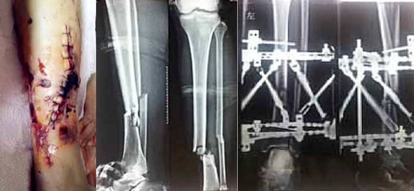
JMIR MEDICAL INFORMATICS                                                                                                                  Sheng et al

     million (range 3.8-97 million). Postoperative wound infection             was provided and internal fixation was performed after the
     occurred in 2 cases, and 13 cases occurred in the needle.                 closure of the wound. All healed well and the average time to
     Infections were cured after wound dressing and oral antibiotics.          weight-bearing was 110.3 days (range 60-185 days). The
     No cases of chronic osteomyelitis occurred. In 1 case, there was          average hospital stay was 18.2 days (range 14-33 days) and
     another fracture after the removal of the external frame, and the         hospitalization costs averaged 6.2 million (range 5.3-11.2
     fracture was healed after internal fixation. At the last follow-up,       million). Postoperative wound infection occurred in 20 cases;
     all patients could step onto the ground and 21 patients could             infections were cured after dressing change, intravenous
     participate in daily housework. There were no patients with               antibiotic, and/or VSD negative pressure treatment. In total, 5
     joint stiffness.                                                          cases of chronic osteomyelitis occurred. One patient’s bone
                                                                               fractured after internal fixation. There was no joint stiffness
     Clinical Follow-up Results of the Internal Fixation                       among patients.
     Group
     A total of 42 patients were followed up for an average of 19.5            Comparison of the Efficacy of TSF and Internal
     months (range 7-34 months). This included 18 patients in the              Fixation in the Treatment of Severe Tibiofibular
     plate fixation group and 24 patients in the intramedullary nail           Fractures
     fixation group. The average preoperative preparation time was             The fracture healing rate was 85.71% (24/28) in the TSF
     10.5 days (range 6-24 days) and the average operation time was            treatment group and 92.86% (39/42) in the internal fixation
     152.4 minutes (range 120-185 minutes). Overall, 3 cases of                group (Table 1). In the TSF treatment group, the time spent on
     nonunion occurred, which healed after internal bone grafting.             preoperative preparation time, operation time, fracture healing
     In total, 4 cases had delayed fracture healing. The fracture              time, total time to full weight-bearing, and hospitalization stays
     healing rate was 92.86% and the average fracture healing time             were shorter than those in the internal fixation group, and the
     was 23.8 weeks (range 17-54 weeks). A total of 6 cases of                 hospitalization cost was lower; the difference was statistically
     compartment syndrome occurred; acute incision decompression               significant (P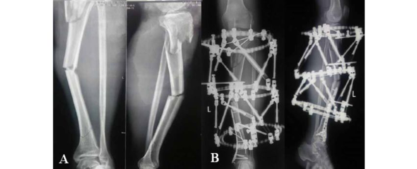
JMIR MEDICAL INFORMATICS Sheng et al
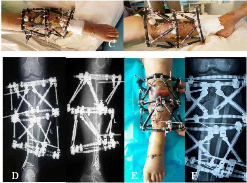
Typical Cases Figures 1 and 2 showed the open fracture of left tibia and
postoperative recovery of patient A.
Figure 1. Patient A: car accident leading to open fracture of the left tibia.
Figure 2. Postoperative recovery of patient A.
performed to achieve a good reduction of the fracture end.
Case-Related Information Finally, 10 months later, a review of computed tomography
Clinicians at an external hospital completed the wound scans showed good fracture healing; the affected limb had
debridement and closure. Later, in our hospital, the soft tissue normal function 11 months after the removal of the outer frame.
injury was found to be considerable. The preoperative x-ray
showed a fracture. The third day of admission, TSF external Figure 3 showed the comminuted fracture of the left tibia of
fixation was performed. Software-assisted adjustments were patient B with severe soft tissue injury. Figures 4 and 5 showed
the patient's TSF treatment and postoperative recovery.
https://medinform.jmir.org/2021/5/e21455 JMIR Med Inform 2021 | vol. 9 | iss. 5 | e21455 | p. 5
(page number not for citation purposes)
XSL• FO
RenderXJMIR MEDICAL INFORMATICS Sheng et al
Figure 3. Patient B had a car accident–caused comminuted fracture of the left tibia, combined with severe soft tissue injury.
Figure 4. Patient with TSF treatment. TSF: Taylor 3D external fixation.
https://medinform.jmir.org/2021/5/e21455 JMIR Med Inform 2021 | vol. 9 | iss. 5 | e21455 | p. 6
(page number not for citation purposes)
XSL• FO
RenderXJMIR MEDICAL INFORMATICS Sheng et al
Figure 5. Postoperative recovery of patients.
incidence of postoperative complications. High-energy damage
Patient-Related Information is mainly seen in traffic accidents, falls from high places, and
The patient was 48 years old. A car accident caused injuries of direct injuries by heavy objects. In contrast, low-energy injuries
the left tibia including severe soft tissue injury and three are more common in sports (about 80.1%) and regular falls. As
comminuted fractures (Tscherne grade 3). The lateral position countries develop, there is a corresponding increase in the
of the left tibia showed three fractures of the proximal, middle, incidence of traffic accidents, leading to an increase in
and distal bone, and the proximal and middle fractures were high-energy fractures. Since there are fewer subcutaneous tissues
clearly displaced. On the fourth day of admission, the proximal on the anterior aspect of the lower leg, these fractures are prone
and middle fractures were treated with TSF external fixation, to be open fractures, and account for 9.72% to 13.7% of open
the distal fracture was fixed with internal fixation, and a lateral fractures.
x-ray was performed. After the operation, soft tissue damage
was severe and many tensional blood vessels could be seen; in An epidemiological survey of 523 cases of tibiofibular fractures
addition, the anterior tibial skin was black and necrotic. showed that 400 cases (76.5%) involved closed fractures and
Combined with the TSF computer software, the positive lateral 123 cases (23.5%) involved open fractures [4]. These were a
radiographs after fracture reduction were good. Finally, 11 result of traffic accident injuries (37.5%), falling (17.8%), sports
months after the operation, the skin was restored to a good (30.9%), and beatings or direct hits (4.5%). The majority of the
condition with soft tissue treatment such as skin grafting. The blood supply of the tibia is provided by the nourishing artery.
limb functioned well 20 months after surgery, and knee joint This artery enters the tibia from the upper one-third, and the
function was good. trophoblast descends into the skeletal cortex. In a fracture, most
of the arteries providing cortical nourishment are broken,
Discussion resulting in insufficient blood supply to the distal one-third of
the tibia, which slows down healing and is not conducive to the
Principal Findings patient’s recovery. There are many treatment methods for tibia
fractures, each with its own advantages and disadvantages. The
In trauma orthopedics, tibiofibular fractures are common,
choice is mainly based on the way the injury occurred, the
accounting for about 12% of total long bone fractures. The
fracture type, other injuries, and the patient’s condition. Gypsum
prognosis after fracture is affected by the energy level of the
or splint fixation is generally suitable for stable fractures from
injury. When the damage energy is higher, the probability of
a low-energy injury and those without obvious displacement.
an open fracture, the degree of fracture complications, and the
Due to the risk of calf compartment syndrome and venous
degree of soft tissue injury increase accordingly, increasing the
thrombosis, it is currently used for fractures of the tibia. If there
https://medinform.jmir.org/2021/5/e21455 JMIR Med Inform 2021 | vol. 9 | iss. 5 | e21455 | p. 7
(page number not for citation purposes)
XSL• FO
RenderXJMIR MEDICAL INFORMATICS Sheng et al
is a lower risk of calf compartment syndrome and venous fixation techniques, both a compression plate as well as steel
thrombosis, there is less indication that the fracture was caused plates and nail tails. The locking component between them is
by a high energy injury. used as an inner bracket. The LCP provides both angular and
axial stability to prevent the screws from slipping. In one study,
Open reduction and internal fixation treatment can achieve good
28 patients with fractures of the lower tibia were treated with
fracture reduction, which is beneficial for early functional
DCP and 20 patients were treated with LCP [7]. The fracture
training of the limb. The intramedullary nail is currently the
healing time was 16.2 months for DCP and 15.4 months for
preferred treatment for humerus shaft fractures. This technique
LCP. The LCP effect was better than the DCP result.
has many advantages and a long history in the treatment of tibial
fractures. It has a central fixed biomechanical advantage, In another study, 25 cases of tibiofibular fracture were treated
involves a minimally invasive operation away from the fracture with the minimally invasive percutaneous plate osteosynthesis
end, retains the hematoma at the fracture end, and involves less (MIPPO) technique, which is considered safe and effective [8].
soft tissue exfoliation, which is conducive to fracture healing. The most ideal fracture treatment should be as minimally
Therefore, it is widely used in clinical practice. However, any invasive as possible and avoid the use of implants such as steel
given treatment is not perfect, and the intramedullary nail still plates and intramedullary nails. The internal fixation treatment
has its limitations. In a previous study, 32 cases of proximal is beneficial for the anatomical reduction of the fracture, but at
humeral fractures were treated with intramedullary nails and the same time, this invasive operation increases the risk of
the malunion rate was 19%, indicating the treatment was not infection. Due to the development of internal fixation equipment
satisfactory [5]. Kumar et al [6] compared the biomechanical and the improvement of fracture fixation, the results of internal
characteristics of the treatment of tibiofibular fractures with fixation for the treatment of severe tibial fractures have been
steel plate, interlocking intramedullary nail, and external greatly improved. However, due to the high incidence of
fixation. The results from the intramedullary nail treatment are complications such as postoperative infection and osteomyelitis,
better than those of the other two techniques, but this treatment the combination of open and severe soft tissue injury, and the
is associated with malunion. treatment of multiple comminuted fractures and infected
tibiofibular fractures, this technique is challenging.
The orthopedic surgeon’s philosophy of fracture treatment has
gone from Association for the Study of Internal Fixation External fixation is a good solution to the abovementioned
(AO)-led anatomical reduction to strong internal fixation to the shortcomings of internal fixation in the treatment of severe tibial
promotion of biological fixation. The four principles of treatment fractures. The external fixator is simple to install and the
of fractures as proposed by the AO concept are as follows: (1) technique is easy to learn. It causes minor secondary damage
anatomical reduction, (2) compression fixations at the fracture to soft tissue and can be used for early fixation of open
end, (3) protection of blood supply, and (4) early functional tibiofibular fractures or fractures with severe soft tissue injury.
exercise. Early dynamic compression plate (DCP) treatment It is beneficial for the early care of the affected limb, such as
increased friction at the end of the fracture through the functional exercise, adjacent joint function, and exercise. This
compression of the fracture end, and achieved first-stage healing study compared the postoperative complications of TSF external
of the fracture. The DCP is in close contact with the bone, and fixation and internal fixation in the treatment of severe complex
the fracture is stabilized by increasing friction, which destroys tibiofibular fractures (Table 2). The incidence of postoperative
the blood supply at the fracture end. Influenced by the AO wound infection and osteomyelitis was significantly lower in
concept, many orthopedic surgeons remove large amounts of TSF than in the internal fixation group (PJMIR MEDICAL INFORMATICS Sheng et al
of fractures aided by a computer. TSF can be the final method length makes postoperative resetting impossible. Overall, our
of fracture fixation due to good fracture stability. It enables the experience shows that TSF is effective in treating patients with
precise treatment of tibial fractures caused by complex severe tibial fractures caused by high-energy injuries. Compared
high-energy injuries. This study compared the preoperative with the internal fixation method, the incidence of postoperative
preparation time, operation time, fracture healing time, total wound infection and osteomyelitis was reduced. TSF can enable
time to weight-bearing, hospitalization days, and other outcomes early fracture fixation surgery and early functional exercise,
of TSF and internal fixation in the treatment of tibial fractures. shorten hospitalization time, and reduce treatment costs.
The operation and healing times of the TSF group were shorter
than in the internal fixation group [11].
Conclusions
TSF has a low complication rate, with the advantages of fracture
The authors’ experience in the treatment of complex tibial closure and accurate reduction, providing a new treatment
fractures with TSF includes the following insights: postoperative method for complex tibiofibular fractures. Compared with the
computer-assisted adjustment of external frame fractures internal fixation method, it has a shorter preoperative preparation
requires the addition of two parallel links on both sides of the time, operation time, fracture healing time, and total time to
outer ring to increase stability; it is important to measure the weight-bearing, as well as shorter hospital stays and lower
fracture displacement parameters; the distance between the hospitalization costs for the treatment of severe complex tibial
proximal and distal rings in the TSF installation process needs fractures. There is a lower chance of complications such as
to be prejudged to avoid the longest distance between the two postoperative infection and osteomyelitis, and there is no
rings being greater than the longest model connecting rod, or significant difference in the incidence of nonunion, delayed
shorter than the minimum model connecting rod, as the short healing, and refracture.
Conflicts of Interest
None declared.
References
1. Laux CJ, Grubhofer F, Werner CML, Simmen H, Osterhoff G. Current concepts in locking plate fixation of proximal
humerus fractures. J Orthop Surg Res 2017 Sep 25;12(1):137 [FREE Full text] [doi: 10.1186/s13018-017-0639-3] [Medline:
28946902]
2. Craig E. Reverse Shoulder Arthroplasty in the Treatment of Proximal Humeral Fractures. Techniques in Shoulder & Elbow
Surgery 2015;16(4):99-102. [doi: 10.1097/bte.0000000000000062]
3. Ribeiro FR, Takesian FH, Bezerra LEP, Filho RB, Júnior ACT, da Costa MP. Impacted valgus fractures of the proximal
humerus. Revista Brasileira de Ortopedia (English Edition) 2016 Mar;51(2):127-131. [doi: 10.1016/j.rboe.2016.01.004]
4. Oura K, Kunihiro O, Okada K, Tanaka H, Murase T. Corrective osteotomy assisted by computer simulation for a malunited
intra-articular fracture of the distal humerus: two case reports. Arch Orthop Trauma Surg 2016 Aug 17;136(11):1499-1505.
[doi: 10.1007/s00402-016-2555-0]
5. Lowry KJ, Hamson KR, Bear L, Peng YB, Calaluce R, Evans ML, et al. Polycaprolactone/glass bioabsorbable implant in
a rabbit humerus fracture model. J Biomed Mater Res 1997 Sep 15;36(4):536-541. [doi:
10.1002/(sici)1097-4636(19970915)36:43.0.co;2-8]
6. Kumar S, Singh S, Kumar D, Kumar N, Verma R. Intercondylar humerus fracture- parallel plating and its results. J Clin
Diagn Res 2015 Jan;9(1):RC01-RC04 [FREE Full text] [doi: 10.7860/JCDR/2014/12137.5479] [Medline: 25738046]
7. Silverstein MP, Yirenkyi K, Haidukewych G, Koval KJ. Analysis of Failure with the Use of Locked Plates for Stabilization
of Proximal Humerus Fractures. Bull Hosp Jt Dis 2015 Jul;73(3):185-189 [FREE Full text] [Medline: 26535597]
8. Cruickshank D, Lefaivre KA, Johal H, MacIntyre NJ, Sprague SA, Scott T, et al. A scoping review of biomechanical testing
for proximal humerus fracture implants. BMC Musculoskelet Disord 2015 Jul 30;16(1). [doi: 10.1186/s12891-015-0627-x]
9. Herrera-Pérez M, Boluda-Mengod J, Muñoz-Ortus R, Gutiérrez-Morales MJ, Pais-Brito J. Continuous pain and swelling
after humerus fracture in an 86-years-old woman. Acta Ortop Mex 2017;31(1):30-34 [FREE Full text] [Medline: 28741325]
10. Sabesan VJ, Lombardo D, Petersen-Fitts G, Weisman M, Ramthun K, Whaley J. National trends in proximal humerus
fracture treatment patterns. Aging Clin Exp Res 2017 Jan 25;29(6):1277-1283. [doi: 10.1007/s40520-016-0695-2]
11. Manoli A, Capriccioso C, Konda S, Egol K. Total shoulder arthroplasty for proximal humerus fracture is associated with
increased hospital charges despite a shorter length of stay. Orthopaedics & Traumatology: Surgery & Research 2016
Feb;102(1):19-24. [doi: 10.1016/j.otsr.2015.11.003]
Abbreviations
AO: Association for the Study of Internal Fixation
BO: biological fixation
DCP: dynamic compression plate
ICF: Ilizarov circular fixator
LCP: locking compression plate
https://medinform.jmir.org/2021/5/e21455 JMIR Med Inform 2021 | vol. 9 | iss. 5 | e21455 | p. 9
(page number not for citation purposes)
XSL• FO
RenderXJMIR MEDICAL INFORMATICS Sheng et al
MIPPO: minimally invasive percutaneous plate osteosynthesis
TSF: Taylor 3D external fixation
VSD: vacuum sealing drainage
Edited by Z Du; submitted 15.06.20; peer-reviewed by J Ding, Y Oannidis, D Thyros; comments to author 22.06.20; revised version
received 26.06.20; accepted 27.06.20; published 14.05.21
Please cite as:
Sheng H, Xu W, Xu B, Song H, Lu D, Ding W, Mildredl H
Application of Intelligent Computer-Assisted Taylor 3D External Fixation in the Treatment of Tibiofibular Fracture: Retrospective
Case Study
JMIR Med Inform 2021;9(5):e21455
URL: https://medinform.jmir.org/2021/5/e21455
doi: 10.2196/21455
PMID:
©Hongfeng Sheng, Weixing Xu, Bin Xu, Hongpu Song, Di Lu, Weiguo Ding, Henry Mildredl. Originally published in JMIR
Medical Informatics (https://medinform.jmir.org), 14.05.2021. This is an open-access article distributed under the terms of the
Creative Commons Attribution License (https://creativecommons.org/licenses/by/4.0/), which permits unrestricted use, distribution,
and reproduction in any medium, provided the original work, first published in JMIR Medical Informatics, is properly cited. The
complete bibliographic information, a link to the original publication on http://medinform.jmir.org/, as well as this copyright and
license information must be included.
https://medinform.jmir.org/2021/5/e21455 JMIR Med Inform 2021 | vol. 9 | iss. 5 | e21455 | p. 10
(page number not for citation purposes)
XSL• FO
RenderXYou can also read