NEOPLASMS: INTRACRANIAL - GammaTile Therapy
←
→
Page content transcription
If your browser does not render page correctly, please read the page content below
CL INICAL WHITE PAPER | BY DAVID BR ACHMAN, MD

INTRACRANIAL
NEOPLASMS:
THE EVOLUTION OF R ADIOTHER APIES

CURRENT STANDARD OF CARE FOR BRAIN TUMORS
The current initial standard of care for aggressive brain tumors—whether they are primary brain tumors
(ie, tumors that originate in the brain), or metastatic tumors from cancers that started outside of the brain—is
resection. After surgery, a follow-up treatment, also referred to as adjuvant treatment, is often recommended
to help eliminate any residual traces of the tumor.1 In the case of brain tumors, more often than not, adjuvant
therapy involves using radiation, in part because few chemotherapy
agents reliably cross the blood brain barrier.1
   Adjuvant radiation is used either alone or in combination with
                                                                              The goal of EBRT, and all
chemotherapy, and the most common method of radiation treatment
is external beam radiation therapy (EBRT).1,2 For EBRT, a large machine       radiation therapy, is to
generates radiation beams and focuses them inward to travel through           deliver the highest-possible
the skin, then through the skull, and finally, into the brain.2 Radiation
therapy including EBRT works by creating double-strand breaks in              safe dose of radiation to
cellular DNA, which interferes with the reproductive integrity of the         achieve maximum
cells.3 Radiation primarily impacts cells that are rapidly undergoing
                                                                              efficacy while limiting
cellular division, which is why it is useful for arresting aggressive tumor
development. However, it can also stop the normal cellular replication        radiation exposure to the
that takes place as part of the body’s healing and repair process after       normal tissues.
surgery. For this reason, external beam radiation treatments are often
delayed for 2 to 3 weeks or longer after a surgery.

EBRT FOR BRAIN TUMORS
As noted above, postsurgical wound healing must occur prior to the initiation of EBRT, leaving a large
window of time for unchecked tumor cell replication.4,5 Additionally, EBRT regimens for primary brain
tumors (ie, fractionated EBRT) are often time and resource intensive (typically requiring daily visits from
Monday through Friday for 4 to 6 weeks), which poses a significant burden on these vulnerable patients and
their caregivers.3 With stereotactic radiosurgery (SRS), a type of EBRT, it is sometimes possible to truncate
the treatment period to a total of 1 to 5 treatments. Involving multiple focused radiation beams, SRS is

                                                             THE EVOLUTION OF RADIOTHERAPIES                |   1
used for both metastasis and for relatively small primary brain tumors with well-defined borders.2 As with
standard fractionated EBRT postsurgery, these treatments must also be delayed for a few weeks to allow
for postsurgery wound healing. For primary brain tumors (gliomas, meningiomas, and similar tumors),
adjuvant fractionated EBRT is administered to a limited region of brain; typically, the tumor bed plus a zone
around the bed to encompass possible tumor spread (FIGURE 1). For the treatment of a single or a limited
number of brain metastases (4 or fewer), SRS delivered in 1 to 5 treatments is often used.3 When more than
4 brain metastases are present, whole-brain radiation therapy (WBRT), given daily over 2 to 4 weeks, is often
recommended.1
KEY TAKEAWAYS
• The standard of care for aggressive brain tumors is resection.1
• Adjuvant therapy (chemotherapy or radiation) is recommended
when surgery is unable to remove all traces of a tumor.1,3
• The most common method of adjuvant radiation treatment is
fractionated EBRT.1,3
• Radiation travels from outside of the body through the skull
and into the brain, exposing healthy tissue.
• EBRT typically involves a 2- to 3-week treatment delay to
allow for postoperative wound healing, and the treatment
regimen is time and resource intensive (typically requiring
daily visits from Monday through Friday for 4 to 6 weeks).3
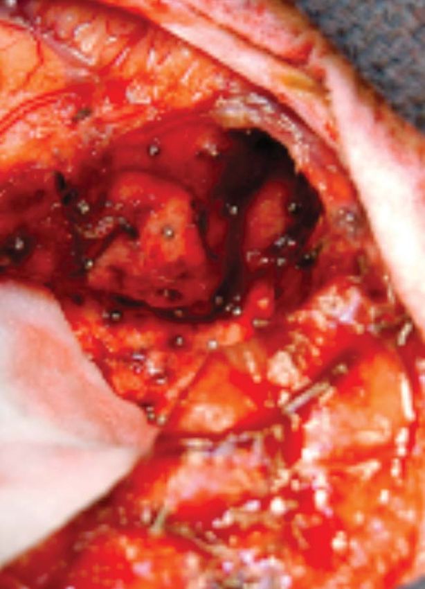
FIGURE 1: A postoperative adjuvant RECURRENT BRAIN TUMORS AND THE UNMET CLINICAL
EBRT treatment plan for a high-grade NEED FOR A NEW POSTOPERATIVE ADJUVANT THERAPY
glioma. Note the surgical treatment bed
When an aggressive tumor returns, resection alone is not usually curative,
(white arrow), zone of concern for traces
of the residual tumor (red area), and but it can provide symptom relief, and the extent of resection correlates
distribution of the actual radiation dose with the likelihood of longer-term control.6 Thus, whenever feasible,
falloff within healthy brain tissue (thin,
recurrent brain tumors are treated by combining maximum safe surgical
colored lines).
resection with an adjuvant therapy.1 As noted previously, few effective
chemotherapies cross the blood brain barrier, and even fewer are
available for recurrent tumors.1 Additionally, for many patients, repeating
adjuvant EBRT is often not an option, as they received a maximum safe
dose of EBRT during their initial treatment.7 If EBRT is repeated, the radiation will pass through the same (or
nearby) skin, skull, and normal brain tissue that were impacted previously. The risks of injury from radiation to
healthy tissues increases with the repeated radiation dose and the total irradiated brain volume treated.8
Consequently, to keep the risk of injury from escalating, we must either lower the radiation dose and/or
lower the volume of brain treated.8 The problem is we know that lower treatment doses are potentially less
effective, and minimizing the treatment volume can lead to missing tumor cells (FIGURE 2A–C). Since EBRT
comes from the outside inward, it typically exposes a large volume of normal brain tissue in proportion to the
actual area in need of treatment.9 As volume has 3 dimensions (H x W x L), even a seemingly small increase in
2 | C L I N I C A L W H I T E PA P E R | D E C 2 0 2 0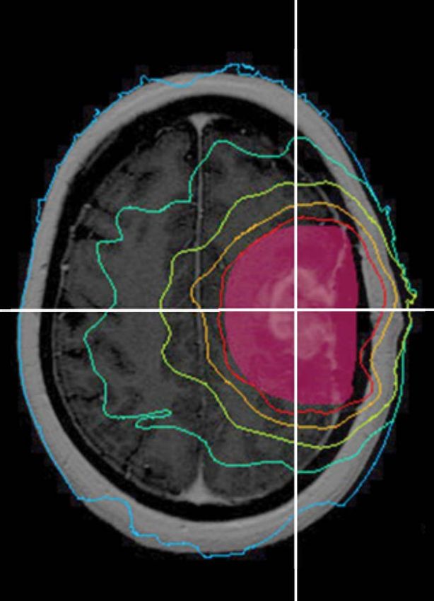
any one of these measurements can lead to a large increase in the end volume. To avoid toxicity with EBRT,
clinicians use treatments in the lower dose range of the highest safe doses.1,8 These lower radiation doses
are temporarily helpful in many cases, but rarely enough to produce reliable tumor control. Hence, there is a
critical need for a new radiation paradigm for recurrent brain tumors.
  Determining the precise treatment zone with postoperative imaging can be an additional challenge, and as
mentioned previously, leaving the residual tumor cell replication unchecked for several weeks until the wound
from the operation has healed is a suboptimal necessity of EBRT.4,5,10 Thus, although many recurrent aggressive
brain tumors may be technically resectable, surgery is frequently not offered at recurrence because: 1) resection
alone is unlikely to remove all tumor cells, and 2) no repeat safe and effective adjuvant therapy has historically
been available to prevent any tumor cells missed at surgery from regrowing.11 For these reasons, effective
re-treatment options are extremely limited, and there is no clearly established standard of care.1,12

Higher                                                                                                                          Higher

                       Change in
 Tumor Control %

                                                                                                                                 Tissue Injury Risk
                         Tumor
                        Control
                                      2A.                                     2B.                                         2C.

Lower                                                                                                                           Lower
                   Lower           Higher          Lower                    Higher        Lower                  Higher
                                                           Radiation Dose

FIGURE 2A–C: A.This radiation dose–response curve (gray line) illustrates how a relatively small change in the radiation dose can
result in a steep increase or decrease in the percentage of neoplastic cells responding to treatment (blue arrow is lower dose; orange
arrow is higher dose). Conversely, even a small decrease in the dose (blue arrow) can potentially lead to a steep decline in tumor
control. With EBRT, doses are often decreased to avoid brain injury when repeat treatments are needed. B. When we add a curve (blue
line) to gauge the impact on the normal tissue of doses used in repeat adjuvant EBRT, we get a set of curves, which illustrate that the
likelihood of tumor control and the likelihood of tissue injury are not too far apart—a relatively unfavorable therapeutic outcome.
C. As compared to repeat EBRT, treatment with GammaTile Therapy reduces the volume of tissue receiving potentially harmful doses
of radiation, which shifts the tissue injury curve to right, creating the potential for a more favorable therapeutic outcome.

KEY TAKEAWAYS
• When possible, recurrent brain tumors are treated combining maximum safe surgical resection
  with an adjuvant therapy such as chemotherapy or radiation.1
• For many patients who would otherwise be good candidates, repeat surgery is not offered
  because repeat adjuvant EBRT is not an option, as they received their maximum safe dose of EBRT
  during their initial treatment.1,7,12
  • The risks of injury from radiation to healthy tissues increases with the repeated radiation dose
		 and the total irradiated brain volume treated.8
  • To avoid toxicity from the interplay of an additional brain radiation dose and additional
		 brain radiation volume, clinicians often need to use a less efficacious, reduced radiation dose
		 in EBRT re-treatment.8
  • No routinely safe and effective repeat adjuvant therapy has historically been available.1,12

                                                                            THE EVOLUTION OF RADIOTHERAPIES                               |           3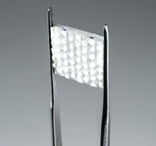
THE UNFULFILLED PROMISE OF BRACHYTHERAPY
IN BRAIN TUMOR TREATMENT
The term brachytherapy is derived from the Greek word for ‘short,’ brachy, which refers to the distance
between the radiation source and the target area.13 Brachytherapy is a specialized type of radiation therapy
that involves placing an emitting radiation source, which is commonly a radioactive isotope, very close to
either a tumor or adjacent tissue that is likely to harbor neoplastic cells.14 It is used to deliver the highest-
possible safe dose of radiation to the tumor site while minimizing a patient’s overall radiation exposure.
  Combining resection with adjuvant reirradiation via brachytherapy represents a theoretically attractive
therapeutic option for several reasons. Early postresection initiation of radiation—when residual tumor
burden is minimal—could produce a higher therapeutic ratio in rapidly proliferating tumors.4,5 Brachytherapy
using a low-energy (ie, short range) isotope exposes less normal tissue to radiation than EBRT techniques,
and it may limit neurocognitive deficits.9,15–17 Importantly, radiation source placement under intraoperative
visualization also allows for more precise identification of the area at risk than the postoperative imaging
utilized for EBRT treatment.10
  Compared to EBRT treatments that come from the outside into the body, traveling through different
healthy tissues and structures not affected by the tumor, brachytherapy offers distinct treatment advantages.
Brachytherapy enables enhanced dose control, as the dose conforms to the treatment area, and dose
intensity, as the highest doses are at the site of the tumor bed. In many ways, brachytherapy can be
considered the ultimate form of conformal radiation therapy, and it functions best when delivered with
specialized applicators that are specifically designed for each anatomic site or clinical circumstance.14
  Brachytherapy employs several different isotopes, and plays a central role in the management of various
tumor types, including prostate, skin, gynecologic, and breast cancers.18,19
  While brachytherapy for the brain has demonstrated considerable efficacy against cancer cells,
unfortunately, the isotopes used and the direct seed-to-tissue contact have resulted in severe complications
due to the difficulty in avoiding toxicity and necrosis.20 The clinical gain achieved has been directly offset by
the adverse impact to the eloquent brain tissue.20 Previous brachytherapy devices, in most cases, were not
specifically designed for use in the brain.20 Refinements for intracranial applications would require more
precise, uniform radiation-source placement and some type of structural offset that would better protect
healthy, eloquent brain tissue while delivering a targeted dose intensity to tumor cells.

KEY TAKEAWAYS
• Brachytherapy is a specialized type of internal radiation therapy that plays a central role in the
  management of various tumor types, including prostate, skin, gynecologic, and breast cancers.18,19
• Historically, brachytherapy has not had significant success in the brain due to the eloquent nature
  of the tissue and the difficulty in avoiding toxicity and necrosis.20
• The dose control and dose intensity of brachytherapy are promising, but refinements for
  intracranial applications are needed.

4   | C L I N I C A L W H I T E PA P E R | D E C 2 0 2 0
THE DRAWBACKS OF TRADITIONAL
INTRACRANIAL BRACHYTHERAPY
Used primarily to treat high-grade gliomas, brachytherapy with
iodine-125 (I-125) has historically demonstrated less than optimal
outcomes due to its high complication rate, which is a result of seed
placement variability, direct seed-to-tissue contact, and lack of dose
control (FIGURE 3).20 Intracranial seeds are most commonly used in
high-grade gliomas, with studies frequently finding high rates of brain
necrosis and reoperation, although poor outcomes have not been
universal.20–22 Consequently, intracranial brachytherapy with I-125
has mostly been relegated to salvage therapy for patients with highly
aggressive brain tumors who are willing to risk the complications.20

CS-131, A MORE IDEAL INTRACRANIAL ISOTOPE
In the early 2000s, a new seed isotope source, Cesium-131 (Cs-131),           FIGURE 3: This image depicts the
became commercially available. Cs-131 is a low-energy source of               traditional brachytherapy approach
                                                                              with seeds implanted directly into
x-radiation with a markedly shorter half-life compared to I-125—9.7           the brain tissue.22 The variable seed
days vs 60 days (TABLE 1). Initially utilized in prostate cancer, Cs-131      spacing, and direct seed-to-brain
has now been investigated for intracranial use. It has a low energy that      contact, lead to inherently variable
                                                                              doses, which both underdose the tumor,
is similar to that of I-125 (30 keV for Cs-131 vs 28 keV for I-125), with a   and overdose the brain.22
depth of penetration that matches up well with the requirement for
a useful intracranial isotope. Notably, its shorter half-life is thought
to offer significant advantages in treating tumors that have relatively
short doubling times, such as recurrent intracranial neoplasms.23
Another advantage is that it lessens the duration of exposure to family caregivers as compared to iodine.24
   Cs-131 delivers a greater dose immediately after resection, when the tumor burden is at a minimum.23
Specifically, the short half-life of Cs-131 means 88% of the therapeutic dose can be delivered within 30 days
(vs approximately 200 with I-125).23 In fact, more than half of the therapeutic dose of Cs-131 is delivered
within the first 10 days after surgery, which helps prevent residual tumor cells from replicating.

                               TABLE 1: COMPARING         CS-131 AND I-12523

                     Half-life (days)                               Time to deliver 88% of
                    		                                              radiation dose (days)

                    Cs-131                          9.7                         30

                    I-125                          59.4                        ~200

                                                               THE EVOLUTION OF RADIOTHERAPIES                  |   5
Only the tissue closest to the radiation source receives the highest
levels of radiation, sparing nearby brain and other tissues.9,16,22 It is
postulated that the rapid, intense, intracranial dose delivery of Cs-131 It is postulated that the
enhances local control and improves efficacy.22,23
rapid, intense, intracranial
Cs-131 offers a high dose of radiation that is delivered to a localized
area with a very steep dose fall-off that spares adjacent normal brain dose delivery of Cs-131
tissue. Cs-131 seeds have been used with traditional brachytherapy enhances local control and
techniques in patients with both initial and recurrent brain metastases
by the Cornell group.25,26
improves efficacy.22,23
Targeting is more localized with Cs-131 than it is with EBRT. EBRT
affects a larger volume of the brain, increasing the risk for high
toxicity rates and brain necrosis.9,27 The dose necessary to control
aggressive brain tumors is typically a minimum of 60 Gy, with the dual
goals of fully treating the areas of concern and minimizing radiation exposure to adjacent healthy tissues.
It is believed that with aggressive brain tumors, doses higher than 60 Gy may enable more reliable tumor
control.28 However, doses above 60 Gy have been shown to increase the risk of brain toxicity when combined
with the volume of brain tissue treated, particularly with EBRT techniques.26
Brachytherapy with an internally placed, low-energy isotope like Cs-131 has been shown to expose a
smaller volume of brain tissue to radiation than EBRT (FIGURE 5), and is able to achieve an increased local
dose of 80 to 120 Gy.25 As potentially useful as brachytherapy with Cs-131 seems, it is still necessary to
overcome several of the current shortcomings of traditional brain brachytherapy techniques including issues
of: 1) direct seed-to-brain contact, which can result in extremely high doses at the sites of contact, 2) the
undesirable spacing variability inherent with the placement of individual seeds (FIGURE 3), and 3) the
time-consuming nature of the traditional placement methods, often adding 30 minutes or more per
case.22,29,30 Thus, to safely take full advantage of the potential therapeutic benefits of brain brachytherapy,
advances beyond just a better isotope are needed.
KEY TAKEAWAYS
• Cs-131 is a low-energy source of x-radiation with a markedly shorter half-life than I-125.23
• This shorter half-life is thought to offer significant advantages in treating tumors that have
relatively short doubling times, such as recurrent intracranial neoplasms.23
• Brachytherapy with Cs-131 has been shown to expose a smaller volume of brain tissue to radiation
than EBRT, and is able to achieve an increased local dose of 80 to 120 Gy compared to
approximately 60 Gy with EBRT.9,25
6 | C L I N I C A L W H I T E PA P E R | D E C 2 0 2 0THE GENESIS OF GammaTile® STaRT: A NEW TREATMENT
OPTION FOR PATIENTS WITH RECURRENT BRAIN TUMORS
A group of brain tumor specialists in Phoenix, Arizona, joined forces to
create a new treatment option to address the critical, unmet need for
a postsurgical adjuvant therapy for patients with recurrent gliomas,
meningiomas, and metastasis. To overcome the drawbacks of previous
adjuvant brain radiation treatment paradigms, the group developed a
modular, permanently implanted collagen-based device. This device,
the GammaTile, functions as a 3D spacer that optimizes interseed
spacing while simultaneously preventing harmful direct seed contact
with the brain. The collagen carrier is easily handled, and it facilitates FIGURE 4: Surgically Targeted Radiation
Therapy (STaRT) with GammaTile is
rapid completion of the implant by allowing simultaneous placement
indicated to deliver radiation therapy
of multiple seeds. GammaTile Therapy is indicated to deliver radiation for patients with newly diagnosed
therapy for patients with newly diagnosed malignant intracranial malignant intracranial neoplasms and
neoplasms and recurrent intracranial neoplasms. recurrent intracranial neoplasms.
THE RIGHT TIME
Fundamentally different from EBRT and traditional brain brachytherapy,
GammaTile Therapy is a surgically targeted radiation therapy (STaRT) (FIGURE 4) that provides immediate,
dose-intense treatment at the completion of resection. By getting a head STaRT on fighting the tumor, resection
plus GammaTile Therapy extends recurrence-free survival with minimal complications, reduced patient burden,
and assured compliance.31,32
EBRT (IMRT) EBRT (3D CONFORMAL) GAMMATILE THERAPY
FIGURE 5: Representative distribution and intensity of radiation doses with 3 different radiation modalities: EBRT with intensity-
modulated radiation therapy (IMRT) treatment, EBRT with 3D conformal treatment, and GammaTile Therapy. Note that
GammaTile Therapy delivers more localized radiation, both in overall extent (blue-green is lower radiation doses) and in the
areas exposed to higher dose radiation (red-orange).
THE EVOLUTION OF RADIOTHERAPIES | 7THE RIGHT TREATMENT AND DOSE INTENSITY
GammaTile Therapy uses Cs-131 to deliver the maximum dose at the
Placement by the surgeon treatment site while minimizing exposure to healthy tissue. With the
under direct operative GammaTile carrier design and seed strength, the radiation dose in the
first few millimeters of the operative bed (the site of greatest concern
visualization virtually
for tumor residual) is 80 to 120 Gy. This dose is 1.3 to 2 times greater
ensures treatment to the than the 60 Gy typically achieved by fractionated EBRT. The shorter
area(s) that is at the highest range afforded by this low-energy brachytherapy isotope limits high-
dose radiation to uninvolved tissues to a greater extent than possible
risk for recurrence.
with intraoperative x-ray treatments or EBRT.9,15,29,33
THE RIGHT PLACE
Keeping the radiation dose in the right place is as critical as keeping
the radiation source from direct contact with the brain tissue. Both of these objectives are achieved with the
tissue-sparing, patented, bioresorbable, conformable, 3D collagen tile that comprises GammaTile Therapy.
Placement by the surgeon under direct operative visualization virtually ensures treatment to the area(s) that
is at the highest risk for recurrence.
GammaTile Therapy enforces uniform radiation-source spacing, both within a single tile of 14 U (2 cm x
2 cm x 4 mm in thickness) and between multiple tiles. This enables rapid, accurate placement of the tile(s)
and a predictable radiation dose in the therapeutic range while reducing local hot and cold spots. With
the GammaTile collagen carrier, source migration postimplant is minimal.34 In addition, because of the
shorter half-life of Cs-131, any dose differences observed from source movement should have a clinically
insignificant impact compared to the dosimetric impact seen with I-125.35
KEY TAKEAWAYS
• Fundamentally different from EBRT and traditional brain brachytherapy, GammaTile Therapy is a
surgically targeted radiation therapy (STaRT) that provides immediate, dose-intense treatment at
the completion of resection.
• The 3D spacer optimizes interseed spacing while simultaneously preventing harmful direct
seed contact with the brain.
• Cs-131 delivers the maximum dose at the treatment site while minimizing exposure to
healthy tissue.36
• Uniform radiation-source spacing enables rapid, accurate placement, delivering a predictable
radiation dose in the therapeutic range while reducing local hot and cold spots.
• By getting a head STaRT on fighting the tumor, resection plus GammaTile Therapy extends
recurrence-free survival with minimal complications, reduced patient burden, and
assured compliance.31,32
8 | C L I N I C A L W H I T E PA P E R | D E C 2 0 2 0CONCLUSION
GammaTile Therapy fulfills the promise of brachytherapy for treatment of newly diagnosed malignant
and recurrent brain tumors in a whole new way, offering significant advantages to patients, clinicians,
and hospitals. In addition to its excellent efficacy and safety profile, GammaTile Therapy advances the
current standard of care for these vulnerable patients by extending their recurrence-free survival and
helping preserve their quality of life.33,34 GammaTile Therapy is a viable, effective treatment option for
individuals with recurrent brain tumors, especially those who have failed treatment with EBRT or for
whom further EBRT is contraindicated.
AUTHOR | DAVID BRACHMAN, MD, FACRO
David G. Brachman, MD, is a nationally recognized radiation oncologist, researcher, and author with more than
50 peer-reviewed publications and extensive neurooncology clinical expertise. Dr Brachman recently retired from practice
after more than 15 years as the chairman and medical director of the Radiation Oncology department and codirector of the
GammaKnife program at St. Joseph’s Hospital and Medical Center and Barrow Neurological Institute in Phoenix, Arizona.
His considerable radiation oncology background includes being named a professor of Radiation Oncology at
the University of Arizona College of Medicine-Phoenix and more than 15 years serving on the NCI-sponsored Radiation
Therapy Oncology Group CNS Tumor Steering Committee. Dr. Brachman was instrumental in the development of
GammaTile Therapy and is cofounder and chief technology officer of GT Medical Technologies.
THE EVOLUTION OF RADIOTHERAPIES | 9REFERENCES
1. National Comprehensive Cancer Network. NCCN Clinical Practice Guidelines in Oncology (NCCN Guidelines®):
Central Nervous System Cancers. Version 1.2018.
2. External beam radiation therapy for cancer. National Cancer Institute Web site. https://www.cancer.gov/about-cancer/treatment/types/
radiation-therapy/external-beam. Published May 1, 2018. Accessed July 9, 2018.
3. Radiation therapy basics. American Cancer Society Web site. https://www.cancer.org/treatment/treatments-and-side-effects/
treatment-types/radiation/basics.html. Updated July 2, 2018. Accessed August 25, 2018.
4. Burnet NG, Jena R, Jefferies SJ, et al. Mathematical modelling of survival of glioblastoma patients suggests a role for radiotherapy dose
escalation and predicts poorer outcome after delay to start treatment. Clin Oncol (R Coll Radiol). 2006;18:93-103.
5. Iorio-Morin C, Masson-Cote L, Ezahr Y, et al. Early Gamma Knife stereotactic radiosurgery to the tumor bed of resected brain metastasis
for improved local control. J Neurosurg. 2014;121 Suppl:69-74.
6. Aizer AA, Bi WL, Kandola MS, et al. Extent of resection and overall survival for patients with atypical and malignant meningioma.
Cancer. 2015;121:4376-4381.
7. Shi W, Scannell Bryan M, Gilbert MR, et al. Investigating the effect of reirradiation or systemic therapy in patients with glioblastoma
after tumor progression: a secondary analysis of NRG Oncology/Radiation Therapy Oncology Group Trial 0525.
Int J Radiat Oncol Biol Phys. 2018;100(1):38-44.
8. Stewart FA. Re-treatment after full-course radiotherapy: is it a viable option? Acta Oncol. 1999;38:855-862.
9. Purdy JA. Dose to normal tissues outside the radiation therapy patient’s treated volume: a review of different radiation therapy
techniques. Health Phys. 2008;95:666-676.
10. Lecchi M, Fossati P, Elisei F, et al. Current concepts on imaging in radiotherapy. Eur J Nucl Med Mol Imaging. 2008;35:821-837.
11. Lu VM, Jue TR, McDonald KL, Rovin RA. The survival effect of repeat surgery at glioblastoma recurrence and its trend: a systematic
review and meta-analysis. World Neurosurg. 2018;115:453-459.e3.
12. Ammirati M, Cobbs CS, Linskey ME, et al. The role of retreatment in the management of recurrent/progressive brain metastases:
a systematic review and evidence-based clinical practice guideline. J Neurooncol. 2010;96(1):85-96.
13. A brief essay on the introduction of brachytherapy. American Brachytherapy Society Web site.
https://www.americanbrachytherapy.org/aboutbrachytherapy/history.cfm. Accessed July 9, 2018.
14. Petereit DG, Frank SJ, Viswanathan AN, et al. Brachytherapy: where has it gone? J Clin Oncol. 2015;33(9):980-982.
15. Ruge MI, Kocher M, Maarouf M, et al. Comparison of stereotactic brachytherapy (125 iodine seeds) with stereotactic radiosurgery
(LINAC) for the treatment of singular cerebral metastases. Strahlenther Onkol. 2011;187:7-14.
16. Pham A, Yondorf MZ, Parashar B, et al. Neurocognitive function and quality of life in patients with newly diagnosed brain metastasis
after treatment with intra-operative cesium-131 brachytherapy: a prospective trial. J Neurooncol. 2016;127:63-71.
17. Rogers LR, Rock JP, Sills AK, et al. Results of a phase II trial of the GliaSite radiation therapy system for the treatment of newly
diagnosed, resected single brain metastases. J Neurosurg. 2006;105:375-384.
18. Skowronek J. Current status of brachytherapy in cancer treatment - short overview. J Contemp Brachytherapy. 2017;9(6):581-589.
10 | C L I N I C A L W H I T E PA P E R | D E C 2 0 2 019. Lukens JN, Gamez M, Hu K, Harrison LB. Modern brachytherapy. Semin Oncol. 2014;41(6):831-847.
20. Combs SE, Debus J, Schulz-Ertner D. Radiotherapeutic alternatives for previously irradiated recurrent gliomas.
BMC Cancer. 2007;(7)167.
21. Magill ST, Lau D, Raleigh DR, Sneed PK, Fogh SE, McDermott MW. Surgical resection and interstitial iodine-125 brachytherapy for
high-grade meningiomas: a 25-year series. Neurosurgery. 2017;80(3):409-416.
22. Dagnew E, Kanski J, McDermott MW, et al. Management of newly diagnosed single brain metastasis using resection and permanent
iodine-125 seeds without initial whole-brain radiotherapy: a two institution experience. Neurosurg Focus. 2007;22(3):E3
23. Armpilia CI, Dale RG, Coles IP, Jones B, Antipas V. The determination of radiobiologically optimized half-lives for radionuclides used
in permanent brachytherapy implants. Int J Radiat Oncol Biol Phys. 2003;55(2):378-385.
24. Parashar B, Wernicke AG, Pavese A, et al. Cesium-131 permanent seed brachytherapy: dosimetric evaluation and radiation exposure
to surgeons, radiation oncologists, and staff. Brachytherapy. 2011;10:508-513.
25. Wernicke AG, Yondorf MZ, Peng L, et al. Phase I/II study of resection and intraoperative cesium-131 radioisotope brachytherapy in
patients with newly diagnosed brain metastases. J Neurosurg. 2014;121(2):338-348.
26. Wernicke AG, Smith AW, Taube S, et al. Cesium-131 brachytherapy for recurrent brain metastases: durable salvage treatment for
previously irradiated metastatic disease. J Neurosurg. 2017;126(4):1212-1219.
27. Pinnaduwage D., Youssef E., Sorensen S., Srivastava S., Yan X., Brachman D. Dosimetric impact of radioisotope type on permanent
brain seed implants. Medical Physics. 2017;44(6):3146.
28. Nguyen D, Rwigema JC, Victoria Y, et al. Feasibility of extreme dose escalation for glioblastoma multiforme using 4π radiotherapy.
Radiat Oncol. 2014; 9:239.
29. Chiu-Tsao ST, Napoli JJ, Davis SD, et al. Dosimetry for 131Cs and 125I seeds in solid water phantom using radiochromic EBT film.
Appl Radiat Isot. 2014;92:102-114.
30. Ware ML, Larson DA, Sneed PK, et al. Surgical resection and permanent brachytherapy for recurrent atypical and malignant
meningioma. Neurosurgery. 2004;54:55-63.
31. Brachman DG, Youssef EY, Dardis CJ, et al. Resection and permanent intracranial brachytherapy using modular, biocompatible
cesium-131 implants: results in 20 recurrent previously irradiated meningiomas. J Neurosurg. In press.
32. Data on file, GT Medical Technologies, Inc.
33. Schueller P, Palkovic S, Moustakis C, et al. Clinical results and isodose planning of neuronavigation-guided intraoperative radiotherapy
(IORT) in 77 brain tumor patients: adequate target volume coverage improves results. Rev Cancer (Madrid). 22(extra):58.
34. Pinnaduwage D. Dosimetric impact of Cesium 131 seed migration in collagen carrier brain brachytherapy implants.
Paper to be presented at: 2018 ASTRO Annual Meeting; October 21-24, 2018; San Antonio, TX.
35. Pinnaduwage D, Sorensen S, Youssef E, et al. Dosimetric impact of source migration and decay based on radioisotope type in collagen
carrier brain brachytherapy implants. Medical Physics. 2018;45(6):e244.
36. Nedialkova L, Sabbas A, Trichter S, et al. Dosimetric comparison of Cs-131 versus I-125 intraoperative brachytherapy for patients
with resected brain metastasis renders Cs-131 more sparing of normal brain tissue: clinical relevance for prevention of radiation
necrosis from a prospective trial. Int J Radiat Oncol Biol Phys. 2013;87:S755-S756.
THE EVOLUTION OF RADIOTHERAPIES | 11gtmedtech.com
©2020 GT Medical Technologies, Inc. | All rights reserved | GammaTile is a registered trademark of GT Medical Technologies,
Inc. MKT-003-1You can also read