An Atypical Case of Neonatal Hypoglycemic Encephalopathy with Extensive White Matter Lesions
←
→
Page content transcription
If your browser does not render page correctly, please read the page content below
Letter to the editor
                                                                                                                     pISSN 2635-909X • eISSN 2635-9103
                                                                                                                              Ann Child Neurol 2023;31(1):70-74
                                                                                                                    https://doi.org/10.26815/acn.2022.00339

An Atypical Case of Neonatal Hypoglycemic
Encephalopathy with Extensive White Matter Lesions
Won Jik Shin, MD, Younghyun Kim, MD, Kye Hyang Lee, MD
Department of Pediatrics, Daegu Catholic University School of Medicine, Daegu, Korea

Received: September 21, 2022              Neonatal hypoglycemia is a common condition                     able protected health information was not dis-
Revised: October 8, 2022                  that constitutes one of the leading causes of brain             closed in this report.
Accepted: October 10, 2022
                                          injury. Symptomatic hypoglycemia may present                       A 2-day-old girl was admitted for refractory hy-
Corresponding author:                     with metabolic encephalopathy characterized by                  poglycemia. The patient was born to a mother
Kye Hyang Lee, MD                         neurological features, including stupor, jitteriness,           with gestational diabetes and hypothyroidism at
Department of Pediatrics, Daegu           seizures, apnea, irritability, and muscular hypoto-             the gestational age of 37 weeks and 6 days and
Catholic University School of             nia mimicking several other diseases. The typical               had a birth weight of 2,460 g. After an uneventful
Medicine, 33 Duryugongwon-ro
                                          magnetic resonance imaging (MRI) patterns of                    delivery at a local hospital, her blood glucose level
17-gil, Nam-gu, Daegu 42472,
                                          neonatal hypoglycemic encephalopathy (NHE)                      was 29 mg/dL. Despite glucose administration,
Korea
Tel: +82-53-650-4242                      are signal abnormalities localized to the parietal              the blood glucose level remained low (range, 23
Fax: +82-53-650-4243                      and occipital lobes and progressive parenchymal                 to 34 mg/dL). She was subsequently transferred
E-mail: rosalia@cu.ac.kr                  loss of the predominant occipital lobe in the new-              to our hospital. On admission, she was alert;
                                          born. Diffusion-weighted imaging (DWI) may                      however, she showed mild chest retraction with
                                          show restricted diffusion in the involved areas.                oxygen saturation of 90% under an oxygen supply
                                          These brain lesions are transient, and the revers-              of 1 L/min. Her heart rate, respiratory rate, body
                                          ibility of DWI has also been reported [1]. The                  temperature, and blood pressure were 147 beats/
                                          patterns of injury associated with NHE are more                 min (range, 120 to 160), 52 breaths/min (range,
                                          diverse than those previously reported, and in-                 40 to 60), 36.7°C (range, 36.5°C to 37.4°C), and
                                          clude white matter, cortical, and basal ganglia/                80/31 mm Hg (range, 60 to 90/30 to 60), respec-
                                          thalamic abnormalities, white matter hemorrhage,                tively. Mechanical ventilation was applied to re-
                                          and middle cerebral artery territory infarctions                lieve her respiratory distress symptoms. The ini-
                                          [2]. However, no reports of NHE with extensive                  tial laboratory findings were as follows: serum
                                          white matter involvement and serial changes on                  glucose, 17 mg/dL; pH, 7.23; pCO2, 56.2 mm
                                          MRI are available. Here, we present a case of                   Hg; and bicarbonate, 23.6 mmol/L. The follow-
                                          NHE with atypical MRI findings along with se-                   ing were the results of the metabolic evaluation
                                          quential neuroradiological changes. This study                  for hypoglycemia: insulin, 13.7 μIU/mL (range,
                                          was approved by the Institutional Review Board                  2.6 to 24.9); insulin/glucose ratio 0.5 (simultane-
                                          of Daegu Catholic University Medical Center                     ous serum glucose, 25 mg/dL, < 0.3); free fatty
                                          (CR-22-014). The requirement for publication                    acids, 93 μEq/L (range, 172 to 586); ketone bod-
                                          consent was waived because personally identifi-                 ies, 0.2 mmol/L (< 0.6); and cortisol, 9.13 μg/dL

Copyright © 2023 Korean Child Neurology Society
This is an Open Access article distributed under the terms of the Creative Commons Attribution Non-Commercial License (http://creativecommons.org/licenses/by-nc/4.0/)
which permits unrestricted non-commercial use, distribution, and reproduction in any medium, provided the original work is properly cited.

70                                                                                                                                        www.annchildneurol.org
Ann Child Neurol 2023;31(1):70-74
(range, 2.47 to 11.9). Even after intravenous glucose administra- level of 68 mg/dL with a serum glucose level of 93 mg/dL. Imme-
tion, her glucose level remained low, with a range of 31 to 52 mg/ diately after the occurrence of seizures, we performed brain MRI
dL. Her glucose level stabilized following the administration of glu- on the 4th day of admission. Brain DWI and an apparent diffusion
cose infusion at a rate of 14 mg/kg/min and hydrocortisone. On coefficient map demonstrated diffuse restricted water diffusion in
the 4th day of admission, she developed multiple seizures with ap- the white matter of the parieto-occipital lobes, centrum semiovale,
nea, eyelid blinking, and lip-smacking under euglycemia with the posterior limb of the internal capsule, and the splenium of the cor-
same glucose management. Cerebrospinal fluid analysis revealed 2 pus callosum. On the 11th day, follow-up MRI showed the spread
white blood cells/μL, a protein level of 81.8 mg/dL, and a glucose of lesions in the corpus callosum and internal capsule (Fig. 1). A
A B C D
E F G H
I J K L
Fig. 1. Serial brain magnetic resonance imaging (MRI) shows the evolution of white matter lesions in a neonate with symptomatic
hypoglycemia from seizure onset to 7 months of age. Axial diffusion-weighted imaging (DWI) (A, B) and apparent diffusion coefficient
(ADC) map (C, D) reveal restricted water diffusion in the white matter of the parieto-occipital lobes, centrum semiovale, posterior limb
of the internal capsule, and the splenium of the corpus callosum on the 4th day of admission. Follow-up DWI (E, F) and ADC map (G, H)
on the 11th day show no interval changes except for the spread of lesions in the corpus callosum and internal capsule. Follow-up MRI at
7 months of age shows mild ventriculomegaly with an outer bulging contour on T1-weighted imaging (K, L), with the disappearance of
restriction of water diffusion observed in the previous images (I, J).
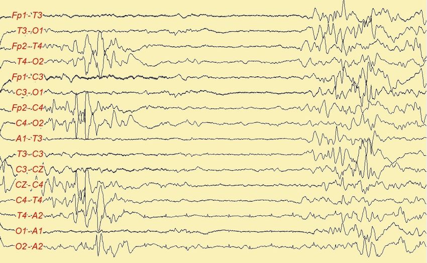
https://doi.org/10.26815/acn.2022.00339 71Shin WJ et al. • Neonatal Hypoglycemic Encephalopathy with Extensive White Matter Lesions stool test for rotavirus antigen was negative. Electroencephalogra- 1212Ile), and MAF bZIP transcription factor A (MAFA) (NM_ phy (EEG) showed a burst-suppression pattern on the 5th day of 201589.4:c.738C> T;p.Gly246 = ). admission (Fig. 2). Intravenous anticonvulsants were administered Parental genetic testing revealed that the MAFA variant was ob- to control seizures. She had five additional seizures after the first served in her mother, and the SCN1A and CACNA1H variants in episode. Except for the first one, the others were subtle seizures her father. Family history revealed her mother and elder brother with autonomic changes that comprised a sudden increase in heart had a history of febrile seizures. On the 26th day of admission, she rate up to 180 to 210 beats/min with decreased responsiveness to was discharged with medications and instructions for follow-up. At noxious stimuli for seconds. After the 12th day of admission, the 7 months of age, brain MRI showed mild ventriculomegaly with seizures were controlled with medications (phenobarbital, 6 mg/ an outer bulging contour, suggesting end-stage periventricular leu- kg/day; levetiracetam, 30 mg/kg/day). Follow-up EEG on the komalacia (Fig. 1). Over 14 months of follow-up, the patient had 12th and 15th days of admission showed an improving status of mild developmental delay with esotropia and hypertropia of the background activity despite persistent multifocal spikes and sharp left eye. On the same follow-up day, her height was 76.2 cm (50th waves. Considering the burst-suppression pattern on EEG and the percentile), and her body weight was 9.3 kg (50th percentile). topography of lesions, we performed whole-exome sequencing MRI studies in patients with symptomatic hypoglycemia through GC Genome (Yongin, Korea) for the molecular diagno- showed a correlation between the pattern of damage and age at sis of possible white matter disorders, including other metabolic clinical presentation: parieto-occipital white matter lesions from disorders causing hypoglycemia. Three heterozygous variants of the neonatal period to 6 months of age versus basal ganglia and pa- unknown significance were revealed in sodium voltage-gated rieto-temporal cortex involvement in older infants [3]. To date, it channel alpha subunit 1 (SCN1A) (NM_001165963.3: remains unclear why the posterior head is more vulnerable to hy- c.4096G > A;p.Val1366Ile), calcium voltage-gated channel sub- poglycemic injury in the neonatal period. The reduction of region- unit alpha1 H (CACNA1H) (NM_021098.3:c. 3635C > T;p.Thr al cerebral glucose use or a local expression deficit of glucose mem- Fig. 2. Initial electroencephalography shows a generalized burst-suppression pattern on the 4th day of admission. 72 https://doi.org/10.26815/acn.2022.00339
Ann Child Neurol 2023;31(1):70-74

brane transporter proteins and active synaptogenesis and axonal          cephalopathies. NHE can present atypical MRI patterns differing
migration in the occipital lobe have been suggested as possible          from those predominantly focused in the posterior area with or
contributors to the localization of damage [4]. Burns et al. [2]         without accompanying hypoxic-ischemic injury.
studied the pattern of injury on early MRI scans following symp-            Our case showed unfamiliar findings caused by hypoglycemia.
tomatic hypoglycemia in 35 term infants. They found that the pat-        This additional information related to NHE could help physicians
tern of brain injury in symptomatic neonatal hypoglycemia was            in medical practice.
not associated with the severity or duration of hypoglycemia. Re-
garding the occurrence of seizures, all patients with seizures had       Conflicts of interest
white matter injuries; even four of the five patients without seizures
had white matter injuries with variable degrees from mild to mod-        No potential conflict of interest relevant to this article was report-
erate [2]. According to other studies, severe or repetitive hypogly-     ed.
cemic brain injury is more often present with atypical MRI, and
the sites of brain injury are numerous and extensive [5]. Diffuse,       ORCID
symmetric diffusion-restricted lesions involving the cerebral white
matter can be observed in patients with neonatal seizures without        Won Jik Shin, https://orcid.org/0000-0001-6026-3476
any structural or metabolic etiology and in neonates with viral in-      Kye Hyang Lee, https://orcid.org/0000-0001-6646-8982
fections, such as rotavirus, human parechovirus, and enterovirus
[6]. Our case had hypoglycemia for 3 days with the lowest level of       Author contribution
17 mg/dL. The patient developed multiple seizures for 9 days un-
der euglycemia; however, she never developed status epilepticus.         Conceptualization: WJS and KHL. Data curation: WJS, YK, and
Her stool rotavirus test was negative. Based on the previous re-         KHL. Formal analysis: WJS, YK, and KHL. Methodology: WJS
ports, the atypical MRI features of our case may have resulted from      and YK. Project administration: KHL. Visualization: WJS, YK, and
various factors, including the duration and severity of hypoglyce-       KHL. Writing-original draft: WJS. Writing-review & editing: WJS
mia, along with the presence of multiple seizures.                       and KHL.
   Although hypoxia-ischemia and hypoglycemia can each have
different MRI patterns of injury, they can exert a combined nega-        References
tive effect on the developing brain. Mild to moderate hypoxic-isch-
emic injury in term neonates produces parasagittal watershed zone         1. Poretti A, Blaser SI, Lequin MH, Fatemi A, Meoded A, North-
infarcts between the anterior/middle and the middle/posterior ce-            ington FJ, et al. Neonatal neuroimaging findings in inborn er-
rebral arteries [7]. Internal watershed strokes involve structures in        rors of metabolism. J Magn Reson Imaging 2013;37:294-312.
the deep white matter that lie between medullary arteries arising         2. Burns CM, Rutherford MA, Boardman JP, Cowan FM. Patterns
from the superficial pial plexus and deep penetrating arteries aris-         of cerebral injury and neurodevelopmental outcomes after
ing from the basal cerebral arteries. These lesions lie in the corona        symptomatic neonatal hypoglycemia. Pediatrics 2008;122:65-
radiata and centrum semiovale adjacent to the lateral ventricles [8].        74.
In a study of 179 term infants, the watershed pattern of hypox-           3. Gataullina S, De Lonlay P, Dellatolas G, Valayannapoulos V, Na-
ic-ischemic injury was most frequently observed to be associated             puri S, Damaj L, et al. Topography of brain damage in metabolic
with severe hypoglycemia [9]. When transferred, our patient                  hypoglycaemia is determined by age at which hypoglycaemia
showed respiratory distress as a possible clinical manifestation of          occurred. Dev Med Child Neurol 2013;55:162-6.
hypoglycemia. Although our case had white matter lesions involv-          4. Filan PM, Inder TE, Cameron FJ, Kean MJ, Hunt RW. Neona-
ing internal watershed zones that were not typical parasagittal ar-          tal hypoglycemia and occipital cerebral injury. J Pediatr 2006;
eas, the MRI findings shown in our case may be an overlapping                148:552-5.
pattern of hypoglycemia and hypoxic-ischemic injuries.                    5. Wang Y, Liu K, Ye X, Lu Y, Zhang X, Zhang C, et al. Typical and
   Owing to atypical MRI features compared to the typical posteri-           non-typical magnetic resonance imaging findings of neonatal
or dominant pattern of NHE, we performed an extensive workup                 hypoglycemic encephalopathy. Zhonghua Yi Xue Za Zhi 2014;
in our patient, including whole-exome sequencing, which showed               94:2938-40.
non-specific findings. Bilateral symmetric white matter changes are       6. Kim M, Hwang JY, Lee YJ, Kim YW, Byun SY, Lee YJ, et al.
typically associated with toxic, metabolic, or hereditary leukoen-           Clinical and imaging findings of neonatal seizures presenting as

https://doi.org/10.26815/acn.2022.00339                                                                                                     73
Shin WJ et al. • Neonatal Hypoglycemic Encephalopathy with Extensive White Matter Lesions

    diffuse cerebral white matter abnormality on diffusion-weight-          8. Marcic M, Marcic L, Kolic K, Titilic M. Internal watershed in-
    ed imaging without any structural or metabolic etiology. Taehan            farction as an imaging and clinical challenge: a case report. Acta
    Yongsang Uihakhoe Chi 2020;81:1412-23.                                     Med Iran 2016;54:222-5.
 7. Bano S, Chaudhary V, Garga UC. Neonatal hypoxic-ischemic                9. Wong DS, Poskitt KJ, Chau V, Miller SP, Roland E, Hill A, et al.
    encephalopathy: a radiological review. J Pediatr Neurosci 2017;            Brain injury patterns in hypoglycemia in neonatal encephalopa-
    12:1-6.                                                                    thy. AJNR Am J Neuroradiol 2013;34:1456-61.

74                                                                                                      https://doi.org/10.26815/acn.2022.00339
You can also read