Allosteric modulation of DNA by small molecules - PNAS
←
→
Page content transcription
If your browser does not render page correctly, please read the page content below
Allosteric modulation of DNA by small molecules
David M. Chenoweth and Peter B. Dervan1
Division of Chemistry and Chemical Engineering, California Institute of Technology, Pasadena, CA 91125
Contributed by Peter B. Dervan, June 17, 2009 (sent for review April 20, 2009)
Many human diseases are caused by dysregulated gene expres-
sion. The oversupply of transcription factors may be required for
the growth and metastatic behavior of human cancers. Cell per-
meable small molecules that can be programmed to disrupt tran-
scription factor-DNA interfaces could silence aberrant gene expres-
sion pathways. Pyrrole-imidazole polyamides are DNA minor-
groove binding molecules that are programmable for a large
repertoire of DNA motifs. A high resolution X-ray crystal structure
of an 8-ring cyclic Py/Im polyamide bound to the central 6 bp of the
sequence d(5ⴕ-CCAGGCCTGG-3ⴕ)2 reveals a 4 Å widening of the
minor groove and compression of the major groove along with a
>18 ° bend in the helix axis toward the major groove. This
allosteric perturbation of the DNA helix provides a molecular basis
for disruption of transcription factor-DNA interfaces by small
molecules, a minimum step in chemical control of gene networks.
DNA binders 兩 gene regulation 兩 minor groove binders 兩
Py-Im polyamides 兩 crystal structure
P y/Im polyamides bind the minor groove of DNA sequence
specifically (1, 2), encoded by side-by-side arrangements of
N-methylpyrrole (Py) and N-methylimidazole (Im) carboxamide Fig. 1. Chemical structure of the cyclic polyamide and DNA sequence. Cyclic
monomers. Im/Py pairs distinguish G䡠C from C䡠G base pairs, polyamide 1 targeting the sequence 5⬘-WGGCCW-3⬘ shown with ball-and-stick
CHEMISTRY
whereas Py/Py pairs are degenerate for T䡠A and A䡠T (3–6). model superimposed onto the DNA oligonucleotide used for crystallization. Black
Antiparallel Py/Im strands are connected by a ␥-aminobutyric circles represent imidazoles, open circles represent pyrroles, and ammonium
acid (GABA) linker to create hairpin-shaped oligomers. Hairpin substituted half circles at each end represent the (R)-␣-amine-␥-turn.
Py/Im polyamides have been programmed to bind a broad library
of different DNA sequences (7). They have been shown to
polyamide binding regions of the NCP-polyamide structures
permeate cell membranes (8–10), access chromatin (11, 12), and
coupled with the perturbations induced by nucleosome bound
disrupt protein-DNA interactions (2). Disruption of transcrip-
DNA, there is a pressing need for higher resolution crystallo-
tion factor-DNA interfaces 6 bp in size such as HIF-1␣ (13–15),
graphic studies to elucidate DNA structural distortions and
androgen receptor (AR) (16), and AP-1 (17, 18) have been
exploited for controlling expression of medically relevant genes molecular recognition details of polyamide-DNA binders at
such as VEGF, PSA, TGF-1, and LOX-1 in cell culture atomic resolution. High-resolution structural studies of poly-
experiments (13–18). X-ray crystallography of antiparallel 2:1 amide-DNA complexes in the absence of nucleosome bound
binding polyamides in complex with DNA reveal a 1–2 Å DNA are needed to adequately evaluate DNA perturbations
widening of the minor groove (5, 6). This modest structural induced by polyamide minor groove binding that result in the
perturbation to the DNA helix by the side-by-side stacked inhibition of transcription factors bound to the DNA major
arrangement of aromatic rings does not explain the large number groove.
of transcription factor-DNA interfaces disrupted by minor- The DNA structural alterations imparted upon polyamide
groove binding hairpin Py/Im polyamides (2, 5, 6, 19). It must be binding can be classified as direct perturbations to the polyamide
that the turn unit in the hairpin oligomer connecting the 2 minor groove binding site, proximal allosteric perturbations, and
antiparallel strands plays a structural role. distal allosteric perturbations. Proximal allosteric perturbations
NMR studies of hairpin polyamide-DNA complexes have occur primarily in the major groove as a result of polyamide
provided valuable insight into polyamide binding site location, binding to the minor groove, whereas distal allosteric perturba-
stoichiometry, and binding orientation (20–22). An NMR struc- tions occur outside of the binding site location displaced in the
ture of a 6-ring GABA-linked cyclic polyamide has confirmed
DNA binding and orientational preference. However, little
Author contributions: D.M.C. performed research; D.M.C. and P.B.D. analyzed the data; and
difference in minor groove width compared to ideal B-form D.M.C. and P.B.D. wrote the paper.
DNA is observed when measuring C1⬘ to C1⬘ distances in these The authors declare no conflict of interest.
models. In addition, the lack of identical DNA structures without
Freely available online through the PNAS open access option.
polyamide ligand have prevented detailed structural compari-
Data deposition: The atomic coordinates have been deposited in the Protein Data Bank,
sons (23). X-ray structures of hairpin polyamides bound to the www.pdb.org (PDB ID codes 3I5L and 3I5E).
nucleosome core particle (NCP) at modest resolution (⬎2 Å) 1To whom correspondence should be addressed. E-mail: dervan@caltech.edu.
have revealed a widening of the minor groove (average ⬍2 Å)
*An average minor groove width of ⬇7 Å has been calculated by subtracting the value of
upon polyamide binding (24, 25).* Interestingly, large perturba- 5.8 Å, to account for the phosphate Van der Waals radii, from the measured phosphate-
tions were observed distal to the polyamide binding sites in
Downloaded by guest on January 20, 2022
phosphate distance of 12.8 Å reported in the NCP-polyamide structures from ref. 25.
addition to long-range structural changes in the NCP. However, This article contains supporting information online at www.pnas.org/cgi/content/full/
given the modest resolution and disorder (high B-factors) in the 0906532106/DCSupplemental.
www.pnas.org兾cgi兾doi兾10.1073兾pnas.0906532106 PNAS 兩 August 11, 2009 兩 vol. 106 兩 no. 32 兩 13175–13179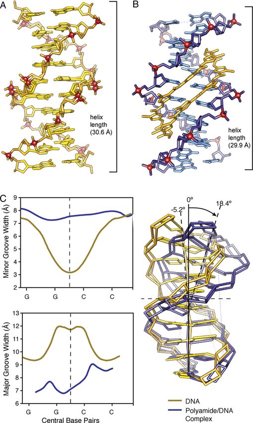
Fig. 2. Comparison of native DNA to polyamide/DNA complex. (A) Native DNA crystal structure at 0.98 Å resolution. (B) Comparison to DNA/polyamide co-crystal
                                          structure at 1.18 Å resolution. (Both structure solved by direct methods.) (C) Analysis of native DNA (yellow) compared to polyamide complexed DNA (blue). Chart
                                          on the top left shows variation in the minor groove width for native DNA (yellow) and polyamide-complexed DNA (blue) over the central core sequence
                                          5⬘-GGCC-3⬘. Chart on the bottom left shows variation in the major groove width for native DNA (yellow) and polyamide complexed DNA (blue) over the central
                                          core sequence 5⬘-GGCC-3⬘. Overlay of the curves calculated geometric helix model from each structure showing a DNA bend of ⬎18° in the polyamide/DNA
                                          complex compared to native DNA.

                                          3⬘ or 5⬘ direction. NCP-polyamide structures have demonstrated                      Here we report the atomic resolution structure (1.18 Å
                                          the possibility of distal allosteric perturbations at moderate                    resolution) of an 8-ring cyclic polyamide in complex with double
                                          resolution (24); however, direct perturbations and proximal                       helical DNA. The cyclic polyamide 1 is comprised of two
Downloaded by guest on January 20, 2022

                                          allosteric DNA perturbations relevant to transcription factor                     antiparallel ImImPyPy strands capped at both ends by (R)-␣-
                                          inhibition have not been characterized at atomic resolution.                      amino-␥ turn units. Polyamide 1, which codes for the sequence

                                          13176 兩 www.pnas.org兾cgi兾doi兾10.1073兾pnas.0906532106                                                                                     Chenoweth and Dervan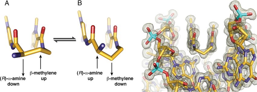
Fig. 3. Conformation of the ␣-amino substituted GABA turn. Two possible Conformations A and B are shown with conformation A directing the -methylene
up and away from the minor groove floor while orienting the ␣-ammonium toward the minor groove wall. Conformation B presents the -methylene down
toward the minor-groove floor while orienting the ␣-ammonium up and out of the minor-groove, relieving possible steric interaction with the sugar-phosphate
backbone (minor-groove wall). View looking down the DNA minor-groove, showing the (R)-␣-amine-␥-turn conformation observed in the X-ray crystal structure,
which matches that of conformation B. Electron density map is contoured at the 1.0 level.
5⬘-WGGCCW-3⬘, was co-crystallized with the palindromic DNA The conformational constraints imposed by the turn unit
oligonucleotide sequence 5⬘-CCAGGCICTGG-3⬘ 10 bp in result in ring placement that is an intermediate of ring-over-ring
length (Fig. 1 and Figs. S1 and S2). Compared with free DNA, and ring-over-amide. This alignment allows the ring pairs to
we observe significant structural changes of the DNA helix remain in phase with the edges of the Watson-Crick base pairs
induced upon binding of the 8-ring cycle in the minor groove. as the polyamide adopts an isohelical conformation complemen-
tary to DNA helix. This is highlighted by comparison to the 2:1
Results and Discussion structure in which the rings lie over the carboxamide linkages of
In the complex, each ImImPyPy strand is bound with N- to C- the adjacent strand (5). The conformational constraints imposed
CHEMISTRY
orientation aligned with the 5⬘ to 3⬘ direction of the DNA. The by the turn and inability of the ligand to slip into a possibly more
cyclic polyamide 1 rigidifies the sugar-phosphate backbone and preferred orientation may impact the overall DNA structure by
strongly perturbs the overall helix structure. The cycle widens the inducing bending and other distortions accommodated by the
minor groove of DNA up to 4 Å while simultaneously compress- plasticity of DNA. The preorganized cycle may have a significant
ing the major groove by 4 Å. The polyamide bends the DNA helix entropic driving force leading to increased affinity by locking out
⬎18° toward the major groove, and shortening the overall length unproductive conformations and alternate binding modes. In
by ⬇1 Å. The cycle is a sequence specific allosteric modulator of addition, we find a shell of highly ordered water molecules
DNA conformation (Fig. 2) (26). around the ␣-ammonium substituent and a water-mediated
Py/Im polyamides linked by a GABA or substituted GABA hydrogen bond from the ammonium to the N3 lone-pair of the
can adopt either of two possible conformations on the floor of adenine under the turn. The hydration pattern around the turn
the DNA minor groove (Fig. 3). In conformation A the amino is highly conserved at both ends of the structure and the
group is directed toward the minor-groove wall of the DNA helix water-mediated hydrogen bonds are within ⬇2.7–2.9 Å from the
with the potential for steric clash with the deoxyribose backbone. ammonium to water to the adenine lone-pair (Fig. 4A).
In alternative conformation B the amine is directed up and out The amide NHs and imidazole lone-pairs form a continuous
of the minor groove forcing the -methylene to the floor of the series of direct hydrogen bonds to the floor of the DNA
minor groove with the potential for steric interaction with the minor-groove, while the imidazoles impart specificity for the
edge of the base pairs and within van der Waals contact distance exocyclic amine of guanine through relief of a steric interaction
of the C2 hydrogen of adenine. We observe the latter confor- and a G(N2-hydrogen)-Im (lone-pair) hydrogen bond. The
mation B in our high resolution X-ray structure at both ends of amides linking the aromatic rings and the turns contribute
the complex (Fig. 3). It is possible that there is an intrinsic hydrogen bonds to the purine N3 and pyrimidine O2 lone-pairs.
preference for conformation A, which relieves the -methylene All amides are within hydrogen bonding distance of a single
interaction with the floor of the minor groove. For turn substi- DNA base (⬇3.0 Å average; Figs. S3 and S4). In all, there are
tution at the ␣-position, however, interaction with the minor- 10 direct amide hydrogen bonds (average distance ⫽ 2.97 Å), 4
groove wall becomes the dominant steric interaction, leading to direct imidazole hydrogen bonds (2 terminal average distance ⫽
conformational inversion. Fig. 3 presents a view of the complex 3.27 Å and 2 internal average distance ⫽ 3.05 Å), and 2
looking down the minor groove directly at the hairpin turn unit. (R)-␣-ammonium turn water-mediated hydrogen bonds (aver-
Significant van der Waals interactions can be observed between age distance amine to water ⫽ 2.75 Å and average distance from
the outside face of the pyrrole-imidazole strands and the walls of the water to adenine N3 ⫽ 2.98 Å) to the floor of the DNA minor
the minor groove, which form a deep binding pocket for the groove with at least 1 interaction for all 12 DNA base pairs in the
cycle. Approximately 40% of the polyamide surface area is 6-bp binding site for a total of 16 hydrogen bond interactions
buried, leaving only the top of the methyl groups on the between the cyclic polyamide and the floor of the DNA minor-
heterocycles, the amide carbonyl oxygens, and the chiral ␣-am- groove. These 16 hydrogen bonds use every heteroatom of the
monium turn solvent exposed. A detailed view of the ␣-amino- polyamide presented to the floor of the DNA minor-groove,
␥-turn conformation and hydration reveal a network of well- which exactly matches the total number of Watson-Crick hydro-
Downloaded by guest on January 20, 2022
ordered water-mediated interactions between the polyamide and gen bonds between all of the DNA base pairs in the 6-bp binding
the minor groove floor of DNA. site (Fig. S5). In addition to these 16 hydrogen bonds, we find
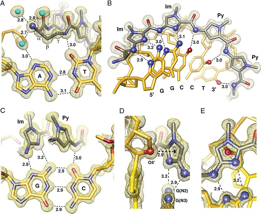
Chenoweth and Dervan PNAS 兩 August 11, 2009 兩 vol. 106 兩 no. 32 兩 13177Fig. 4. Direct and water-mediated noncovalent molecular recognition interactions. (A) Geometry of the ␣-amino turn interacting with the AT base pair through
water-mediated hydrogen bonding interactions. Structural basis for the turn preference for AT versus GC is demonstrated by the -methylene conformational
preference, which points down toward the DNA minor-groove floor within van der Waals contact distance of the adenine base. (B) Isolated view of one-half
of the macrocyclic-polyamide showing hydrogen bond distances made to the DNA minor groove floor by the imidazoles and amides of compound 1. (C) Im-Py
pair showing the mechanism for GC specificity. (D) Interaction of the O4⬘ oxygen of a deoxyribose sugar with the terminal imidazole aromatic ring through a
lone-pair- interaction. The sugar conformation is C2⬘-endo at the N-terminal imidazole of the polyamide with the sugar oxygen lone-pair pointing directly to
the centroid of the imidazole ring. The distance between the sugar oxygen and the ring centroid is 2.90 Å, which is less than the sum of the van der Waals radii
to any atom in the imidazole ring. Electrostatic potential maps calculated at the HF/3–21g* level of theory show the slightly electropositive nature of the
imidazole ring under these conditions (Fig. S6). (E) View of the O4⬘ deoxyribose oxygen atom looking through the imidazole ring showing the ring centroid
superimposed on the oxygen atom. All distances are reported in angstroms (Å), and all electron density maps are contoured at the 1.0 level (Im, imidazole;
Py, pyrrole).
unique weak interactions in the form of lone-pair- interactions Materials and Methods
(27, 28) between the center of the leading imidazole ring and the General. Chemicals and solvents were purchased from Sigma-Aldrich and
lone-pair of the adjacent deoxyribose O4⬘ oxygen (Fig. 4D and Hampton Research and were used without further purification. Water (18
E). This interaction is only observed for the terminal imidazole M⍀) was purified using a Millipore MilliQ purification system. Analytical
aromatic ring. Analysis of qualitative electrostatic potential high-performance liquid chromatography (HPLC) analysis was conducted on a
Beckman Gold instrument equipped with a Phenomenex Gemini analytical
surfaces substantiates the electropositive nature of the imidazole
column (250 ⫻ 4.6 mm, 5 m), a diode array detector, and the mobile phase
when buried in the minor groove of DNA and electrostatically consisted of a gradient of acetonitrile (MeCN) in 0.1% (vol/vol) aqueous
preturbed by specific interaction of the lone-pair with the trifluoroacetic acid (TFA). Preparative HPLC was performed on an Agilent 1200
exocyclic amine of guanine (Fig. 4 and Fig. S6) (29). system equipped with a solvent degasser, diode array detector, and a Phe-
The crystal structure highlights the DNA structural distortion nomenex Gemini column (5-m particle size, C18 110A, 250 ⫻ 21.2 mm, 5 m).
induced upon polyamide minor-groove binding and provides an A gradient of MeCN in 0.1% (vol/vol) aqueous TFA was used as the mobile
allosteric model for disrupting transcription factor-DNA inter- phase. UV-Vis measurements were made on a Hewlett-Packard diode array
spectrophotometer (Model 8452 A) and polyamide concentrations were mea-
faces in the promoters of selected genes. Allosteric control over
sured in 0.1% (vol/vol) aqueous TFA using an extinction coefficient of 69,200
transcription factor regulatory networks (30–32) by small mol- M⫺1䡠cm⫺1 at max near 310 nm. Matrix-assisted laser desorption/ionization
ecules that bind distinct locations on promoter DNA provides a
Downloaded by guest on January 20, 2022
time-of-flight mass spectrometry (MALDI-TOF MS) was performed on an
mechanism for inhibiting excess transcription factor activity (1, Applied Biosystems Voyager DR Pro spectrometer using ␣-cyano-4-hydroxy-
2, 19, 33). cinnamic acid as matrix.
13178 兩 www.pnas.org兾cgi兾doi兾10.1073兾pnas.0906532106 Chenoweth and DervanSynthesis and Purification. Polyamide 1 was synthesized by solid-phase syn- asymmetric unit. This data set was collected at SSRL beamline 11–1 with a MAR
thesis methods (34, 35) on oxime resin (SI Text and Fig. S1) and purified by Research imaging plate detector at wavelength 0.999 Å (Table S1).
reverse-phase HPLC (Fig. S2). Data were processed with MOSFLM (36) and SCALA (37) from the CCP4 suite
of programs (37). Both crystals were solved by direct methods using the SHELX
Oligonucleotide Purification and Crystallization. Oligonucleotides were pur- suite of programs (SHELXD) (38, 39). Model building and structure refinement
chased HPLC-purified from Trilink Biotechnologies. Before use, oligonucleo- was done with Coot (40) and REFMAC5 (41). The final polyamide-DNA complex
tides were de-salted using a Waters Sep-Pak cartridge (5 g, C-18 sorbent). The was refined to an R factor of 9.8% and an Rfree of 13.6%. The final DNA structure
Sep-Pak was prewashed with acetonitrile (25 mL, 3⫻) followed by MilliQ water was refined to an R factor of 10.9% and an Rfree of 14.3%. Anisotropic B factors
(25 mL, 3⫻). The oligonucleotide was dissolved in 5 mL 2.0 M NaCl and loaded were refined in the final stages and riding hydrogens included (Table S1).
directly onto the sorbent followed by a wash with 5 mL 2.0 M NaCl and 250 mL
MilliQ water. The oligonucleotide was eluted with acetonitrile:water (1:1) and Structure Analysis and Figure Preparation. DNA helical parameters were cal-
lyophilized to dryness. Single strand DNA was quantitated by UV-Vis spectros- culated using the program Curves (42). Molecular electrostatic potential maps
copy. Crystals were obtained after 2– 8 weeks from a solution of 0.5 mM duplex were calculated at the HF/3–21g* level using the Gamess program (Fig. S6)
DNA, 0.65 mM polyamide, 21% 2-methyl-2,4-pentanediol (MPD), 35 mM (43– 45). Distance measurements and least squares fitting procedures for
calcium acetate, 10 mM Tris, pH 7.5, equilibrated in sitting drops against a ring-centroid measurements were performed using UCSF Chimera (46) and
reservoir of 35% MPD at 4 °C. Crystals were collected in Hampton nylon Mercury (47). Structural figures were prepared using UCSF Chimera.
CryoLoops (10 m, 0.1 mm) and flash-cooled to 100 K before data collection.
ACKNOWLEDGMENTS. Synchrotron data were collected at Stanford Synchro-
Polyamides Data Collection, Structure determination, and Refinement. Poly- tron Radiation Laboratory (SSRL) beamlines 11–1 and 12–2. We thank Douglas
amide-DNA crystals grew in space group P1 with unit cell dimensions a ⫽ Rees valuable discussions, Jens Kaiser and Michael Day for their guidance with
22.500, b ⫽ 25.140, c ⫽ 29.090, ␣ ⫽ 66.53,  ⫽ 79.28, ␥ ⫽ 79.57, and 1 data collection and structure determination, and the staff of the SSRL for their
assistance during crystal screening and data collection. Operations at SSRL are
polyamide-duplex DNA complex in the asymmetric unit. This data set was
supported by the U.S. Department of Energy and the National Institutes of
collected at Stanford Synchrotron Radiation Laboratory (SSRL) beamline 12–2 Health. We acknowledge the Gordon and Betty Moore Foundation for sup-
with a MAR Research imaging plate detector at wavelength 0.97 Å. DNA only port of the Molecular Observatory at Caltech. This work was supported by
crystals grew in space group C2 (C 1 2 1) with unit cell dimensions a ⫽ 31.827, National Institutes of Health and a Kanel Foundation predoctoral fellowship
b ⫽ 25.636, c ⫽ 34.173, ␣ ⫽ 90,  ⫽ 116.72, ␥ ⫽ 90, and 1 DNA strand in the (to D.M.C.).
1. Dervan PB (2001) Molecular recognition of DNA by small molecules. Bioorg Med Chem 23. Zhang Q, et al. (2004) NMR structure of a cyclic polyamide-DNA complex. J Am Chem
9:2215–2235. Soc 126:7958 –7966.
2. Dervan PB, Edelson BS (2003) Recognition of the DNA minor groove by pyrrole- 24. Suto RK, et al. (2003) Crystal structures of nucleosome core particles in complex with
imidazole polyamides. Curr Opin Struct Biol 13:284 –299. minor groove DNA-binding ligands. J Mol Biol 326:371–380.
3. Trauger JW, Baird EE, Dervan PB (1996) Recognition of DNA by designed ligands at 25. Edayathumangalam RS, Weyermann P, Gottesfeld JM, Dervan PB, Luger K (2004)
subnanomolar concentrations. Nature 382:559 –561. Molecular recognition of the nucleosomal ‘‘supergroove.’’ Proc Natl Acad Sci USA
4. White S, Szewczyk JW, Turner JM, Baird EE, Dervan PB (1998) Recognition of the four 101:6864 – 6869.
Watson-Crick base pairs in the DNA minor groove by synthetic ligands. Nature 26. Heinemann U, Alings C (1989) Crystallographic study of one turn of G/C-rich B-DNA. J
CHEMISTRY
391:468 – 470. Mol Biol 210:369 –381.
5. Kielkopf CL, Baird EE, Dervan PB, Rees DC (1998) Structural basis for G.C recognition in 27. Egli M, Sarkhel S (2007) Lone pair-aromatic interactions: To stabilize or not to stabilize.
the DNA minor groove. Nat Struct Biol 5:104 –109. Acc Chem Res 40:197–205.
6. Kielkopf CL, et al. (1998) A structural basis for recognition of A.T and T.A base pairs in 28. Gallivan JP, Dougherty DA (1999) Can lone pairs bind to a system? The water-
the minor groove of B- DNA. Science 282:111–115. hexafluorobenzene interaction. Org Lett 1:103–106.
7. Hsu CF, et al. (2007) Completion of a programmable DNA- binding small molecule 29. Mecozzi S, West AP, Dougherty DA (1996) Cation- interactions in aromatics of
library. Tetrahedron 63:6146 – 6151. biological and medicinal interest: Electrostatic potential surfaces as a useful qualitative
8. Edelson BS, et al. (2004) Influence of structural variation on nuclear localization of guide. Proc Natl Acad Sci USA 93:10566 –10571.
DNA-binding polyamide-fluorophore conjugates. Nucleic Acids Res 32:2802–2818. 30. Hogan M, Dattagupta N, Crothers DM (1979) Transmission of allosteric effects in DNA.
9. Nickols NG, Jacobs CS, Farkas ME, Dervan PB (2007) Improved nuclear localization of Nature 278:521–524.
DNA-binding polyamides. Nucleic Acids Res 35:363–370. 31. Panne D, Maniatis T, Harrison SC (2007) An atomic model of the interferon-beta
10. Hsu CF, Dervan PB (2008) Quantitating the concentration of Py-Im polyamide- fluo-
enhanceosome. Cell 129:1111–1123.
rescein conjugates in live cells. Bioorg Med Chem Lett 18:5851–5855.
32. Lavelle C (2008) DNA torsional stress propagates through chromatin fiber and partic-
11. Gottesfeld JM, et al. (2001) Sequence-specific recognition of DNA in the nucleosome
ipates in transcriptional regulation. Nat Struct Mol Biol 15:146 –154.
by pyrrole-imidazole polyamides. J Mol Biol 309:615– 629.
33. Moretti R, et al. (2008) Targeted chemical wedges reveal the role of allosteric DNA
12. Suto RK, et al. (2003) Crystal structures of nucleosome core particles in complex with
modulation in protein-DNA assembly. ACS Chem Biol 3:220 –229.
minor groove DNA-binding ligands. J Mol Biol 326:371–380.
34. Baird EE, Dervan PB (1996) Solid phase synthesis of polyamides containing imidazole
13. Olenyuk BZ, et al. (2004) Inhibition of vascular endothelial growth factor with a
and pyrrole amino acids. J Am Chem Soc 118:6141– 6146.
sequence-specific hypoxia response element antagonist. Proc Natl Acad Sci USA
35. Belitsky JM, Nguyen DH, Wurtz NR, Dervan PB (2002) Solid-phase synthesis of DNA
101:16768 –16773.
binding polyamides on oxime resin. Bioorg Med Chem 10:2767–2774.
14. Kageyama Y, et al. (2006) Suppression of VEGF transcription in renal cell carcinoma cells
36. Leslie AGW (1992) Recent changes to the MOSFLM package for processing film and
by pyrrole-imidazole hairpin polyamides targeting the hypoxia responsive element.
image plate data. Newsletter on Protein Crystallography 26.
Acta Oncol 45:317–324.
37. Collaborative Computational Project, Number 4 (1994) The CCP4 suite: Programs for
15. Nickols NG, Jacobs CS, Farkas ME, Dervan PB (2007) Modulating hypoxia-inducible
protein crystallography. Acta Crystallogr D 50:760 –763.
transcription by disrupting the HIF-1-DNA interface. ACS Chem Biol 2:561–571.
16. Nickols NG, Dervan PB (2007) Suppression of androgen receptor-mediated gene ex- 38. Sheldrick GM (2008) A short history of SHELX. Acta Crystallogr A 64:112–122.
pression by a sequence-specific DNA-binding polyamide. Proc Natl Acad Sci USA 39. Schneider TR, Sheldrick GM (2002) Substructure solution with SHELXD. Acta Crystallogr
104:10418 –10423. D 58:1772–1779.
17. Matsuda H, et al. (2006) Development of gene silencing pyrrole-imidazole polyamide 40. Emsley P, Cowtan K (2004) Coot: Model-building tools for molecular graphics. Acta
targeting the TGF-beta1 promoter for treatment of progressive renal diseases. J Am Crystallogr D 60:2126 –2132.
Soc Nephrology 17:422– 432. 41. Murshudov GN, Vagin AA, Dodson EJ (1997) Refinement of macromolecular structures
18. Yao EH, et al. (2008) Novel gene silencer pyrrole- imidazole polyamide targeting by the maximum-likelihood method. Acta Crystallogr D 53:240 –255.
lectin-like oxidized low-density lipoprotein receptor-1 attenuates restenosis of the 42. Lavery R, Sklenar H (1997) Curves 5.2: Helical analysis of irregular nucleic acids.
artery after injury. Hypertension 52:86 –92. Biochimie Theorique CNRS URA 77.
19. Nguyen-Hackley DH, et al. (2004) Allosteric inhibition of zinc-finger binding in the 43. Schmidt MW, et al. (1993) General atomic and molecular electronic structure system.
major groove of DNA by minor-groove binding ligands. Biochemistry 43:3880 –3890. J Comput Chem 14:1347–1363.
20. de Clairac RPL, Geierstanger BH, Mrksich M, Dervan PB, Wemmer DE (1997) NMR 44. Binkley JS, Pople JA, Hehre WJ (1980) Self-consistent molecular orbital methods. 21.
characterization of hairpin polyamide complexes with the minor groove of DNA. J Am Small split-valence basis sets for first-row elements. J Am Chem Soc 102:939 –947.
Chem Soc 119:7909 –7916. 45. Pietro WJ, et al. (1982) Self- consistent molecular orbital methods. 24. Supplemented
21. Hawkins CA, et al. (2000) Controlling binding orientation in hairpin polyamide DNA small split-valence basis sets for second-row elements. J Am Chem Soc 104:5039 –5048.
complexes. J Am Chem Soc 122:5235–5243. 46. Pettersen EF, et al. (2004) UCSF chimera—a visualization system for exploratory re-
22. Hawkins CA, Baird EE, Dervan PB, Wemmer DE (2002) Analysis of hairpin polyamide search and analysis. J Comput Chem 25:1605.
Downloaded by guest on January 20, 2022
complexes having DNA binding sites in close proximity. J Am Chem Soc 124:12689 – 47. Macrae CF, et al. (2006) Mercury: Visualization and analysis of crystal structures. J Appl
12696. Cryst 39:453– 457.
Chenoweth and Dervan PNAS 兩 August 11, 2009 兩 vol. 106 兩 no. 32 兩 13179You can also read


