Adipose tissue is a source of bro-adipogenic progenitors for regenerating skeletal muscles
←
→
Page content transcription
If your browser does not render page correctly, please read the page content below
Adipose tissue is a source of fibro-adipogenic
progenitors for regenerating skeletal muscles
Quentin Sastourne-Arrey
 INSERM
Amandine Girousse
 INSERM
Virginie Bourlier
 INSERM, UMR1048, Institute of Metabolic and Cardiovascular Diseases, Obesity Research Laboratory,
Toulouse
Sylvie Monferran
 INSERM
Marta Gil-Ortega
 INSERM
Enda Murphy
 School of Health and Human Performance
Claire Laurens
 INSERM, UMR1048, Institute of Metabolic and Cardiovascular Diseases, Obesity Research Laboratory,
Toulouse
Audrey Varin
 STROMALab
Guissard Christophe
 INSERM/RESTORE
Corinne Barreau
 INSERM/RESTORE
Mireille André
 INSERM/RESTORE
Noémie Juin
 INSERM/RESTORE
Marie Marques
 Institut National de la Santé et de la Recherche Médicale (Inserm), UMR1048, Institute of Metabolic and
Cardiovascular Diseases, Toulouse
Benoit Chaput
 Toulouse Hospital
Cedric Moro

                                                Page 1/27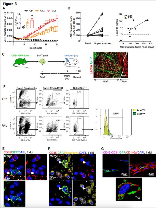
Institute of Metabolic and Cardiovascular Diseases, Inserm/Paul Sabatier University UMR1297
https://orcid.org/0000-0003-4294-0597
Donal O’Gorman
Dublin City University
Louis Casteilla
Toulouse University
Coralie Sengenes ( coralie.sengenes@inserm.fr )
INSERM https://orcid.org/0000-0002-4820-6660
Article
Keywords: Adipose stromal cells, Fibro/adipogenic progenitors, platelets, cell migration, acute muscle
stress
Posted Date: August 12th, 2021
DOI: https://doi.org/10.21203/rs.3.rs-754516/v1
License: This work is licensed under a Creative Commons Attribution 4.0 International License.
Read Full License
Version of Record: A version of this preprint was published at Nature Communications on January 5th,
2023. See the published version at https://doi.org/10.1038/s41467-022-35524-7.
Page 2/27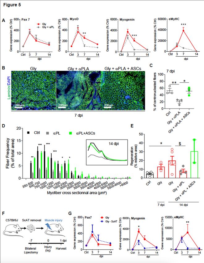
Abstract
Fibro adipogenic progenitors (FAPs) play a crucial role in skeletal muscle regeneration, as they generate a
favorable niche that allows satellite cells to perform efficient muscle regeneration. After muscle injury,
FAP content increases rapidly within the injured muscle, the origin of which has been attributed to their
proliferation. Recently, single-cell RNAseq approaches have revealed phenotype and functional
heterogeneity in FAPs. Here we report that FAP-like cells residing in subcutaneous adipose tissue (ScAT),
the adipose stromal cells (ASCs), are rapidly released from ScAT in response to muscle injury. In parallel,
we show in healthy humans that exercise-induced muscle stress response triggers the migration of
human native ASCs. Additionally, we find that released ASCs infiltrate the damaged muscle, via a platelet
dependent mechanism and that blocking ASC infiltration impairs muscle regeneration. Collectively, our
data reveal that ScAT is an unsuspected physiological reservoir of regenerative cells that support skeletal
muscle regeneration.

Introduction
Skeletal muscle exhibits a remarkable regenerative capacity in adult mammals and a large number of
studies are running to better characterize and understand the underlying mechanisms controlling this
process. The regenerative potential of skeletal muscle relies on a pool of resident adult stem cells, the
satellite cells which proliferate and differentiate to allow muscle growth and remodeling in response to
exercise or following trauma 1–3. Recent studies identified mesenchymal progenitors, termed “fibro-
adipogenic progenitors” (FAPs), providing a key functional support to satellite cells 4–7. Beside their
supportive role, the regulation of FAP content is also crucial since their absence leads to regeneration
impairment, whereas FAP maintenance in the late phase of regeneration leads to fibrosis and/or fatty
degeneration of the injured muscle 8–10. The evolution of FAP number upon damage exhibits distinct
wave 9. A first wave occurring 1 day post-injury (dpi) followed by a second wave at 3 dpi 9. Interestingly,
single cell analysis technology largely revealed the heterogeneity of FAPs 11–14 and also reported the first
wave of FAP. Indeed, Oprescu et al. elegantly identified a subpopulation of “activated” FAPs in the very
early phases following muscle injury (0.5 dpi)14. These “activated” FAPS were considered by the authors
to be muscle residents, although direct demonstration was not done. Moreover, it is very well described
that FAPs proliferation starts 2dpi for a period of 72–96 h 4,13,15−17. Consequently, the cellular origin of
the first wave of FAP increase observed at 1dpi is not characterized and has not been studied so far 8,9.

White adipose tissue (AT) houses a mesenchymal cell population, which resembles FAPs, the adipose
stromal cells (ASCs). Various studies, including ours show that ASCs exhibit similar cell surface antigen
combination, same clonogenic activity and differentiation potentials 18–20. ASCs attract a lot of interest
due to their regenerative potential and represent very promising tools for cell-based therapies 21. Whether
FAPs and ASCs are distinct cell types remains unclear 22–24. The circulation of stromal cells has been
reported 25,26 and we and others have shown that ectopic fat cells partly arise from these circulating
stromal cells particularly upon weight gain27–29. We demonstrated that subcutaneous AT (ScAT) releases
                                                   Page 3/27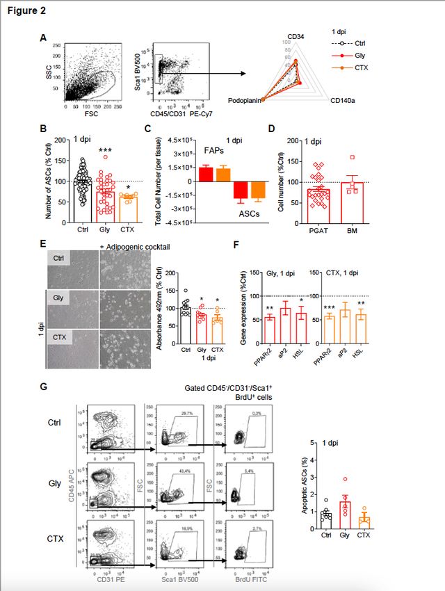
ASCs in response to inflammatory stimuli18,30 or to high fat diet28. Consequently, we questioned here
whether the first wave of FAP number increase in response to acute muscle injury is the result of ASC
release from ScAT followed by their further infiltration into the damaged muscle.
In the present study, we report that ScAT releases ASCs in response to acute muscle damage and that
released ASCs infiltrate the injured muscle via a platelet dependent mechanism. We also show that
blunting the infiltration of ASCs impairs muscle regeneration suggesting that ScAT is an unsuspected
partner of muscle regeneration.
Results
Acute muscle injury induces a rise in FAP muscle content mirrored by a specific and rapid decrease in
ASC content in the ScAT
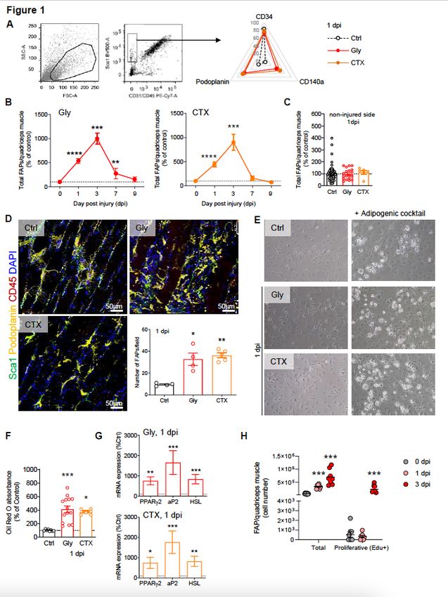
The number of FAPs was evaluated in the quadriceps muscle 24h following damage using glycerol (Gly)
or myotoxin (cardiotoxin, CTX) both well characterized murine models of muscle injury (Figure S1A,B).
The cell composition of whole muscle-derived stroma-vascular fraction (SVF) was analyzed by flow
cytometry and FAPs content (identified as Sca1+/CD34+/CD140a+/CD31−/CD45− cells (Fig. 1A)) was
measured from 1 to 9 days post-injury (dpi) (Fig. 1B). As previously reported 4,5,31 and whatever the
muscle injury model, muscle FAP number peaked at 3 dpi and reached back basal value at 9 dpi (Fig. 1B).
Also in accordance with previous studies, FAP number drastically increased by 1 dpi in the injured muscle
(> 5 fold, Fig. 1A-B, without being affected in the controlateral non-injured side, Fig. 1C) and we noticed
that most of them were podoplanin-positive (Fig. 1A). The rise in FAPs at 1 dpi was not due to their
proliferation, since no differences in EdU incorporation -a nucleotide analogue- between FAPs from
injured and control mice were detected (Fig. 1C, S1C), whereas other cell population, such as leucocytes
start proliferating by 1 dpi (Figure S1D). The FAP increase evidenced by flow cytometry 1 dpi was then
confirmed in situ, by immuhistochemistry of the damaged muscle (Fig. 1D, S1E). Moreover, given that
FAPs are the only cell type expressing adipogenic potential in the muscle 5,24,31,32, in vitro adipogenesis of
whole muscle-derived SVF from control or 1 dpi injured animals would directly and functionally reflects
the quantity of FAPs inside (Fig. 1E). Upon adipogenic induction muscle-derived SVF originating from
injured animals clearly accumulated more lipids and expressed higher levels of adipogenic markers
(Fig. 1F) confirming the rise in FAP content in the injured muscle at 1 dpi.
In parallel, we studied the evolution of ASCs content in ScAT following muscle injury. ASCs were identified
as Sca1+/CD34+/CD140a+/CD31−/CD45− cells as previously reported (Fig. 2A)18,19. Strikingly,
concomitant with the increase in FAP number in the injured muscle, the number of ASCs significantly
decreased by 20–25% in the ScAT at 1dpi (Fig. 2B), both following Gly or CTX damage. Importantly, the
gain in muscle FAP number was found equivalent to the loss in ScAT ASCs, whatever the injury model
used (Fig. 2C). To verify that muscle injury specifically triggered the decrease in ASCs and to rule out any
impact of the injury procedure on the cell content of ScAT, NaCL was injected into the quadriceps muscle.
NaCL neither induced muscle damage (Figure S2A, B) nor modified ASC and/or leucocyte content in ScAT
Page 4/27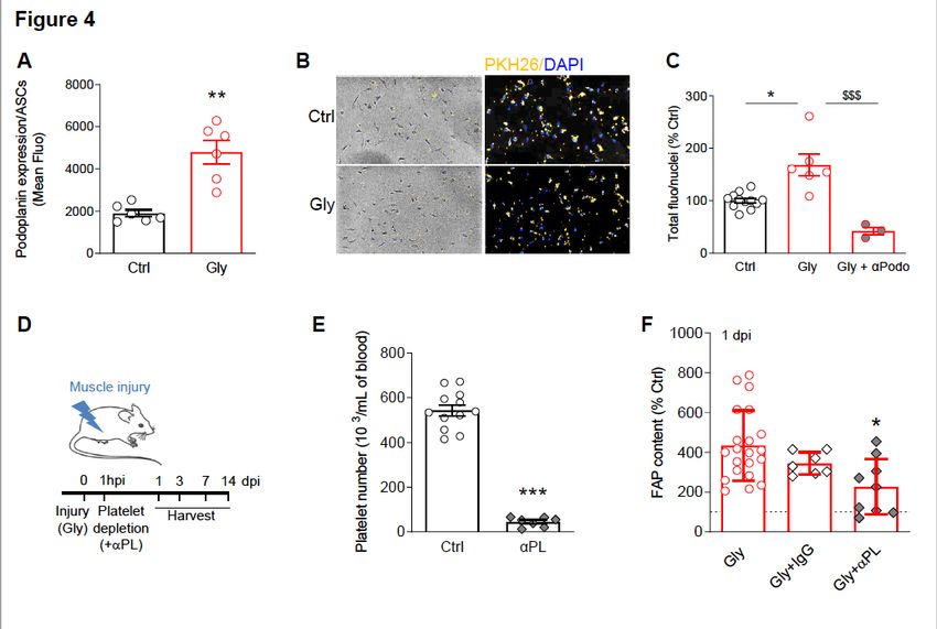
(Figure S2C, D), demonstrating that muscle injury per se triggers the fall in ASCs content observed in the
ScAT at 1 dpi. Moreover, the ScAT was specifically impacted by muscle damage since neither perigonadic
AT (PGAT) nor the bone marrow (BM), another tissue source of MSCs, were affected (Fig. 2D). To
examine whether programmed cell death could be responsible of the drop of ASCs content in the ScAT,
we measured DNA fragmentation (TUNEL) after Gly or CTX muscle damage at 1 dpi. Frequencies of
TUNEL positive ASCs were not modified in the ScAT (Fig. 2E-F). As for the muscle, ASCs are the only cell
population in AT-derived SVF capable of adipogenic potential 19,33. Therefore, in vitro adipogenesis of
whole ScAT-derived SVF from control or injured animals were tested and compared (Fig. 2G) to assess
ASC content. In agreement with flow cytometry results the drop in ASCs content was associated with a
decrease in lipid accumulation and expression of adipogenic markers in ScAT-derived SVF originating
from injured animals (Fig. 2G-H). To rule out putative effects of CTX or Gly on the metabolic status of the
animals, and as thus on the biology of AT, the body weights as well as plasmatic glucose levels of the
animals were monitored during 28 days following muscle injury. Neither the body weights of the animals
nor their glycaemia were modified by muscle lesion (Figure S2E, F). Collectively our data strongly suggest
that in response to quadriceps injury the ScAT releases ASCs that might infiltrate the injured muscle
within 24h after injury, corresponding to the first wave of FAP rise.

The muscle stress response after injury or exercise triggers ASC chemotaxis in humans and mice

We hypothesized that the drop in ASCs in response to muscle injury resulted from their egress from the
ScAT. Consequently, we investigated in vitro whether serum from injured animals could trigger ASC
chemotaxis. We thus collected the serum from control or injured (1dpi) animals and ASCs chemotaxis
was studied and compared. We found that whatever the injury model, serum from injured animals
strongly induced the chemotaxis of ASCs (Fig. 3A-B), supporting the notion that muscle injury may trigger
the egress of ASCs from ScAT via the production of blood circulating factors. Whether such mechanism
may occur in human was also questioned. To do so, we took advantage of blood samples originating
from a clinical study investigating the impact of an acute bout of continuous exercise 34 in young active
men (Table 1) as a putative source of muscle damage and/or fatigue. As for mice, the chemotactic
response of human native ASCs towards the serum of each individuals before and after the exercise wad
studied. As shown in Fig. 3C, the chemotactic response of ASCs toward serum at post-exercise was
increased by 2 to 5 fold compared to the pre-exercise condition, though heterogeneity was observed from
one individual to another. We then wondered whether such a disparity could be explained by inter-
individual variability to exercise-mediated stress response. Growth differentiation factor 15 (GDF-15) is
induced under stress conditions, to maintain cell and tissue homeostasis 35. Recent studies also
suggested that GDF15 is an exerkine 34 exhibiting possible protective role in exercise-induced muscle
injury or inflammation36,37. Therefore, circulating blood levels of GDF15 were measured before and after
exercise to assess the exercise-induced stress response intensity of each individuals (Table 2). In
agreement with our hypothesis and the results obtained in mice, the chemotactic activity of ASCs is
strongly correlated with GDF15 levels akin to an index of the exercise-induced stress response (Fig. 3D).
These findings indicate that blood factors released after muscle injury/stress may trigger the

                                                 Page 5/27
mobilization of ASCs from the ScAT, in both mice and humans, further reaching the injured or stressed
muscle.
Table 1
Anthropometry and body composition of the individuals enrolled in the clinical study
Subject Age Weight Height BMI %Fat Mass Fat Mass Free Fat Mass
ID (kg) (cm) (kg/m²) (kg) (kg)
A 22 79 184 23.33 12.16 8.5 62.1
B 19 87 193 23.36 17 13.6 63.9
D 19 70 171 23.94 17.8 11.1 49.4
I 27 87.2 185 24,54 15.9 12.8 64.8
J 24 79.3 191 21.7 15.6 10.6 60.7
M 29 71.3 176 23.01 14.3 10.4 59.2
Table 2
GDF 15 blood levels in individuals before (pre)
and after (post) an exercise on a bicycle
ergometer at 60% VO2 peak for 1-hr. Δ
corresponds to the difference of GDF15 blood
level, before and after the exercise.
GDF15 (pg/ml)
Subject ID Pre Post Δ (Post - Pre)
A 153,4 168,1 14,7
B 417,0 457,5 40,5
D 209,4 313,2 103,8
I 341,2 446,5 105,3
J 451,6 437,7 -14,0
M 263,8 329,5 65,6
Upon muscle injury ASCs egress the ScAT and infiltrate the damaged muscle
To determine whether ASCs can leave the ScAT in response to muscle injury to further infiltrate it, we
performed in vivo experiment in mice. However, due to the absence of unique specific ASC marker, no
mouse model is so far available to visualize, in vivo, the trafficking of native ASCs from AT to any other
tissue compartments. To overcome this obstacle we took advantage of the Tg(Cd34-
EGFP)MF6Gsat/Mmucd (referred to as CD34-GFP, an ASC surface marker) from which a piece of ScAT
was grafted into the ScAT of a non-GFP recipient mouse (Fig. 3E). Fat graft revascularization, an index of
Page 6/27viability, was verified by using retro-orbital injection of Rhodamin-lectin into grafted animals (Fig. 3E,
S3A). At day 7 post-graft, muscle was damaged and the presence of GFP+ cells into the injured muscles
was assessed at 1 dpi. CD31−/CD45−/Sca-1+/GFP+ cells were found in injured muscles by flow cytometry
(roughly 8% of CD31−/CD45−/Sca-1+ cells, Fig. 3F, S3B) while by contrast no GFP cells were detected
neither in the muscle of a CD34-GFP ScAT grafted non-injured control animal (Fig. 3F) nor in a non-
grafted mouse (Figure S3B). Further analysis demonstrated that GFP+-ASCs were mainly in the Sca-1 low
subpopulation (Fig. 3D, right panel). This result clearly demonstrates that following muscle injury ScAT
releases CD34+ cells, which infiltrate specifically the damaged muscle. Further analysis of their cell
surface immunophenotype showed that the infiltrated GFP cells were
Sca1+/CD34+/Podoplanin+/CD140a+/CD31−/CD45−, corresponding to ASCs (Fig. 3E-G). To verify that
GFP expression was specific, FAPs from grafted injured (1 dpi) or non-injured animals were sorted and
genomic GFP expression was analysed. In agreement with flow cytometry and immunohistochemistry
data, genomic GFP expression was found in FAPs of injured muscles (Figure S3C), in contrast to control
muscles from grafted animals (Fig. 3G) or other distant organs (Figure S3D). Finally, we performed
immunohistochemistry analysis of muscle originating from grafted animals at 1dpi and showed the
presence of GFP+-ASC- (Fig. 3E) that also expressed podoplanin (Fig. 3F) and CD140a (Fig. 3G) such as
observed in previous cytometry experiments. Of note, the observation of infiltrated ASCs post muscle
injury (1 dpi) was also made with the ScAT graft from another fluorescent mouse model, the
Rosa26mT/mG animals (Figure S3E). Here again, we observed ~ 10% of Tomato+-cells among the
CD45−/CD31−/Sca-1+ cells, corresponding to infiltrated ASCs originating from the red fluorescent mT/mG
graft pad (Figure S3F). Interestingly, the Tomato+-cells laid preferentially among the CD45-/CD31-/Sca-
1low cell subpopulation (Figure S3F, lower panel) such as observed previously in Fig. 3D. Altogether, these
findings demonstrate that the first wave of FAP increase following muscle injury at 1dpi largely results
from ASC mobilization and infiltration into the damaged muscle.
ASC early infiltration into the injured muscle involves platelets and is crucial for effective muscle
regeneration
Our current understanding of in vivo trafficking of native ASCs/MSCs is still very incomplete38 and mostly
derives from in vitro experiments. However, some studies indicate that platelets can control MSC
trafficking and/or recruitment to sites of injury 39–41. Interestingly, we observed an up-regulation of
podoplanin expression at the cell surface of ASCs from injured animals measured by flow cytometry
(Fig. 4A). Podoplanin is an endogenous ligand for C-type lectin-like receptor 2 (CLEC-2) which is an
essential platelet-activating receptor 42. Consequently, we next assessed whether ASCs and platelets
interaction is necessary for the recruitment of ASCs to the injured muscle. We used an in vitro assay
whereby platelets, isolated from control or injured animals (1 dpi) were fluorescently labelled and co-
incubated with ASCs (isolated from ScAT). The adhesion of platelets to ASCs was evaluated and
compared. The results show that platelets originating from injured animals adhered more to ASCs in vitro
(Fig. 4B-C), a physical interaction that was completely abolished when an antibody directed against
Page 7/27podoplanin was added (Fig. 4C). Given our results showing that both ASC mobilization and infiltration
strongly account for the first wave of FAP rise in injured muscle and that platelets originating from injured
animals interact more with ASCs, we next investigated the consequences of platelet depletion in vivo on
FAP augmentation following muscle injury. Muscle damage was induced as described earlier and platelet
depletion was performed 1h later (Fig. 4D-E, S4A). FAP content was then quantified by flow cytometry at
1dpi, according to the immunophenotype described earlier. Our data show that platelet depletion
diminished by more than twofold the rise of FAPs in the muscle at 1 dpi (Fig. 4F) suggesting that the
infiltration of ASCs into the injured muscle involves platelets.
Previous studies have demonstrated that FAP facilitate myogenesis by promoting differentiation of
muscle progenitors and myotube formation 4,10,17. Hence, we next studied the impact of FAP rise
disruption on the muscle regeneration process. To do so, the expression of genes involved in the
molecular program of muscle regeneration was studied following muscle injury and concomitant platelet
depletion until 14 dpi. Though the expression patterns of early genetic markers of the muscle
regeneration program (Pax7, Myf5) were not modified (Fig. 4G), the expression of late myogenic markers
(MyoD, Myogenin, eMyHC) was clearly modified in platelet depleted animals compared with controls
(Fig. 4H). These results strongly suggest that when the early infiltration of ASCs is disrupted, the
regeneration program is impaired. To reinforce these results, we quantified the number of newly
synthesized centrally nucleated fibers, a marker of the regenerative process and found that the proportion
of centrally nucleated fibers was dramatically diminished in platelet depleted animals (Fig. 5A-B). Next,
we quantified the cross-sectional area distribution of regenerating fibers with centralized nuclei, as well as
the total area of these regenerating fibers and we showed that they were dramatically diminished in
animals where the rise in FAP was impaired (Fig. 5C, D). Upon activation, platelets secrete more than 300
active substances, among which is PDGF, an important factor for FAPs expansion and survival 15,43−45.
To rule out that a reduction in PDGF was the main effect of platelet depletion rather than the absence of
platelet per se, ScAT from both sides were removed in order to take away the reservoir of ASCs. Muscle
injury was performed and the expression of genes involved in the molecular program of muscle
regeneration was studied. As for platelet depleted animals, the expression pattern of Pax7 was not
modified (Fig. 5E) while the one of late myogenic markers (myogenin, eMYHC) was clearly affected
(Fig. 5E), strongly suggesting that an interaction between platelets and ASCs is necessary. To finally
demonstrate that ASCs early infiltration was responsible for both early FAP rise and effective muscle
regeneration, ASCs were injected into the injured muscle (1h after muscle injury) of platelet depleted
animals in order to mimic the first wave of FAP increase. The number of newly synthesized centrally
nucleated fibers, the cross-sectional area distribution of regenerating fibers and the total area of
regenerating fibers were evaluated and the injection of ASCs completely restored those parameters
(Fig. 5B-D) strongly supporting a proregenerative role of ASC early infiltration in the damaged skeletal
muscle.7
Discussion
Page 8/27The regenerative capacity of skeletal muscle mostly relies on satellite cells (SCs), which proliferate in
response to exercise or following myotrauma to repair the injured muscle 3,46. However, in the past
decade many other cell types have been shown to contribute to this process in order to maintain skeletal
muscle integrity and functions 47.
Among those diverse and heterogeneous cell types, resident mesenchymal progenitors named fibro-
adipogenic progenitors (FAPs) have emerged as key players in skeletal muscle regeneration and disease
by providing functional support to SCs to perform efficient muscle regeneration 7,48. Upon muscle injury,
FAPs become activated and expand rapidly 4,5. Indeed, the number of FAPs peaks 96h post-muscle
damage 9 together with their proliferative activity 4. Surprisingly, while FAPs proliferation is not yet
activated 4,5,8,9,49, FAPs number dramatically increases within the 24h following muscle injury 8,9,11,47. The
results presented here provide a mechanism that explains the rapid FAP number increase observed 24h
after muscle injury without proliferation and could correspond to the subpopulation of “activated” FAPs
reported by Oprescu et al14. Indeed, we show that in response to acute muscle injury ASCs egress ScAT
and infiltrate the damaged muscle, a mechanism necessary for efficient muscle regeneration. Our data
also suggest that such a mechanism may also occur in humans in response to exercise-induced muscle
stress response.
FAPs and ASCs share many molecular and functional properties with MSCs 47,50. Importantly, the lack of
unique specific marker so far prevents the specific genetic targeting of these cell types and as thus the
simultaneous investigation of their fates in vivo. To overcome this obstacle, we have developed and used
a ScAT grafting approach, to track specifically endogenous ASCs in vivo. To the best of our knowledge,
using this model allowed us to visualize for the first time the specific mobilization and homing of ASCs
from ScAT within injured muscle. Interestingly, it was recently identified that Drosophila fat body cells, the
equivalent to the vertebrate adipocyte precursors, are also motile and migrate to distant wounded sites to
drive repair 51. Until now, FAPs are considered as resident-muscle progenitors23, however, we propose here
that a subpopulation of mesenchymal progenitors originating from ScAT participates to the pool of FAPs.
Various recent studies addressed the question of muscle resident cell heterogeneity in homeostasis and
regenerative conditions using single cell analysis 11,14,52−54. In that context, Malecova et al. identified in
the mouse the appearance of a subpopulation of Tie2lowFAPs within the 24h following muscle injury,
which they considered as reflecting cell state of FAPs during muscle regeneration 11. The analogous in
human samples was recently identified and it expresses FBN1 gene (fibrillin 1) 54. Based on our results it
is tempting to speculate that this FAP subpopulation originates from ScAT rather than being a transitory
cell state of resident FAPs. Also, it has been reported that FAPs can differentiate into brown-like
adipocytes 55,56, whether the muscle infiltrating ASCs exhibit a brown/beige adipogenic potential remains
to be determined particularly in light of the closer association between brown adipocytes and the skeletal
muscle lineage 31,57.
The mechanisms controlling the trafficking of endogenous MSCs and/or ASCs are poorly understood and
our current understanding mostly derives from in vitro studies 38. We show here that in response to
Page 9/27muscle injury, ASCs within ScAT overexpress podoplanin, which is a ligand for the platelet receptor CLEC-
2 (for C-type lectin-like receptor 2) 58. In agreement with this result, several studies reported that the
majority of infused MSCs can be found within the circulation in close contact with platelets 40,41 to
facilitate their homing to inflamed tissues59. Our data show that platelets originating from muscle-injured
animals adhere more in vitro to ASCs and that neutralizing the CLEC-2/podoplanin interaction completely
abolished this effect, suggesting a role for platelets in ASC trafficking in vivo. This was confirmed in
platelet-depleted animals where FAP increase was strongly inhibited in response to muscle injury.
Platelets maintain endothelial permeability and the trafficking of infused MSCs to inflamed sites is
facilitated by increased endothelial permeability 60. However, it was recently demonstrated that the
extravasation of infused MSCs to inflamed sites is not dependent on endothelial gaps or permeability 41.
Hence, the precise mechanisms by which platelets control ASC egress and/or infiltration are still unclear.
In conclusion, our work identifies a novel and unsuspected role of ScAT. These findings introduce the
concept of adipose tissue as an endogenous supplier of regenerative cells allowing skeletal muscle to
regenerate efficiently. The well-timed clearance of FAPs during the regeneration process is essential 9.
Indeed, abnormal persistence of FAPs leads to excessive matrix deposition and thus to muscle fibrosis
contributing to subsequent pathogenesis 7,61. Our work strongly supports a model in which the finely
tuned progression of FAP increase is necessary for healthy muscle regeneration where ScAT plays a
crucial role in this precise temporal succession.
Declarations
Acknowledgments.
We thank the Cell sorting platform of RESTORE and Marie-Laure Renoud for technical help. We also
thank the Cellular Imaging Facility-I2MC/TRI platform and Alexia Zakaroff-Girard and Elodie Riant as well
as Imag’IN platform and François-Xavier Frenois for helpful assistance.
References
1. Chargé, S. B. P. & Rudnicki, M. A. Cellular and molecular regulation of muscle regeneration. Physiol.
Rev. 84, 209–238 (2004).
2. Hawke, T. J. & Garry, D. J. Myogenic satellite cells: physiology to molecular biology. J Appl Physiol
(1985) 91, 534–51 (2001).
3. Mauro, A. Satellite cell of skeletal muscle fibers. J Biophys Biochem Cytol 9, 493–5 (1961).
4. Joe, A. W. B. et al. Muscle injury activates resident fibro/adipogenic progenitors that facilitate
myogenesis. Nat. Cell Biol. 12, 153–163 (2010).
5. Uezumi, A., Fukada, S., Yamamoto, N., Takeda, S. & Tsuchida, K. Mesenchymal progenitors distinct
from satellite cells contribute to ectopic fat cell formation in skeletal muscle. Nat Cell Biol 12, 143–
Page 10/2752 (2010).
6. Wosczyna, M. N., Biswas, A. A., Cogswell, C. A. & Goldhamer, D. J. Multipotent progenitors resident in
the skeletal muscle interstitium exhibit robust BMP-dependent osteogenic activity and mediate
heterotopic ossification. J Bone Miner Res 27, 1004–17 (2012).
7. Theret, M., Rossi, F. M. V. & Contreras, O. Evolving Roles of Muscle-Resident Fibro-Adipogenic
Progenitors in Health, Regeneration, Neuromuscular Disorders, and Aging. Front. Physiol. 12, (2021).
8. Fiore, D. et al. Pharmacological blockage of fibro/adipogenic progenitor expansion and suppression
of regenerative fibrogenesis is associated with impaired skeletal muscle regeneration. Stem Cell Res
17, 161–169 (2016).
9. Lemos, D. R. et al. Nilotinib reduces muscle fibrosis in chronic muscle injury by promoting TNF-
mediated apoptosis of fibro/adipogenic progenitors. Nat. Med. 21, 786–794 (2015).
10. Murphy, M. M., Lawson, J. A., Mathew, S. J., Hutcheson, D. A. & Kardon, G. C. Satellite cells,
connective tissue fibroblasts and their interactions are crucial for muscle regeneration. Development
(Cambridge, England) 138, 3625–3637 (2011).
11. Malecova, B. et al. Dynamics of cellular states of fibro-adipogenic progenitors during myogenesis
and muscular dystrophy. Nature communications 9, 3670 (2018).
12. Stumm, J. et al. Odd skipped-related 1 (Osr1) identifies muscle-interstitial fibro-adipogenic
progenitors (FAPs) activated by acute injury. Stem Cell Research 32, 8–16 (2018).
13. Scott, R. W., Arostegui, M., Schweitzer, R., Rossi, F. M. V. & Underhill, T. M. Hic1 Defines Quiescent
Mesenchymal Progenitor Subpopulations with Distinct Functions and Fates in Skeletal Muscle
Regeneration. Cell Stem Cell 25, 797–813.e9 (2019).
14. Oprescu, S. N., Yue, F., Qiu, J., Brito, L. F. & Kuang, S. Temporal Dynamics and Heterogeneity of Cell
Populations during Skeletal Muscle Regeneration. iScience 23, 100993 (2020).
15. Contreras, O. et al. Cross-talk between TGF-β and PDGFRα signaling pathways regulates the fate of
stromal fibro–adipogenic progenitors. Journal of Cell Science 132, (2019).
16. Dulauroy, S., Di Carlo, S. E., Langa, F., Eberl, G. & Peduto, L. Lineage tracing and genetic ablation of
ADAM12(+) perivascular cells identify a major source of profibrotic cells during acute tissue injury.
Nat Med 18, 1262–70 (2012).
17. Mathew, S. J. et al. Connective tissue fibroblasts and Tcf4 regulate myogenesis. Development 138,
371–84 (2011).
18. Gil-Ortega, M. et al. Native adipose stromal cells egress from adipose tissue in vivo: evidence during
lymph node activation. Stem Cells 31, 1309–1320 (2013).
19. Rodeheffer, M. S., Birsoy, K. & Friedman, J. M. Identification of white adipocyte progenitor cells in
vivo. Cell 135, 240–9 (2008).
20. Tang, W. et al. White fat progenitor cells reside in the adipose vasculature. Science 322, 583–6
(2008).
Page 11/2721. Gimble, J. M., Bunnell, B. A., Chiu, E. S. & Guilak, F. Concise review: Adipose-derived stromal vascular
fraction cells and stem cells: let’s not get lost in translation. Stem Cells 29, 749–54 (2011).
22. Arrighi, N. et al. Characterization of adipocytes derived from fibro/adipogenic progenitors resident in
human skeletal muscle. Cell Death Dis 6, e1733 (2015).
23. Judson, R. N., Zhang, R.-H. H. & Rossi, F. M. Tissue-resident mesenchymal stem/progenitor cells in
skeletal muscle: collaborators or saboteurs? The FEBS journal 280, 4100–4108 (2013).
24. Laurens, C. et al. Adipogenic progenitors from obese human skeletal muscle give rise to functional
white adipocytes that contribute to insulin resistance. Int J Obes (Lond) 40, 497–506 (2016).
25. Bellows, C. F., Zhang, Y., Simmons, P. J., Khalsa, A. S. & Kolonin, M. G. Influence of BMI on level of
circulating progenitor cells. Obesity 19, 1722–6 (2011).
26. Kolonin, M. G., Evans, K. W., Mani, S. A. & Gomer, R. H. Alternative origins of stroma in normal organs
and disease. Stem Cell Res 8, 312–23 (2012).
27. Crossno, J. T., Jr., Majka, S. M., Grazia, T., Gill, R. G. & Klemm, D. J. Rosiglitazone promotes
development of a novel adipocyte population from bone marrow-derived circulating progenitor cells.
J Clin Invest 116, 3220–8 (2006).
28. Girousse, A. et al. The Release of Adipose Stromal Cells from Subcutaneous Adipose Tissue
Regulates Ectopic Intramuscular Adipocyte Deposition. Cell Rep 27, 323–333.e5 (2019).
29. Rydén, M. On the origin of human adipocytes and the contribution of bone marrow-derived cells.
Adipocyte 5, 312–317 (2016).
30. Gil-Ortega, M., Fernández-Alfonso, M. S., Somoza, B., Casteilla, L. & Sengenès, C. Ex vivo
microperfusion system of the adipose organ: a new approach to studying the mobilization of
adipose cell populations. Int J Obes (Lond) 38, 1255–1262 (2014).
31. Pannérec, A., Formicola, L., Besson, V., Marazzi, G. & Sassoon, D. A. Defining skeletal muscle resident
progenitors and their cell fate potentials. Development 140, 2879–2891 (2013).
32. Schulz, T. J. et al. Identification of inducible brown adipocyte progenitors residing in skeletal muscle
and white fat. Proc. Natl. Acad. Sci. U.S.A. 108, 143–148 (2011).
33. Sengenès, C., Lolmède, K., Zakaroff-Girard, A., Busse, R. & Bouloumié, A. Preadipocytes in the human
subcutaneous adipose tissue display distinct features from the adult mesenchymal and
hematopoietic stem cells. J. Cell. Physiol. 205, 114–122 (2005).
34. Laurens, C. et al. Growth and differentiation factor 15 is secreted by skeletal muscle during exercise
and promotes lipolysis in humans. JCI Insight 5, (2020).
35. Wischhusen, J., Melero, I. & Fridman, W. H. Growth/Differentiation Factor-15 (GDF-15): From
Biomarker to Novel Targetable Immune Checkpoint. Front Immunol 11, 951 (2020).
36. Gil, C. I. et al. Role of GDF15 in active lifestyle induced metabolic adaptations and acute exercise
response in mice. Sci Rep 9, 20120 (2019).
37. Klein, A. B. et al. Pharmacological but not physiological GDF15 suppresses feeding and the
motivation to exercise. Nat Commun 12, 1041 (2021).
Page 12/2738. Girousse, A. et al. Endogenous Mobilization of Mesenchymal Stromal Cells: A Pathway for Interorgan
Communication? Front. Cell Dev. Biol. 8, (2021).
39. Massberg, S. et al. Platelets secrete stromal cell-derived factor 1alpha and recruit bone marrow-
derived progenitor cells to arterial thrombi in vivo. J Exp Med 203, 1221–33 (2006).
40. Sheriff, L. et al. Origin-Specific Adhesive Interactions of Mesenchymal Stem Cells with Platelets
Influence Their Behavior After Infusion. Stem Cells 36, 1062–1074 (2018).
41. Teo, G. S. L., Yang, Z., Carman, C. V., Karp, J. M. & Lin, C. P. Intravital imaging of mesenchymal stem
cell trafficking and association with platelets and neutrophils. Stem Cells 33, 265–277 (2015).
42. Suzuki-Inoue, K. et al. Involvement of the snake toxin receptor CLEC-2, in podoplanin-mediated
platelet activation, by cancer cells. J Biol Chem 282, 25993–6001 (2007).
43. Piñol-Jurado, P. et al. Platelet-Derived Growth Factor BB Influences Muscle Regeneration in Duchenne
Muscle Dystrophy. The American Journal of Pathology 187, 1814–1827 (2017).
44. Uezumi, A. et al. Fibrosis and adipogenesis originate from a common mesenchymal progenitor in
skeletal muscle. J. Cell. Sci. 124, 3654–3664 (2011).
45. Contreras, O., Córdova-Casanova, A. & Brandan, E. PDGF-PDGFR network differentially regulates the
fate, migration, proliferation, and cell cycle progression of myogenic cells. Cellular Signalling 84,
110036 (2021).
46. Sambasivan, R. et al. Pax7-expressing satellite cells are indispensable for adult skeletal muscle
regeneration. Development 138, 3647–56 (2011).
47. Wosczyna, M. N. & Rando, T. A. A Muscle Stem Cell Support Group: Coordinated Cellular Responses
in Muscle Regeneration. Developmental cell 46, 135–143 (2018).
48. Biferali, B., Proietti, D., Mozzetta, C. & Madaro, L. Fibro–Adipogenic Progenitors Cross-Talk in Skeletal
Muscle: The Social Network. Front. Physiol. 10, (2019).
49. Dong, Y., Silva, K. A., Dong, Y. & Zhang, L. Glucocorticoids increase adipocytes in muscle by affecting
IL-4 regulated FAP activity. FASEB journal: official publication of the Federation of American Societies
for Experimental Biology 28, 4123–4132 (2014).
50. Gimble, J. M., Grayson, W., Guilak, F., Lopez, M. J. & Vunjak-Novakovic, G. Adipose tissue as a stem
cell source for musculoskeletal regeneration. Front Biosci (Schol Ed) 3, 69–81 (2011).
51. Franz, A., Wood, W. & Martin, P. Fat Body Cells Are Motile and Actively Migrate to Wounds to Drive
Repair and Prevent Infection. Dev. Cell 44, 460–470.e3 (2018).
52. De Micheli, A. J. et al. Single-Cell Analysis of the Muscle Stem Cell Hierarchy Identifies Heterotypic
Communication Signals Involved in Skeletal Muscle Regeneration. Cell Rep 30, 3583–3595.e5
(2020).
53. De Micheli, A. J., Spector, J. A., Elemento, O. & Cosgrove, B. D. A reference single-cell transcriptomic
atlas of human skeletal muscle tissue reveals bifurcated muscle stem cell populations. Skeletal
Muscle 10, 19 (2020).
Page 13/2754. Rubenstein, A. B. et al. Single-cell transcriptional profiles in human skeletal muscle. Sci Rep 10, 229
(2020).
55. Gorski, T., Mathes, S. & Krützfeldt, J. Uncoupling protein 1 expression in adipocytes derived from
skeletal muscle fibro/adipogenic progenitors is under genetic and hormonal control. J Cachexia
Sarcopenia Muscle 9, 384–399 (2018).
56. Lau, A.-M., Tseng, Y.-H. & Schulz, T. J. Adipogenic Fate Commitment of Muscle-Derived Progenitor
Cells: Isolation, Culture, and Differentiation. Methods Mol Biol 1213, 229–243 (2014).
57. Hepler, C., Vishvanath, L. & Gupta, R. K. Sorting out adipocyte precursors and their role in physiology
and disease. Genes Dev. 31, 127–140 (2017).
58. Astarita, J. L., Acton, S. E. & Turley, S. J. C. Podoplanin: emerging functions in development, the
immune system, and cancer. Frontiers in immunology 3, 283 (2012).
59. Jiang, L. et al. Platelet-mediated mesenchymal stem cells homing to the lung reduces monocrotaline-
induced rat pulmonary hypertension. Cell Transplant 21, 1463–75 (2012).
60. Karp, J. M. & Leng Teo, G. S. Mesenchymal stem cell homing: the devil is in the details. Cell Stem Cell
4, 206–16 (2009).
61. Kramann, R., Dirocco, D. P. & Humphreys, B. D. Understanding the origin, activation and regulation of
matrix-producing myofibroblasts for treatment of fibrotic disease. J Pathol 231, 273–289 (2013).
Material And Methods
Animal experiments
This work was submitted to and approved by the Regional Ethic Committee and registered to the French
Ministère de la Recherche. For muscle injuries, 8-12 weeks old male C57BL/6 mice (Janvier) were
anesthetized with isoflurane and 80µL of 10 µM cardiotoxin (Sigma, C9759) or 50% glycerol in saline
solution (NaCl 0.9%) were injected into the right quadriceps. For ScAT grafting, small pieces of ScAT
(10mg) from Tg(Cd34-EGFP)MF6Gsat/Mmcd referred to as CD34-GFP mice (MMRRC) were grafted into
ScAT of WT C57BL/6 mice by surgically skin incision, and after 7 days grafted mice were injured. For
platelet depletion, mice were injected intraperitoneally with platelet depleting antibody (anti-GPIb,
EMFRET) in saline solution (2mg/kg). Blood was collected under anesthesia in inferior cava vein with
G25 needle and 1mL syringe coated with PBS/Heparin (20U/mL), and then samples were stored at 4°C to
perform plasma or platelets isolation. Quadriceps muscle, sub cutaneous (Sc) and perigonadic (PG)
adipose tissues (AT) were harvested for cell isolation. Liver, heart, kidney and front limb were dissected
and fresh frozen at -80°C for genomic DNA or RNAs extraction. For lipectomy model, animals were
anesthetized with isoflurane and a skin incision was performed above ScAT lymph node. ScAT was then
removed using forceps to disrupt conjunctive tissue adherences, and blood vessels located at the
extremities were cauterized. Wound was closed using surgical clips, and animals were monitored daily for
5 to 7 days before removal of the clips. Muscle injured results were compared to non-injured animals, and
lipectomy results were compared to sham (skin incision only) animals.
Page 14/27Human Clinical Study.
Young active men (age 23.3 ± 1.7 years; BMI 23.3 ± 0.4 kg.m-²; VO2max 47.5 ± 1.6 ml.kg-1.min-1) were
recruited to partake in this study. The protocol was approved by the Dublin City University Ethics
Committee and all subjects gave written informed consent. Participants were instructed to refrain from
exercise and to replicate food intake the day before each trial. In the morning, following an overnight fast,
participants lay on a bed for 1-hr after arriving at the lab. A blood sample was taken and they then
exercised on a bicycle ergometer at 60% VO2 peak for 1-hr. VO2 peak was determined on a cycle
ergometer starting at 70 watts and increasing in 30-watt increments every 3 minutes until exhaustion. A
blood sample was taken at the end of the exercise. The intensity was determined using the results of an
incremental exercise test to exhaustion. GDF15 protein levels in blood samples were determined by ELISA
(R&D Systems).
Human adipose tissue samples.
AT was obtained from patients who provided prior written informed consent according to the ethics
committees of Toulouse Hospitals. AT was harvested during plastic surgery (abdominoplasty) from 3
adult patients (female, age 47.6 ± 3.7 years, BMI 26.7 ± 0.9 kg.m-²) at Rangueil Hospital (part of CHU of
Toulouse).
Human AT-SVF isolation.
Human adipose tissue was digested for 45 minutes in α-MEM (GIBCO) containing collagenase (NB 4 ,
Coger, 0.4 IU/mL), Dispase II (Roche, 1.6 UI/mL, Basel), in a water-shaking bath at 37°C. After digestion,
an equal volume of α-MEM was added to stop enzymatic digestion. The cells were passed through a 100
μm filter (Steriflip, Millipore, Billerica, MA) and then centrifuged. The pellet was resuspended in α-MEM
containing 2% PLP and the total number of cells in the SVF was counted.
Murine AT- or muscle- SVF isolation.
Freshly harvested tissues were minced and stroma vascular fractions (SVF) were obtained by enzymatic
digestion. PGAT and ScAT were digested with collagenase (NB4, Coger; 0,4 U/mL) and DNAse (1%,
Roche) in αMEM (GIBCO) at 37°C for 45 and 60 min respectively under constant agitation. After
centrifugation (300g, 10min, RT) and elimination of the floating adipocytes and the supernatant, the
pellet containing the SVF was resuspended in erythrocyte lysis buffer (155 mmol/L NH4Cl; 5,7 mmol/L
K2HPO4; 0,1 mmol/L EDTA, pH 7.3). After filtration through 34µm sieve and centrifugation (300g, 10min,
RT), cells were resuspended in autoMACS® Running Buffer (Miltenyi Biotec). Quadriceps muscles were
digested with collagenase B (0,5 U/mL, Roche) and dispase II (2,4 U/mL; Roche) in Hank’s Balanced
Saline Solution (HBSS)+2,5 mM Ca2+ for 2 rounds of 30 min at 37°C under agitation separated by
mechanical dissociation through G18 syringe. Reaction was stopped by adding a 2 volumes of αMEM +
10%NCS, then samples were filtered through 34µm sieve and centrifuged (300g, 10 min, RT) to eliminate
supernatant and to resuspend the pellets in autoMACS® Running Buffer.
Page 15/27ASCs, FAPs, Platelets isolation.
For ASCs sorting, ScAT-derived SVF was depleted in CD45+ and CD31+ cells using anti-CD45-FITC and
anti-CD31-FITC microbeads and autoMACS® Pro Separator (MACS Cell Separation, Miltenyi Biotec SAS)
according to the manufacturer’s instructions. ASCs were plated at a density of 80,000 cells/cm2 in αMEM
+ 10% NCS for 3 days with 5% CO2 for further co-incubation with platelets.
For FAPs sorting, muscle derived SVF cells were stained with anti-CD45-PE, anti-CD31-PE and anti-Sca1-
BV421 for 20 min and sorted with FACS ARIA (BD Biosciences). Sorted cells were stored at -20°C for
further DNA extraction.
For platelets isolation, blood was centrifuged (5 min,500g, RT), and the plasma rich platelets (PRP) was
resuspended in Tyrode’s Buffer. Samples were centrifuged for 2 rounds (1900g, 8 min) and platelets were
stained with PKH26 for 5min at RT°. After another centrifugation step (1900, 8 min, twice), platelets were
resuspended in αMEM + 10%NCS at 106 platelets/mL.
Cell culture.
Plated ASCs were incubated with blocking antibody anti-Podoplanin (10µg/mL) before being co-cultured
with 100.000 platelets for 1 hour (37°C, 5%CO2). Then, medium was removed, cells were rinsed with
phosphate-buffered saline (PBS) and fixed with 3,7% PFA. Nuclei were stained with DAPI. Acquisition and
analysis of data were performed with Operetta® system (Perkin Elmer).
For adipogenic differentiation, SVF cells were plated at a density of 60 000 cells/cm² in αMEM + 10%NCS
at 37°C with 5% CO2. After 24 hours, medium was removed to eliminate the non-adherent cells, and
replaced by adipogenic medium (αMEM, 2% NCS, Dexamethazone 33nM, Insulin 5mg/mL, Rosiglitazone
1µM, T3 10µM, Transferrin 10µg/mL). After 3 days of incubation, medium was removed and cells were
frozen at -20°C for RNAs extraction.
Cell Migration Assay.
ASC migration assay was performed using the IncuCyte® S3 Live-Cell Analysis System
(Essenbioscience). ASCs were isolated from mice ScAT or human samples as described above and were
plated at a density of 2000 cell per well in a 96 well plate including a reservoir and an optically clear
membrane insert assembly with laser-etched 8µm pore. For mice migration assay, the reservoirs were
loaded with 200 µl of plasma (50%) obtained from animals injured or not. For human migration assay,
the reservoirs were loaded with 200 µl of human serum at 25 % from healthy volunteers before or 1 hour
after exercise. Cell migration across the pores was automatically quantified for over 30h or 40h for
human and mouse ASCs respectively. Migration was analyzed via the Incucyte S3 software
(Essenbioscience) and area under curves (AUC) were calculated and compared using GraphPad Prism
software (GraphPad Software).
Page 16/27Flow Cytometry Analysis.
Isolated cells from ATs or BM were incubated (25 minutes, 4°C, dark) with Phycoerythrin (PE) CD31, CD45
or α7int, Allophycocyanin (APC)-Podoplanin or CD140a, Fluorescein isothiocyanate (FITC)-CD31, CD45 or
Sca1, Sca-1-V500 (BD Bio-sciences), Brilliant violet (BV) 421-CXCR4 or Sca1 antibodies or the appropriate
isotype controls. After washing, the labeled cells were quantified on LSR Fortessa flow cytometer and
analyzed using FACSDiva software (BD Biosciences).
Proliferation in vivo was assessed with the Click Plus EdU 488 Flow Kit (Life Technologies) following the
manufacturer’s instructions. Briefly, animals were injected IP with Edu (40 mg/g) just after the muscle
injury, (i.e. 24 hr before sacrifice, such as described by Lemos et al. 9). Isolated muscle SVF-derived cells
were fixed and permeabilized and further incubated for 30min with a Click It reaction cocktail to reveal
Edu staining with Alexa Fluor 488 in proliferative cells.
For apoptose/necrose evaluation, In Situ Cell Death Detection Kit (ROCHE) was used following
manufacturer’s instructions. Shortly, isolated SVF-derived cells were fixed and permeabilized before the
TUNEL staining (labelling solution + enzyme) for 1 hour at 37°C. Stained cells were quantified by flow
cytometry on LSR Fortessa (BD Biosciences).
RNAs and genomic DNA extraction, and RT-qPCR.
Extraction was realized on frozen cells or whole tissues using RNA Extractions Mini kit (QiaGEN)
following manufacturer’s instructions. Briefly, samples were unfrozen in RLT and lysed with tissue lyser®
(QIAGEN). Samples were passed through columns with washing steps and DNA incubation to purify
RNAs. Elution was performed with RNAse free water, and RNAs concentration was evaluated with
Nanodrop® 2000c (Thermo Scientific).
Genomic DNA was obtained with QiaAmp DNA Mini Kit (QiaGEN) following manufacturer’s instructions.
Cells were lysed and passed through column to bind DNA, and after two washing steps genomic material
was eluted in Elution Buffer. Genomic DNA concentration was measured using n Nanodrop® 2000c; and
then stored at -20°C.
For qPCR, RNAs were reverse transcripted using High capacity reverse transcriptase (Invitrogen) and
diluted at 50ng/µL in RNAse fre water. Then, qPCR was performed using Fast SYBR® Green Mix (Applied
Biosystems) on 394 wells plate, results were acquired with Viia7 device (Life Technologies) and data
were analyzed with Real-Time qPCR Studio (Life Technologies) using the 2-ΔΔCt method compared to CTL
condition.
Immunohistochemistry.
FAP/ASC immunohistology
Page 17/27Paraformaldehyde (PFA) (4%) fixed muscles and ScAT were embedded in agarose 3% for 1 hour and sliced with Vibroslicer® 5100mz (Campden instruments) (300µm thick). Samples were permeabilized with Triton X100 (0,2% in PBS) before non-specific antigen saturation with BSA 3% and Goat serum in PBS. Tissue sections were incubated with Hamster anti mouse podoplanin, anti Sca1, anti CD45, anti- GFP primary antibodies for 24 hours at 4°C under constant agitation. Several washing with PBS were performed, and then sections were incubated with secondary antibodies (see Supplementary Informations) for 8 hours at RT. After several washes, nucleus were stained with DAPI and images were obtained using ZEISS LSM780 Confocal microscope (Plateforme I2MC) and analyzed with IMARIS® 8 (Bitplane). Regeneration evaluation Mouse muscle samples were cryopreserved in OCT frozen in liquid nitrogen–cooled isopentane. Samples were then sectioned at 10 μ on a cryostat and post fixed with 4% PFA for 15 min at RT. Slides were incubated with PBS containing Triton 0.5% (10 min, RT). They were incubated with 2% BSA (1h, RT) and overnight with a rabbit anti laminin antibody (1:200, Sigma Aldrich) (4°C, moist chamber) Slides were washed three times with PBS and incubated with FITC conjugated anti-rabbit secondary antibody (1:200, Invitrogen) (37°C, 45 min). Section were soaked for 5 min in DAPI solution and washed once with PBS before mounting with antifading mounting medium. Images were captured using Operetta High Content Imaging System (Perkin Elmer). The size and distribution of myofibers with central nuclei was calculated from laminin–DAPI staining on all fibers of the section and area determination were performed across the entire sections using an automated image processing algorithm (Harmony 4.5 software, Perkin Elmer). Statistical Analysis. Data are expressed as the mean ± s.e.m. Statistical analyses were performed using Student’s t-test (two- tailed paired or unpaired) or one-way ANOVA followed by the post hoc Dunnett’s and Tukey’s test with GraphPad Prism software (GraphPad Software). P values less than * p
Subject    Age     Weight      Height     BMI              %Fat Mass       Fat Mass   Free Fat Mass
      ID                 (kg)        (cm)       (kg/m²)                          (kg)       (kg)

      A          22      79          184        23.33            12.16           8.5        62.1

      B          19      87          193        23.36            17              13.6       63.9

      D          19      70          171        23.94            17.8            11.1       49.4

      I          27      87.2        185        24,54            15.9            12.8       64.8

      J          24      79.3        191        21.7             15.6            10.6       60.7

      M          29      71.3        176        23.01            14.3            10.4       59.2

Table 2: GDF 15 blood levels in individuals before (pre) and after (post) an exercise on a bicycle
ergometer at 60% VO2 peak for 1-hr. Δ corresponds to the difference of GDF15 blood level, before and
after the exercise.

                                              GDF15 (pg/ml)

                                Subject ID    Pre        Post           Δ (Post - Pre)

                                A             153,4      168,1          14,7

                                B             417,0      457,5          40,5

                                D             209,4      313,2          103,8

                                I             341,2      446,5          105,3

                                J             451,6      437,7          -14,0

                                M             263,8      329,5          65,6

Supplementary Figure Legends
Figure S1 :

(A) Representative images of Hemalun-Eosine stained muscles from uninjured mice (0 dpi) and during
muscle regeneration (3 to14 dpi). Regenerating fibers are detected with the presence of centronuclei.
Scale bar 200µm. (B) Expression of pro inflammatory cytokines at 1 dpi in injured muscles (Gly, CTX)
compared to uninjured muscle (Ctrl). (C) Representative image of positive and negative gates which were
set by analysing isotype or unstained control sample in each analysis. (D) Comparison of in vivo EdU
incorporation in FAPs by flow cytometry after Gly damage at 1 dpi. (E) Split channels of

                                                    Page 19/27
immunofluorescent staining performed in muscle at 1dpi. FAPs are described as Podoplanin+/Sca- 1+/CD45-, nuclei are stained with DAPI. White arrows point infiltrated ASCs. Bar scale 50 mm. Results are expressed as mean ± SEM of 6 to 8 animals; *p
Figure 1

FAPs content increases within 24h after muscle injury. (A-C) Myofibre damage was induced by
intramuscular injection of glycerol (Gly) or cardiotoxin (CTX) into the quadriceps and FAP number was
quantified from 1 to 9 dpi by flow cytometry and compared between control (uninjured, Ctrl), Gly or CTX
injected animals (A-B) as well as in controlateral non-injured quadriceps (C). Quadriceps-derived SVF were
analysed for CD31, CD45, Sca1, CD34, CD140α and podoplanin expression by flow cytometry. (D)
                                                Page 21/27
Immunohistological analysis of injured quadriceps at 1 dpi and quantification of Sca1+/Podoplanin+/CD45- cells in situ. Bar scale 50 μm. (E) Representative phase contrast images of Ctrl, Gly or CTX muscle-derived SVF cells under adipogenic culture conditions. (F) Cells were fixed at day 4 and stained with oil red O. (G) Expression of adipogenic genes in muscle-derived SVF cells from Ctrl, CTX or Gly animals. (H) Comparison of in vivo EdU incorporation in FAPs by flow cytometry after Gly damage. Results are expressed as mean ± SEM of 7 to 30 animals; *p
ASCs content diminishes in ScAT within 24 hours after muscle injury. (A) Flow cytometry analyses of
CD31, CD45, Sca1, CD34, CD140α and podoplanin expression in ScAT derived SVF. (B) ASCs were
quantified at 1 dpi in SVF from Gly or CTX injured animals and compared to uninjured animals (Ctrl). (C)
Relative cell numbers in injured muscle and ScAT at 1 dpi. (D) ASCs and MSCs content in perigonadic
adipose tissue (PGAT) and bone marrow (BM) respectively using flow cytometry at 1 dpi in Gly-injured
animals. (E) Phase contrast images of adipogenic challenged ScAT-derived SVF from Ctrl, Gly and CTX -
injured animals. Cells were fixed at day 4 of differentiation and stained with oil red O. (F) mRNA
expression of adipogenic genes in ScAT-derived SVF from Ctrl, Gly and CTX--injured animals. (G) TUNEL
staining of ScAT derived SVF from Gly- or CTX-injured, or non-injured animals (Ctrl) at 1 dpi. Results are
expressed as mean ± SEM of 3 to 30 animals; *p
Figure 3

ASCs leave the ScAT and infiltrate the injured muscle. (A) Time course evaluation of murine in vitro ASCs
chemotaxis in response to plasma isolated from Ctrl, CTX and Gly animals at 1 dpi. (B) In vitro human
ASCs chemotaxis in response to serum of individuals collected 1 hour after an acute bout of continuous
exercise (60% VO2 max). (C) Correlation of human ASCs chemotaxis with 1h-post exercise GDF15 blood
levels. (D) Model of ScAT grafting from CD34-GFP mouse into WT C57Bl/6 mice (left panel).
                                                Page 24/27
Immunohistofluorescence image of the ScAT depot 7 days post-graft surgery (right panel). (E) Flow cytometry analysis of the SVF from injured muscle of the grafted mice (1 dpi). (F-H) Immunohistological analysis of injured quadriceps in grafted mice 1 dpi in situ (arrowheads point CD34- GFP+/Podoplanin+/CD45- or CD34-GFP+/CD140α+/CD31-/CD45- cells). Results are expressed as mean ± SEM; *p
Figure 5

The interaction of blood platelets with ASCs is crucial for effective muscle regeneration. (A) Time course
of mRNA expression of myogenic genes in quadriceps muscle from injured animals. (B-C)
Immunohistological-based quantification of regenerative (laminin centronucleated fibers) muscle fibers
at 7 dpi. Scale bar : 200µm. (D) Size distribution of regenerating muscle fibers in control (black), platelet
depleted (gray) and ASCs supplemented (red) animals at 7 dpi. (E) Time course of regeneration
                                                  Page 26/27
(expressed in relative area) in the presence or not of platelets and the impact of ASCs injection. Insert : Linearized representation of regenerating fibers size distribution. (F) Model of ScAT lipectomy. (G) Time course of mRNA expression of myogenic genes in quadriceps muscle from injured animals containing (Inj) or not (Inj-ScAT) their ScAT. Results are expressed as mean ± SEM of 3 to 7 animals; *p
You can also read