PI3K- driven HER2 expression is a potential therapeutic target in colorectal cancer stem cells
←
→
Page content transcription
If your browser does not render page correctly, please read the page content below
Colon
Original research
PI3K-driven HER2 expression is a potential
Gut: first published as 10.1136/gutjnl-2020-323553 on 12 January 2021. Downloaded from http://gut.bmj.com/ on January 21, 2022 by guest. Protected by copyright.
therapeutic target in colorectal cancer stem cells
Laura Rosa Mangiapane,1 Annalisa Nicotra,1 Alice Turdo ,2 Miriam Gaggianesi,1
Paola Bianca,1 Simone Di Franco,1 Davide Stefano Sardina,1 Veronica Veschi,1
Michele Signore ,3 Sven Beyes,4 Luca Fagnocchi ,4 Micol Eleonora Fiori ,5
Maria Rita Bongiorno,2 Melania Lo Iacono,1 Irene Pillitteri,1 Gloria Ganduscio,1
Gaspare Gulotta,1 Jan Paul Medema,6,7 Alessio Zippo,4 Matilde Todaro,2
Ruggero De Maria,8,9 Giorgio Stassi 1
►► Additional material is ABSTRACT surgery are very often lethal and require a substan-
published online only. To view, Objective Cancer stem cells are responsible for tumour tial improvement of current therapeutic regimens.
please visit the journal online
(http://dx.doi.o rg/10.1136/ spreading and relapse. Human epidermal growth factor Over the past decade, the scientific community
gutjnl-2020-323553). receptor 2 (HER2) expression is a negative prognostic focused on different mechanisms found to be
factor in colorectal cancer (CRC) and a potential target responsible for the development of therapy resis-
For numbered affiliations see
end of article. in tumours carrying the gene amplification. Our aim was tance, such as genetic heterogeneity and activation
to define the expression of HER2 in colorectal cancer of alternative survival pathways.2 Despite the avail-
Correspondence to stem cells (CR-CSCs) and its possible role as therapeutic ability of a large repertoire of new targeted thera-
Professor Giorgio Stassi, target in CRC resistant to anti- epidermal growth factor peutics, there are not many options to treat patients
Department of Surgical, receptor (EGFR) therapy. with chemoresistant tumours, particularly if asso-
Oncological and Stomatological Design A collection of primary sphere cell cultures ciated with activation of the signal pathway of rat
Sciences, Università degli Studi
di Palermo, Palermo, Italy; obtained from 60 CRC specimens was used to generate sarcoma (RAS) or epidermal growth factor receptor
g iorgio.stassi@u nipa.it and CR-CSC mouse avatars to preclinically validate (EGFR) resistance.
Professor Ruggero De Maria, therapeutic options. We also made use of the ChIP-seq The expansion of resistant subclones due to
Institute of General Pathology, analysis for transcriptional evaluation of HER2 activation tumour heterogeneity is now regarded as the major
Università Cattolica del Sacro
Cuore, Rome, Italy;
and global RNA-seq to identify the mechanisms clinical hurdle in patient management.3 About
r uggero.demaria@u nicatt.it underlying therapy resistance. 36%–40% of patients with CRC are characterised
Results Here we show that in CD44v6-positive CR- by Kras-activating mutation at codons 12, 13 and
LRM and AN contributed CSCs, high HER2 expression levels are associated with 61, while 8%–15% present mutations in the Braf
equally. an activation of the phosphatidylinositol 3-kinase (PI3K)/ gene. In advanced stages, the presence of either
Received 8 November 2020 AKT pathway, which promotes the acetylation at the Braf or Ras mutations correlates with a particularly
Revised 15 December 2020 regulatory elements of the Erbb2 gene. HER2 targeting poor prognosis.4 Several studies have shown the
Accepted 18 December 2020 in combination with phosphatidylinositol 3-kinase predictive and prognostic roles of different gene
Published Online First (PI3K) and mitogen-activated protein kinase kinase mutations belonging to mitogen-activated protein
12 January 2021
(MEK) inhibitors induces CR-CSC death and regression kinase (MAPK) and phosphatidylinositol 3-kinase
of tumour xenografts, including those carrying Kras and (PI3K)/AKT pathways, such as Ras, Braf, Pik3ca and
Pik3ca mutation. Requirement for the triple targeting PTEN.5 6 EGFRs are the most important actionable
is due to the presence of cancer-associated fibroblasts, targets identified so far in CRC. Although the addi-
which release cytokines able to confer CR-CSC resistance tion of EGFR-targeting antibodies to chemotherapy
to PI3K/AKT inhibitors. In contrast, targeting of PI3K/AKT is the most effective current therapy for Ras wild-
as monotherapy is sufficient to kill liver-disseminating type (wt) metastatic CRC, the therapeutic response
CR-CSCs in a model of adjuvant therapy. is temporary and restricted to a limited number of
Conclusions While PI3K targeting kills liver-colonising patients due to primary or acquired resistance.7
CR-CSCs, the concomitant inhibition of PI3K, HER2 and Repeated liquid biopsies accompanied by the anal-
MEK is required to induce regression of tumours resistant ysis of tumour-associated genetic alterations would
►► http://dx.doi.org/10.1136/ to anti-EGFR therapies. These data may provide a be needed to monitor the treatment responses and
gutjnl-2 020-323924 rationale for designing clinical trials in the adjuvant and to adapt new targeted therapies.8 The heteroge-
metastatic setting. neity in the clinical responses of these patients with
Kras-wt CRC has pointed out the contribution of
© Author(s) (or their other genetic mutations or amplifications.9 For
employer(s)) 2022. Re-use instance, beyond the Ras mutation, the activation of
permitted under CC BY. INTRODUCTION alternative or parallel downstream signalling inside
Published by BMJ.
Despite major advances in terms of prevention and or outside the MAPK pathways is involved in the
To cite: Mangiapane LR, treatment, colorectal cancer (CRC) is one of the anti-EGFR treatment inefficacy.7 10 Thus, simulta-
Nicotra A, Turdo A, et al. Gut major causes of cancer-related death worldwide.1 neous inhibition of the EGFR family members and
2022;71:119–128. Diagnosis at stage IV and tumour progression after alternative signalling pathways has been adopted to
Mangiapane LR, et al. Gut 2022;71:119–128. doi:10.1136/gutjnl-2020-323553 119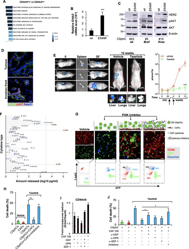
Colon
EGFR/HER2 dual inhibitors has shown a synergistic activity in
Significance of this study preclinical models based on CRC cell lines bearing Kras muta-
tion.21 The investigation on EGFR family members in CRC has
Gut: first published as 10.1136/gutjnl-2020-323553 on 12 January 2021. Downloaded from http://gut.bmj.com/ on January 21, 2022 by guest. Protected by copyright.
What is already known on this subject? been recently focused on HER2. Erbb2 amplification occurs in
►► Advanced colorectal cancer (CRC) remains essentially approximately 3%–10% of patients with CRC and may promote
incurable, particularly in the presence of genomic alterations resistance to EGFR inhibitors.22 23 Moreover, HER2 expression
in the signalling pathway of rat sarcoma (RAS). appears as a negative prognostic factor that correlates with the
►► Human epidermal growth factor receptor 2 (HER2) expression stage and survival in patients with CRC.24 This hypothesis has
seems to correlate with the stage of disease and reduced been recently validated by the clinical trial Heracles, which
survival in CRC. showed that the combination of trastuzumab and lapatinib in
►► Targeting Erbb2 amplification has a significant therapeutic Erbb2-amplified patients with CRC can induce the regression of
activity in patients with CRC. tumours resistant to anti-EGFR therapies.25 Constitutive expres-
►► The phosphatidylinositol 3-kinase (PI3K)/AKT pathway is sion of HER2 can be also driven by the degree of its enhancer
constitutively activated in colorectal cancer stem cells (CR- and promoter activities.26
CSCs) and sustains the expression of CD44v6, which drives Myc overexpression may contribute to promote therapy
the metastatic dissemination. resistance in Kras-mutant CRCs.27 While the MAPK effector
What are the new findings? promotes Myc stabilisation, its proteosomal degradation is
►► The constitutive activation of PI3K/AKT is associated with
mediated by GSK3β.28 29 The role of Myc in the tumourigenesis
high expression levels of HER2 in CD44v6-positive CR-CSCs. programme is mediated by the upregulation of the miR-17–92
►► HER2, in combination with PI3K and mitogen-activated
cluster, which is associated with a poor prognosis.30
protein kinase kinase (MEK) inhibitors, leads to cancer stem Colorectal cancer stem cells (CR-C SCs) are responsible
cell death and tumour regression in CRC avatars resistant to for tumour development, spreading and resistance to chemo-
anti-epidermal growth factor receptor (EGFR) therapy and to therapy.31 32 We have created a large collection of primary
combinations of PI3K, BRAF and HER2 targeting. CRC cells growing as spheroids and able to reproduce the
►► Liver disseminated CR-CSCs can be effectively killed by PI3K/
patients’ tumour in mouse avatars. We have recently shown
AKT inhibitors in an experimental model of adjuvant therapy. that CR-C SCs express CD44v6 and depend on the PI3K/
►► Cytokines released by cancer-associated fibroblasts,
AKT pathway for survival and spreading.31 33 Herein, we
particularly hepatocyte growth factor (HGF), stromal cell- have studied the molecular pathways that should be targeted
derived factor-1 (SDF-1) and osteopontin (OPN), confer to kill CR-C SCs both in the adjuvant and metastatic settings.
resistance to the targeting of the PI3K/AKT pathway and While targeting the PI3K/AKT pathway is sufficient to kill
surrogate the protective effect of tumour microenvironment. disseminating CR-C SCs, we found that HER2 is constitu-
tively expressed in CR- C SCs and that the simultaneous
How might it impact on clinical practice in the foreseeable targeting of HER2, PI3K and MEK neutralises the protective
future? effect of tumour stroma and induces tumour regression, even
►► PI3K/AKT inhibitors could be effective in the adjuvant setting in the presence of aggressive mutational backgrounds.
for CRC.
►► Targeting HER2, MEK and PI3K may provide a valuable
METHODS
therapeutic strategy against anti-EGFR-resistant advanced
A detailed description of the methods can be found in online
CRCs.
supplemental information.
overcome EGFR therapy resistance determined by the amplifi-
cation of receptor tyrosine kinases.11 12 In contrast to melanoma, RESULTS
CRCs harbouring Braf mutations are refractory to BRAF inhibi- CD44V6-positive CRC cells express high levels of HER2 and
tors, such as vemurafenib.13 CRC resistance to BRAF inhibitors are cetuximab resistant
remains a major obstacle in clinical settings.14 This unresponsive- EGFR inhibitors promote an effective therapeutic response
ness is sustained by the activation of a feedback loop involving in about 50%–55% of the patients with Ras/Braf-wt CRC,
EGFR and downstream pathways.15 Although a recent phase II whereas Braf- and Kras- mutant CRC cells are completely
clinical study showed that addition of vemurafenib and cetux- resistant.9 In accordance with clinical data, treatment with
imab to chemotherapy prolonged the progression-free survival cetuximab affected the cell viability of about a half of the
of metastatic patients with Braf-mutated tumours by 2.4 months, Ras/Braf-wt primary colorectal cancer sphere cells (CSphCs)
the prognosis of these patients remains poor.16 Moreover, and delayed the outgrowth of tumour xenografts (figure 1A,
another recent study reported that the combined BRAF, EGFR online supplemental figure 1A,B and online supplemental
and mitogen-activated protein kinase kinase (MEK) inhibition table 1).
in patients with BrafV600E-mutant CRC could increase survival, Primary resistance to the EGFR blockade is mostly due
even if patients experienced primary and acquired resistance due to a constitutive activation of the RAS- MAPK signalling
to a positive feedback regulation of MAPK pathway in tumour network.9 Accordingly, a global RNA- Seq transcriptome
cells.17 analysis of cetuximab- resistant versus sensitive Ras/Braf-
Given that Kras- mutant CRCs display different resistance wt CSphCs showed 252 differentially expressed genes
mechanisms to EGFR and MAPK inhibitors, many alternative (DEGs) (online supplemental figure 1C and online supple-
strategies are currently under investigation.18 In addition to mental table 2). The gene set enrichment analysis (GSEA)
acquired genetic mutations, the activation of a positive human computed with the Molecular Signatures Database displayed
epidermal growth factor receptor 2 (HER2) and HER3 feed- the enrichment of genes associated with activation of the
back loop seems to be one example of antitumour therapy MAPK-signalling pathway, including the negative feedback
escape.19 20 Accordingly, the combined treatment with MEK and regulator DUSP434 (figure 1B and online supplemental
120 Mangiapane LR, et al. Gut 2022;71:119–128. doi:10.1136/gutjnl-2020-323553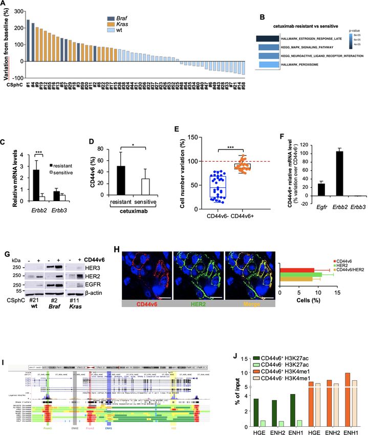
Colon
Gut: first published as 10.1136/gutjnl-2020-323553 on 12 January 2021. Downloaded from http://gut.bmj.com/ on January 21, 2022 by guest. Protected by copyright.
Figure 1 High expression of HER2 confers resistance to anti-epidermal growth factor receptor (EGFR) treatment in CD44v6-positive CR-CSCs.
(A) Waterfall plot of cetuximab response in Ras/Braf-wt, Braf-mutant and Kras-mutant CSphC lines following 72 hours of treatment. (B) Top four
significantly enriched gene sets in hallmark, canonical pathways MSigDB collections (false discovery rate (FDR) Q-value≤0.05) identified through
the analysis of differentially expressed genes between cetuximab resistant versus sensitive Ras/Braf-wt sphere cells. P values related to each
enriched gene set are indicated. (C) Erbb2 and Erbb3 mRNA expression levels in Ras/Braf-wt sphere cells resistant and sensitive to cetuximab. Gapdh
amplification was used as endogenous control. Data are represented as ±SD of three experiments performed with 31 Ras/Braf-wt. (D) CD44v6
expression performed in cells as in (C). (E) Viable cell number variation in enriched CD44v6− and CD44v6-positive Ras/Braf-wt treated with cetuximab
for 72 hours and normalised with the values of cells treated with vehicle (indicated as 100%, red dotted line). Boxes and whiskers represent
mean±SD of six experiments performed with 15 resistant and 16 sensitive Ras/Braf-wt sphere cells. (F) Variation of Egfr, Erbb2 and Erbb3 mRNA
expression levels in CD44v6-positive versus CD44v6− cells. Gapdh amplification was used as endogenous control. Data are represented as mean±SD
of nine experiments performed with three Ras/Braf-wt (CSphC#14, 21 and 33), three Braf-mutants (CSphC#1, 2 and 5) and three Kras-mutants
(CSphC#10, 11 and 16). (G) Immunoblot analysis of HER3, HER2 and EGFR on purified CD44v6− and CD44v6-positive Ras/Braf-wt (CSphC#21),
Braf-mutant (CSphC#2) and Kras-mutant (CSphC#11) CR-CSphC populations. β-Actin was used as loading control. (H, left panel) Representative
immunofluorescence analysis of CD44v6 and HER2 on paraffin embedded sections from human CRC tissue specimen. Nuclei were counterstained with
TOTO-3. Scale bars, 20 µm. Percentages of CD44v6, HER2 and CD44v6/HER2 positivity in eight human CRC tissues are shown on the right panel. Data
are mean±SD of eight different samples. (I) Browser view of the Erbb2 locus, showing different isoforms of Erbb2 and chromatin states (ChromHMM
tracks). Two promoters and three potential enhancers are highlighted (Prom1, Prom2, ENH1, ENH2 and HGE). (J) ChIP-qPCR for the histone marks
H3K27ac and H3K4me1 at the indicated enhancer regions (ENH1, ENH2 and HGE) in Braf-mutant cells positive or negative for CD44v6. Enrichment is
indicated as % relative to the input. CR-CSC, colorectal cancer stem cell; CSphC, colorectal cancer sphere cell; ENH1, intron 1 enhancer; ENH2, intron 2
enhancer; HER2, human epidermal growth factor receptor 2; HGE, HER2 gene body enhancer; MSigDB, Molecular Signatures Database; wt, wild type.
*indicates P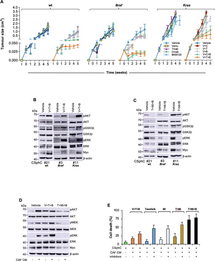
Colon
figure 1D). Besides the activation of signalling pathway of cells were mainly prominent in the tumour invasion front and
MAPK, Ras/Braf-wt CSphCs resistant to cetuximab showed displayed activation of the PI3K/AKT pathway (figure 2D). The
higher mRNA expression levels of Erbb2 compared with essential role of PI3K in the transcriptional regulation of Erbb2 was
Gut: first published as 10.1136/gutjnl-2020-323553 on 12 January 2021. Downloaded from http://gut.bmj.com/ on January 21, 2022 by guest. Protected by copyright.
those sensitive (figure 1C). In line with literature,23 our further supported by ChIP-qPCR analysis showing that overexpres-
CSphC collection showed a 9.7% of Erbb2 amplification sion of the mostly represented Pik3ca activating mutation in breast
(online supplemental table 1). We previously reported that cancer, the Pik3caH1047R, in mammary IMEC-MYC cells enhances
while CD44v6 is a functional marker that identifies tumour- the transcriptional activity of both Erbb2 promoters (prom 1 and
initiating CR- C SCs, the CD44v6- negative population prom 2) and ENH1 and HGE (online supplemental figure 2E). Since
represents the progenitor and differentiated fraction.31 33 A the inhibition of the PI3K/AKT pathway hampers the cell viability
cohort of 31 out of 47 primary CSphC lines showed that of CD44v6-positive cells,33 we evaluated whether the addition of a
the high percentage of CD44v6 expression levels resided PI3K inhibitor to the combination therapy could affect the viability
in the Ras/Braf-wt cells resistant to cetuximab, even though of both CD44v6-positive and CD44v6− cells. To confirm the depen-
these expression levels are similarly distributed between Ras/ dence of CR-CSCs on the PI3K activity, we tested an AKT (miran-
Braf-wt, Braf-mutated and Kras-mutated cell lines (figure 1D sertib) and two PI3K (BKM120 and taselisib) inhibitors on several
and online supplemental figure 1E). Because the CD44v6- primary CSphC lines. Both miransertib and PI3K inhibitors reduced
positive population, within the Ras/Braf-wt cells, is resistant considerably the viability of CD44v6-positive cells in vitro regardless
and increases after treatment with cetuximab (figure 1E and of the mutational background (online supplemental figure 2F–H),
online supplemental figure 1F), we investigated whether the confirming that the PI3K/AKT pathway plays a key role on CR-CSC
expression in signalling pathways associated with drug resis- survival.
tance may differ between the stem and differentiated cell We previously showed that CRC development is sustained by
compartments. Reverse phosphoproteomic analysis (RPPA) cancer stem cells (CSCs), whose dissemination is responsible for
of CSphCs showed that, while MAPK pathways and HER2 CRC metastasisation,33 suggesting that targeting disseminated
are highly regulated, EGFR and HER3 are expressed in a CR- CSCs may prevent tumour relapse and increase survival of
lesser extent in the CD44v6-positive than in the CD44v6− patients with CRC.31 Thus, we investigated the ability of PI3K inhib-
fraction (online supplemental figure 1G). The analysis at itors to target disseminated sphere cells in the liver before they were
mRNA and protein levels confirmed an increased HER2 able to make metastases in a model of adjuvant treatment. We found
expression in the tumourigenic CD44v6- positive popula- that the administration of taselisib in immunocompromised mice
tion of CRC cells, independently of the mutational back- was able to prevent the formation of liver metastases after dissem-
ground (figure 1F,G and online supplemental figure 1H,I). ination of CSphCs by spleen injection (figure 2E). These findings
Accordingly, immunofluorescence analysis of patient tumour support the investigation of PI3K/AKT inhibitors in clinical trials
sections and tumour spheres indicated that the majority of aiming at killing disseminated metastasis-initiating CR-CSCs. The
CD44v6- positive cells coexpressed HER2 (figure 1H and different survival properties of CD44v6 cells in vitro and in estab-
online supplemental figure 1J). lished tumours are likely due to the protective activity of the tumour
We next investigated the transcriptional regulation of Erbb2 microenvironment.31 Outside the protective tumour context, PI3K
expression in CD44v6-positive fraction by evaluating its 3′ regulatory and AKT inhibitors can kill CR-CSCs. In contrast, the protective
elements. Interestingly, H3K27 acetylation (H3K27ac) was enriched activity of cells and cytokines present in the tumour microenviron-
at the analysed regions of HER2 gene body enhancer (HGE), intron ment may require the targeting of multiple pathways to overcome
1 enhancer (ENH1) and intron 2 enhancer in CD44v6- positive the enhanced survival of CR-CSCs. This hypothesis is supported by
compartment, whereas H3K4me1 enrichment was equally found the significant therapeutic activity of PI3K inhibitors on micrometas-
in both CD44v6− and CD44v6-positive cell fractions at all three tases and small tumour lesions.36 In order to identify some possible
enhancer regions (figure 1I,J and online supplemental figure 1K). soluble mediators of such protective activity, we then measured
These data indicate that the enhancers are poised in both cellular the release of cytokines from cancer-associated fibroblasts (CAFs).
fractions but only fully activated in the CD44v6-positive compart- Among the cytokines more abundantly produced by CAFs, we
ment, suggesting that the activation of Erbb2 transcription mediated selected HGF, SDF-1 and OPN (figure 2F) to further investigation,
by the increased acetylation is restricted to the CD44v6-positive based on their ability to support PI3K/AKT activity and stemness
CRC cell compartment. properties in CSphCs.33 We next investigated whether the presence
of CAFs would influence the survival of CSphCs exposed to the PI3K
inhibitor. The coculture of GFP-labelled tumour spheres with CAFs
PI3K/AKT pathway activation is associated with the protected cells from taselisib treatment (figure 2G), suggesting that
transcriptional regulation of Erbb2 in CD44v6-positive cells CAFs could play a critical role in opposition to the killing activity of
The transcriptomic analysis of CD44v6high versus CD44v6low cells PI3K inhibitors in CRC. Moreover, neutralisation of HGF, SDF-1
highlighted the presence of 173 DEGs (online supplemental figure and OPN completely prevented the protective activity of CAFs
2A and online supplemental table 3). In CD44v6high cells, the GSEA (figure 2H), indicating that these cytokines are responsible for deliv-
enriched for genes related to the activation of PI3K/AKT signalling ering a survival signal in CR-CSCs that makes ineffective the PI3K
pathway, such as NOS335 (figure 2A and online supplemental figure targeting. Exposure of CSphCs, CD44v6- positive and CD44v6−
2B). To investigate the role of PI3K in the transcriptional regulation cell fractions to CAF-released cytokines enhanced the expression of
of Erbb2, we induced an activating Pik3ca mutation into Pik3ca-wt Erbb2 mRNA (figure 2I and online supplemental figure 2I). Inter-
low expressing HER2 CSphC lines by using CRISPR nuclease in estingly, in the presence of tumour microenvironmental cytokines,
combination with a specific donor DNA that introduced the E545K Erbb2 expression levels were not affected by the treatment with the
point mutation (online supplemental figure 2C and online supple- PI3K inhibitor (online supplemental figure 2J–L). We also observed
mental table 1). The presence of a Pik3caE545K is associated with an that HGF plays a major role in CAF-mediated protection of CSphCs
increased expression of HER2 and phospho-AKT (figure 2B,C and treated with taselisib (figure 2J). Taken together, these data suggest
online supplemental figure 2D). Interestingly, immunofluorescence that the tumour microenvironment protects CR- CSCs from the
analysis of primary tumour sections indicated that HER2-positive targeting of the PI3K/AKT pathway.
122 Mangiapane LR, et al. Gut 2022;71:119–128. doi:10.1136/gutjnl-2020-323553Colon
Gut: first published as 10.1136/gutjnl-2020-323553 on 12 January 2021. Downloaded from http://gut.bmj.com/ on January 21, 2022 by guest. Protected by copyright.
Figure 2 Activation of PI3K/AKT pathway is accompanied by elevated Erbb2 expression levels in CD44v6-positive CRC cells. (A) Top 10 significantly
enriched gene sets in hallmark, canonical pathways MSigDB collections (FDR Q-value≤0.05) computed by the analysis of differentially expressed
genes between CD44v6high and CD44v6low cells. (B) mRNA relative levels of Erbb2 in CSphCs and their corresponding CRISPR/Cas9-Pik3caE545K cells.
Data are represented as mean±SD of six independent experiments performed with Ras/Braf-wt (CSphC#23), Braf-mutant (CSphC#5) and Kras-mutant
(CSphC#15) cells and their corresponding CRISPR/Cas9-Pik3caE545K cells. (C) Immunoblot analysis of HER2, pAKT and AKT on Ras/Braf-wt (CSphC#23),
Braf-mutant (CSphC#5) Kras-mutant (CSphC#15) cells. β-Actin was used as loading control. (D) Representative immunofluorescence analysis of
HER2 and pAKT on paraffin-embedded sections from six human CRC tissue specimens. Nuclei were counterstained with TOTO-3. Scale bars, 20 µm.
(E, left panels) In vivo whole-body imaging analysis of mice at 0 and 30 min and 12 weeks injected with sphere cells into the spleen. Five days after
cell injection, mice were treated daily with taselisib for 3 weeks. Signal within the red dotted area represents the bioluminescence quantification.
Kinetics of metastasis formation at the indicated time points (right panels). Black arrows indicate the start and end of treatment (from day 6 to week
4). Data are mean±SD of four independent experiments of six mice per group using Kras-mutant (CSphC#8 and 11) sphere cell lines. (F) Lollipop
plot representing the amount of cytokines released by immortalised CAFs. Data are mean of six independent experiments using cells purified from
six different patients. (G) Cell death (blue colour) evaluated by immunofluorescence (upper panels) and flow cytometry (lower panels) in sphere cells
(CSphC#8) transduced with GFP (green colour) cocultured with CAFs CD90 positive (red colour) and treated with a PI3K inhibitor (taselisib) for 72
hours in the presence or absence of hepatocyte growth factor (HGF), stromal cell-derived factor-1 (SDF-1) and osteopontin (OPN) blocking antibodies
(inhibitors). Scale bars, 40 µm. (H) Percentage of cell death in cells as in (G). Data are mean±SD of three independent experiments using Ras/Braf-wt
(CSphC#14, 21 and 33), Braf-mutant (CSphC#1, 2 and 5) and Kras-mutant (CSphC#8, 10 and 11) sphere cell lines. (I) Erbb2 mRNA expression levels
in CD44v6− enriched cells treated with CAF CM and the indicated cytokines. Data are mean±SD of three independent experiments performed with
cells derived from Ras/Braf-wt (CSphC#14 and 33), Braf-mutant (CSphC#1 and 5) and Kras-mutant (CSphC#10 and 11) sphere cell lines. (J) Cell
death in sphere cells exposed to CAF CM and treated with taselisib for 72 hours in the presence of cytokine neutralising antibodies as indicated.
Data are mean±SD of three independent experiments performed with Ras/Braf-wt (CSphC#6, 14, 21 and 33), Braf-mutant (CSphC#1, 2, 4 and 5) and
Kras-mutant (CSphC#8, 10, 11 and 17) sphere cell lines. CAF, cancer-associated fibroblast; CM, conditioned medium; CRC, colorectal cancer; CSphC,
colorectal cancer sphere cell; HER2, human epidermal growth factor receptor 2; MSigDB, Molecular Signatures Database; wt, wild type. *indicates
PColon
MEK sustains CR-CSCs resistance to the triple targeting of as shown by the massive tumour regression and lack of regrowth even
HER2, BRAF and PI3K 6 weeks after treatment suspension (figure 4E). Next, we examined
To further analyse the therapeutic potential of PI3K inhibitor in whether this MEK-targeted triplet was effective on a large number
Gut: first published as 10.1136/gutjnl-2020-323553 on 12 January 2021. Downloaded from http://gut.bmj.com/ on January 21, 2022 by guest. Protected by copyright.
combination with MAPK pathway targeting, tumour xenografts, of primary CSphC cultures of different mutational backgrounds and
generated by the subcutaneous injection of CSphCs, were initially their corresponding tumour xenografts. To confirm the effectiveness
treated with either trastuzumab or cetuximab in combination with of this treatment, we tested other MEK and PI3K inhibitors (cobi-
BRAF and PI3K inhibitors. These treatments were largely ineffec- metinib and taselisib) in combination with trastuzumab. Importantly,
tive. We observed only a transient stabilisation of Braf-mutated tumour size generated by the subcutaneous injection of primary
tumours and a short delay in the disease progression of Ras/Braf- sphere cells was significantly hampered by the treatment with
wt and Kras-mutated tumours (figure 3A and online supplemental either trametinib in combination with trastuzumab and BKM120
figure 3A). These experiments allowed us to evaluate the potential or cobimetinib plus trastuzumab and taselisib, independently of the
mechanisms of acquired resistance to such triple combinations. mutational status (figure 4A and online supplemental figure 4D,E).
CSphCs surviving the combinatorial treatment showed a significant Of note, this latter combination remarkably reduced the CD44v6
phosphorylation of p235–236 S6 kinase (online supplemental figure expression level on xenograft- derived CRC cells (online supple-
3B), which could follow the activation of RAS/ERK and mammalian mental figure 4F). Consistently, cobimetinib plus trastuzumab and
target of rapamycin (mTOR) and result in the engagement of the taselisib induced the death of a conspicuous number of cells that
Myc pathway.37 38 The activation of PI3K/AKT and MAPK pathways were substituted by fibrosis, resulting in a considerable decrease in
were confirmed by western blot in tumour specimen-derived subcu- the amount of Ki67-positive and CK20-positive cells (online supple-
taneous xenograft treated with the triple combination (figure 3B and mental figure 4G). Thus, simultaneous MEK/HER2/PI3K inhibition
online supplemental figure 3C). This phenomenon was paralleled by exerted a potent antitumour activity in CRC xenografts regardless
a strong activation of the PI3K/AKT and MAPK pathways, particu- of the mutational status. Altogether these data demonstrate that the
larly in the presence of Braf or Kras mutations (figure 3B). Moreover, combination treatment with HER2, PI3K and MEK inhibitors is
cells surviving the combinatorial treatment showed high expression synthetically lethal for CRC cells (figure 4F).
levels of the miR-17–92 cluster (online supplemental figure 3D),
whose upregulation is associated with Myc expression.39 Altogether,
these findings indicate that PI3K and MEK promote CR-CSC resis- DISCUSSION
tance to the targeting of BRAF, HER2 and PI3K signalling pathways. The currently available targeted therapies for advanced CRC have
Replacement of BRAF targeting with a MEK inhibitor caused a a limited effect, particularly on the survival of patients carrying
marked reduction of the PI3K/AKT and MAPK pathway activation tumours with Kras mutation.5 We recently demonstrated that
and a decrease of phosphorylation of S6 kinase (figure 3C and online CD44v6-positive CR-CSCs are responsible for metastatic spreading
supplemental figure 3E,F). Viability of CSphCs was severely affected and have a constitutive activation of the PI3K/AKT pathway that
by the use of trametinib combination regardless of the mutational appears essential for their survival.33 Here, we demonstrate that
status and remarkably diminished the Myc-regulated miRNAs in CR-CSCs express high levels of HER2, which are associated with
cells previously exposed to vemurafenib-based combination (online a constitutive activation of the PI3K/AKT pathway. Inhibition of
supplemental figure 3G,H). HER2, MEK and PI3K kills CR-CSCs and promotes a long-lasting
We then investigated whether the MEK inhibitor-based combi- regression of all the tumour xenografts tested, regardless of their
nation is also able to overcome the protective effect mediated by mutational background.
the tumour microenvironment. Beside PI3K, CAF- released cyto- Among the attempts to target the actionable mutations in CRC,
kines boosted MAPK pathway activation, which persisted after the the treatment with anti-HER2 in patients carrying Erbb2 amplifi-
treatment of CSphCs with vemurafenib-based combination therapy cation has been successful in clinical trials, whereas patients with
(figure 3D). Conversely, pharmacological targeting of MEK, instead BrafV600E-mutant CRC are poorly responsive to the administration of
of BRAF, promoted a considerable cell death, paralleled with a vemurafenib or dabrafenib.13 40
marked reduction of MEK/ERK, AKT activation and Myc expres- Although the existence of synthetic lethality between BRAF and
sion in CSphCs, independently of the presence of Erbb2 amplifica- EGFR in Braf-mutated CRC cells would predict the potential ther-
tion and the exposure to CAF conditioned medium (figure 3D,E and apeutic effect of a combined targeting, we found that CR-CSCs
online supplemental figure 3I). Of note, sphere cells able to survive are resistant to the combination of anti-EGFR or anti-HER2 and
to a prolonged exposure to the vemurafenib-based treatment remain BRAF inhibitors due to the constitutive activation of the PI3K/AKT
sensitive to the triplet containing trametinib (online supplemental pathway. This could be the reason why vemurafenib, in combination
figure 3J). Altogether, these data suggest that the tumour microen- with irinotecan and cetuximab, showed a weak therapeutic effect in
vironment confers therapy resistance mediated by Myc through the patients with metastatic CRC.41
activation of MAPK and PI3K–AKT pathways. Here, we found that the regulatory elements of Erbb2 transcrip-
tion are acetylated in CD44v6-positive cells. While this can explain
why CR-CSCs are remarked by high HER2 expression, the poten-
MEK inhibition-based therapy is synthetically lethal in CR- tial ability of PI3K to promote a transcriptional activation of Erbb2
CSCs corroborates the hypothesis that both of these oncogenic pathways
In line with these results, we found that the replacement of vemu- should be targeted simultaneously. Since HER2 expression is lost on
rafenib with MEK inhibitors in the triple combination prevented CR-CSC differentiation, it is likely that the specific expression of
the tumourigenic activity retained by sphere cells (figure 4A,B) and HER2 in the CD44v6-positive cell compartment results from the
tumour progression when delivered in vivo, as indicated by the considerable reduction of the PI3K/AKT signalling pathway and
decrease in Ki67, CD44v6 and CK20 expression (figure 4C,D and β-catenin activity observed in their differentiated progeny.33
online supplemental figure 4A–C). Of note, Braf-mutated or Kras- Although sphere culture models mostly recapitulate the genetic
mutated xenograft tumours that recurred following the treatment landscape and the transcriptomic profile of parental tumour, repre-
with the vemurafenib-based triple combination, tumor xenografts senting valuable preliminary tools to identify potentially effec-
resulted very sensitive to the trametinib-based combination therapy, tive targeted therapies,42 43 it is fundamental to dissect the tumour
124 Mangiapane LR, et al. Gut 2022;71:119–128. doi:10.1136/gutjnl-2020-323553Colon Figure 3 HER2/MEK/PI3K combinatorial targeting counteracts the protective effect of cytokines produced by CAF. (A) Size of xenograft tumours Gut: first published as 10.1136/gutjnl-2020-323553 on 12 January 2021. Downloaded from http://gut.bmj.com/ on January 21, 2022 by guest. Protected by copyright. generated by subcutaneous injection of Ras/Braf-wt (CSphC#14, 21 and 33), Braf-mutant (CSphC#1, 2, 3 and 5) or Kras-mutant (CSphC#8, 11 and 16) sphere cells. Mice were treated for the first 4 weeks with vehicle (vehicle) or Vemu (or V), cetuximab (Cmab or C), Tmab (or T) and a PI3K inhibitor (B) alone or in combination as indicated. ‘I’ indicates the time of cell injection. Treatment was started at time 0. Data are mean values of six independent experiments (n=6 mice per group). (B) Immunoblot analysis of pAKT, AKT, pGSK3β, GSK3β, pERK, ERK and Myc on tumour xenograft-derived cells of mice injected with RasBraf/Braf-w t (CSphC#21), Braf-mutant (CSphC#3), Kras-mutant (CSphC#11) sphere cells. Mice were treated with vehicle or V in combination with T and B, and sacrificed 1 week after the treatment suspension (5 weeks). β-Actin was used as loading control. (C) Representative Western blot analysis of pAKT, AKT, pGSK3β, GSK3β, pERK, ERK and Myc in Ras/Braf-wt (CSphC#21), Braf-mutant (CSphC#3), Kras-mutant (CSphC#11) sphere cells treated for 24 hours with vehicle or T+Mi+B. β-Actin was used as loading control. (D) Immunoblot analysis of the indicated proteins in Kras-mutant (CSphC#9) sphere cells treated with vehicle or V in combination with T and B or T+Mi+B cultured in fetal bovine serum (FBS)- free Dulbecco’s modified eagle medium (DMEM) or CAF CM for 24 hours. (E) Cell death percentage in CSphCs exposed to hepatocyte growth factor (HGF), stromal cell-derived factor-1 (SDF-1) and osteopontin (OPN) blocking antibodies (inhibitors) and treated as indicated for 72 hours. Data are mean±SD of three independent experiments performed with Ras/Braf-wt (CSphC#14, 21 and 33), Braf-mutant (CSphC#1, 2 and 5) and Kras-mutant (CSphC#8, 10 and 11) sphere cell lines. B, BKM120; CAF, cancer-associated fibroblast; CSphC, colorectal cancer sphere cell; T, trastuzumab; T+Mi+B, trastuzumab in combination with MEKi and BKM120; V, vemurafenib; V+T+B, vemurafenib in combination with trastuzumab and BKM120; wt, wild type; CM, conditioned medium. Mangiapane LR, et al. Gut 2022;71:119–128. doi:10.1136/gutjnl-2020-323553 125
Colon
Gut: first published as 10.1136/gutjnl-2020-323553 on 12 January 2021. Downloaded from http://gut.bmj.com/ on January 21, 2022 by guest. Protected by copyright.
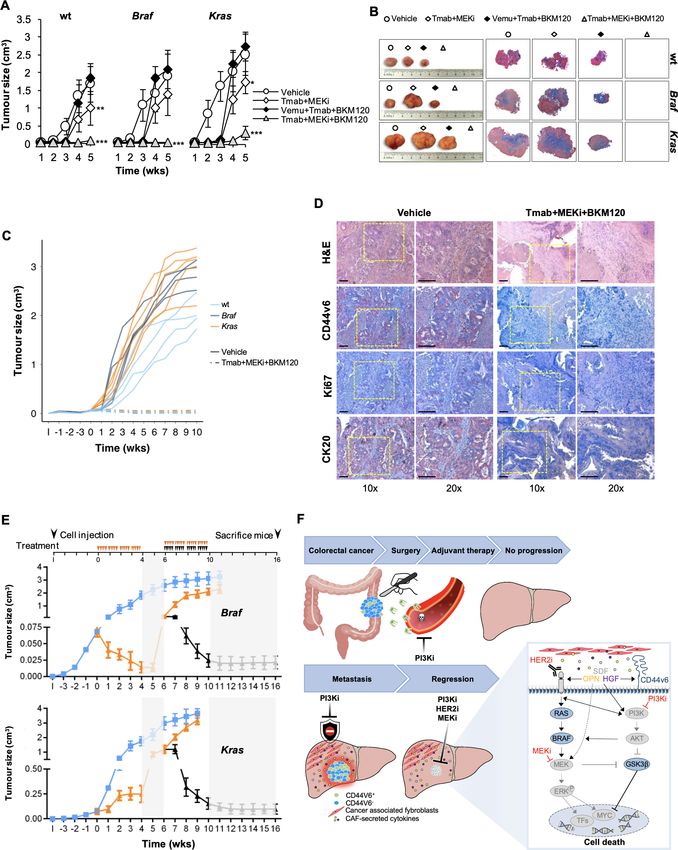
Figure 4 Therapeutic potential of HER2, PI3K and MEK targeting in CRC. (A) Size of tumours generated by subcutaneous injection of surviving
Ras/Braf-wt (CSphC#14, 21 and 33), Braf-mutant (CSphC#1, 2 and 5) and Kras-mutant (CSphC#8, 11 and 16) sphere cells after 5 days of in vitro
combination treatment as indicated. Data reported are mean±SD of tumour size for each cell lines (n=6 mice per group). (B) Representative
macroscopic and Azan-Mallory analysis on tumour xenografts at 5 weeks treated as in (A). (C) Individual subcutaneous tumour volume plots of mice
generated by the injection of four CSphC lines bearing the indicated different mutational background and treated for 4 weeks (0–4 weeks) with
vehicle (vehicle) or Tmab plus MEKi plus BKM120. ‘I’ indicates the time of cell injection. Treatment was started at time 0. Data show kinetic growth
of xenograft tumours generated by the injection of Ras/Braf-wt (CSphC#14, 21, 33 and 56), Braf-mutant (CSphC#1, 2, 3 and 5) and Kras-mutant
(CSphC#8, 9, 11 and 16) CSphCs. (D) Representative H&E and immunohistochemical analysis of CD44v6, Ki67 and CK20 on tumour xenografts
generated by the injection of Kras-mutant (CSphC#11) sphere cells treated as in (C) at the time of sacrifice (10 weeks). Scale bars, 200 µm. (E) Tumour
size of mice xenografted with Braf-mutant (CSphC#1–5) and Kras-mutant (CSphC#8, 9, 11, 13 and 16) mutant sphere cells. Mice were treated with
vehicle (vehicle, blue lines) or sequential treatments. A combination of Vemu, Tmab, BKM120 (Vemu+Tmab+BKM120, orange line) was used as first
line (0–4 weeks, orange arrowheads) and after 2 weeks off-treatments, Tmab in combination with MEKi and BKM120 (Tmab+MEKi+BKM120, black
lines and arrowheads) or the same Vemu combination used in the first 4 weeks (orange arrowheads) was administered from weeks 6 to 10. Off-
treatments are highlighted with grey regions. ‘I’ indicates the time of cell injection. Data are expressed as mean±SD of subcutaneously implanted
CSphC lines for each mutational status (n=6 mice per group). (F) Scheme of the signalling axis illustrating the site of action of the triple combination
therapy. Surgery is the main treatment for primary CRC followed by adjuvant therapy. PI3Ki has shown efficacy in targeting disseminating CRC cells,
impeding tumour progression (upper panel). However, PI3Ki as single agents are unable to counteract the TME protective influence in metastatic
lesions. Triple combination treatment (PI3Ki, HER2i and MEKi) induces tumour regression by overcoming CAF-secreted cytokine effect (lower left
panel). In CD44v6-positive CR-CSCs characterised by high PI3K pathway activity, TME-derived cytokines upregulate HER2 and CD44v6 expression
levels, activate mitogen-activated protein kinase (MAPK) pathway and increase Myc protein levels, jeopardising the potential therapeutic efficacy of
PI3Ki. The additional targeting of HER2 and the Myc upstream kinase MEK achieves a synthetic lethal effect in CR-CSCs (lower right panel). HER2,
BRAF, PI3K and MEK inhibitors are indicated as I. CAF, cancer-associated fibroblast; CRC, colorectal cancer; CR-CSC, colorectal cancer cancer stem
cell; CSphC, colorectal cancer sphere cell; HER2, human epidermal growth factor receptor 2; MEKi, trametinib; PI3Ki, PI3K inhibitors; TF, transcriptional
factor; Tmab, trastuzumab; TME, tumour microenvironment; Vemu, vemurafenib; wt, wild type. *indicates PColon
microenvironment contribution in mediating resistance of cancer EGFR inhibitors are commonly associated with adverse events,
cells to therapeutic drugs. including the inhibition of the MEK/ERK signal pathway, which
According to our previous observation, we found that tumour compromises the epidermis cell differentiation leading to skin
Gut: first published as 10.1136/gutjnl-2020-323553 on 12 January 2021. Downloaded from http://gut.bmj.com/ on January 21, 2022 by guest. Protected by copyright.
microenvironmental cytokines produced by CAFs contribute to reca- lesions.46 Given that HER2 inhibitors generally display minimal
pitulate a protective effect against antitumour drugs expanding the dermatological side effects as compared with those induced by
CD44v6-positive compartment expressing HER2. We showed that EGFR inhibitors,23 as shown by current clinical studies for the treat-
HGF and to a lesser extent OPN and SDF-1 make CR-CSCs resis- ment of advanced CRC,25 we foresee that triple targeting of HER2,
tant to the targeting of the PI3K/AKT pathway, possibly explaining MEK and PI3K may have a superior patient compliance and overall
the disappointing results obtained in the clinical trials that evaluated treatment outcome.
the therapeutic effects of PI3K inhibitors in metastatic patients.44 Here, we have shown that some biological features of CR-CSCs
Such vulnerability of CR-CSCs in the absence of CAFs suggests that have the potential to be exploited in the clinics. The specific expres-
PI3K/AKT inhibitors can contribute to kill cells disseminated into the sion of HER2 in CR-CSCs, independently of gene amplification,
liver as part of adjuvant treatment due to the absence of a protective suggests that HER2 should be regarded as key therapeutic target
microenvironment. This hypothesis is strengthened by the obser- that deserves further preclinical and clinical investigations in CRC.
vation that treatment with taselisib prevents the formation of liver The good therapeutic response, observed in clinical trial by HER2
metastases in mice receiving sphere cells by spleen injection. targeting in patients with amplified tumours, increases the feasibility
In a subsequent set of experiments, we show that the addi- of this approach. Moreover, we provide evidence that targeting of
tion of PI3K inhibitors to the combination of vemurafenib with the PI3K/AKT pathway could be exploited both in advanced disease
trastuzumab or cetuximab induces a partial response of Braf- and in the adjuvant setting. These findings may help define new ther-
mutated tumours and a temporary stabilisation followed by a apeutic strategies based on CR-CSC targeting.
slower progression of Ras/Braf-wt and Kras-mutated tumours.
Such transient therapeutic effect induces the rapid accumulation Author affiliations
1
of tumour-initiating cells resistant to this triplet likely due to the Department of Surgical, Oncological and Stomatological Sciences, Università degli
Studi di Palermo, Palermo, Italy
presence of tumour microenvironmental cytokines. 2
Department of Health Promotion Sciences, Internal Medicine and Medical
The RPPA analysis in residual CSphCs spared by the HER2/BRAF/ Specialties, Università degli Studi di Palermo, Palermo, Italy
PI3K targeting allowed us to identify, through the regulation of S6 3
Core Facilities, Istituto Superiore di Sanità, Roma, Italy
4
kinase phosphorylation, MEK and PI3K as major components of the Department of Cellular, Computational, and Integrative Biology (CIBIO), University
resistance pathway. Accordingly, we observed increased levels of Myc of Trento, Trento, Italy
5
Department of Oncology and Molecular Medicine, Istituto Superiore di Sanita,
in cells simultaneously exposed to agents targeting HER2, BRAF and Roma, Italy
PI3K. The concomitant activation of S6 kinase and MEK in sphere 6
Laboratory for Experimental Oncology and Radiobiology, Center for Experimental
cells resistant to the vemurafenib-based triple combinations suggests and Molecular Medicine, University of Amsterdam, Amsterdam, Noord-Holland, The
that the failure to target both RAF and PI3K downstream pathways Netherlands
7
Oncode Institute, University of Amsterdam, Amsterdam, Noord-Holland, The
is responsible for maintaining activation of ERK and high Myc levels Netherlands
and promoting the pharmacological resistance of CR-CSCs to this 8
Institute of General Pathology, Universita Cattolica del Sacro Cuore Facolta di
triplet. Medicina e Chirurgia, Roma, Italy
9
MEK is a key downstream element of the RAS-RAF pathway able Policlinico A Gemelli, Roma, Lazio, Italy
to indirectly activate Myc.15 Replacement of vemurafenib with MEK Correction notice This article has been corrected since it published Online First.
inhibitors in the triple combination was able to significantly limit The funding statement has been amended.
ERK activation and downregulate Myc expression while inducing a Acknowledgements We thank Francesco Calò for graphic image editing and
considerable therapeutic response in Braf-mutated and Kras-mutated Alice Alferi for technical assistance. LRM is a student of the Molecular and Clinical
tumours progressing after the vemurafenib-based combination. Of Medicine PhD Program. AT and VV are research fellows funded by European Union-
note, our data showed that MEK inhibition-based triplets were able FESR FSE, PON Ricerca e Innovazione 2014–2020 (AIM line 1).
to kill CR-CSCs in the presence of cytokines released by CAFs and Contributors LRM, AN, RDM and GS conceived and designed the project.
to induce tumour regression in all CR-CSC-based xenografts tested, Experiments were conducted by LRM, AN, AT, MG, PB, SDF, VV, MS, SB, LF, MEF, MLI,
IP, GGa and MT. Data provision and bioinformatic analysis were carried out by DSS,
regardless of the mutational status and Erbb2 amplification. Hence,
MS, SB, JPM and AZ. Pathology support, tissue provision and intellectual input were
HER2, PI3K and MEK appear as critical therapeutic targets in from MEF, MRB and GGu. RDM and GS wrote the manuscript.
CR-CSCs, independently of the genomic abnormalities developed
Funding This work was supported by AIRC 5x1000 (9979) to GS and RDM, AIRC
in patients’ tumours. This combination appears the most active both IG (22911) to AZ, RF2018-12367044 to MT and RDM, AIRC IG (21445) and PRIN
in tumour xenografts and in the in vitro experiments designed in the (2017WNKSLR) to GS.
presence of the CAF-released cytokines. Competing interests None declared.
The advent of targeted therapies and the study of the associated
Patient consent for publication Not required.
resistance mechanisms revealed the presence of clonal heteroge-
neity in CRC.45 Most of the current therapeutic strategies, including Provenance and peer review Not commissioned; externally peer reviewed.
targeted combination treatments, affect differentiated cells and spare Data availability statement Data are available in a public, open access
repository. All data relevant to the study are included in the article or uploaded
CSCs that eventually reinitiate tumour growth. It is therefore clear
as supplementary information. The data that support the findings of this study
that the identification of the critical pathways responsible for the are available from the corresponding author (GS) upon reasonable request. RNA
increase of survival and therapy resistance of CR-CSCs appears as sequencing data of CR-CSphCs have been deposited in a public, open access GEO
a major priority to define possible effective treatments for patients repository, under accession number GSE162104 (link to data: https://www.ncbi.nlm.
with advanced CRC. This is particularly true for metastatic patients nih.gov/geo/query/acc.cgi?acc=GSE162104).
carrying oncogenic alterations in the RAS pathway, who have very Supplemental material This content has been supplied by the author(s). It
limited therapeutic options. Our data show that MEK inhibition has not been vetted by BMJ Publishing Group Limited (BMJ) and may not have
been peer-reviewed. Any opinions or recommendations discussed are solely those
in association with PI3K and HER2 targeting can induce tumour of the author(s) and are not endorsed by BMJ. BMJ disclaims all liability and
regression even in tumours carrying mutations in the RAS pathway. responsibility arising from any reliance placed on the content. Where the content
Although targeted therapy is less toxic than standard chemotherapy, includes any translated material, BMJ does not warrant the accuracy and reliability
Mangiapane LR, et al. Gut 2022;71:119–128. doi:10.1136/gutjnl-2020-323553 127Colon
of the translations (including but not limited to local regulations, clinical guidelines, 21 Sun C, Hobor S, Bertotti A, et al. Intrinsic resistance to MEK inhibition in KRAS
terminology, drug names and drug dosages), and is not responsible for any error mutant lung and colon cancer through transcriptional induction of ErbB3. Cell Rep
and/or omissions arising from translation and adaptation or otherwise. 2014;7:86–93.
Gut: first published as 10.1136/gutjnl-2020-323553 on 12 January 2021. Downloaded from http://gut.bmj.com/ on January 21, 2022 by guest. Protected by copyright.
22 Personeni N, Fieuws S, Piessevaux H, et al. Clinical Usefulness of EGFR Gene
Open access This is an open access article distributed in accordance with the
Copy Number as a Predictive Marker in Colorectal Cancer Patients Treated with
Creative Commons Attribution 4.0 Unported (CC BY 4.0) license, which permits
Cetuximab: A Fluorescent In situ Hybridization Study. Clinical Cancer Research
others to copy, redistribute, remix, transform and build upon this work for any
2008;14:5869–76.
purpose, provided the original work is properly cited, a link to the licence is given,
23 Siena S, Sartore-Bianchi A, Marsoni S, et al. Targeting the human epidermal growth
and indication of whether changes were made. See: https://creativecommons.org/
factor receptor 2 (HER2) oncogene in colorectal cancer. Annals of Oncology
licenses/by/4.0/.
2018;29:1108–19.
ORCID iDs 24 Kapitanovic S, Radosevic S, Kapitanovic M, et al. The expression of p185(HER-2/neu)
Alice Turdo http://orcid.org/0000-0002-6152-4903 correlates with the stage of disease and survival in colorectal cancer. Gastroenterology
Michele Signore http://orcid.org/0 000-0002-0262-842X 1997;112:1103–13.
Luca Fagnocchi http://orcid.org/0000-0002-9 551-5474 25 Sartore-Bianchi A, Trusolino L, Martino C, et al. Dual-Targeted therapy with
Micol Eleonora Fiori http://orcid.org/0000-0002-1813-7035 trastuzumab and lapatinib in treatment-refractory, KRAS codon 12/13 wild-type,
Giorgio Stassi http://o rcid.org/0000-0002-1016-9059 HER2-positive metastatic colorectal cancer (HERACLES): a proof-of-concept,
multicentre, open-label, phase 2 trial. Lancet Oncol 2016;17:738–46.
REFERENCES 26 Liu Q, Kulak MV, Borcherding N, et al. A novel HER2 gene body enhancer contributes
1 Siegel RL, Miller KD, Jemal A. Cancer statistics, 2019. CA: A Cancer Journal for to HER2 expression. Oncogene 2018;37:687–94.
Clinicians 2019;69:7–34. 27 Wang Q, Tan R, Zhu X, et al. Oncogenic K-ras confers SAHA resistance by up-
2 Dagogo-Jack I, Shaw AT. Tumour heterogeneity and resistance to cancer therapies. Nat regulating HDAC6 and c-myc expression. Oncotarget 2016;7:10064–72.
Rev Clin Oncol 2018;15:81–94. 28 Sears R, Nuckolls F, Haura E. Multiple Ras-dependent phosphorylation pathways
3 Russo M, Siravegna G, Blaszkowsky LS, et al. Tumor heterogeneity and lesion-specific regulate Myc protein stability. Genes Dev 2000;14:2501–14.
response to targeted therapy in colorectal cancer. Cancer Discov 2016;6:147–53. 29 Yeh E, Cunningham M, Arnold H, et al. A signalling pathway controlling c-myc
4 Dienstmann R, Mason MJ, Sinicrope FA, et al. Prediction of overall survival in stage degradation that impacts oncogenic transformation of human cells. Nat Cell Biol
II and III colon cancer beyond TNM system: a retrospective, pooled biomarker study. 2004;6:308–18.
Ann Oncol 2017;28:1023–31. 30 Diosdado B, van de Wiel MA, Terhaar Sive Droste JS, et al. Mir-17-92 cluster is
5 Van Cutsem E, Cervantes A, Adam R, et al. ESMO consensus guidelines for the associated with 13q gain and c-myc expression during colorectal adenoma to
management of patients with metastatic colorectal cancer. Annals of Oncology adenocarcinoma progression. Br J Cancer 2009;101:707–14.
2016;27:1386–422. 31 Zeuner A, Todaro M, Stassi G, et al. Colorectal cancer stem cells: from the crypt to the
6 Mei ZB, Duan CY, Li CB, et al. Prognostic role of tumor PIK3CA mutation in colorectal clinic. Cell Stem Cell 2014;15:692–705.
cancer: a systematic review and meta-analysis. Ann Oncol 2016;27:1836–48. 32 Todaro M, Alea MP, Di Stefano AB, et al. Colon cancer stem cells dictate tumor growth
7 Misale S, Di Nicolantonio F, Sartore-Bianchi A, et al. Resistance to anti-EGFR therapy and resist cell death by production of interleukin-4. Cell Stem Cell 2007;1:389–402.
in colorectal cancer: from heterogeneity to convergent evolution. Cancer Discov 33 Todaro M, Gaggianesi M, Catalano V, et al. Cd44V6 is a marker of constitutive and
2014;4:1269–80. reprogrammed cancer stem cells driving colon cancer metastasis. Cell Stem Cell
8 Siravegna G, Mussolin B, Buscarino M, et al. Erratum: clonal evolution and resistance 2014;14:342–56.
to EGFR blockade in the blood of colorectal cancer patients. Nat Med 2015;21:827. 34 Cagnol S, Rivard N. Oncogenic KRAS and BRAF activation of the MEK/ERK signaling
9 De Roock W, Claes B, Bernasconi D, et al. Effects of KRAS, BRAF, NRAS, and PIK3CA pathway promotes expression of dual-specificity phosphatase 4 (DUSP4/MKP2)
mutations on the efficacy of cetuximab plus chemotherapy in chemotherapy-refractory resulting in nuclear ERK1/2 inhibition. Oncogene 2013;32:564–76.
metastatic colorectal cancer: a retrospective Consortium analysis. Lancet Oncol 35 Lim K-H, Ancrile BB, Kashatus DF, et al. Tumour maintenance is mediated by eNOS.
2010;11:753–62. Nature 2008;452:646–9.
10 Tricker EM, Xu C, Uddin S, et al. Combined EGFR/MEK Inhibition Prevents 36 Veschi V, Mangiapane LR, Nicotra A, et al. Targeting chemoresistant colorectal cancer
the Emergence of Resistance in EGFR -Mutant Lung Cancer. Cancer Discov via systemic administration of a BMP7 variant. Oncogene 2020;39:987–1003.
2015;5:960–71. 37 Roux PP, Shahbazian D, Vu H, et al. Ras/Erk signaling promotes site-specific ribosomal
11 Arena S, Siravegna G, Mussolin B, et al. MM-151 overcomes acquired resistance protein S6 phosphorylation via RSK and stimulates cap-dependent translation. J Biol
to cetuximab and panitumumab in colorectal cancers harboring EGFR extracellular Chem 2007;282:14056–64.
domain mutations. Sci Transl Med 2016;8:324ra14. 38 Gera JF, Mellinghoff IK, Shi Y, et al. AKT Activity Determines Sensitivity to Mammalian
12 Van Emburgh BO, Arena S, Siravegna G, et al. Acquired Ras or EGFR mutations Target of Rapamycin (mTOR) Inhibitors by Regulating Cyclin D1 and c- myc
and duration of response to EGFR blockade in colorectal cancer. Nat Commun Expression. J Biol Chem 2004;279:2737–46.
2016;7:13665. 39 He L, Thomson JM, Hemann MT, et al. A microRNA polycistron as a potential human
13 Kopetz S, Desai J, Chan E, et al. Phase II Pilot Study of Vemurafenib in Patients oncogene. Nature 2005;435:828–33.
With Metastatic BRAF -Mutated Colorectal Cancer. Journal of Clinical Oncology 40 Falchook GS, Long GV, Kurzrock R, et al. Dabrafenib in patients with melanoma,
2015;33:4032–8. untreated brain metastases, and other solid tumours: a phase 1 dose-escalation trial.
14 Ahronian LG, Sennott EM, Van Allen EM, et al. Clinical Acquired Resistance to RAF The Lancet 2012;379:1893–901.
Inhibitor Combinations in BRAF -Mutant Colorectal Cancer through MAPK Pathway 41 Hong DS, Morris VK, El Osta B, et al. Phase IB Study of Vemurafenib in Combination
Alterations. Cancer Discov 2015;5:358–67. with Irinotecan and Cetuximab in Patients with Metastatic Colorectal Cancer with
15 Prahallad A, Heynen GJJE, Germano G, et al. Ptpn11 is a central node in intrinsic and BRAF V600E Mutation. Cancer Discov 2016;6:1352–65.
acquired resistance to targeted cancer drugs. Cell Rep 2015;12:1978–85. 42 Schütte M, Risch T, Abdavi-Azar N, et al. Molecular dissection of colorectal cancer in
16 Kopetz S, McDonough SL, Morris VK, et al. Randomized trial of irinotecan and pre-clinical models identifies biomarkers predicting sensitivity to EGFR inhibitors. Nat
cetuximab with or without vemurafenib in BRAF -mutant metastatic colorectal cancer Commun 2017;8:14262.
(SWOG 1406). Journal of Clinical Oncology 2017;35:520. 43 Linnekamp JF, Hooff SRvan, Prasetyanti PR, et al. Consensus molecular subtypes of
17 Corcoran RB, André T, Atreya CE, et al. Combined BRAF, EGFR, and MEK Inhibition in colorectal cancer are recapitulated in in vitro and in vivo models. Cell Death Differ
Patients with BRAF V600E -Mutant Colorectal Cancer. Cancer Discov 2018;8:428–43. 2018;25:616–33.
18 Douillard J-Y, Oliner KS, Siena S, et al. Panitumumab–FOLFOX4 Treatment 44 Vermeulen L, De Sousa E Melo F, van der Heijden M, et al. Wnt activity defines
and RAS Mutations in Colorectal Cancer. New England Journal of Medicine colon cancer stem cells and is regulated by the microenvironment. Nat Cell Biol
2013;369:1023–34. 2010;12:468–76.
19 Kavuri SM, Jain N, Galimi F, et al. Her2 activating mutations are targets for colorectal 45 Siravegna G, Mussolin B, Buscarino M, et al. Clonal evolution and resistance to EGFR
cancer treatment. Cancer Discov 2015;5:832–41. blockade in the blood of colorectal cancer patients. Nat Med 2015;21:795–801.
20 Zhang L, Castanaro C, Luan B, et al. ERBB3/HER2 signaling promotes resistance to 46 Segaert S, Van Cutsem E. Clinical signs, pathophysiology and management of skin
EGFR blockade in head and neck and colorectal cancer models. Mol Cancer Ther toxicity during therapy with epidermal growth factor receptor inhibitors. Annals of
2014;13:1345–55. Oncology 2005;16:1425–33.
128 Mangiapane LR, et al. Gut 2022;71:119–128. doi:10.1136/gutjnl-2020-323553You can also read