A Novel Hybrid Convolutional Neural Network for Accurate Organ Segmentation in 3D Head and Neck CT Images
←
→
Page content transcription
If your browser does not render page correctly, please read the page content below
A Novel Hybrid Convolutional Neural Network
                                            for Accurate Organ Segmentation in 3D Head
                                                        and Neck CT Images

                                               Zijie Chen∗1,2,3,4 , Cheng Li∗5 , Junjun He∗1,2,6,7 , Jin Ye1,2 , Diping Song1,2 ,
                                                          Shanshan Wang5,8,9 , Lixu Gu6,7 , and Yu Qiao1,2(B)
arXiv:2109.12634v1 [eess.IV] 26 Sep 2021

                                           1
                                               Shenzhen Key Lab of Computer Vision and Pattern Recognition, SIAT-SenseTime
                                           Joint Lab, Shenzhen Institute of Advanced Technology, Chinese Academy of Sciences,
                                                                        Shenzhen, Guangdong, China
                                                                             yu.qiao@siat.ac.cn
                                                                     2
                                                                       Shanghai AI Lab, Shanghai, China
                                             3
                                                Shenzhen Yino Intelligence Techonology Co., Ltd., Shenzhen, Guangdong, China
                                            4
                                               Shenying Medical Technology (Shenzhen) Co., Ltd., Shenzhen, Guangdong, China
                                            5
                                               Paul C. Lauterbur Research Center for Biomedical Imaging, Shenzhen Institute of
                                             Advanced Technology, Chinese Academy of Sciences, Shenzhen, Guangdong, China
                                           6
                                               School of Biomedical Engineering, Shanghai Jiao Tong University, Shanghai, China
                                              7
                                                 Institute of Medical Robotics, Shanghai Jiao Tong University, Shanghai, China
                                                            8
                                                              Peng Cheng Laboratory, Shenzhen, Guangdong, China
                                                                9
                                                                  Pazhou Lab, Guangzhou, Guangdong, China

                                                   Abstract. Radiation therapy (RT) is widely employed in the clinic for
                                                   the treatment of head and neck (HaN) cancers. An essential step of RT
                                                   planning is the accurate segmentation of various organs-at-risks (OARs)
                                                   in HaN CT images. Nevertheless, segmenting OARs manually is time-
                                                   consuming, tedious, and error-prone considering that typical HaN CT
                                                   images contain tens to hundreds of slices. Automated segmentation al-
                                                   gorithms are urgently required. Recently, convolutional neural networks
                                                   (CNNs) have been extensively investigated on this task. Particularly, 3D
                                                   CNNs are frequently adopted to process 3D HaN CT images. There are
                                                   two issues with naı̈ve 3D CNNs. First, the depth resolution of 3D CT
                                                   images is usually several times lower than the in-plane resolution. Direct
                                                   employment of 3D CNNs without distinguishing this difference can lead
                                                   to the extraction of distorted image features and influence the final seg-
                                                   mentation performance. Second, a severe class imbalance problem exists,
                                                   and large organs can be orders of times larger than small organs. It is
                                                   difficult to simultaneously achieve accurate segmentation for all the or-
                                                   gans. To address these issues, we propose a novel hybrid CNN that fuses
                                                   2D and 3D convolutions to combat the different spatial resolutions and
                                                   extract effective edge and semantic features from 3D HaN CT images.
                                                   To accommodate large and small organs, our final model, named Organ-
                                                   Net2.5D, consists of only two instead of the classic four downsampling
                                                   operations, and hybrid dilated convolutions are introduced to maintain
                                                   the respective field. Experiments on the MICCAI 2015 challenge dataset

                                               * These authors contributed equally to this work.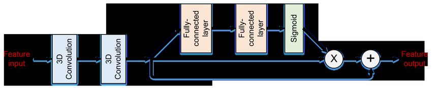
2      Z. Chen et al.

      demonstrate that OrganNet2.5D achieves promising performance com-
      pared to state-of-the-art methods.

      Keywords: Segmentation of organs-at-risks · Hybrid 2D and 3D con-
      volutions · 3D HaN CT images.

1   Introduction

Head and neck (HaN) cancers, such as oral cavity and nasopharynx, are one
of the most prevalent cancer types worldwide [19]. Treatment of HaN cancers
relies primarily on radiation therapy. To prevent possible post-treatment com-
plications, accurate segmentation of organs-at-risks (OARs) is vital during the
treatment planning [6]. In the clinic, computed tomography (CT)-based treat-
ment planning is routinely conducted because of its high efficiency, high spatial
resolution, and the ability to provide relative electron density information. Man-
ual delineation of OARs in CT images is still the primary choice regardless
of the time-consuming and tedious process. Several hours are required to pro-
cess the images of only one patient [7]. Besides, it subjects to high inter- and
intra-observer variations, which can significantly influence the prognosis of the
treatment [1]. Automatic segmentation methods are in urgent need to speed up
the process and achieve robust outcomes.
    The low contrast of soft tissues in HaN CT images and the large volume size
variations of different organs make it challenging to achieve automatic and accu-
rate segmentation of all OARs in an end-to-end fashion. Conventional learning
approaches often rely on one or multiple atlases or require the extraction of hand-
crafted image features [2,21], which is difficult to be enough comprehensive and
distinctive for the segmentation task. Deep neural networks, especially convolu-
tional neural networks (CNNs), have proved to be highly effective for medical
image segmentation in different applications [9,13]. Many efforts have been de-
voted to CNN-based segmentation of OARs in HaN CT images. To deal with the
class imbalance issue caused by the differently sized organs, image patches based
on certain prior knowledge were extracted before conducting CNN-based segmen-
tation [8,15]. Two-step CNNs consisting of a region detector and a segmentation
unit were also employed [12,17]. To make full use of the image information, a
joint localization and segmentation network with a multi-view spatial aggrega-
tion framework was proposed [10]. The inputs to these models were either 3D
image patches lacking the global features or 2D images without the depthwise
information. AnatomyNet was designed to specifically process whole-volume 3D
HaN CT images [22]. The major contributions of AnatomyNet include a novel
network architecture for effective feature extraction and a combined loss func-
tion to combat the class imbalance problem. Following AnatomyNet, FocusNet
was proposed to better handle the segmentation of both large and small organs
with a delicate network structure design [4].
    Despite the inspiring results achieved, several issues exist in the developed
approaches. First, some studies dealt with only 2D inputs and thus, did not
Hybrid convolutional neural network 3
fully exploit the 3D image information [8,10,12]. Others conducted 3D convolu-
tions but without paying attention to the different in-plane and depth resolutions
[4,15,22]. The in-plane resolution of 3D HaN CT images is normally several times
higher than the depth resolution. The direct employment of 3D convolutions can
probably lead to the extraction of distorted image features, which might not be
optimal for the segmentation task. Anisotropic convolutions have been proposed
to solve this issue but without distinguishing the low-level and high-level features
[11]. Second, for networks processing whole volume 3D CT images (AnatomyNet
and FocusNet), only one downsampling layer was used to preserve the informa-
tion of small anatomies. Consequently, the receptive fields of these networks are
limited. To increase the receptive field, DenseASPP with four dilation rates (3,
6, 12, and 18) was introduced to FocusNet [4]. However, when the dilation rates
of cascaded dilated convolutions have a common factor relationship, the gridding
issue may appear that influence the segmentation accuracy [20]. Besides, pure
3D networks also suffer from increased parameters and computational burden
issues, which also limit the network depth and performance.
To address these issues, a hybrid convolutional neural network, OrganNet2.5D,
is proposed in this work to improve the segmentation performance of OARs in
HaN CT images. OrganNet2.5D integrates 2D convolutions with 3D convolu-
tions to simultaneously extract clear low-level edge features and rich high-level
semantic features. The hybrid dilated convolution (HDC) module is introduced
to OrganNet2.5D as a replacement for the DenseASPP in FocusNet. HDC mod-
ule is able to increase the network receptive field without decreasing the image
resolutions and at the same time, avoid the gridding issue. OrganNet2.5D has
three blocks: the 2D convolution block for the extraction of clear edge image
features, the coarse 3D convolution block for the extraction of coarse high-level
semantic features with a limited receptive field, and the fine 3D convolution
block for the extraction of refined high-level semantic features with an enlarged
receptive field through the utilization of HDC. Similar to AnatomyNet and Fo-
cusNet [4,22], a combined loss of Dice loss and focal loss is employed to handle
the class imbalance problem. The effectiveness of the proposed OrganNet2.5D is
evaluated on two datasets. On the publicly available MICCAI Head and Neck
Auto Segmentation Challenge 2015 dataset (MICCAI 2015 challenge dataset),
promising performance is achieved by OrganNet2.5D compared to state-of-the-
art approaches.
2 Method
2.1 Dataset
We evaluate the performance of our proposed model on two datasets. The first
dataset is collected from two resources of 3D HaN CT images (the Head-Neck
Cetuximab collection (46 samples) [3] and the Martin Vallières of the Medical
Physics Unit, McGill University, Montreal, Canada (261 samples)1 ). This first
1
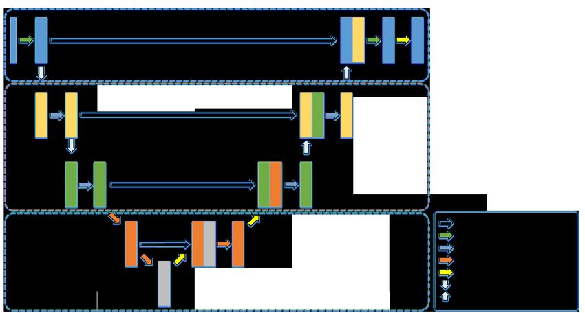
https://wiki.cancerimagingarchive.net/display/Public/Head-Neck-PET-CT4 Z. Chen et al. Fig. 1. An illustration of the proposed OrganNet2.5D network architecture. The blue, yellow, and green boxes indicate the 2D convolution block, the coarse 3D convolution block, and the fine 3D convolution block, respectively. Fig. 2. The 2 × Conv + ResSE unit. “X” refers to element-wise multiplication and “+” is element-wise summation. dataset is utilized to validate the effectiveness of the different blocks of our model. Segmentation annotations of 24 OARs are provided by experienced radiologists with quality control management. We randomly grouped the 307 samples into a training set of 240 samples, a validation set of 20 samples, and a test set of 47 samples. To compare the performance of our proposed method to the existing approaches, we utilize the MICCAI 2015 challenge dataset [14]. There are 48 samples, among which 33 samples are provided as the training set, 10 as the offset test set, and the remaining 5 as the onsite test set. Manual segmentation of 9 OARs is available for the 33 training samples and 10 offset test samples. Similar to previous studies, we optimize our model with the training samples and report the model performance on the 10 offset test samples. 2.2 Network architecture The overall network architecture of our proposed OrganNet2.5D is shown in Fig. 1. OrganNet2.5D follows the classic encoder-decoder segmentation network
Hybrid convolutional neural network      5

structure [16]. The inputs to our network are the whole volume 3D HaN CT
images and the outputs are the segmentation results of the 25 categories for the
first dataset (24 OARs and background) or 10 categories (9 OARs and back-
ground) for the MICCAI 2015 challenge dataset. OrganNet2.5D contains three
major blocks, the 2D convolution block, the coarse 3D convolution block, and
the fine 3D convolution block.

2D convolution block. The 2D convolution block is designed for the extraction
of clear edge image features. It is widely accepted that during image encoding,
the low-level features extract the geometric information and the high-level fea-
tures extract the semantic information. Therefore, in our model, only the first
two convolutions near the inputs and the corresponding last two convolutions
near the outputs are replaced with 2D convolutions. Without the direct applica-
tion of 3D convolutions, distorted image edge feature extraction can be avoided.
Meanwhile, considering the different in-plane and depth image resolutions, in-
plane downsampling is conducted with the 2D convolution block to calibrate the
image features for the following 3D convolution operations.

Coarse 3D convolution block. The 2D convolution block is followed by the
coarse 3D convolution block. To prevent information loss, especially for the small
anatomies, only one downsampling is preserved. The coarse 3D convolution block
is designed to extract rich semantic features that are important for the pixel-
wise distinction task. Following the successful practice of existing methods, the
basic unit of our coarse 3D convolution block is composed of two standard 3D
convolution layers and one squeeze-and-excitation residual module (ResSE mod-
ule, Fig. 2). The ResSE module is responsible for feature filtering to highlight
the important features and suppress the irrelevant ones. With the filtered im-
age features, the final segmentation step can concentrate more on the important
features and better results can be expected.

Fine 3D convolution block. With the 2D convolution block and coarse 3D
convolution block, clear edge and rich semantic image features are extracted.
However, since only two downsampling layers are used (one 2D downsampling
and one 3D downsampling), the receptive field of the network is limited. Without
the global image information, the segmentation accuracy may be compromised.
As such, a series of hybrid dilated convolution (HDC) modules is employed
to integrate the global image information with the semantic features and at
the same time, to prevent the gridding issue [20]. Moreover, by using different
dilation rates, multi-scale image features are extracted, which can better process
the OARs of different sizes.

2.3   Loss function
A combination of focal loss and Dice loss is employed to prevent the model from
biasing the large objects.
6       Z. Chen et al.

   Focal loss forces the network to focus on the hard samples, which refers to
the samples predicted by the network with high uncertainty. It is improved from
the cross-entropy loss with both fixed and dynamic loss weighting strategies.
The focal loss is calculated as:
                                       N   C
                                    1 XX
                      Lf ocal = −             αc (1 − pcn )γ ync logpcn                (1)
                                    N n=1 c=1

where N refers to the sample size, C refers to the different categories (25 for the
first dataset and 10 for the second), αc is the fixed loss weight of the cth OAR,
p ∈ [0, 1] is the network prediction, (1 − pcn )γ is the dynamic loss weight, and
y ∈ {0, 1} is the manual label.
    Dice loss deals with the class imbalance problem by minimizing the distribu-
tion distance between the network prediction and the manual segmentation. For
multi-class segmentation, one Dice loss should be calculated for each class and
the final Dice loss is the average over all the classes. In this work, the average
Dice loss is calculated as:
                                             C N
                                          1 X X 2 × pcn × ync
                         Lavgdice = 1 −                                                (2)
                                          C c=1 n=1 pcn + ync

   The final loss function for our network training is a weighted summation of
the two losses:
                             L = Lf ocal + λLavgdice                       (3)
    For our experiments, we empirically set γ = 2 and λ = 1.0. The fixed weights
αc in the focal loss for the first dataset are 0.5, 1.0, 1.0, 1.0, 4.0, 4.0, 4.0, 4.0, 4.0,
1.0, 1.0, 4.0, 1.0, 1.0, 3.0, 3.0, 1.0, 1.0, 1.0, 1.0, 1.0, 1.0, 3.0, 1.0, and 1.0 for the
25 categories (background, brain stem, eye left, eye right, lens left, lens right,
optic nerve left, optic nerve right, optic chiasma, temporal lobes left, temporal
lobes right, pituitary, parotid gland left, parotid gland right, inner ear left, inner
ear right, middle ear left, middle ear right, tongue, temporomandibular joint left,
temporomandibular joint right, spinal cord, mandible left, and mandible right),
and for the second dataset are 0.5, 1.0, 4.0, 1.0, 4.0, 4.0, 1.0, 1.0, 3.0, and 3.0 for
the 10 categories (background, brain stem, optic chiasma, mandible, optic nerve
left, optic nerve right, parotid gland left, parotid gland right, submandibular
left, submandibular right.)

2.4   Implementation details
All our models are implemented with PyTorch on an NVIDIA GeForce GTX
1080Ti GPU (11G) with a batch size of 2. The inputs to the networks are
resized to 256 × 256 × 48. Adam optimizer is utilized to train the models. The
step decay learning rate strategy is used with an initial learning rate of 0.001
that is reduced by a factor of 10 every 50 epochs until it reaches 0.00001. Two
evaluation metrics are calculated to characterize the network performance, the
Dice score coefficient (DSC) and the 95% Hausdorff distance (95HD).
Hybrid convolutional neural network 7
Table 1. Segmentation performance on the first dataset averaged over the 24 OARs
with different network configurations
3DUNet-SE- 3DUNet-SE- 3DUNet-SE-
Models 3DUNet-SE Proposed
2D 2D-C2 2D-DC2
DSC (%) 83.9 ± 2.0 84.4 ± 2.0 84.2 ± 2.1 84.3 ± 2.1 84.6 ± 1.9
95HD 3.38 3.39 3.24 3.41 3.07
Table 2. DSC of 10 small organs on the first dataset with different network configu-
rations (%)
3DUNet- 3DUNet-SE- 3DUNet-SE- 3DUNet-SE-
Models Proposed
SE 2D 2D-C 2D-DC
Lens L 82.7 ± 7.8 84.5 ± 6.8 83.5 ± 7.5 83.2 ± 6.5 84.4 ± 6.6
Lens R 82.6 ± 5.6 84.1 ± 5.8 83.3 ± 6.9 83.9 ± 5.4 83.9 ± 5.3
Opt. Ner. L 70.9 ± 10.0 71.0 ± 10.1 70.7 ± 10.2 70.5 ± 10.8 71.1 ± 10.3
Opt. Ner. R 70.1 ± 8.7 71.7 ± 8.9 71.8 ± 9.3 70.4 ± 9.1 71.2 ± 9.2
Opt. Chiasm 57.2 ± 14.3 59.6 ± 15.1 57.8 ± 15.4 58.0 ± 14.9 59.8 ± 14.8
Pituitary 74.4 ± 11.6 75.1 ± 13.0 74.3 ± 12.4 75.1 ± 11.4 75.6 ± 12.3
Mid. Ear L 86.4 ± 5.1 86.6 ± 4.8 86.5 ± 5.1 86.7 ± 4.9 86.9 ± 5.1
Mid. Ear R 85.4 ± 4.1 85.5 ± 4.3 85.8 ± 4.6 85.6 ± 4.7 85.9 ± 4.3
T.M.J. L 83.5 ± 7.2 83.8 ± 7.5 83.8 ± 7.1 82.7 ± 7.8 83.8 ± 7.2
T.M.J. R 82.1 ± 8.3 81.6 ± 8.8 82.8 ± 7.9 82.7 ± 8.2 82.9 ± 7.9
3 Experimental results
3.1 Results on the collected public dataset
Ablation studies regarding our network design are conducted. Average DSC and
95HD on the test set are listed in Table 1. DSC values of the 10 small or-
gans are presented in Table 2. See supplementary material for results on all 24
organs. Four network configurations are involved. 3DUNet-SE refers to the base-
line where 3D UNet is combined with the ResSE module. 3DUNet-SE includes
only the coarse 3D convolution block in Fig. 1. Introducing the 2D convolution
block to 3DUNet-SE, we obtain the 3DUNet-SE-2D model. 3DUNet-SE-2D-C
replaces the HDC module in the proposed OrganNet2.5D (Fig. 1) with standard
3D convolutions, and 3DUNet-SE-2D-DC replaces the HDC module with dilated
convolutions of the same dilation rate of 2.
Overall, our proposed model achieves the highest mean DSC and lowest mean
95HD. Statistical analysis confirms that our model performs significantly better
than the other network configurations (p < 0.05 with paired t-tests of the DSC
values). These results reflect that both the 2D convolution block and the fine 3D
convolution block can enhance the segmentation results. Furthermore, our pro-
posed OrganNet2.5D gives excellent performance on small organ segmentation
by generating the best results for 7 of the 10 small organs (Table 2).8 Z. Chen et al.
Table 3. Segmentation results on the MICCAI 2015 challenge dataset
MICCAI AnatomyNet FocusNet SOARS SCAA
Models Proposed
2015 [22] [4] [5] [18]
Brain Stem 88.0 86.7 ± 2 87.5 ± 2.6 87.6 ± 2.8 89.2 ± 2.6 87.2 ± 3.0
Opt. Chiasm 55.7 53.2 ± 15 59.6 ± 18.1 64.9 ± 8.8 62.0 ± 16.9 66.3 ± 7.4
Mandible 93.0 92.5 ± 2 93.5 ± 1.9 95.1 ± 1.1 95.2 ± 1.3 92.2 ± 2.1
Opt. Ner. L 64.4 72.1 ± 6 73.5 ± 9.6 75.3 ± 7.1 78.4 ± 6.1 75.0 ± 7.8
Opt. Ner. R 63.9 70.6 ± 10 74.4 ± 7.2 74.6 ± 5.2 76.0 ± 7.5 74.1 ± 5.1
Parotid L 82.7 88.1 ± 2 86.3 ± 3.6 88.2 ± 3.2 89.3 ± 1.5 86.7 ± 2.6
Parotid R 81.4 87.4 ± 4 87.9 ± 3.1 88.2 ± 5.2 89.2 ± 2.3 85.8 ± 4.9
Subman. L 72.3 81.4 ± 4 79.8 ± 8.1 84.2 ± 7.3 83.2 ± 4.9 82.1 ± 5.8
Subman. R 72.3 81.3 ± 4 80.1 ± 6.1 83.8 ± 6.9 80.7 ± 5.2 82.1 ± 4.1
Mean DSC 74.9 79.2 80.3 82.4 82.6 81.3
3.2 Results on MICCAI 2015 challenge dataset
We compare the performance of our proposed model to the state-of-the-art meth-
ods on the MICCAI 2015 challenge dataset (Table 3). It should be noted that in
the table, the MICCAI 2015 results were the best results obtained for each OAR
possibly by different methods. AnatomyNet was trained with additional samples
except for the MICCAI 2015 challenge dataset. All the results of existing meth-
ods are adopted from the respective papers without method re-implementation
to avoid implementation biases.
Segmentation results show that our proposed model achieves better perfor-
mance than the three most prevalent methods in the field (MICCAI 2015, Anoto-
myNet, and FocusNet) indicated by the mean DSC, which confirms the effective-
ness of the proposed network. Compared to the two recently published methods,
SOARS and SCAA, our method is slightly worse. However, it should be noted
that SOARS utilized neural network search to find the optimal network architec-
ture [5], which is more computationally intensive. SCAA combined the 2D and
3D convolutions with a very complicated network design [18]. Nevertheless, with
the simple and easy-to-implement architecture, our OrganNet2.5D still performs
the best when segmenting the smallest organ, optic chiasma. This observation
reflects the suitability of our network modifications and training strategy for our
task. Visual results lead to similar conclusions as to the quantitative results (See
supplementary material for details).
4 Conclusion
In this study, we present a novel network, OrganNet2.5D, for the segmentation
of OARs in 3D HaN CT images, which is a necessity for the treatment planning
of radiation therapy for HaN cancers. To fully utilize the 3D image information,
deal with the different in-plane and depth image resolutions, and solve the dif-
ficulty of simultaneous segmentation of large and small organs, OrganNet2.5DHybrid convolutional neural network 9 consists of a 2D convolution block to extract clear edge image features, a coarse 3D convolution block to obtain rich semantic features, and a fine 3D convolu- tion block to generate global and multi-scale image features. The effectiveness of OrganNet2.5D was evaluated on two datasets. Promising performance was achieved by our proposed OrganNet2.5D compared to the state-of-the-art ap- proaches, especially on the segmentation of small organs. Acknowledgements. This research is partially supported by the National Key Research and Development Program of China (No. 2020YFC2004804 and 2016YFC0106200), the Scientific and Technical Innovation 2030-”New Genera- tion Artificial Intelligence” Project (No. 2020AAA0104100 and 2020AAA0104105), the Shanghai Committee of Science and Technology, China (No. 20DZ1100800 and 21DZ1100100), Beijing Natural Science Foundation-Haidian Original Inno- vation Collaborative Fund (No. L192006), the funding from Institute of Medical Robotics of Shanghai Jiao Tong University, the 863 national research fund (No. 2015AA043203), Shenzhen Yino Intelligence Techonology Co., Ltd., Shenying Medical Technology (Shenzhen) Co., Ltd., the National Natural Science Foun- dation of China (No. 61871371 and 81830056), the Key-Area Research and De- velopment Program of GuangDong Province (No. 2018B010109009), the Basic Research Program of Shenzhen (No. JCYJ20180507182400762), and the Youth Innovation Promotion Association Program of Chinese Academy of Sciences (No. 2019351). References 1. Brouwer, C.L., Steenbakkers, R.J.H.M., Heuvel, E.v.d., et al.: 3D variation in de- lineation of head and neck organs at risk. Radiation Oncology. 7(1), 32 (2012) 2. Chen, A., Niermann, K.J., Deeley, M.A., Dawant, B.M.: Evaluation of multiple- atlas-based strategies for segmentation of the thyroid gland in head and neck CT images for IMRT. Phys. Med. Biol. 57(1), 93–111 (2012) 3. Clark, K., Vendt, B., Smith, K., et al.: The Cancer Imaging Archive (TCIA): Main- taining and operating a public information repository. J. Digit. Imaging 26(6), 1045– 1057 (2013) 4. Gao, Y., Huang, R., Chen, M., Wang, Z., Deng, J., Chen, Y., Yang, Y., Zhang, J., Tao, C., Li, H.: FocusNet: Imbalanced large and small organ segmentation with an end-to-end deep neural network for head and neck CT images. In: Shen, D. (eds.) MICCAI 2019, LNCS, vol. 11766, pp. 829–838. Springer, Cham (2019). https://doi.org/10.1007/978-3-030-32248-9 92 5. Guo, D., Jin, D., Zhu, Z., Ho, T., Harrison, A.P., Chao, C., Xiao, J., Lu, L.: Organ at risk segmentation for head and neck cancer using stratified learning and neural architecture search. In: 2020 IEEE Conference on Computer Vision and Pattern Recognition (CVPR), pp. 4223–4232. Virtual Conference (2020) 6. Han, X., Hoogeman, M.S., Levendag, P.C., Hibbard, L.S., Teguh, D.N., Voet, P., Cowen, A.C., Wolf, T.K.: Atlas-based auto-segmentation of head and neck CT images. In: Metaxas D., Alex L., Fichtinger G., Szekely G. (eds.) MIC- CAI 2008, LNCS, vol. 5242, pp. 434–441. Springer, Berlin, Heidelberg (2008). https://doi.org/10.1007/978-3-540-85990-1 52
10      Z. Chen et al.

7. Harari, P.M., Song, S., Tome, W.A.: Emphasizing conformal avoidance versus target
   definition for IMRT planning in head-and-neck cancer. Int. J. Radiation Oncology
   Biol. Phys. 77(3), 950–958 (2010)
8. Ibragimov, B., Xing, L.: Segmentation of organs-at-risks in head and neck CT images
   using convolutional neural networks. Med. Phys. 44(2), 547–557 (2017)
9. Li, C., Sun, H., Liu, Z., Wang, M., Zheng, H., Wang, S.: Learning cross-
   modal deep representations for multi-modal MR image segmentation. In: Shen,
   D. (eds.) MICCAI 2019, LNCS, vol. 11765, pp. 57–65. Springer, Cham (2019).
   https://doi.org/10.1007/978-3-030-32245-8 7
10. Liang, S., Thung, K.-H., Nie, D., Zhang, Y., Shen, D.: Multi-view spatial aggre-
   gation framework for joint localization and segmentation of organs at risk in head
   and neck CT images. IEEE Trans. Med. Imaging 39(9), 2794–2805 (2020)
11. Liu, S., Xu, D., Zhou, S.K., Pauly, O., Grbic, S., Mertelmeier, T., Wicklein, J., Jere-
   bko, A., Cai, W., Comaniciu, D.: 3D anisotropic hybrid network: Transferring convo-
   lutinoal features from 2D images to 3D anisotropic volumes. In: Frangi, A., Schnabel,
   J., Davatzikos, C., Alberola-López, C., Fichtinger, G. (eds) MICCAI 2018, LNCS,
   vol. 11071, pp. 851–858. Springer, Cham (2018). https://doi.org/10.1007/978-3-030-
   00934-2 94
12. Men, K., Geng, H., Cheng, C., et al.: More accurate and efficient segmentation of
   organs-at-risk in radiotherapy with convolutional neural networks cascades. Med.
   Phys. 46(1), 286–292 (2019)
13. Qi, K., Yang, H., Li, C., Liu, Z., Wang, M., Liu, Q., Wang, S.: X-Net: Brain
   stroke lesion segmentation based on depthwise separable convolution and long-range
   dependencies. In: Shen, D. (eds.) MICCAI 2019, LNCS, vol. 11766, pp. 247–255.
   Springer, Cham (2019). https://doi.org/10.1007/978-3-030-32248-9 28
14. Raudaschl, P.F., Zaffino, P., Sharp, G.C., et al.: Evaluation of segmentation meth-
   ods on head and neck CT: Auto-segmentation challenge 2015. Med. Phys. 44(5),
   2020–2036 (2017)
15. Ren, X., Xiang, L., Nie, D., Shao, Y., Zhang, H., Shen, D., Wang, Q.: Interleaved
   3D-CNNs for joint segmentation of small-volume structures in head and neck CT
   images. Med. Phys. 45(5), 2063–2075 (2018)
16. Ronneberger, O., Fischer, P., Brox, T.: U-Net: Convolutional networks for biomed-
   ical image segmentation. In: Navab, N., Hornegger, J., Wells, W., Frangi, A.
   (eds.) MICCAI 2015, LNCS, vol. 9351, pp. 234–241. Springer, Cham (2015).
   https://doi.org/10.1007/978-3-319-24574-4 28
17. Tang, H., Chen, X., Liu, Y., et al.: Clinically applicable deep learning framework
   for organs at risk delineation in CT images. Nat. Mach. Intell. 1(10), 480–491 (2019)
18. Tang, H., Liu, X., Han, K., et al.: Spatial context-aware self-attention model for
   multi-organ segmentation. In: 2021 IEEE Winter Conference on Applications of
   Computer Vision (WACV), pp. 939–949. Virtual Conference (2021)
19. Torre, L.A., Bray, F., Siegel, R.L., Ferlay, J., Lortet-Tieulent, J., Jemal, A.: Global
   cancer statistics, 2012. Ophthalmology 65(2), 87–108 (2015)
20. Wang, P., Chen, P., Yuan, Y., et al.: Understanding convolution for semantic seg-
   mentation. In: 2018 IEEE Winter Conference on Applications of Computer Vision
   (WACV), pp. 1451–1460. Lake Tahoe, NV, USA (2018)
21. Wang, Z., Wei, L., Wang, L., Gao, Y., Chen, W., Shen, D.: Hierarchical vertex
   regression-based segmentation of head and neck CT images for radiotherapy plan-
   ning. IEEE Trans. Image. Process. 27(2), 923–937 (2018)
22. Zhu, W., Huang, Y., Zeng, L., Chen, X., Liu, Y., Qian, Z., Du, N., Fan, W.,
   Xie, X.: AnatomyNet: Deep learning for fast and fully automated whole-volume
   segmentation of head and neck anatomy. Med. Phys. 46(2), 576–589 (2019)
You can also read


