A new species of Hydrochoerus (Rodentia: Caviidae: Hydrochoerinae) from the Pleistocene of San Diego County, California, USA with remarks on ...
←
→
Page content transcription
If your browser does not render page correctly, please read the page content below
Vertebrate Anatomy Morphology Palaeontology 9:131–155 131
ISSN 2292-1389
A new species of Hydrochoerus (Rodentia: Caviidae:
Hydrochoerinae) from the Pleistocene of San Diego
County, California, USA with remarks on capybara
biogeography and dispersal in the Pleistocene of
Western North America
Richard S. White1*, Jim I. Mead1,2, Thomas A. Deméré3, and Gary S. Morgan4
1
The Mammoth Site, Hot Springs, South Dakota, USA 57747; RSWhite@mammothsite.org
2
Desert Laboratory on Tumamoc Hill, University of Arizona, Tucson, Arizona, USA 85745
3
San Diego Natural History Museum, San Diego, California, USA 92112
4
New Mexico Museum of Natural History and Science, Albuquerque, New Mexico, USA 87104
Abstract: We describe a new species of capybara from late Pleistocene deposits (Rancholabrean NALMA) in
northern San Diego County, California, USA which tentatively dates to Marine Isotope Stage (MIS) 5 interglacial
(~130 ka to 80 ka). The specimen represents a new species of Hydrochoerus based on morphological characters of
the upper incisor (I1) and the upper (maxillary) third molar (M3). Hydrochoerus hesperotiganites sp. nov. differs
from other described species of Hydrochoerus in its larger size, wider skull roof, more robust zygomatic process of
the maxilla and descending zygomatic process of the lacrimal and in details of the otic region. The new species is
the only confirmed record of fossil Hydrochoerus in North America and is the northwestern-most record of any
capybara in North America. All previous records of fossil capybara from North America represent one of two
extinct genera, Neochoerus or Phugatherium. Northward dispersal of capybaras from central and southern México
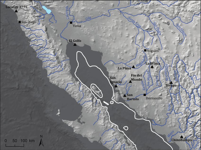
probably occurred along the coasts of Sinaloa and Sonora, entering the north or northeast flowing drainages which
entered the Gulf of California, then further north into the San Simon drainage to the Gila River and ultimately into
the Colorado River, or directly northward along the coast of Sonora to the mouth of the Colorado River.
lsid: urn:lsid:zoobank.org:pub:BD2DA9EB-32EF-4761-8551-E585D6AF317C
INTRODUCTION and Miller 1988). Neochoerus has also been recorded from
northern México in the Irvingtonian NALMA, as well as
Three genera of capybaras (Caviidae: Hydrochoerinae:
from the United States in the middle Blancan of South
Phugatherium, Neochoerus and Hydrochoerus, the latter
Carolina (Sanders 2002; Albright et al. 2019) and in
being today’s largest living rodent) dispersed into North
the late Blancan through Rancholabrean in Florida and
America after the establishment of the Panama Land
the Rancholabrean in Texas (Morgan and White 1995;
Bridge about 5 Ma (Morgan 2008; Woodburne 2010;
Morgan 2005; Baskin et al. 2020). Hydrochoerus has
O’Dea et al. 2016). Phugutherium was present in cen-
been thought to have been present from the late Pliocene
tral México by the Pliocene (early Blancan NALMA) at
(late Blancan) to the latest Pleistocene (Rancholabrean)
3.6 Ma (Vucetich et al. 2015). Neochoerus is found in
in the United States, but not in the Recent (Ahearn
southern México in the Rancholabrean NALMA (Carbot-
1981; Morgan 2005). The fact that the earliest records
Chanona et al. 2020) and in Central México in the early
of Neochoerus in North American are in the north rath-
Blancan at 3.5 Ma. These are the oldest verified record
er than the south would seem to suggest that its origin
of any capybara in North America (Carranza-Castañeda
was in the north; but the relative lack of fossil producing
Published February 28, 2022
*corresponding author. © 2022 by the authors; submitted September 30, 2021; revisions received February 15, 2021; accepted
February 15, 2021. Handling editor: Robert Holmes. DOI 10.18435/vamp29379
Vertebrate Anatomy Morphology Palaeontology is an open access journal http://ejournals.library.ualberta.ca/index.php/VAMP Article copyright by the author(s). This open access work is distributed under a Creative
Commons Attribution 4.0 International (CC By 4.0) License, meaning you must give appropriate credit, provide a link to the license, and indicate if changes were made. You may do so in any reasonable manner, but not
in any way that suggests the licensor endorses you or your use. No additional restrictions — You may not apply legal terms or technological measures that legally restrict others from doing anything the license permits.
Vertebrate Anatomy Morphology Palaeontology 9:131–155

localities of appropriate age in southern México and           onomy was followed by subsequent authors. Mones (1991),
Central America has likely produced a skewed record. The       however, noted that the two species were clearly separable
taxonomy and affinities of the two lineages (Phugatherium      based on cranial measurements of 17 specimens of H.
and Neochoerus + Hydrochoerus) were the subject of much        isthmius. Aeschbach et al. (2016) made a detailed analysis of
research in the last decade (Vucetich et al. 2013; Vucetich    ontogenetic changes in cranial size and morphology of both
et al. 2015; Carranza-Castañeda 2016; Albright et al.          species based on 44 specimens of H. isthmius and 171 of H.
2019; Baskin et al. 2020; Carbot-Chanona et al. 2020).         hydrochaeris, demonstrating that the two are morphologically
Fossils of capybaras from North America previously allo-       distinct. We follow recent authors (Ahearn 1981; Mones
cated to the extant genus Hydrochoerus have subsequent-        1991; Baskin et al. 2020) in considering the fossil taxon H.
ly been referred to Neochoerus, leaving Neochoerus and         holmesi to be properly allocated to Neochoerus.
Phugatherium as the only capybaras known in the North            One additional fossil species of Hydrochoerus (H. gaylordi)
American fossil record (Ahearn 1981; Mones 1991).              has been described by MacPhee et al. (2000) from Pliocene
 Capybaras reached as far north and east as Florida and        deposits on the island of Grenada at the southern end of the
South Carolina, and as far north and west as Texas,            Lesser Antilles, about 160 km (100 miles) off the coast of
Arizona, and Sonora, México (Ahearn 1981; Sanders              Venezuela. We do not consider this species further here, as it
2002; Baskin et al. 2020; Mead et al. 2006). The Colorado      is based on a partial right maxilla with M1-3 and diagnosed
River seems to have been a barrier beyond which capy-          by a single character of the M2. We observe that the M2 of
baras and glyptodonts (Cingulata: Glyptodontinae) did          neonate extant Hydrochoerus have the two prisms on M2
not extend in the Pliocene or Pleistocene (Morgan 2008).       united buccally as in H. gaylordi; by the age of 4 weeks the
The western-most occurrence of Glyptotherium texanum           two laminae are separate as in the adults (Mones 1991: fig.
the Blancan and Irvingtonian species, is in Arizona, while     7A, B). Thus, it is possible that the condition seen in the
the western-most occurrences of G. floridanum, or any          type and only specimen of H. gaylordi may be an individ-
Rancholabrean species, are in central Texas (Kurtén and        ual neotenic variant. Should further specimens be found to
Anderson 1980) and in Sonora, Mexico (Mead et al. 2007.        have the same condition and given the insular occurrence
One capybara specimen has been collected from extreme          (Grenada) of H. gaylordi, it would stand as a valid species.
northwestern Sonora, México. However, that specimen,
found in the early Irvingtonian locality of El Golfo, in       MATERIALS AND METHODS
deltaic deposits of the ancestral Colorado River (Lindsay
1984; Croxen et al. 2007; Carranza-Castañeda 2016) may           Catalogued specimens of extant Hydrochoerus hydrocha-
have floated down from drainages to the north and east.        eris are in the comparative collections of The Mammoth
 Here we present the discovery of a nearly complete al-        Site, Hot Springs, South Dakota. Age classes are as de-
though badly crushed skull of a capybara collected in 1994     fined by Ojasti (1973, 2011) and Gorosabel et al. (2017).
from upper Pleistocene lacustrine deposits in the San Luis     Restrictions in access to museum collections due to
Rey River Valley in San Diego County, California, USA.         COVID-19 prevented us from examining a larger sample
This specimen (SDSNH 50000) provides an opportunity            of Hydrochoerus skulls. This was especially unfortunate in
to re-examine the evolution, historical biogeography, and      that skulls of the northern-most species of extant capybara,
dispersal of capybaras in North America.                       H. isthmius, were not available to us.
 Previous work: For the purposes of the present study, we        Abbreviations: ka, kilo annum; Ma, mega-annum;
adopt the taxonomic classification of capybaras proposed       MSCC, Mammoth Site Comparative Collection, The
by Vucetich et al. (2013, 2014, 2015) and followed in the      Mammoth Site at Hot Springs, SD; NALMA, North
three most recent papers on North American (NA) capybaras      American Land Mammal Age; SDSNH, San Diego Society
(Albright et al. 2019; Baskin et al. 2020; Carbot-Chanona      of Natural History, San Diego, California. Descriptive
et al. 2020). Two lineages with a total of three genera are    terminology for the capybara skull and dentition (upper
recognized by Vucetich et al. (2015): (1) Phugatherium,        M2 and M3) discussed is presented in Figures 1 and 2. L,
with P. dichroplax as the only NA species; and (2) a lineage   left; R, right.
consisting of Neochoerus, with the NA species N. aesopi; and
a Central and South American sister taxon, Hydrochoerus,       GEOLOGICAL AND CHRONOLOGICAL
including the extant H. hydrochaeris and H. isthmius.          CONTEXT
 The diminutive capybara H. isthmius was described by
Goldman (1912) from six specimens collected in eastern          Geological context: The new fossil specimen de-
Panama. Cabrera (1961) considered H. isthmius as only          scribed here was collected from SDSNH Locality 3775 by
subspecifically distinct from H. hydrochaeris and this tax-    Bradford O. Riney on March 5, 1994, during paleonto-

132
White et al. — new Pleistocene Hydrochoerus from California

Figure 1. Cranial terminology utilized in this paper. Museum of Veterinary Anatomy FMVZ USP / Wagner Souza e Silva (https://
commons.wikimedia.org/wiki/File:Capybara_skull._Hydrochoerus_hydrochaeris_02.jpg), adding labels and arrows by Richard
S. White, https: // creativecommons .org / licenses / by-sa / 4.0 / legalcode

logical mitigation monitoring of mass grading operations        the middle to upper Eocene Santiago Formation. In turn,
at the Town Center North shopping center in Oceanside,          an irregular erosional surface marks the upper contact
San Diego County, California, USA (Fig.3). The site is          between the older Pleistocene sedimentary sequence and
located on the south side of the San Luis Rey River Valley,     an overlying younger Pleistocene sequence. This younger
a generally east-west trending coastal valley that preserves    sequence consisted of up to 15.2 m of fluvial and lacustrine
late Pleistocene invertebrate and vertebrate fossils from       sediments beginning with a basal transgressive cross-bed-
elevated and dissected river terrace deposits at a series of    ded light gray sandstone gradationally overlain by 5.4 m of
discovery sites along its length (Guthrie 2010; Deméré et       lacustrine green laminated mudstones and siltstones. The
al. 2013). At the Town Center North shopping center site,       lacustrine deposits were overlain by 8.5 m of gray laminat-
the Pleistocene stratigraphic sequence consisted of two         ed and cross-laminated, fine-grained sandstones.
distinct fluvial-lacustrine sequences (Fig. 4). The capy-         The basal cross-bedded sandstone produced a diverse
bara fossil (SDSNH 50000) was collected from the lower          aquatic assemblage of freshwater snails, clams, bony fishes,
(older) sequence, which consisted of up to 7.3 m (24 feet)      amphibians, and pond turtle, as well as a diverse terrestrial
of interbedded gray-green mudstones, laminated carbon-          vertebrate assemblage of lizards, snakes, birds, and mammals
aceous siltstones, and white to orange, friable medium- to      (Tab. 1). Guthrie (2010) reported on the avifauna from this
coarse-grained graded sandstones with dispersed calcrete        basal sandstone, which consists of over 19 species including
nodules. The skull was found palate up and collected from       grebes, pelican, ducks, rails, sandpiper, quail, roadrunner,
one of the gray-green mudstone beds, which also produced        and passerines. The dominance of the avifauna by waterfowl
associated maxilla, mandibular, and dental fragmentary          is consistent with the sedimentology and aquatic molluscan,
remains of an extinct pronghorn (cf. Stockoceros sp.) (Tab.     fish, and turtle fossils also recovered from this stratum. The
1). This lower lacustrine sequence was deposited along an       terrestrial mammal assemblage from the basal sandstone
irregular erosional surface cut into fluvial sandstones of      includes isolated skeletal elements of mole, rabbit, rodents,

                                                                                                                          133
Vertebrate Anatomy Morphology Palaeontology 9:131–155

Figure 2. Dental terminology utilized in this paper. A, Hydrochoerus hydrochaeris after Mones (1991) : R M2 and M3 of SDSNH
50000. Abbreviations: RM2, second right upper molar; RM3, third right upper molar; HFE, Hendadura Fundamental Externa
(Fundamental External Flexus); HPE, Hendadura Primeria Externa (Primary External Flexus); HSE, Hendadura Secundaria
Externa (Secondary External Flexus); BLFP, BiLobed First Prism; IL1-12, Independent Prism 1-12.

fox, tapir, horse, mastodon, and ground sloths (Tab. 1). The     stones, laminated carbonaceous siltstones, and medium- to
overlying lacustrine laminated mudstones produced a much         coarse-grained graded sandstones likely represent seasonal
less diverse fossil assemblage that consisted of freshwater      changes in sediment input to this fluvial-lacustrine setting.
snails and clams, bony fishes, and horse, as well as leaf com-    The erosion surface that cuts the older Pleistocene lacus-
pressions of vascular plants including oak (Quercus sp.) and     trine sequence is here interpreted to have formed as the riv-
sycamore (Platanus sp.) (Tab. 1).                                er meandered back to the south during a later time of the
 Depositional environment: The sedimentology and                 same interglacial period. The basal cross-bedded sandstone
stratigraphy (Fig. 4) of the Town Center North Pleistocene       resting on this unconformity likely represents a coarse-
sequence suggests deposition in a freshwater pond or ox-         grained “beach” facies that was deposited as the floodplain
bow lake on the southern margin of the ancestral San Luis        was aggrading and another ox-bow lake was forming. The
Rey River Valley. The older lacustrine sequence preserving       mixture of terrestrial and aquatic taxa in this basal trans-
the capybara skull, although only exposed in a small area,       gressive unit and the occurrence of isolated and non-articu-
appears to have been deposited on a relatively high relief       lated skeletal elements suggests a possible scenario whereby
unconformity eroded into older Eocene strata. This erosion       bloated, floating mammalian carcasses were successively
surface likely formed as the ancestral San Luis Rey River        being “beached,” shedding bones, and refloated. The
was beginning to aggrade and deposit fine-grained sedi-          gradational contact between the sandstone “beach” facies
ments on its floodplain during a eustatic rise in sea level      and the laminated mudstone-siltstone “lake” facies suggests
(i.e., an interglacial). The interbedded sequence of mud-        continuous interglacial floodplain aggradation.

134
White et al. — new Pleistocene Hydrochoerus from California

Figure 3. Map of northwestern México and southwestern United States, showing key cities for landmarks (solid square) and
key paleontological sites mentioned in the text (solid triangles). Major river valleys indicated and labeled. Glacial maximum
shoreline indicated by white line, based on bathymetry. The US/Mexico border is approximated by the location of San Diego,
Yuma and Nogales. Map courtesy of Matthew C. Pailes, Department of Anthropology, University of Oklahoma.

  Chronology: Unfortunately, none of the recovered fos-            SYSTEMATIC PALEONTOLOGY
sils provide clear biochronological control concerning the
                                                                                  RODENTIA Bowdich, 1821
age of the enclosing strata. Although no remains of Bison
                                                                                   CAVIOIDEA Gray, 1821
(an “index” fossil for the Rancholabrean NALMA) were
                                                                                    CAVIIDAE Fisher, 1817
recovered from these strata, there are a number of extinct
                                                                               HYDROCHOERINAE Gray 1825
mammalian taxa that typically occur in Rancholabrean
                                                                                   Hydrochoerus Brisson, 1762
age faunas of southern California (e.g., Mammut ameri-
                                                                               Hydrochoerus hesperotiganites sp. nov.
canum, Megalonyx jeffersonii), or only in the Rancholabrean
                                                                                        Figures 2, 5, 6, 7
(Nothrotheriops shastensis). Further, fossils of Bison latifrons
                                                                                    3D Animated Rendering
have been recovered from potentially correlative strata
located in the San Luis Rey River Valley, approximately 18
                                                                    Holotype: SDSNH 50000, a nearly complete skull lack-
km upstream from the Town Center North fossil localities
                                                                   ing mandible.
(Deméré et al. 2013). Considering the fauna and strata
together suggests that deposition of the Town Center                Type locality and horizon: SDSNH Locality 3775,
North Pleistocene stratigraphic sequence occurred during           San Diego County, California, USA. The fauna and stra-
an interglacial interval and most likely during the early          tigraphy together suggest that deposition occurred during
Rancholabrean MIS 5 interglacial (~130 ka to 80 ka).               an interglacial interval and most likely during the early
                                                                   Rancholabrean MIS 5 interglacial (~130 ka to 80 ka).

                                                                                                                          135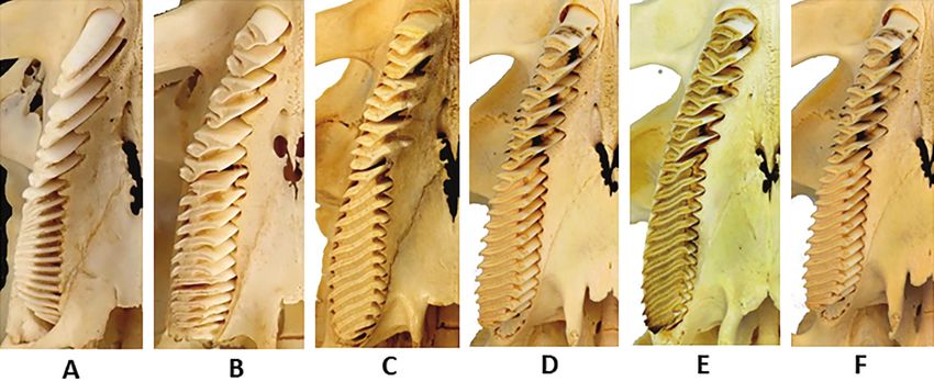
Vertebrate Anatomy Morphology Palaeontology 9:131–155

Table 1. Faunal lists for the three depositional units producing fossils

Lower Lacustrine Unit                     Upper Lacustrine Unit (basal sandstone)   Upper Lacustrine Unit (mudstone/siltstone)

		                                    Mollusca		                                Mollusca
			Gastropoda		 Gastropoda
				Physa sp.			                                                                            Physa sp.
				Fossaria sp.
				Gyraulus sp.
			Pelecypoda		 Pelecypoda
				Anodonta sp.			                                                                         Anodonta sp.
Vertebrata                            Vertebrata		                              Vertebrata
			Osteichthyes		 Osteichthyes
				Mugil sp. cf. M. cephalus			                                                            Mugil sp. cf. M. cephalus
				Gila sp.			                                                                             Gasterosteus sp.
				Gasterosteus sp.			                                                                     Gobiidae
			Amphibia
				Bufo sp.
			Chelonia
				Actinemys marmorata
			Squamata
				Thamnophis sp.
				Colubridae
		                                          Aves
				Aechmophorus occidentalis
				                                             Podilymbus podiceps
				                                             Podiceps parvus
				                                             Pelecanus erythrorhynchus
				                                             Aythya affinis
				                                             Bucephala albeola fossilis
				                                             Oxyura jamaicensis
				                                             Rallus limicola
				                                             Fulica americana
				                                             Phalaropus lobatus
				                                             Callipepla californica
				                                             Geococyx californicus
				                                             Aphelocoma californica
				Vireo sp.
				Toxostoma redivivum
				                                             Agelaius phoeniceus
				Meospiza sp. cf. M. melodi
Mammalia		Mammalia		 Mammalia
        Hydrochoerus hesperotiganites			Talpidae			 Equus sp.
        cf. Stockoceros sp.			                   Sylvilagus sp. cf. S. auduboni
				Thomomys sp.
				Peromyscys sp.
				Microtus sp. cf. M. californicus
				Urocyon cinereoargenteus                                                    Plantae
				Tapirus sp.		                                                                      Tracheophyta
				                                             Equus sp.			                               Quercus sp.
				Mammut americanum			                                                                    Platanus sp.
				                                             Megalonyx jeffersonii
				                                             Nothrotheriops shastensis

136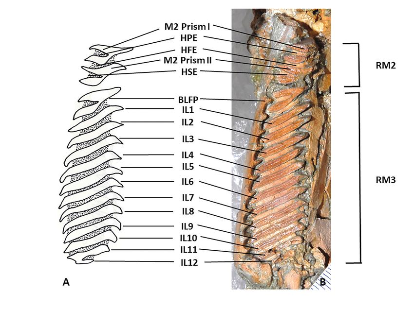
White et al. — new Pleistocene Hydrochoerus from California

Figure 4. Stratigraphic section for SDSNH locality 3775 Oceanside, San Diego County, California, USA.

                                                                                                                       137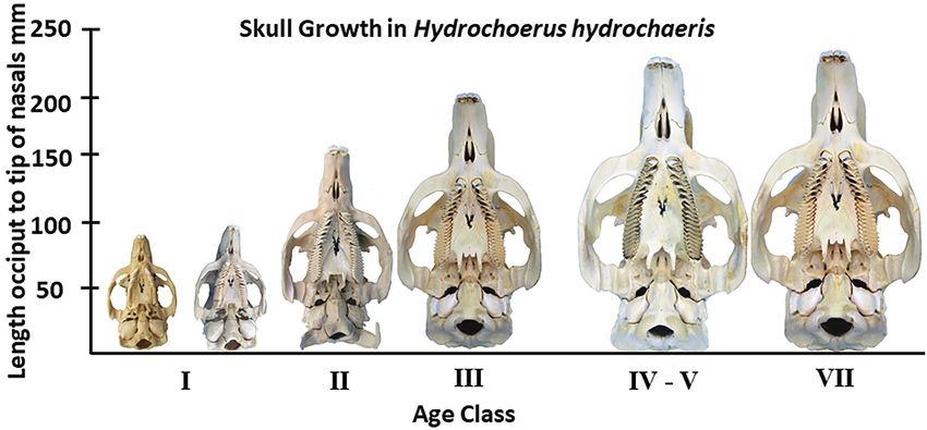
Vertebrate Anatomy Morphology Palaeontology 9:131–155

 Etymology: The specific epithet is from the Greek                    of the intrapremaxillary suture, the anterolateral margin of
'hesperos', meaning western, and the Greek 'tiganites',               the incisive foramen, and the ventrolateral external surface.
meaning pancake, in reference to its geographic location as           Portions of the right maxilla are visible in dorsal view but
the northwestern-most occurrence of a capybara in North               badly fragmented. However, enough is preserved to allow
America, as well as the crushed condition of the holotype.            the infraorbital canal to be visualized. As viewed anteriorly,
 Diagnosis: A hydrochoerine caviid rodent referred to                 the canal opening has the form of an acute triangle about
Hydrochoerus because it has an anteriorly grooved upper               34.9 mm wide at the base. The preserved height of the
incisor (I1) with faint striations within and lateral to the          canal is 46.3 mm, but this represents only the dorsomedial
groove and an M3 with a Bi-Lobed First Prism (BLFP) fol-              height of the canal as measured on the lacrimal and not the
lowed by 12 independent prisms. It differs from other de-             maximum height as measured on the maxilla.
scribed species of Hydrochoerus in its larger size, wider skull         The frontals are badly fragmented; the right frontal more
roof, more robust zygomatic process of the maxilla, and               so than the left. Neither the fronto-nasal nor the fronto-pa-
more robust descending zygomatic process of the lacrimal.             rietal sutures are clearly visible; we interpret them as un-
In the otic region, the anteromedial part of the petrosal             fused, given the unfused condition of other cranial sutures.
reaches and overlaps the alisphenoid; in the extant species           However, this cannot be determined definitively given the
H. hydrochaeris the anterolateral process of the petrosal does        damaged nature of the specimen. The fronto-squamosal su-
not reach the alisphenoid because the rostral process of the          tures are also unfused, as evidenced by the mortised sutural
malleus separates the two.                                            surface on the ascending processes of the squamosals that
                                                                      are elevated above the diagenetically depressed frontals. The
                                                                      dorsal surface of the frontals is planar and marked along
DESCRIPTION                                                           the position of the completely fused interfrontal suture
Skull                                                                 by several minute foramina. A slight sagittal ridge occurs
 SDSNH 50000 is a nearly complete, although badly crushed             behind the posterior-most foramen and extends 32.8 mm
skull (Figs. 2, 5, 6, 7; link to 3D scan). To better reference the    to the broken posterior margin. A portion of the right
position of the structures described, as well as the direction        lacrimal is present, where it remains in articulation with the
and extent of distortion during crushing, we established the          ascending process of the maxilla.
dorsal midline of the skull (Fig. 5A) as a line passing through         The dorsal surface of the fused parietals is also relatively pla-
the midline of the nasal bones and the midline of the occipital       nar and does not appear to slope ventrally towards its contact
bone. Ventrally we established the midline as a line passing          with the occipital, although this is difficult to judge given the
between the nasals, and the midline of the basisphenoid and           crushed condition of the skull. There is no indication that an
basioccipital (Fig. 5C). Neither line is entirely straight, nor are   inter-parietal was present. Anteriorly, the lateral edges of the
the two lines congruent, due to the differential distortion of        dorsal parietal table are bounded by sharply defined, medial-
the ventral versus the dorsal aspect of the skull.                    ly convex parasagittal ridges, that mark the juncture of the
                                                                      lateral and horizontal surfaces of the parietal. These features
  Dorsal view: In dorsal view (Fig. 5A, B), the skull seems
                                                                      have been referred to as the sagittal crest by Simpson (1930),
relatively undeformed; the long axis of the specimen as
                                                                      although they are not a true sagittal crest, as they never unite
preserved approximates the sagittal axis of the skull. The
                                                                      at the midline. This same feature has also been termed the
nasal, frontal, parietal, and occipital bones are present
                                                                      temporal line in suid crania (e.g., Doley et al. 2018). Hulbert
although fragmented. The condition of the naso-frontal
                                                                      et al. (2009) described a similar feature in Tapirus polkensis
and fronto-parietal sutures suggests that they were un-
                                                                      (Tapiridae), terming them parasagittal ridges; we follow this
fused; the parietal-occipital suture is clearly unfused. The
                                                                      terminology. The parasagittal ridge extends from the occipi-
left and right nasal bones are present, although only the
                                                                      to-parietal suture towards the fronto-parietal suture at the
right preserves the anterior margin. The maximum length
                                                                      postorbital projection of the frontal. The posterior portion of
of the right nasal is 87.8 mm. The maximum width of the
                                                                      each parasagittal ridge is sharply marked and produced into
left nasal is 30.0 mm and the right nasal, 30.7 mm. The
                                                                      a ridge not more than 1 mm in height above the dorsal table.
left premaxilla is represented by two unattached fragments
                                                                      As the ridge extends further anteriorly toward the postorbit-
preserving portions of the incisor alveolus. The upper incis-
                                                                      al projection, it becomes lower and broader until it nearly
or can be accurately placed in anatomical position between
                                                                      disappears. The parasagittal ridges never unite to form a true
these fragments, allowing visualization of the form of the
                                                                      sagittal crest; their closest approximation to each other is
left premaxilla. The dorsal premaxilla fragment preserves
                                                                      located 10.1 mm anterior to the occipito-parietal suture. The
the ventrolateral margin of the external nares and a portion
                                                                      posterior width of the dorsal surface of the parietal table at
of the sharply defined nasal process of the premaxilla. The
                                                                      its narrowest is 23.5 mm. The parasagittal ridges are continu-
ventral premaxilla fragment preserves the medial surface

138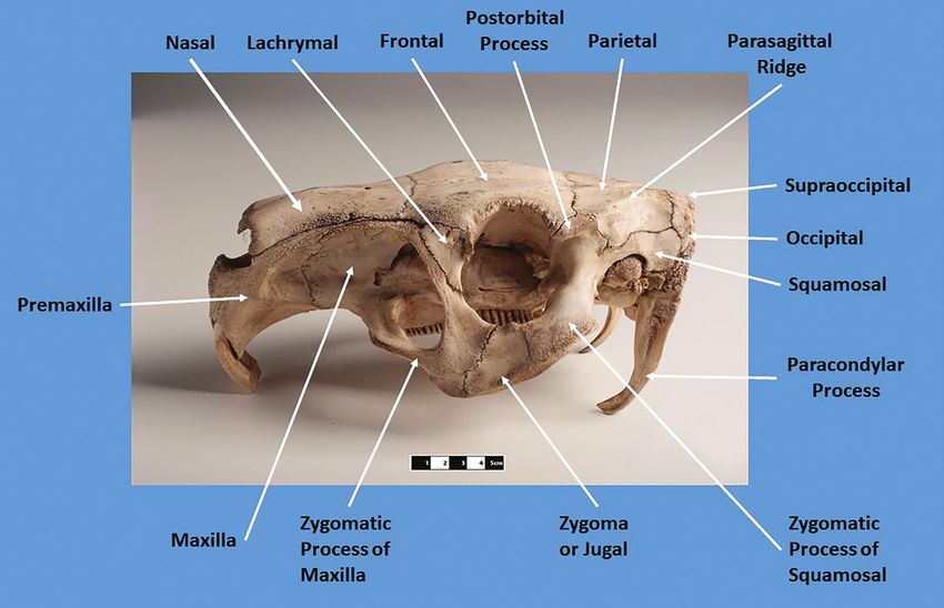
White et al. — new Pleistocene Hydrochoerus from California

Figure 5. SDSNH 50000. A, B: dorsal view; C, D: ventral view. In B and D, the boundaries of the bones are approximations, as
the crushed condition of the skull prevented determining actual suture lines in many cases. Abbreviations: BO, basioccipital;
BS, basisphenoid; F, frontal; GF, glenoid fossa; J, jugal; L, lachrymal; LB, left auditory bulla; LN, left nasal; M, maxilla; MTR,
maxillary tooth row; MZ, zygomatic process of maxilla; M2, fragment of second upper molar; M3, fragment of third upper
molar; O, occipital; OC, occipital condyle; P, parietal; Pa, palatine; Pe, petrosal; PR, parasagittal ridge; RN, right nasal; RO,
right occipital condyle; RPt, right petrosal; S, squamosal; SO, supraoccipital; SZ, zygomatic process of squamosal. Unlabeled
areas are either fragments of bone which could not be allocated to a specific element, areas of crushed and comminuted
bone, matrix, or are gaps between bones where fragments have separated or are missing.

                                                                                                                               139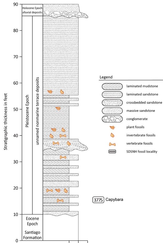
Vertebrate Anatomy Morphology Palaeontology 9:131–155

Figure 6. SDSNH 50000. A & B, lateral view; C, posterior view. In B, the boundaries of the bones are approximations, as the
crushed condition of the skull prevented determining actual suture lines in many cases. Abbreviations: F, frontal; GF, glenoid
fossa; J, jugal; L, lachrymal; MTR, maxillary tooth row; MZ, zygomatic process of maxilla; O, occipital; P, parietal; Pe, petrosal;
RM, right maxilla; RN, right nasal; ROC, right occipital condyle; RPt, right petrosal; S, squamosal; SO, supraoccipital; SZ,
zygomatic process of squamosal. Unlabeled areas are either fragments of bone which could not be allocated to a specific
element, areas of crushed and comminuted bone, and matrix, or are gaps between bones where fragments have separated
or are missing.

140
White et al. — new Pleistocene Hydrochoerus from California
ous with the temporal crests marking the lateral edges of the       relatively long and narrow. The suture between the palatal
posterior facing portion of the supraoccipital and occipital.       bone and the palatal portion of the maxillary bones is not
The temporal crests appear to be continuous with the poster-        visible. The area of the ventral exposure of the premaxillae
ior edge of the paracondylar process as far as can be seen on       and maxillae is largely missing and filled with matrix; the
the portion of the process preserved on the right side.             palatine fissure is not visible.
 There is no indication of a supraoccipital-occipital suture; we     The basisphenoid is well preserved, as is the anterior half of
interpret the suture to be completely ossified in contrast with     the basioccipital. We are unable to determine whether the
the suture between the supraoccipital and the parietal which        suture between the two bones is present and open, or if the
appears to be unfused. In posterior view, the dorsal margin of      bones were fused in life but are now broken. It is likely that,
the occipital forms the nuchal crest, which is broadly rounded      given the size of this individual, the suture was fused, as the
and evenly curved laterally and ventrally. Immediately below the    basicranial sutures in modern capybara are all fused by 4 years
nuchal crest, there is a small deep depression on either side of    of age, while many of the cranial sutures persist throughout
the vertical median crest of the supraoccipital. A second pair of   the life of the individual (Ojasti 1973, 2011; Gorosabel et al.
shallower but larger depressions is present on the supraoccipital   2017). The paracondylar process is present on the right side
immediately above and lateral to the midline of the foramen         but largely missing on the left, where only a small fragment of
magnum. The occipital is preserved, although the elongated          the dorsal-most part of the process remains.
paracondylar processes have been separated from the skull and        The mandibular fossa for the articulation of the lower jaw
cannot be reattached because of missing pieces. The right para-     is preserved on the right side. As in all hydrochoerines, the
condylar process is more complete than the left and is relatively   squamosal and the jugal participate in the formation of this
robust. We interpret the supraoccipital—paraoccipital suture as     fossa. The fossa is roofed dorsally by the zygomatic process
unfused. The dorsal margin of the foramen magnum is pre-
served, having a small, inverted V-shaped notch at its apex.
  Lateral view: In lateral view (Fig. 6 A, B), the skull
shows the crushed condition, with the right squamosal
pushed up above the parietal table, and overriding parts of
both the frontal and parietal. The zygomatic arch compris-
es the laterally and posteriorly directed zygomatic process
of the maxilla, the jugal, and the laterally and anteriorly
directed zygomatic process of the squamosal. The lateral zy-
gomatic processes of both squamosals are preserved, while
only the zygomatic process of the right maxilla is present.
The zygomatic process of the left maxilla is missing, as is
the left jugal (Fig. 6A). The anterior body of the right jugal
remains attached to the posteriorly directed zygomatic pro-
cess of the maxilla (not visible in Fig. 6 A, B). A small frag-
ment of the posterior portion of the right jugal is preserved
where it overlaps the anterior part of the zygomatic process
of the squamosal, forming the mandibular fossa.
 Ventral view: The ventral aspect of the skull (Fig. 5C,
D) is more deformed than the dorsal, with the left side
more distorted, fragmented, and more widely disarticulated
than the right. The somewhat fractured palate is preserved
with the individual fragments separated from each other by
narrow bands of matrix. The entire palate, along with both
tooth rows, is shifted to the right of the midline as defined
by the basioccipital and basisphenoid, but is nearly com-
plete. The right M2 and M3 are well preserved. The LM3
is represented by the posterior-most 4 prisms. Fragments of
what is likely the LM1 and/or LM2 are present anterior to
the remnants of the LM3 (Fig. 5C, D). The posterior por-
tion of the palate has a rounded U-shaped posterior margin          Figure 7. Hydrochoerus hesperotiganites SDSNH 50000 Right
demarcating the nasal choanae.The pterygoid processes are           otic region of skull. Anterior toward top.

                                                                                                                               141
Vertebrate Anatomy Morphology Palaeontology 9:131–155
Figure 8. Skull growth in Hydrochoerus hydrochaeris. Specimens are, from left to right: MSCC 456, MSCC 457, MSCC 463,
MSCC 461, MSCC 462, MSCC 460.
of the squamosal, which has a ridge produced ventrally squamosal, reaching and overlapping the alisphenoid. Medial
forming the medial wall of the mandibular fossa. The pos- to the anterolateral-most part of the petrosal lies the rostral
terior end of the jugal has a ridge produced ventrally which process of the malleus. There is a space between the alisphen-
forms the lateral wall of the mandibular fossa. The two oid and the petrosal between the two anterior processes of
ridges serve to prevent any side-to-side movement of the the petrosal, which forms the pyriform fenestra. Posterior and
mandible and limit its motion to the fore and aft move- slightly medial to the pyriform fenestra is what we interpret to
ment characterizing propalinal mastication (Ahearn 1981). be either the oval window or the stapedius fossa or both. The
The otic region is variably preserved (Fig. 7). On the left side, anteromedial part of the petrosal reaches and overlaps the ali-
the auditory bulla is complete, although fractured. The right sphenoid; in the extant species H. hydrochaeris the anterolateral
bulla is lacking, exposing the petrosal, which is unbroken. process of the petrosal does not reach the alisphenoid because
Anteriorly, the lateral margin of the petrosal lies against the the rostral process of the malleus separates the two completely.
Table 2. Skull measurements (in mm) of hydrochoerine specimens
Hydrochoerus hesperotiganites Hydrochoerus hydrochaeris Neochoerus aesopi
SDSNH MSCC 50000 MSCC 460 MSCC 575
Length of Skull Nasal to Occipital 250.3 230.2 -
Length of R Nasal 85.5 90.2 -
Width of R Nasal 33.5 27.3 -
Length of Frontal, approximate1 102.1 91.6 121.2
Length of Parietals 53.9 59.0 65.8
Posterior Width of Parietal Table 45.6 26.0 34.1
Anterior Width of Parietal Table 76.4 76.8 108.6
Length of Occipital 18.5 21.3 28.1
Width of Occipital, estimated2 45.1 56.3 73.2
Depth of Occipital excluding notch 31.7 31.9 42.1
1
Approximate measurement due to uncertainty of exact position of suture.
2
Estimated using bilateral symmetry where one side is complete and the other incomplete.
142White et al. — new Pleistocene Hydrochoerus from California
Figure 9. Ontogenetic series of right mandibular tooth rows in Hydrochoerus hydrochaeris. A, Age Class I with no wear on anter-
ior teeth (MSCC 456); B, Age Class I with wear on all teeth (MSCC 457); C, Age Class II (MSCC 463); D, Age Class III (MSCC 461); E,
Age Class IV – V (MSCC 4620; F, Age Class VII (MSCC 460). Measurements of the upper third molar (M3) are provided in Table 5.
Mones (1974) described and beautifully illustrated the counts of the M3 have figured prominently in the litera-
auditory region of the extant Hydrochoerus hydrochaeris. His ture and have been used as diagnostic at both the specific
drawings (Mones 1974:figs.3−22) of the auditory region of and generic level for hydrochoerids, the method of count-
neonate, juvenile, and adult specimens match closely the ing prisms and reporting those counts has varied con-
specimens in our ontogenetic series (Fig. 8). Given that the siderably among the various authors, who were not always
auditory region is well preserved in SDSNH 50000, it is specific about the method used (Ahearn 1981; Mones
possible that there may be generically diagnostic characters 1991). For the purpose of this paper, we count, and
present. We are unable to compare this with Neochoerus, report, the number of independent (free) enamel prisms
since a comparably detailed description of the auditory (laminae) posterior to the BLFP as illustrated in Figure
region for Neochoerus has not been published. 2. The posterior prism sometimes has a tiny additional
Relatively few accurate measurements can be taken of prism joined to it on the buccal side; we count them as
SDSNH 50000 because of the crushed condition of the two independent prisms following Mones (1991). Thus,
skull; those which can be either directly measured or ap- our count is one less than reported by those authors who
proximated are provided in Table 2. counted BLFP as a single prism (Ahearn 1981; Mones
Dentition: SDSNH 50000 possesses three teeth that are 1991). The RM3 of SDSNH 50000 has 12 enamel prisms
well enough preserved to provide adequate description of the posterior to the BLFP (Fig. 2).
dentition. The RM2 and RM3 (Fig. 2) are preserved in their M2 measures 12.6 mm anteroposteriorly and 10.0
entirety. The RM3 has sunk into its alveolus due to compac- mm in maximum transverse width at the second prism.
tion with its occlusal surface resting ~4 mm below the lateral The M1 and M2 are so nearly identical in Neochoerus
rim of the maxilla. The incomplete left upper incisor (I1) is and Hydrochoerus that they are not usually separated
preserved with a partial premaxilla (Fig. 10B), which cannot out in discussions of isolated teeth. In Hydrochoerus and
be attached to the skull because of missing pieces. Neochoerus, M1 and M2 are composed of two prisms,
The RM3 measures 49.6 mm in the anteroposterior both in a V-shape open buccally and joined lingually.
length and 15.7 mm in maximum transverse width. We follow Mones (1991) in counting the two lamina
There are 12 enamel prisms posterior to the BLFP, which which make up each prism as a single prism, even where
has one enamel column on the lingual side of the tooth, they lose their connection during wear. The re-entrant
and 2 columns on the buccal side separated by a distinct between the two lamina of each prism is termed the HPE
Hendedura Primera Externa (HPE, Fig. 2). The BLFP (Hendedura Primera Externa) for the anterior prism I
has been described as “V-shaped” (Ahearn 1981:64), or and the HSE (Hendadura Secundaria Externa) for the
“Y-shaped” (Kerber and Ribeiro 2011:7); we follow Perez posterior Prism II (after Mones 1991, in turn based
et al. (2017) and describe it as bi-lobed. While prism upon Rusconi (1939) and Kraglievich (1941) (Fig. 2).
143Vertebrate Anatomy Morphology Palaeontology 9:131–155
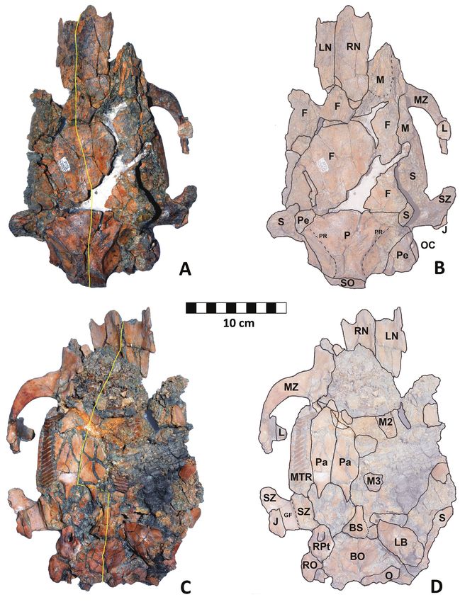
Table 3: Tooth measurements (in mm) of SDSNH 50000 The upper and lower incisors of both genera have a wide
anteromedial longitudinal groove on the enamel surface of
Anterior-Posterior Length of RM2 12.60 the tooth. Hydrochoerus was diagnosed by Ahearn (1981)
Medial-Lateral Width of RM2 10.03 as grooved but lacking ridging on the enamel surface, while
Anterior-Posterior Length of RM3 49.60
Neochoerus has both the groove and ridged enamel (Fig.
Medial-Lateral Width of RM3 15.70
Width of Upper L Incisor 13.90
10). Based on our examination of six extant Hydrochoerus
Anterior-Posterior Depth of Upper L incisor 9.72 hydrochaeris, this character needs slight revision. Careful
examination of those six specimens in incident light reveals
that faint longitudinal striations are visible both within
In SDSNH 50000 the connections between the laminae the groove and to the lateral and medial side of the groove
making up each prism have been lost, likely due to wear, (Fig. 10C). We propose changing the character state seen
extending the HPE and HSE across the entire width of in Neochoerus to “pronounced ridging” of the enamel.
the tooth. Albright et al. (2019) figure a left M2 from SDSNH 50000 is grooved, and lacks the pronounced
an individual of uncertain age from the Cooper River in ridging seen in Neochoerus, but does have faint striations
South Carolina that they refer to Neochoerus pinckneyi visible, as in Hydrochoerus (Fig. 9C). Thus, SDSNH 50000
which has the laminae of both prisms joined lingually. is referrable to Hydrochoerus based in part on this character
Unfortunately, they do not provide measurements of of the upper incisor.
this tooth. Mones (1991) provides drawings of M2 in The character most often used to allocate specimens to
Hydrochoerus hydrochaeris at birth, 4 weeks, 6 weeks, and hydrochoerine genera has been the M3, specifically the
adult; all have the laminae of both prisms joined lingually. number of enamel prisms present. Dental characters have
The incomplete left I1 (Fig. 10B) has a curvilinear length been discounted by some workers who note that size and
of 51.00 mm (as measured along the anterior enamel occlusal pattern change markedly through ontogeny (Prado
margin), a width of 13.90 mm, and a depth of 9.72 mm et al. 1998; Vucetich et al. 2005). Additionally, confu-
distally, with a width of 14.24 mm and a depth of 9.77 sion has been caused by the different methods employed
mm proximally. It has a wide, shallow groove on the to count the number of prisms as noted above. Ahearn
anterior enamel covered surface. Visible in incident light (1981:62) characterizes the genus Neochoerus as having an
are fine longitudinal striations, both within the groove M3 “…composed of an anterior Prism with a V-shaped
and lateral to it; no sharply marked longitudinal ridges cross-section followed by 13 to 16 separate laminae.” fol-
are present. The root end of the tooth has a damaged but lowing the BLFP, while Hydrochoerus has 11 or 12 separate
open pulp cavity. When fitting the partial tooth to the lamina. According to Ahearn (1981:62), Neochoerus pinck-
isolated ventral and dorsal portions of the left premaxilla, neyi has an M3 “…composed of seventeen prisms (lam-
it is clear that the preserved portion of the incisor was inae)”. We interpret this to mean that it has a BLFP and 16
unerupted and entirely within the incisor alveolus. Dental independent laminae.
measurements are provided in Table 3. Table 4 presents descriptions of M3 as used by Ahearn
(1981), Mones (1991) and Vucetich et al. (2015) to charac-
TAXONOMIC ASSIGNMENT terize the genera and species of North American hydrocho-
erines. We interpret the terminology of both Ahearn (1981)
Identifying North American Pleistocene capybara fossils and Mones (1991) based on their written descriptions as well
has long been a contentious undertaking (Ahearn 1981; as the illustrations they provided. It should be noted that the
Mones 1991; Vucetich et al. 2015; Carranza-Castañeda low prism count for Neochoerus reported by Ahearn (1981)
2016). In the past, taxonomic assessment has relied primar- and Mones (1991) is due to the inclusion of South American
ily on three morphological features, one on the mandible, species of Neochoerus, particularly N. tarijensis and N sulci-
one on the skull, and one on the upper and lower incisors. dens. No North American specimens of Neochoerus have been
The masseteric ridge of the mandible has been used to sep- reported with fewer than 15 post-BLFP prisms. The Central
arate Neochoerus (originally including N. dichroplax, now American skulls of Neochoerus examined by us directly or
assigned to the genus Phugatherium) from Hydrochoerus. In in photographic images also have 15 or more post-BLFP
Neochoerus (less N. dichroplax), the masseteric ridge begins prisms. A more complete review of the South American spe-
lateral to the middle of the last prism (PIII) of p4, accord- cies is needed to determine their status. We here consider 15
ing to Ahearn (1981), while in Hydrochoerus, the masse- or more post-BLFP prisms as diagnostic for North American
teric ridge begins lateral to the middle prism (PII). Since species of Neochoerus.
SDSNH 50000 lacks the lower jaws, it cannot be allocated Several authors have remarked that tooth morphology,
to either genus based on characters of the lower jaw. including size, prism counts, and occlusal pattern, vary
144White et al. — new Pleistocene Hydrochoerus from California
Table 4. Description of M3 in hydrochoerines by previous authors and as used in this study.
Taxon Ahearn 1981 Mones 1991 Vucetich 2015 This study
Hydrochoerus BLFP + 11-12 BLFP + 9 - 13 - BLFP + 10 - 13
H. hydrochaeris BLFP + 11-12 BLFP + 10 - 13 - -
H. holmesi BLFP + 11-12 - - -
Neochoerus BLFP + 13 -16 BLFP + 12 - 16 - BLFP + 15- 161
N. dichroplax BLFP + 14 -15 BLFP + 15 - -
N. pinckneyi BLFP + 16 - - -
N. aesopi - BLFP + 15 - 16 - BLFP + 15 - 16
N. tarijensis2 - BLFP + 13 - 14 - -
N. sulcidens2 - BLFP + 12 - 13 - -
Phugatherium - - BLFP + 14 -18 BLFP + 14 - 18
P. cataclisticum2 - - BLFP + 16 - 18 -
P. dichroplax - - - BLFP + 15
1
Excludes South American species
2
Indicates South American species
Figure 10. Anterior enamel surface of incisors. A, Neochoerus aesopi, MSCC 581, Florida; B, Hydrochoerus hesperotiganites,
SDSNH 50000, California; ,. Hydrochoerus hydrochaeris, MSCC 461, zoo specimen.
145Vertebrate Anatomy Morphology Palaeontology 9:131–155 Table 5. Ontogenetic series of Hydrochoerus hydrochaeris M3s. Specimen Number Age Class Calendar Age Post- BLFP Independent Prisms Length of M3 (mm) Width of M3 (mm) MSCC 456 I 0-4 months 13 13.5 3.5 MSCC 457 I 0-4 months 12 15.9 5.6 MSCC 463 II 4 –12 months 11 30.4 12.0 MSCC 461 III 1 – 1.5 years 12 35.1 12.9 MSCC 462 IV-V 1.5 – 2 years 11 43.3 15.8 MSCC 460 VII 4 years and up 12 48.2 17.2 Table 6. M3 prism counts in Hydrochoerus hydrochaeris, data from Mones (1991); N = 57. Number of prisms Number of Post-BLFP Prisms Count in sample % of sample 11 10 1 1.8 12 11 20 34.9 13 12 34 59.4 14 13 2 3.7 ontogenetically, and thus should not be given much taxo- (Age Class VII) based on the scheme of Ojasti (1973, 2011) nomic weight (Prado et al. 1998; Vucetich et al. 2005). as utilized by Gorosabel et al. (2017). Counts of the in- Mones (1991:fig. 7) illustrated the ontogenetic changes in dependent prisms posterior to the BLFP and the length and M3 of extant Hydrochoerus hydrochaeris. width of the M3 are presented in Table 5. Figure 9 shows In order to further evaluate the usefulness of prism counts enlarged views of the individual tooth rows; Figure 8 shows as a taxonomic character, we examined an ontogenetic series the 6 skulls to scale to illustrate skull growth. As can be seen of six modern skulls of Hydrochoerus hydrochaeris (Figs. 8, 9), from this relatively small sample, the number of independ- ranging in age from about 4 months (Age Class I) to 4+ years ent prisms ranges from 11 to 13 but is not correlated with Figure 11. Bivariate plot of M3 measurements of capybaras. 146
White et al. — new Pleistocene Hydrochoerus from California
ontogeny. Mones (1991) reported on a sample of 57 M3s of         and Ribiero (2011), Vucetich et al. (2005), Carranza-
Hydrochoerus hydrochaeris from Uruguay. Table 6 summar-          Castaneda and Miller (1988), Hay (1923) and our own
izes those counts and their percentage in the entire sample.     measurements. It is curious that no smaller specimens have
Additional data on the M3 of extant Hydrochoerus hydro-          been assigned to that taxon. In contrast, smaller speci-
chaeris were published by Hooijer (1959) who measured a          mens have been assigned to both Neochoerus aesopi and
series of 20 capybara skulls in the Leiden Museum, recording     Hydrochoerus holmesi. Ahearn (1981) considered H. holmesi
the length of the M3 and the basilar length of the skull. We     to be a nomen dubium but noted that the ridged incisor
have plotted his data, with the data from our 6 specimens,       of the type might indicate a small or juvenile Neochoerus
in Figure 12. It is clear from this plot that the length of M3   (in which she also included N. dichroplax). Mones (1991)
increases throughout ontogeny.                                   included both H. holmesi and N. pinckneyi in Neochoerus
                                                                 aesopi. Baskin et al. (2020) considered H. holmesi to be a
                                                                 junior synonym of N. aesopi. These authors recognized just
                                                                 one species of Neochoerus from the late Blancan through the
                                                                 late Rancholabrean of North America, Neochoerus aesopi.
                                                                 Given the existence of juvenile specimens of both Neochoerus
                                                                 and Hydrochoerus, it seems unlikely that only fully adult
                                                                 Neochoerus pinckneyi have been found. We are not convinced
                                                                 by the arguments of Mones (1991), accepted by most recent
                                                                 authors, that all of the specimens of N. pinckneyi referred by
                                                                 him to N. aesopi are correctly allocated, particularly those
                                                                 dating to the Blancan NALMA. Given the significant range
                                                                 in size of the three species allocated to Hydrochoerus, the
                                                                 most parsimonious interpretation, and the one followed
                                                                 here, is that the specimens identified as H. holmesi in the bi-
                                                                 variate plot of Ahearn (1981), and those described by others
Figure 12. Bivariate plot of the length of M3 plotted against
the basilar length of the skull. Data from a sample of extant    as N. aesopi (Baskin et al. 2020; Carbot-Chanona et al.
H. hydrochaeris in Hooijer (1959) and from our sample of         2020) are simply younger conspecifics of the large, presum-
extant H. hydrochaeris (Tab. 5). N=26.                           ably fully adult individuals identified as Neochoerus pinck-
                                                                 neyi. This confusion is exacerbated by the difficulty in aging
                                                                 capybara specimens independent of tooth size. Nevertheless,
 Gorosabel et al. (2017) examined 250 skulls collected in        we advocate for a more thorough study of this problem that
northern Argentina, stating that the skulls corresponding        includes Florida Blancan and Irvingtonian samples.
to different age categories exhibited the same number of          Another potentially diagnostic character was first de-
prisms. It is unclear whether they intended to indicate          scribed by Simpson (1930:9) involving the skull roof.
that the entire sample has the same number of prisms, or           “It is the skull roof that most obviously distinguishes
that each age category has the same range of plate counts.       fossil and recent animals. The frontal region on the fos-
However, either interpretation suggests that variation in        sil is very broad and nearly flat. The sagittal crests on the
prism count in the teeth of extant Hydrochoerus hydrochaeris     parietals narrow very rapidly: at the postorbital processes
is unrelated to ontogenetic age. Aeschbach et al. (2016)         they are nearly twice as wide as in Hydrochoerus, while at
examined the growth of the cheek teeth in their samples          the contact with the supraoccipital (or interparietal?) they
of 117 H. hydrochaeris and H. isthmus and concluded that         are of almost the exact same width as in the latter. The roof
the number of prisms did not change through post-natal           of the parietals is not arched and does not curve downward
ontogeny. These data all indicate that the changes in oc-        posteriorly but continues the plane of the frontals and rises
clusal pattern occur within the first 3 to 4 months; and that    to a point at the supraoccipital suture. The interparietal
after that time, the occlusal pattern, particularly the prism    part of the supraoccipital is relatively longer in the fossil,
count, is remarkably stable.                                     and the occiput relatively higher.”
 Other characters have been utilized to differentiate             Mones (1991:39, translation by RSW) included characters
Hydrochoerus from Neochoerus. We note here that all speci-       of the skull roof in his generic diagnoses of Hydrochoerus
mens we were able to find in the literature that have been       and Neochoerus:
referred to Neochoerus pinckneyi are exceedingly large. Figure         [Hydrochoerus with] “… face relatively shorter than
11 presents a bivariate plot of the length and width of the           Neochoerus, with the skull roof proportionally short-
upper third molar (M3) based on measurements in Kerber                er at the level of the frontals and nasals, and slightly

                                                                                                                            147
Vertebrate Anatomy Morphology Palaeontology 9:131–155
descending towards the occiput; thinner anteorbital bar extant H. hydrochaeris, as does the more robust zygomatic
and less robust lacrimal; supraoccipital narrower and less process of the maxilla and descending zygomatic process
excavated. of the lacrimal. Characteristics of the otic region are also
[Neochoerus differs] “… from Hydrochoerus in: size one different from those in extant Hydrochoerus, although the
third to two times larger; proportionally longer face; true taxonomic significance of these differences cannot be
anterior portion of the zygomatic arch more rounded meaningfully evaluated until descriptions of this area in
and directed backwards, less transverse; more robust other hydrochoerines are available. Although we cannot ad-
anteorbital bar and lacrimal; skull roof very broad, pro- equately quantify the difference in the breadth of the skull
portionally wider through the nasals and frontals, with because it is crushed and distorted, the other differences
these proportionally shorter and the parietals longer, are clearly defined and justify the establishment of the new
occiput less convergent dorsally, supraoccipital wider species Hydrochoerus hesperotiganites.
and relatively more excavated; …” (Mones 1991:49)
BIOGEOGRAPHY
Interpreting this character complex is somewhat sub-
jective in SDSNH 50000 since the skull has been exten- Capybaras (Caviidae: Hydrochoerinae) and the porcu-
sively crushed dorso-ventrally. In addition, Mones (1991) pine Erethizon (Erethizontidae) are the only two groups of
included “Neochoerus dichroplax” in his diagnosis of the South American caviomorph rodents that reached temper-
genus in which he discussed only dental characters, so ate North America in the late Pliocene to early Pleistocene
we are unable to determine how the rest of his diagno- (late Blancan and early Irvingtonian) phase of the Great
sis for the genus would be affected by the reallocation of American Biotic Interchange (GABI) (Frazier 1981; Morgan
N. dichroplax to the genus Phugatherium (Vucetich et al. 2005, 2008; Woodburne 2010; Vucetich et al. 2015). This
2015). SDSNH 50000 is somewhat larger than the largest later phase of the GABI began in the early Pliocene about 5
of the modern Hydrochoerus hydrochaeris specimens avail- Ma with the final connection of North America and South
able to us (MSCC 460). The skull length, measured from America at the Isthmus of Panama (O’Dea et al. 2016).
the posterior edge of the occipital to the anterior end of During an earlier phase of the GABI in the late Miocene
the nasals is 9% larger than MSCC 460 (250.3 mm, versus (early Hemphillian NALMA, ~9 Ma), two genera of ground
230.2 mm). Other measurements vary from smaller to sloths in the families Megalonychidae and Mylodontidae ap-
larger in SDSNH 50000 than MSCC 460 (Table 2), but parently reached North America by overwater dispersal from
how much of this is due to the distortion in the fossil is South America (Morgan 2005; 2008). Although our paper
unclear. SDSNH 50000 is clearly somewhat larger than our describing a new species of the extant genus Hydrochoerus
largest modern specimen, MSCC 460, but nowhere near from the late Pleistocene is not the appropriate place for
the 33-50% larger condition described by Mones (1991) a detailed discussion of the biogeography and systematics
for Neochoerus. The parietals and frontals appear broad- of capybaras associated with the late Pliocene and early
er in SDSNH 50000 than in modern Hydrochoerus. In Pleistocene phase of the GABI, it is nevertheless important
addition, the supraoccipital of SDSNH 50000 is broader to establish the evolutionary history of Hydrochoerus and
and gently rounded laterally and ventrally, as opposed to other hydrochoerines in North America.
MSCC 460, where the supraoccipital is higher, less broad Three genera of capybaras, the extinct Neochoerus and
and has a straighter slope laterally. Our ontogenetic sample Phugatherium and the extant Hydrochoerus, have been
of modern H. hydrochaeris shows that the supraoccipital reported from the North American fossil record (Ahearn
does not unite with the occipitals until Age Class IV where 1981; Mones 1991; Morgan 2005, 2008; Vucetich et al.
it is partly, but not completely, fused. In Age Class VII no 2015). Phugatherium was named from the Pliocene of
suture is visible. Argentina and is represented in North America by the
Since we cannot reliably separate the effect of distortion species P. dichroplax (Vucetich et al., 2015), originally
and the difference in size in skull roof proportions, we are described as Neochoerus dichroplax from three late Blancan
not confident that this character complex is well-enough faunas in the southern USA, 111 Ranch (= Dry Mountain)
defined to be considered in our taxonomic assignment. in Arizona (type locality) and two sites in southern Florida
Considering all the above, SDSNH 50000 can confident- (Ahearn and Lance 1980). Morgan and Hulbert (1995)
ly be allocated to Hydrochoerus, based on the morphol- and Hulbert (2010) reported several additional late
ogy of M3 and I1 as described. The fact that the skull is Blancan records of N. (= P.) dichroplax from Florida. The
somewhat larger, and the likelihood that the skull roof earliest well-dated capybara from North America is N. cor-
is relatively broader in SDSNH 50000 than in living H. dobai from the early Blancan (~3.6 Ma) of central México
hydrochaeris indicates its specific distinctiveness from the (Carranza-Castañeda and Miller 1988). Although Vucetich
148White et al. — new Pleistocene Hydrochoerus from California
et al. (2015) synonymized N. cordobai with the late States and Central America, with Phugatherium known
Blancan (~2.5−2.7 Ma) N. dichroplax from the southern only from the Blancan and Neochoerus occurring in the
USA, Carranza-Castañeda (2016) recognized N. cordobai Blancan, Irvingtonian and Rancholabrean. Following the
as a valid species of Neochoerus and also described a new current taxonomy, with referral of Hydrochoerus holmesi to
species N. occidentalis from several late Blancan and early Neochoerus aesopi (Mones 1991; Baskin et al. 2020), none
Irvingtonian faunas in México. Neochoerus (not including of the previously described North American capybaras
Phugatherium but including most previous North American belong to the extant genus Hydrochoerus. Consequently,
records of Hydrochoerus-see below) first appeared in either the new species described here, H. hesperotiganites, from
the early Blancan of México or the late Blancan of Florida the Rancholabrean of southern California appears to be
and South Carolina (Ahearn 1981; Morgan 2005; Vucetich the only valid fossil record of Hydrochoerus from North
et al. 2015; Carranza-Castañeda 2016; Albright et al. America, causing us to rethink the biogeography of this
2019). Several of these North American Blancan capybaras genus. With the northern-most range extension of the liv-
appear to be older than the oldest records of Neochoerus ing capybara H. isthmius in eastern Panama, there appears
from South America (Vucetich et al. 2015). That begs the to be no record of Hydrochoerus, living or fossil, between
question-did Neochoerus evolve in South America and Panama and southern California.
disperse to North America in the Pliocene as a participant Determining which species of capybara crossed the
in the GABI, or did Neochoerus evolve in North America Panamanian Land Bridge, and when, as well as what route
from Phugatherium dichroplax or a species closely related to they took in their dispersal northwards into North America
N. cordobai? An answer to this question will require more would naturally begin, of course, with Central America and
detailed systematic studies of North American Blancan southern México. However, the fossil record of capybaras is
capybaras, which is beyond the scope of this paper. extremely limited for Central America, with just five rec-
More relevant to our current study is the status of fossil ords (one unpublished), all probably, but not certainly, of
capybaras from North America previously referred to the Pleistocene age. Importantly, all are identified as Neochoerus
living genus Hydrochoerus. Simpson (1928) described an rather than Hydrochoerus. The unpublished record is from
extinct species of Hydrochoerus, H. holmesi, from the late Guatemala, in the American Museum of Natural History
Pleistocene (Rancholabrean) Sabertooth Cave in Florida. (FM 94034; RSW notes), the other in the Paleontological
Many other Florida fossil capybaras have since been Museum in Estanzuela, Guatemala (PV-H-45; McDonald
referred to H. holmesi, including specimens as old as late and Davila A 2017; H.G. McDonald, personal communi-
Blancan (Ahearn 1981; Morgan 2005). However, the taxo- cation). Judging from the available photographs, both
nomic studies we follow here (e.g., Mones 1991; Baskin et specimens appear to be Neochoerus, as they have M3s with
al. 2020) transferred H. holmesi to Neochoerus and have also 15 independent prisms following the BLFP. The published
synonymized this species with N. aesopi, in which they also records include one each in San Salvador (Webb and Perrigo
included N. pinckneyi. Although these authors proposed 1984), Nicaragua (Leidy 1887; Lucas et al. 2008), Honduras
that N. aesopi occurred over a time period of nearly 2 mil- (Ahearn 1981; Webb and Perrigo 1984) and Guatemala
lion years, from the late Blancan to the late Rancholabrean, (Lucas et al. 2021; McDonald and Davila A 2017; H.G.
it is worth noting that few Rancholabrean species of North McDonald, personal communication). All four were identi-
American mammals have chronologic ranges that extend fied as Neochoerus and dated as late Pleistocene. McDonald
back into the Blancan (Kurtén and Anderson, 1980). and Davila A (2010) map the occurrence of Mammuthus
Furthermore, Vucetich et al. (2015:325) stated “…extinct columbi in Central America; all known occurrences are
species [of capybaras] had short stratigraphic ranges (partly confined to the Pacific lowlands of El Salvador, Honduras,
because of their physiological requirements) and relatively Nicaragua, and Costa Rica. This suggests that the corridor
wide geographic ranges, rendering them useful as biostrati- for southward dispersal (and presumably northward as well)
graphic tools…”. Most late Blancan and Irvingtonian sam- was along the Pacific Coast. Alternate explanations could be
ples of Neochoerus from the southeastern United States, in that there has been less paleontological exploration along the
particular Florida, have not been studied in detail and may Atlantic coast and the interior highlands; or that an Atlantic
represent a species of Neochoerus distinct from N. aesopi (= dispersal corridor lies east of the present shoreline and was
N. pinckneyi = H. holmesi). submerged by rising sea levels after the last glacial high sea
In summary, it appears that all previous fossil records of level. Certainly, intensive exploration of the interior and the
capybaras from North America represent one of two extinct Atlantic coast areas is desirable.
genera, Neochoerus or Phugatherium. These records span The only record of Rancholabrean capybaras from
the time period from the late Pliocene (~3.6 Ma) of central southern México is that reported by Carbot-Chanona et
México to the late Pleistocene of the southeastern United al. (2020) from Chiapas and discussed below. Given the
149You can also read