A Comparative Study on Low and High Salinity Tolerance of Two Strains of Pinctada fucata
←
→
Page content transcription
If your browser does not render page correctly, please read the page content below
ORIGINAL RESEARCH
                                                                                                                                                     published: 27 July 2021
                                                                                                                                           doi: 10.3389/fmars.2021.704907

                                            A Comparative Study on Low and
                                            High Salinity Tolerance of Two
                                            Strains of Pinctada fucata
                                            Jing Sun 1,2,3,4† , Mingqiang Chen 2,3,4† , Zhengyi Fu 2,4 , Jingru Yang 2,4 , Shengjie Zhou 2,3,4 ,
                                            Gang Yu 2,3 , Wenli Zhou 1* and Zhenhua Ma 2,3,4*
                                            1
                                              College of Fisheries, Tianjin Agricultural University, Tianjin, China, 2 Sanya Fisheries Research Institute, Sanya, China, 3 Key
                                            Laboratory of South China Sea Fishery Resources Exploitation and Utilization, Ministry of Agriculture, Guangzhou, China,
                                            4
                                              Tropical Aquaculture Research and Development Center, South China Sea Fisheries Research Institute, Chinese Academy
                                            of Fishery Sciences, Sanya, China

                            Edited by:      This study compares salinity tolerance between red and black shell Pinctada fucata
                                Yan Shi,
                                            salinity stress of 20 and 50h, while 35h was used as a control. The hemolymph
                 Hohai University, China
                                            osmotic pressure, inorganic ion concentration, the activities of Na+ -K+ -ATPase,
                     Reviewed by:
                     You-Ting Zhu,          respiratory metabolism related enzymes and liver tissue antioxidant related enzymes
    Shanghai Ocean University, China        were measured at 12 and 24 h after salinity stress. The osmotic pressure and inorganic
                    Jeswin Joseph,
        Cochin University of Science
                                            ion concentration of hemolymph of two strains P. fucata increased significantly with the
              and Technology, India         increase of salinity. The activity of Na+ -K+ -ATPase of red P. fucata only decreased under
                *Correspondence:            low salinity at 24 h, and was significantly higher than that the control under low salinity
                        Wenli Zhou
                                            at 12 h and high salinity at 12 and 24 h. The succinate dehydrogenase (SDH) activities
                wlzhou69@126.com
                       Zhenhua Ma           of the P. fucata treatment groups were significantly higher than those the control at 12 h.
           zhenhua.ma@hotmail.com           The lactate dehydrogenase (LDH) activity increased significantly with salinity at 12 h. and
     † These   authors have contributed     the black P. fucata LDH activity was significantly higher than the control at 24 h, while the
                    equally to this work
                                            LDH activity of red P. fucata was significantly lower than that the control in 50h salinity.
                   Specialty section:       The superoxide dismutase (SOD) activity of black P. fucata was significantly lower than
         This article was submitted to      that the control, while that of red P. fucata was significantly higher than that the control
                   Aquatic Physiology,
               a section of the journal     within 24. At 12 h, the catalase (CAT) activity of red P. fucata increased significantly
          Frontiers in Marine Science       with salinity, but decreased significantly with salinity at 24 h. The CAT activity of black
               Received: 04 May 2021        P. fucata was highest at 24 h under low salinity. Glutathione peroxidase (GSH-Px) and
               Accepted: 08 July 2021
               Published: 27 July 2021
                                            alkaline phosphatase (AKP) activities of red P. fucata were significantly higher than those
                             Citation:
                                            the control under low or high salinity. At high salinity for 24 h, the GSH-Px activity was
        Sun J, Chen M, Fu Z, Yang J,        lowest in black P. fucata, AKP activity was highest. The present study indicates that
    Zhou S, Yu G, Zhou W and Ma Z           the physical responses of P. fucata to the salinity stress vary with shell colors. The red
 (2021) A Comparative Study on Low
   and High Salinity Tolerance of Two       P. fucata can quickly respond positively to the change of environmental salinity and
           Strains of Pinctada fucata.      reduce the damage caused by the change of environmental salinity.
           Front. Mar. Sci. 8:704907.
    doi: 10.3389/fmars.2021.704907          Keywords: Pinctada fucata, salinity, osmotic regulation, respiratory metabolism, antioxidant

Frontiers in Marine Science | www.frontiersin.org                                    1                                                 July 2021 | Volume 8 | Article 704907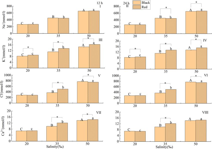
Sun et al. Pinctada fucata Salinity Tolerance
INTRODUCTION was significantly higher than that under low and high salt stress
(Huang et al., 2016). In American oysters, C. virginica, rapid
The Pinctada fucata is an important marine pearl producing changes in salinity affected the rate of hemocytes locomotion and
molluscan, distributes in tropical and subtropical areas in the extended the spreading time of hemocytes (Fisher and Newell,
coast or offshore seabed (Meng et al., 1996). Since 1949, P. fucata 1986). Although salinity significantly affects various physiological
has been cultured in Hainan, Guangdong and Guangxi in China functions of marine shellfish, physiological responses to salinity
(Ai et al., 2003), and the pearl production peaked in the 1990s, stress varied between species.
which has brought substantial economic income to the pearl Shellfish shell color can be inherited, and there are significant
industry (Yang et al., 2017; Yang et al., 2017). In recent years, differences in growth, survival and nutritional performance of
massive mortality frequently occurs in P. fucata, and has brought individuals with different shell colors (Huaiping et al., 2005;
considerable economic losses. Evidence has suggested that such Liu et al., 2005; Gu, 2014). The growth environment has an
massive mortality is often associated with environmental changes important influence on the choice of shell color, and salinity
(Zhang et al., 2015; Chen et al., 2016). Studies have shown that is one of the important reasons that affect the shell color
under the influence of salinity, temperature, pH and dissolved polymorphism among different populations or within the same
oxygen, physiological indicators, such as osmotic pressure, population (Guan and He, 2009). The studies on Littorina
cellular immunity level, oxygen consumption and ammonia obtusata and Littorina saxatilis also showed that salinity had
excretion, can be altered, resulting in physiological metabolism a high contribution to the selection of shell color phenotype
imbalance, growth inhibition and even death (Ghiselli et al., 2000; (Sergievskii and Berger, 1984; Sokolova and Berger, 2000; Phifer-
Nan et al., 2004; Liu and Yan, 2006; Ocaño-Higuera et al., 2011). Rixey et al., 2008). Evidence has suggested that the changes of
As one of the vital ecological factors in the marine salinity can have regulated the physiology and biochemistry of
ecosystem, salinity affects the metabolism, osmotic adjustment P. fucata. Liu et al. (2011) found that the oxygen consumption
and biological rhythm of marine organisms and plays a decisive rate and ammonia excretion rate of P. fucata decreased with the
role in their distribution (Navarro, 1988; Kim et al., 1998). Due increase of salinity in the range of 21–36h. Under short-term
to the influence of seasonal rainfall, evaporation and tide, the low salinity stress, P. fucata might be vulnerable to diseases due to
salinity of seawater usually changes over season. The salinity level low activity of lysozyme and catalase (Arisman et al., 2018). Pan
of the external water environment directly affects the osmotic et al. (2020) found that the aquaporin expression level of P. fucata
pressure of aquatic animals and then regulates their survival, returned to the control level (27h) under high salinity (36h)
growth and reproduction (Cheng et al., 2002; Garçon et al., 2009; stress for 168 h and low salinity (16h) stress for 72 h. However,
Jasmani et al., 2010). Studies have shown that when the osmotic there are relatively few studies on the effects of salinity on
pressure of aquatic animals’ body fluid is equal to the osmotic P. fucata with different shell colors. Therefore, the purpose of this
pressure of the ambient environment, the adjustment of osmotic study was to compare and analyze the salinity tolerance of two
pressure consumes the least amount of energy, and the energy different shell-colored P. fucata populations by measuring the
conversion efficiency is the highest, thus achieving maximum effects of salinity on osmotic regulation, respiratory metabolism
growth (Chen and Lin, 1995; Masui et al., 2009). Alagarswami and antioxidant function. Results of the present study can provide
and Victor (1976) found that the filtration rate of P. fucata a reference for further study of salt tolerance mechanism of
decreased with decreasing salinity. Widdows (1985) found that P. fucata with different shell colors.
when salinity was < 20h, the feeding activity and growth rate
of Mytilus edulis decreased. Inorganic ions are the main osmotic
factors in the hemolymph of marine shellfish, and K+ , Cl− , and MATERIALS AND METHODS
Na+ ions are mainly involved in osmotic regulation. In M. edulis
and Littorina littorea, Natochin et al. (1979) found that besides Source and Acclimation of Pinctada
the Na+ pump and K+ pump, both species have a Cl− related fucata
sodium ion exchange system, which could regulate cell volume In total, 360 P. fucatas (F11 ) used in this study were obtained
and osmotic pressure. from the Lingshui Station (Hainan, China), Tropical Aquaculture
Low-salt or high-salt stress on marine shellfish can reduce Research and Development Center, South China Sea Fisheries
shellfish activities and changes physiological status. The Research Institute, Chinese Academy of Fishery Sciences. Upon
hemolymph volume of Haliotis rubra and H. laevigata can transfer to the laboratory, the attached organisms on the shell
increase by 25% in a short time of salinity decrease, while with surface were removed, and healthy individuals with distinct shell
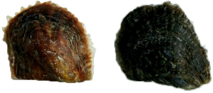
increasing salinity, their hemolymph volume decreased, and color (Figure 1) characteristics and similar size (shell length:
the adhesive force, movement of gill cilia, and heart beat were 50.75 ± 1.43 mm, body weight: 34.19 ± 1.39 g) were collected.
affected (Edwards, 2003). The metabolic activities of Haliotis The P. fucata were acclimated in a cement tank (5,000 L) for
cracherodii and Haliotis rufescens under high-salinity stress were 7 days. During the acclimation, the water temperature was
swiftly and significantly impacted within 10 min and began to maintained at 30 ± 1◦ C; salinity was 35h; pH was 8.0 ± 0.1;
resume in 6–8 h (Berger and Kharazova, 1997). At low salinity, DO was > 6.5 mg/L; light intensity was < 500 Lx and natural
mitochondria in gills of Crassostrea virginica showed a higher photoperiod was used. During the acclimation, 50% water was
ratio of glutamic acid oxidation (Ballantyne and Moyes, 1987). replaced daily, and the P. fucata was fed with Platymonas
The expression level of α -amylase in P. fucata at salinity 27h subcordiformis at 09:00-09:30 daily with a concentration of
Frontiers in Marine Science | www.frontiersin.org 2 July 2021 | Volume 8 | Article 704907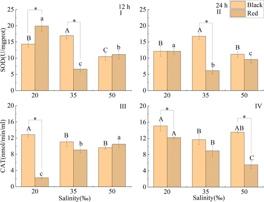
Sun et al.                                                                                                         Pinctada fucata Salinity Tolerance

2 × 10⁵ cell/mL. The number of dead P. fucata was recorded                 reaction. O2 − reacted with a tetrazolium salt WST-8 dye to form a
and removed daily. Feeding was stopped 1 day before the                    water-soluble colored formazan product (λmax = 450 nm). When
experiment began.                                                          the inhibition percentage in the above-mentioned xanthine
                                                                           oxidase coupling reaction system was 50%, the SOD enzyme
Experiment Design                                                          activity in the reaction system was defined as an enzyme activity
The experiment was carried out in nine cement tanks (tank                  unit (U/mL). Catalase (EC 1.11.1.6) activity: This assay kit was
volume: 800 L). According to the natural living conditions of              based on the reaction of catalase with methanol, with an optimal
P. fucata and the preliminary experimental results, experiment             concentration of H2 O2 . The formaldehyde produce could be
water was adjusted to the desired salinity of 20, 50h by mixing            measured colorimetrically at OD 540 nm. One unit was defined as
natural seawater (35h), tap water with 24 h aeration and sea               the amount of enzyme that could cause the formation of 1.0 nmol
salts. Natural double-filtered seawater served as the control.             of formaldehyde per minute at 25◦ C (nmol/min/ml). Glutathione
There were three salinity treatments in the experiment, each               Peroxidase activity: GSH-Px catalyzed H2 O2 to oxidize GSH to
treatment had three replicates, and each replicate contained 15            produce GSSG, glutathione reductase (GR) catalyzed NADPH
P. fucata with red shell and black shell, respectively. Besides            to reduce GSSG to regenerate GSH, while NADPH oxidized to
the salinity, water quality parameters were maintained at the              produce NADP+ , NADPH had a characteristic absorption peak
same level used in the acclimation period. Three shellfish were            at 340 nm, while NADP+ did not. One unit of enzyme activity was
randomly collected from each parallel at 12 and 24 h, respectively.        defined at 25◦ C or 37◦ C, 1 mol NADPH oxidation per milligram
                                                                           of protein per minute was catalyzed at pH 8.0 (U/mgprot).
Hemolymph Sample Collection                                                Alkaline Phosphatase activity: in an alkaline environment, AKP
The shells were slightly opened by a shell opener, and the                 catalyzed phthalate disodium to generate free phenol; phenol
adductor muscle was cut off by a scalpel on the left shell, and the        reacts with 4-aminoantipyrine and potassium ferricyanide to
pericardial cavity was exposed. Hemolymph was withdrawn from               produce a red quinone derivative, which had characteristic light
the pericardial cavity of each P. fucata with a 2 mL syringe and           absorption at 510 nm. One unit of enzyme activity was defined at
was quickly expelled into a 2 mL centrifuging tube. Hemolymph              37◦ C, 1 µmol phenol produced per min in 1 mg protein reaction
Na+ , K+ , Cl− , and Ca2+ and osmotic pressure were measured by            system is defined as a unit of enzyme activity (U/mgprot). Total
the PL2000Plus blood gas biochemical analyzer (Nanjing Pulang              protein quantification: The content of total protein in gill and
Medical Equipment Co., Ltd.).                                              liver tissue was determined by BCA method. Under the alkaline
                                                                           condition, protein reduced Cu2+ to Cu+ , and Cu+ formed
Tissue Sample Collection                                                   a purple complex with BCA reagent. There was a maximum
After the hemolymph was collected, the gill and liver tissues were         absorption peak at 562 nm.
cut off with scissors on an ice tray. Rinsed with cold physiological
saline (0.9% NaCl), blotted with filter paper, the tissue samples          Statistical Analysis
were placed in a 2 mL centrifugal tube and stored at −80◦ C.               All experimental data were expressed as mean ± standard
The activities of Na+ -K+ -ATPase (Item No. KTB1800) and                   deviation and SPSS 22.0 was used to analyze the hemolymph
lactate dehydrogenase (Item No. KTB1110) in gills tissue and               biochemical indexes and related indexes of gill and liver tissues
the activities of superoxide dismutase (Item No. KTB1030),                 of P. fucata with the same shell color under different salinity
catalase (Item No. KTB1040), glutathione peroxidase (Item No.              by one-way ANOVA and Duncan multiple comparisons. T-test
KTB1640) and alkaline phosphatase (Item No. KTB1700) in the                was used to analyze the differences of hemolymph biochemical
liver tissue were determined according to the reagent instructions         indexes gill and liver tissues related indexes with the same salinity
of Abbkine reagent company. The activity of succinic acid                  and different shell colors, and P < 0.05 was set as the significant
dehydrogenase (Item No. A022-1-1) in the gill tissue and total             difference level.
protein (Item No. A045-4-4) was determined according to the
instructions of the manufacturer (Nanjing Jiancheng Institute of
Biological Engineering).
    In the assay, Na+ -K+ -ATPase catalyzed ATP hydrolysis                 RESULTS
to produce ADP and inorganic phosphorus. The content of
inorganic phosphorus could reflect the activity of ATPase. The             Determination of Osmotic Pressure in
amount of inorganic phosphorus produced by the Na+ -K+ -                   Hemolymph
ATPase per mg tissue protein per hour was defined as the enzyme            The response of osmotic pressure in hemolymph to salinity
activity unit (U/mg). Lactate activity (U/mL): LDH reduced NAD             changes was shown in Figure 2. At 12 and 24 h, the hemolymph
to NADH, which then interacted with a probe to produce color               osmotic pressure increased gradually with the increase of salinity
(λmax = 450 nm). Succinic acid dehydrogenase activity: for                 in the range of 20 to 50h (P < 0.05). After exposed to 50h
determination of SDH activity, one unit of enzyme activity was             salinity for 12 h, the hemolymph osmotic pressure of black
defined as the reduction of absorbance by 0.01 per minute by               P. fucata was significantly lower than the red shell (P < 0.05,
1 mg of tissue protein in the entire chemical reaction medium              Figure 2A). Under the same salinity, there was no significant
(U/mgprot). Superoxide dismutase (EC1.15.1.1): superoxide                  difference in hemolymph osmotic pressure of red shell and black
anion (O2 − ) was provided by xanthine oxidase (XO) catalyzes              shell P. fucata at 24 h (P < 0.05, Figure 2B).

Frontiers in Marine Science | www.frontiersin.org                      3                                        July 2021 | Volume 8 | Article 704907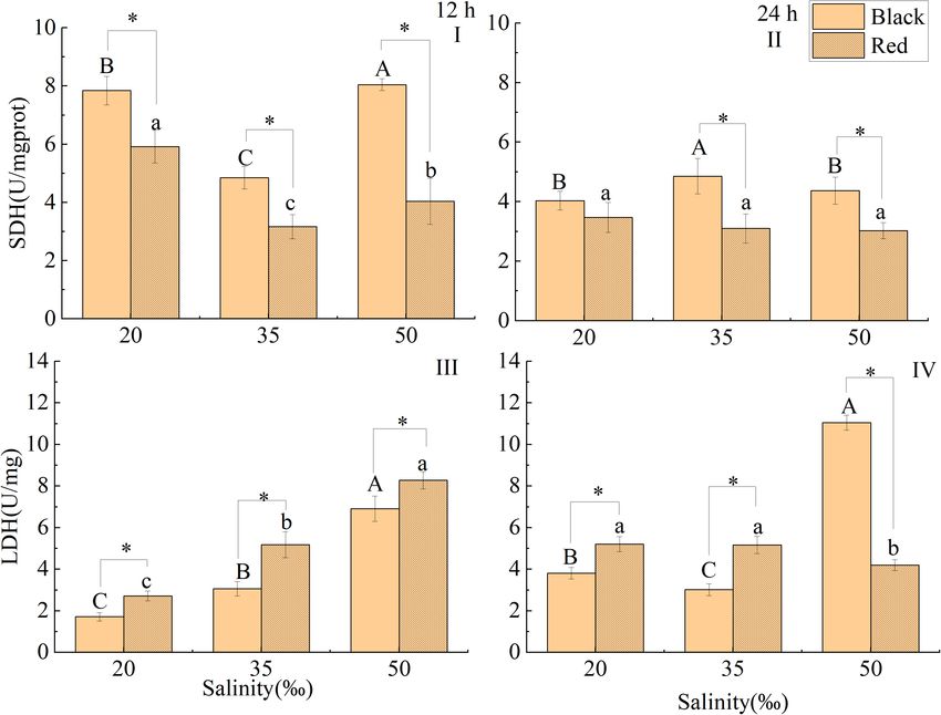
Sun et al.                                                                                                                                   Pinctada fucata Salinity Tolerance

  FIGURE 1 | (A) Red shell; (B) Black shell.

  FIGURE 2 | Effect of salinity on the osmotic pressure of hemolymph of P. fucata with two shell colors (I: 12h; II: 24h). Capital letters indicate the significant difference
  between salinities of black P. fucata (P < 0.05). Lowercase letters indicate the significant difference between salinities of red P. fucata (P < 0.05). The asterisk “*”
  indicates significant difference between shell colors under the same salinity (P < 0.05).

Frontiers in Marine Science | www.frontiersin.org                                      4                                                 July 2021 | Volume 8 | Article 704907
Sun et al. Pinctada fucata Salinity Tolerance
Determination of Related Ion salinity of 20 and 50h (P < 0.05, Figure 5A). At the same
Concentration in Hemolymph salinity, the SDH activity of black was significantly higher than
The response of Na+ concentration in hemolymph to salinity that of the red shell (P < 0.05). After exposure to different salinity
was shown in Figure 3. At 12 and 24 h, the Na+ concentration for 24 h, the activity of SDH in black P. fucata was significantly
increased gradually with salinity (P < 0.05). Under the same lower than that in the control at the salinity of 20 and 50h
salinity, there was no significant difference in Na+ concentration (P < 0.05), but there was no significant difference in red P. fucata.
of red shell and black shell P. fucata at 12 h (Figure 3A). At the same salinity, SDH activity of black shell was significantly
However, the treatment groups of red shell showed a higher Na+ higher than that of red shell (P < 0.05, Figure 5B).
concentration than black shell P. fucata in the salinity 35 and After 12 h, the LDH activity of red P. fucata and black P. fucata
50h at 24 h (P < 0.05, Figure 3B). gradually increased (P < 0.05, Figure 5C). At the same salinity,
At 12 and 24 h, the K+ concentration increased gradually LDH activity of black shell was significantly lower than that of red
with salinity (P < 0.05). Under the same salinity and time (12 shell (P < 0.05). After exposure to different salinity for 24 h, the
or 24 h), the K+ concentration in the hemolymph of red shell activity of LDH in black P. fucata was significantly higher than
was significantly higher than in the black P. fucata (P < 0.05, that in the control at the salinity of 20 and 50h (P < 0.05).
Figures 3C,D). Compared to the control, the LDH activity of red P. fucata
The Cl− concentration increased gradually with salinity at decreased significantly at salinity 50h (P < 0.05), but there was
12 and 24 h (P < 0.05), but there was no significant difference no significant difference at salinity 20h (P < 0.05). At salinity of
between red and black P. fucata at a salinity of 20h (P < 0.05). 20 and 35h, LDH activity of black shell was significantly lower
The Cl− concentration in the hemolymph of red shell was than that of red shell, and significantly higher than that in red
significantly higher than in the black P. fucata at 35h (P < 0.05). shell at salinity of 50h (P < 0.05, Figure 5D).
However, the Cl− concentration in the hemolymph of red shell
was significantly lower than that in the black shell at 50h Effect of Salinity on the Activities of
(P < 0.05, Figures 3E,F). Antioxidant Related Enzymes in Liver
At 12 and 24 h, the Ca2+ concentration increased gradually
with the increase of salinity (P < 0.05). Exposed to 35 and 50h
Tissue
After exposure to salinity of 20 and 50h for 12 or 24 h, the SOD
salinity, the red shell showed a higher Ca2+ concentration than
activity of black P. fucata was significantly higher than that in
the black P. fucata at 12 h. At 24 h (P < 0.05), the red shell
the control (P < 0.05, Figures 6A,B), while that of red shell was
showed a higher Ca2+ concentration than the black P. fucata at
significantly lower than that in the control (P < 0.05). At 12 h, the
salinity 35h. However, when salinity is 20 and 50h, there was
SOD activity of black P. fucata was significantly lower than that of
no significant difference of Ca2+ concentration between red and
red shell at salinity 20h, but was significantly higher than that of
black P. fucata (P < 0.05, Figures 3G,H).
red shell at salinity 35h (P < 0.05, Figure 6A). At 24 h, the SOD
Effect of Salinity on the Gill activity of black P. fucata was significantly higher than that of red
shell at salinity of 35h, but there was no significant difference in
Na+ -K+ -ATPase Activity the salinity of 20 and 50h (P < 0.05, Figure 6B).
At 12 h, the activity of Na+ -K+ -ATPase in black P. fucata was After treated with different salinity for 12 h, the CAT activity
significantly lower than that in the control at the salinity of 20 of black P. fucata was significantly higher than in the control at
and 50h (P < 0.05, Figure 4A). However, the gill Na+ -K+ - salinity 20h, lower than that of the control at salinity 50h but
ATPase activity of red P. fucata was significantly higher than that no significant difference (P < 0.05, Figure 6C). The CAT activity
in the control (P < 0.05). At 12 h within the same salinity, the of red P. fucata was significantly lower than that in the control at
activity of Na+ -K+ -ATPase of red P. fucata was significantly salinity 20h, but significantly higher than that of the control at
higher than that in the black shell at salinity 20h (P < 0.05), and the salinity of 50h (P < 0.05). The CAT activity of black P. fucata
significantly lower than that in the black shell at salinity 35 and was significantly higher than that of red shell at salinity 20h
50h (P < 0.05). At 24 h, the activity of Na+ -K+ -ATPase in black (P < 0.05, Figure 6C). After exposure to the salinity of 20 and
P. fucata was significantly lower than that in the control at the 50h for 24 h, the CAT activity of black P. fucata was significantly
salinity of 20 and 50h. The Na+ -K+ -ATPase activity of red shell higher than that in the control, the CAT activity of red P. fucata
increased significantly with the increase of salinity in the range of was significantly higher than that in the control at salinity 20h
20 to 50h (P < 0.05). At 24 h, the Na+ -K+ -ATPase activity of (P < 0.05, Figure 6D), but significantly lower than that of the
red P. fucata was significantly lower than that in the black shell at control at a salinity of 50h (P < 0.05). The CAT activity of black
salinity 20 and 35h salinity, and significantly higher than that in P. fucata was significantly higher than that of red shell in salinity
the black shell at salinity 50h (P < 0.05, Figure 4B). 20 and 50h (P < 0.05, Figure 6D).
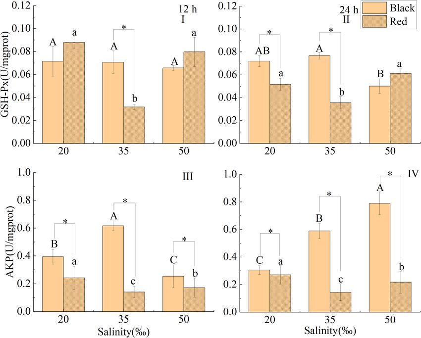
After treated with different salinity for 12 h, the GSH-Px
Effect of Salinity on the Activities of activity of red P. fucata was significantly higher than that in the
Enzymes Related to Respiratory control, while that of black P. fucata was no significant difference
Metabolism in Gills from that in the control (P < 0.05, Figure 7A). At the salinity
After 12 h, the activity of SDH in the black shell and red shell of 35h, the GSH-Px activity of black P. fucata was significantly
P. fucata was significantly higher than that in the control at the higher than that of red shell (P < 0.05, Figure 7A). After exposure
Frontiers in Marine Science | www.frontiersin.org 5 July 2021 | Volume 8 | Article 704907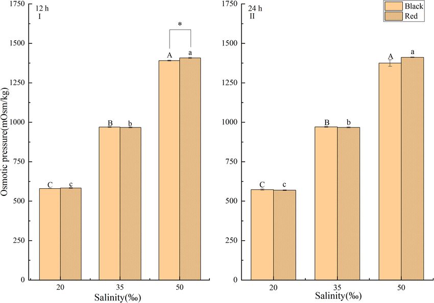
Sun et al.                                                                                                                               Pinctada fucata Salinity Tolerance

  FIGURE 3 | Effect of salinity on inorganic ion concentration in the hemolymph of P. fucata with two shell colors (I: 12h, Na+ concentration; II: 24h, Na+
  concentration; III: 12h, K+ concentration; IV: 24h, K+ concentration; V: 12h, Cl− concentration; VI: 24h, Cl− concentration; VII: 12h, Ca2+ concentration; VIII: 24 h,
  Ca2+ concentration).

to salinity of 20 and 50h for 24 h, the GSH-Px activity of black                         of seasonal rainfall and continental runoff, seawater salinity
P. fucata was significantly lower than that in the control, while                        in coastal and estuarine areas tends to change dynamically.
that of red shell was significantly higher than that in the control                      When marine bivalves encounter salinity stress, they usually
(P < 0.05, Figure 7B). At the same salinity of 20 and 50h, the                           respond to the changes in environmental salinity by closing
activity of GSH-Px of black P. fucata was significantly higher than                      shells and sealing the mantle cavity, and then by adjusting
that of red P. fucata (P < 0.05, Figure 7B).                                             the hemolymph osmotic pressure to cope with salinity stress
    After exposure to salinity of 20 and 50h for 12 h, the AKP                           (Davenport, 1979; Navarro, 1988; Shui, 2007). For most aquatic
activity of black P. fucata was significantly lower than that in                         animals, osmotic pressure regulation is a basic physiological
the control, while that of red P. fucata was significantly higher                        process, which enables the body to adapt to the difference
than that in the control (P < 0.05, Figure 7C). After exposure to                        of internal and external ion concentration. However, osmotic
salinity of 20 and 50h for 24 h, the AKP activity of red P. fucata                       pressure regulation is complicated, because the requirement
was significantly higher than that in the control, the AKP activity                      of living environment varies between organism. The osmotic
of black P. fucata was significantly lower than that in the control                      regulation of marine bivalves has been reported in several species,
at salinity 20h (P < 0.05), but significantly higher than that of                        including common mussel M. edulis (Willmer, 1978; Davenport,
the control at salinity of 50h (P < 0.05, Figure 7D). After 12 and                       1979), horse mussels Modiolus sp. (Pierce, 1970, 1971), soft
24 h, the AKP activity of black P. fucata was significantly higher                       clam Mya arenaria (Shumway, 1977; Deaton, 1992), arcid clam
than that of red P. fucata (P < 0.05, Figures 7C,D).                                     Noetia ponderosa (Amende and Pierce, 1980), and Pacific oyster
                                                                                         Crassostrea gigas (Shumway, 1977). These studies indicated that
                                                                                         the hemolymph osmolality varies directly with seawater density,
DISCUSSION                                                                               and is either equal to the ambient osmolality, and or slightly
                                                                                         hyper-osmotic (5–50 mOsm/kg) to ambient media over a range
Salinity is an important ecological variable that impacts the                            of non-lethal salinity. In the present study, the osmotic pressure
growth and survival of aquatic organisms. Due to the influence                           of hemolymph of P. fucata was significantly lower than that

Frontiers in Marine Science | www.frontiersin.org                                    6                                               July 2021 | Volume 8 | Article 704907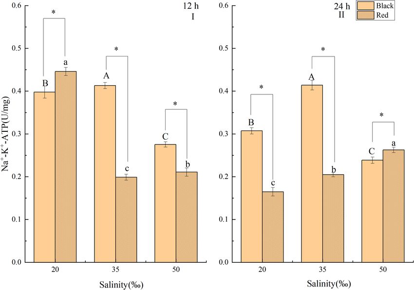
Sun et al.                                                                                                                                        Pinctada fucata Salinity Tolerance

  FIGURE 4 | Effects of salinity on the activity of Na+ -K+ -ATPase in the gill tissue of P. fucata with two shell colors (I: 12h; II: 24h).

of the control at salinity 20h, and significantly higher than                                   The constant osmotic pressure of shellfish mainly refers to the
that of the control at salinity 50h. The osmotic pressure of                                 balance between the main osmotic effector in the hemolymph
P. fucata with two shell colors increased significantly with the                             of shellfish and the content of organic ions and inorganic
increase of environmental salinity at 12 h and 24 h. After 12 h                              ions in the external water environment, which will not lead
stress at 50h salinity, the osmotic pressure of red P. fucata was                            to the expansion or shrinkage of cells and can carry on a
significantly higher than that of black shell, indicating that the                           normal life. Inorganic ions are the main osmotic effectors in
tolerance to high salinity of two shell colors P. fucata is different.                       the hemolymph of marine shellfish, among which K+ , Na+ ,
The metabolic activities of H. cracherodii and H. rufescens under                            Cl− , and other ions mainly participate in osmotic adjustment
high-salinity stress were swiftly and significantly impacted within                          (Cheng et al., 2002). K+ play an important role in maintaining
10 min, and began to resume in 6–8 h (Berger and Kharazova,                                  osmotic pressure in neurons and the normal function of the
1997). Liu et al. (2008) found that the hemolymph osmotic                                    nervous system (Cooper and Morris, 1997). Intracellular changes
pressure of C. japonica tended to be stable within 0.5 days in                               of Na+ concentration impinge the osmotic pressure response
each treatment group with a sudden change of salinity. In this                               of the organism, seal the mantle cavity, and preserve cells from
study, the osmotic pressure of P. fucata with two shell colors in                            extreme salinity (Berger and Kharazova, 1997). Most shellfish
the experimental group was significantly different from that of                              keep the concentration of K+ in hemolymph always higher
the control group at 24 h, which may be due to the large range                               than that in the external environment, but the concentrations
of salinity changes in the experimental group, leading to a wider                            of sodium and Cl− are lower than those in the external
range of hemolytic osmotic pressure changes, thus increasing the                             environment. These three inorganic ions change directly with
adjustment and recovery time of P. fucata osmotic pressure. In                               the external salinity and are not regulated by the nervous system
general, physical responses of shellfish to salinity stress become                           and hormones (Natochin et al., 1979; Hildreth and Stickle,
significantly between 24 and 48 h, and such responses will                                   1980; Deaton, 1992). In the present study, the concentrations
gradually approach or return to the normal level. The duration                               of Na+ , K+ , Cl− , and Ca2+ in the low salinity stress group
of this process is species-specific (Castagna, 1973).                                        were significantly lower than those in the control, while those

Frontiers in Marine Science | www.frontiersin.org                                        7                                                     July 2021 | Volume 8 | Article 704907
Sun et al.                                                                                                                                     Pinctada fucata Salinity Tolerance

  FIGURE 5 | Effects of salinity on SDH and LDH activities in the gill tissue of P. fucata with two shell colors (I: 12h, SDH activity; II: 24h, SDH activity; III: 12h, LDH
  activity; IV: 24h, LDH activity).

in the high salinity stress group were significantly higher than                            P. fucata (F3 ) were 95.83 and 100%, respectively. Chen et al.
those in the control. The concentrations of inorganic ions in                               (2016) found that that there were significant differences in
the hemolymph of P. fucata with two shell colors increased                                  shell height, hinge length, shell width, body mass, shell mass,
significantly with the increases of salinity, which is consistent                           tissue mass, adductor mass, adductor muscle tension and other
with the changing trend of osmotic pressure. The results of                                 major traits between black shell and red shell selected lines of
this study are similar to the changes of inorganic ions in                                  P. fucata (F6 ). Bauchau (2001) proposed that pigmentation is
hemolymph after salinity stress of Haliotis Discus reported by                              closely related to the regulation of the shell growth to achieve
Gao et al. (2017), that the concentration of inorganic ions                                 developmental stability. The results showed that the physiological
in hemolymph and the changing trend of osmotic pressure                                     parameters of P. fucata (F11 ) with red shell and black shell were
are consistent. However, Ding et al. (2013) found that the                                  changed based on the stable inheritance of shell color. Next, the
inorganic ions concentration of Ruditapes philippinarum in the                              changes of other physiological indexes in this study also fully
control (32h) and the experimental group (15h) reached                                      reflect this point.
the maximum concentration at 48 and 24 h, respectively. The                                     Na+ -K+ -ATPase is an ion channel that uses the energy
decrease of osmotic pressure is not accompanied by the decrease                             generated by ATP hydrolysis to maintain membrane potential
of inorganic ion concentration. The reason may be that P. fucata’s                          by driving three Na+ outlets and two K+ intents, which is
tolerance to salinity is different from R. philippinarum. During                            essential to osmotic regulation (Evans and Lambert, 2015; Abdel-
the experiment, P. fucata did not close the shells to form a                                Mohsen, 2016). In this study, after 12 h and 24 h of salinity
closed space but maintained the osmotic pressure balance inside                             stress, Na+ -K+ -ATPase activity of P. fucata with black shell
and outside the cell through ion exchange with the external                                 in low-salt and high-salt groups was significantly lower than
environment through ion channels on the cell membrane. In                                   that in the control, showing an inverted U-shape distribution.
addition, at the same time and under the same salinity, the                                 After 12 h, P. fucata with red shell was significantly higher in
concentration of inorganic ions in the hemolymph of P. fucata                               low-salt and high-salt treatment groups than in the control,
with black shell color and red shell color was significantly                                showing a U-shaped distribution. Zhang et al. (2015) found that
different. Chen et al. (2010). found that the shell color purification                      the influence of salinity on the activity of Na+ -K+ -ATPase in
rates of black shell color and red shell color breeding lines of                            the gill filamentum of Siganus guttatus also showed a U-shaped

Frontiers in Marine Science | www.frontiersin.org                                       8                                                 July 2021 | Volume 8 | Article 704907
Sun et al.                                                                                                                                    Pinctada fucata Salinity Tolerance

  FIGURE 6 | Effects of salinity on SOD and CAT activities in the liver tissue of P. fucata with two shell colors (I: 12h, SOD activity; II: 24h, SOD activity; III: 12h, CAT
  activity; IV: 24h, CAT activity).

distribution. The reason for the inverted “U” distribution of Na+ -                         adaptability to external environmental conditions (Marqueze
K+ -ATPase activity in black P. fucata may be that it is weak                               et al., 2006). Aerobic respiration is the main type of respiratory
in self-adaptation to the change of salinity, which temporarily                             metabolism in aquatic animals, but anaerobic respiration can
inhibits the activity of Na+ -K+ -ATPase. After 24 h, the Na+ -                             also provide energy for the body under low salt conditions. SDH
K+ -ATPase activity in red P. fucata increased significantly with                           not only plays a key role in tricarboxylic acid cycle, but also
the increase of salinity, which is consistent with the changing                             participates in oxidative phosphorylation. Therefore, the activity
trend of osmotic pressure. Liu et al. (2008) found that Na+ -                               of SDH reflects the aerobic metabolism to a certain extent (Gao
K+ -ATPase activity of Eriocheir sinensis is negatively correlated                          et al., 2016). LDH can catalyze the anaerobic metabolite lactic
with salinity, which is different from the results of our study,                            acid to pyruvate and release energy, and its activity can reflect
indicating that osmotic regulation of P. fucata is not completely                           the anaerobic metabolism ability to a certain extent. In our study,
implemented by Na+ -K+ -ATPase in the gill tissue. It is possible                           after salinity stress for 12 h, the SDH activity of P. fucata with
that in a short time of salinity stress, biogenic amines (dopamine                          two shell colors was significantly higher than that in the control,
and serotonin) could stimulate phosphorylation of the enzyme in                             which is different from the result reported by Nie et al. (2018)
the gills including the Na+ -K+ -ATPase, and then ATPases could                             that aerobic respiratory metabolism level gradually increased
adjust the ion concentration both in vivo and in vitro and make                             when the clam was close to the isotonic point. The aerobic
the osmotic pressure of hemolymph attain a balance. After a long                            metabolism of P. fucata was enhanced under salinity stress,
time of adaptation, the penetrability of gill epithelium to water                           which may be related to the large amount of energy required
and ions changed and then the activity of gill Na+ -K+ -ATPase                              for the transmembrane transport of inorganic ions. After 24 h,
reached a steady level (Kamemoto, 1991; Mo et al., 1998; Lucu                               the SDH activity of black P. fucata decreased significantly,
and Flik, 1999; Morris, 2001). Under the same salinity stress for                           while that of red P. fucata had no significant difference. It is
the same time, the Na+ -K+ -ATPase in gill tissues of P. fucata                             possible that long-term salinity stress has a certain influence
with two shell colors were significantly different, which indicated                         on the aerobic metabolism level of black P. fucata, while red
that the ion regulation in P. fucata with two shell colors played                           P. fucata gradually adapt to salinity stress. In this study, after
different roles.                                                                            12 h of different salinity stress, LDH activity of P. fucata of
   Respiratory metabolism is one of the basic physiological                                 both shell colors was significantly increased with the increase of
activities of animal energy metabolism, reflecting animal                                   salinity, and anaerobic metabolism was enhanced accordingly.
metabolic characteristics, physiological conditions and                                     Shi et al. (2017) found in their study on Epinephelus moara that

Frontiers in Marine Science | www.frontiersin.org                                       9                                                 July 2021 | Volume 8 | Article 704907
Sun et al.                                                                                                                                    Pinctada fucata Salinity Tolerance

  FIGURE 7 | Effects of salinity on GSH-Px and AKP activities in the liver tissue of P. fucata with two shell colors (I: 12h, GSH-Px activity; II: 24h, GSH-Px activity; III:
  12h, AKP activity; IV: 24h, AKP activity).

LDH activity first decreased and then increased under salinity                              capabilities (Martínez-Álvarez et al., 2005; Okoye et al., 2019).
stress, which together with aerobic respiration provided energy                             In the long process of evolution, aquatic animals have an
for resisting environmental stress. Combined with the change                                evolutionarily conserved antioxidant defense system that can
of SDH activity for 12 h, it was suggested that the respiratory                             eliminate superfluous ROS. In particular, as antioxidant enzymes,
metabolism of red shell and black shell P. fucata increased under                           SOD and CAT are regarded as the first line of defense against
high salt stress to meet the energy consumption of the body.                                oxygen intoxication (Bhagat et al., 2016; Wang et al., 2020). SOD
After 24 h stress, LDH activity of black P. fucata was significantly                        can catalyze O2− to produce H2 O2 and remove O2− , while CAT
higher than that of the control, while red shell was significantly                          can catalyze H2 O2 to produce water and oxygen. Therefore, the
lower than that of the control when salinity was 50h. Our                                   changes in CAT and SOD enzyme activities can reflect changes
results indicate that under salinity stress for 24 h, the anaerobic                         in the body’s antioxidant system under environmental stress to
metabolism of black shell was enhanced while the aerobic                                    a certain extent (Burgeot et al., 1996; Peters and Livingstone,
metabolism of the body was weakened, which will provide energy                              1996). Li et al. (2012) explores the impacts of salinity on the
for the body. Under the same salinity stress for the same time,                             changes of SOD and CAT activities in Cyclina sinensis, and found
the SDH activity and LDH activity in gill tissue of P. fucata with                          that the fluctuations of antioxidant enzymes of C. sinensis under
two shell colors showed significant differences, which indicated                            salinity stress were basically completed within 24 h after stress.
that the respiratory metabolic intensity of P. fucata with two shell                        In the present study, after salinity stress for 12 h and 24 h, SOD
colors changed in different degrees under salinity stress.                                  activity in the liver of black P. fucata was significantly lower
    Aerobic organisms unceasingly generate ROS by metabolism.                               than that the control, while SOD activity of red P. fucata was
Massive accumulation of ROS damages macromolecular                                          significantly higher than that the control. The reason might be
substances, such as nucleic acids, carbohydrates, proteins, and                             that the salinity stress caused a large number of free radicals
lipids (Finkel and Holbrook, 2000; Lushchak, 2011). As a result,                            in the black P. fucata, and the production rate of free radicals
equilibrium between the production and removal of ROS will                                  was much faster than the scavenging rate, and the unscavenged
protect oxidative damage and maintain normal physiological                                  free radicals caused oxidative damage to cells so that the SOD

Frontiers in Marine Science | www.frontiersin.org                                      10                                                 July 2021 | Volume 8 | Article 704907
Sun et al. Pinctada fucata Salinity Tolerance
activity in the black P. fucata was always lower than that in the oxidative damage to cells in the body. However, prolonged low
control. However, the red P. fucata had adaptability to salinity salinity stress resulted in excessive energy consumption and many
stress and maintained high SOD activity to compensate for the blood cell apoptosis in black P. fucata, which leads to AKP activity
SOD consumed by the organism to produce free radicals. In decreased, ultimately. Under the same salinity stress for the same
this study, the CAT activity of red P. fucata was lowest after time, the activities of SOD, CAT, GSH-Px, and AKP in liver
12 h of low salt stress, but after 24 h of low salt stress, the CAT tissue of P. fucata with two shell colors showed different degrees
activity recovered and was significantly higher than that of the of differences, which indicated that the antioxidant defense
control, possibly because the SOD activity increased after 12 h mechanism of P. fucata with two shell colors played different roles
of low salt stress produced a lot of H2 O2 to inhibit the CAT under salinity stress.
activity. The study of Zhang et al. (2020) also shows that the In summary, the osmotic pressure of hemolymph, ion
increase of SOD expression level leads to excessive accumulation concentration, Na+ -K+ -ATPase and enzymes related to
of hydrogen peroxide, thus inhibiting the function of CAT. respiratory metabolism in gill tissue, enzymes related to
In addition, as the second line of preserve against oxidative an antioxidant in liver tissue of P. fucata with two shell
trauma, GSH-Px also has a significant role in cell metabolism colors under salinity stress were compared and analyzed.
and scavenging of free radicals. In cells, GSH-Px can catalyze the We found that salinity changes had different effects on
reduction of hydroperoxides to hydroxy compounds (Cnubben the physiology and biochemistry of the two shell colors
et al., 2001; Peña-Llopis et al., 2003). In the present study, the P. fucata. However, red P. fucata can quickly respond
variation trend of GSH-Px activity and the changing trend of positively to the change of environmental salinity and
SOD activity in red-shell P. fucata were consistent. GSH-Px reduce the damage caused by the change of environmental
activity increased, and CAT activity decreased under 20h salinity salinity. The results can provide a reference for further
for 12 h and salinity 50h stress for 24 h, respectively. At this study of salt tolerance mechanism of P. fucata with
point, the scavenging of H2 O2 was mainly dominated by GSH- different shell colors.
Px. Regarding enzyme kinetics, GSH-Px in mammals and other
vertebrates has a comparatively large affinity for H2 O2 compared
with CAT (Reddy et al., 1998; Avanzo et al., 2001). GSH-Px is DATA AVAILABILITY STATEMENT
thus largely conscientious for the removal of H2 O2 in vertebrates,
while CAT and GSH-Px are play complementarily responsible for The raw data supporting the conclusions of this article will be
H2 O2 removal (Mourente et al., 2002; Jo et al., 2008). The activity made available by the authors, without undue reservation.
of GSH-Px, in P. fucata with black shell, was significantly lower
than that in the control at 24 h, which also has the complementary
effect of GSH-Px and CAT. In response to sudden varies in AUTHOR CONTRIBUTIONS
salinity, CAT and GSH-Px had a decisive action in mediating the
scavenging of H2 O2 to maintain the mobile equilibrium between JS and MC conceived and designed the project, and wrote
internal oxidation and reduction. AKP is involved in various the manuscript. ZF and JY measured and collected the data.
metabolic processes such as metabolism, detoxification, and the SZ collected the samples and carried out the analysis. ZM,
biosynthesis of macromolecules for diverse fundamental function WZ, and GY supervisioned, wrote—reviewed and edited the
(Suzuki and Mori, 1990). AKP is vital lysosomal enzymes in manuscript. All authors contributed to the article and approved
marine invertebrates (Rahman and Siddiqui, 2004) and can take the submitted version.
part in non-specific immunity, participate in the degradation of
foreign proteins, carbohydrates, and lipids or in phagocytosis
(Liang et al., 2014). Li et al. (2015) found that the AKP activity FUNDING
of Chlamys Nobilis first decreased and then increased with the
increase of salinity. In this study, after 12 or 24 h, the AKP activity This research was financially supported by Central Public-
of red P. fucata was significantly higher than that of the control, interest Scientific Institution Basal Research Fund, CAFS (No.
while that of black P. fucata was significantly higher than that 2020TD55), Guangxi Key Research and Development Program
of the control after high salt stress for 24 h. The reason may be (Guike AB18221090), Natural Science Foundation of Hainan
that P. fucata produces a large number of free radicals under low Province (319QN338), and Beihai Science and Technology
or high salinity stress, which increases AKP activity and avoids Planning Project (Beikehe 201995002).
REFERENCES Alagarswami, K., and Victor, A. (1976). Salinity tolerance and rate of filtration of
the pearl oyster Pinctada fucata. J. Mar. Biol. Assoc. India 18, 149–158.
Abdel-Mohsen, H. (2016). Assessment of respiratory and ion transport potential of Amende, L. M., and Pierce, S. K. (1980). Cellular volume regulation in salinity
Penaeus japonicus gills in response to environmental pollution. Mediter. Mar. stressed molluscs: the response of Noetia ponderosa (Arcidae) red blood cells to
Sci. 10, 5–18. doi: 10.12681/mms.118 osmotic variation. J. Compar. Physiol. 138, 283–289. doi: 10.1007/bf00691562
Ai, H., Li, Y., and Ding, Y. (2003). A review of study on diseases in pearl oyster. Arisman, N., Istiqomad, N., and Yoshimatsu, T. (2018).
Shanghai Fish. Univ. 12, 61–64. Impact of short-term hyposalinity stress on Akoya pearl
Frontiers in Marine Science | www.frontiersin.org 11 July 2021 | Volume 8 | Article 704907Sun et al. Pinctada fucata Salinity Tolerance oyster, Pinctada fucata (Gould 1850). Asian Fish. Sci. 31, Gao, X., Li, Y., Li, X., Wu, F., Song, C., and Liu, Y. (2017). The response and 265–275. osmotic pressure regulation mechanism of Haliotis discus hannai (Mollusca, Avanzo, J. L., de Mendonça, C. X. Jr., Pugine, S. M. P., and de Cerqueira Cesar, Gastropoda) to sudden salinity changes. Hydrobiologia 795, 181–198. doi: M. (2001). Effect of vitamin E and selenium on resistance to oxidative stress in 10.1007/s10750-017-3129-z chicken superficial pectoralis muscle. Compar. Biochem. Physiol. Part C Toxicol. Gao, X., Zhang, M., Li, X., Shi, C., Song, C., and Liu, Y. (2016). Effects of LED light Pharmacol. 129, 163–173. doi: 10.1016/s1532-0456(01)00197-1 quality on the growth, metabolism, and energy budgets of Haliotis discus discus. Ballantyne, J. S., and Moyes, C. D. (1987). Osmotic effects on fatty acid, pyruvate, Aquaculture 453, 31–39. doi: 10.1016/j.aquaculture.2015.11.033 and ketone body oxidation in oyster gill mitochondria. Physiol. Zool. 60, Garçon, D. P., Masui, D. C., Mantelatto, F. L., Furriel, R. P., McNamara, J. C., 713–721. doi: 10.1086/physzool.60.6.30159987 and Leone, F. A. (2009). Hemolymph ionic regulation and adjustments in gill Bauchau, V. (2001). Developmental stability as the primary function of the (Na+ , K+ )-ATPase activity during salinity acclimation in the swimming crab pigmentation patterns in bivalve shells? Belgian J. Zool. 131, 23–28. Callinectes ornatus (Decapoda, Brachyura). Compar. Biochem. Physiol. Part A Berger, V. J., and Kharazova, A. (1997). “Mechanisms of salinity adaptations in Mol. Integr. Physiol. 154, 44–55. doi: 10.1016/j.cbpa.2009.04.624 marine molluscs,” in Interactions and adaptation strategies of marine organisms. Ghiselli, A., Serafini, M., Natella, F., and Scaccini, C. (2000). Total antioxidant Cham: Springer, 115–126. capacity as a tool to assess redox status: critical view and experimental data. Bhagat, J., Ingole, B., and Singh, N. (2016). Glutathione S-transferase, catalase, Free Rad. Biol. Med. 29, 1106–1114. doi: 10.1016/s0891-5849(00)00394-4 superoxide dismutase, glutathione peroxidase, and lipid peroxidation as Gu, X. (2014). The variation analysis of nutrient composition and the growth trait of biomarkers of oxidative stress in snails: A review. Invertebrate Surv. J. 13, clams with different shell color and decorative pattern. China: Ninbo University. 336–349. Guan, Y., and He, M. (2009). Research progress on shell color polymorphism of Burgeot, T., Bocquéné, G., Porte, C., Dimeet, J., Santella, R., Pfhol-Leszkowicz, marine economic shellfish. Mar. Sci. Bull. 28, 108–114. A., et al. (1996). Bioindicators of pollutant exposure in the northwestern Hildreth, J. E., and Stickle, W. B. (1980). The effects of temperature and salinity on Mediterranean Sea. Mar. Ecol. Prog. Ser. 131, 125–141. doi: 10.3354/ the osmotic composition of the southern oyster drill, Thais haemastoma. Biol. meps131125 Bull. 159, 148–161. doi: 10.2307/1541015 Castagna, M. (1973). Salinity tolerance of some marine bivalves from inshore and Huaiping, Z., Guofan, Z., and Xiao, L. (2005). Comparison of growth and survival estuarine environments in Virginia waters on the western mid-Atlantic coast. of larvae among different shell color stocks of bay scallop Argopecten irradians Malacologia 12, 47–96. irradians (Lamarck 1819). Chinese J. Oceanol. Limnol. 23:183. doi: 10.1007/ Chen, J., Liu, Z., Sun, X., Wang, H., and Du, X. (2010). Analysis on bf02894236 growth characteristic and genetic diversity in the selected lines (F3 ) of Huang, G., Guo, Y., Li, L., Fan, S., Yu, Z., and Yu, D. (2016). Genomic structure four shell colors of Pincatada martensii. J. Shanghai Ocean Univ. 19, of the α-amylase gene in the pearl oyster Pinctada fucata and its expression in 588–595. response to salinity and food concentration. Gene 587, 98–105. doi: 10.1016/j. Chen, J.-C., and Lin, C.-Y. (1995). Responses of oxygen consumption, ammonia- gene.2016.04.044 N excretion and urea-N excretion of Penaeus chinensis exposed to ambient Jasmani, S., Jayasankar, V., and Wilder, M. (2010). Na/K-ATPase activity and osmo- ammonia at different salinity and pH levels. Aquaculture 136, 243–255. doi: ionic regulation in adult whiteleg shrimp Litopenaeus vannamei exposed to low 10.1016/0044-8486(95)01060-2 salinities. Aquaculture 304, 88–94. doi: 10.1016/j.aquaculture.2010.03.025 Chen, M., Liu, B., Yan, J., Guo, H., Wu, K., and Li, Y. (2016). Comparison analysis Jo, P. G., Choi, Y. K., and Choi, C. Y. (2008). Cloning and mRNA expression of major traits among four shell color selective lines of pearl oyster (Pinctada of antioxidant enzymes in the Pacific oyster, Crassostrea gigas in response to fucata). South China Fish. Sci. 12, 118–122. cadmium exposure. Compar. Biochem. Physiol. Part C Toxicol. Pharmacol. 147, Cheng, W., Yeh, S.-P., Wang, C.-S., and Chen, J.-C. (2002). Osmotic and ionic 460–469. doi: 10.1016/j.cbpc.2008.02.001 changes in Taiwan abalone Haliotis diversicolor supertexta at different salinity Kamemoto, F. I. (1991). Neuroendocrinology of osmoregulation in crabs. Zool. Sci. levels. Aquaculture 203, 349–357. doi: 10.1016/s0044-8486(01)00606-8 8, 827–833. Cnubben, N. H., Rietjens, I. M., Wortelboer, H., van Zanden, J., and van Bladeren, Kim, W., Kim, J., Kim, M., Park, C., and Huh, H. (1998). Effects of sudden changes P. J. (2001). The interplay of glutathione-related processes in antioxidant in salinity on endogenous rhythms of the spotted sea bass Lateolabrax sp. Mar. defense. Environ. Toxicol. Pharmacol. 10, 141–152. doi: 10.1016/s1382- Biol. 131, 219–225. doi: 10.1007/s002270050314 6689(01)00077-1 Li, Z., Qian, J., Lao, C., and Liu, Z. (2015). Synergistic effects of temperature Cooper, A. R., and Morris, S. (1997). Osmotic and ionic regulation by Leptograpsus and salinity on the activities of immune-related enzymes of Chlamys nobilis variegatus during hyposaline exposure and in response to emersion. J. Exper. (Reeve). Adv. Mar. Sci. 32, 227–237. Mar. Biol. Ecol. 214, 263–282. doi: 10.1016/s0022-0981(96)02778-5 Li, Z.-N., Lin, T., Yao, Z., Lai, Q.-F., Lu, J.-X., Wang, H., et al. (2012). Effects of Davenport, J. (1979). Is Mytilus edulis a short term osmoregulator? Compar. water salinity on the antioxidant enzyme activities and growth of clam Cyclina Biochem. Physiol. Part A Mol. Integr. Physiol. 64, 91–95. doi: 10.1016/0300- sinensis. Chinese J. Ecol. 31, 2625–2630. 9629(79)90436-5 Liang, S., Luo, X., You, W., Luo, L., and Ke, C. (2014). The role of hybridization in Deaton, L. E. (1992). Osmoregulation and epithelial permeability in two euryhaline improving the immune response and thermal tolerance of abalone. Fish Shellfish bivalve molluscs: Mya arenaria and Geukensia demissa. J. Exper. Mar. Biol. Ecol. Immunol. 39, 69–77. doi: 10.1016/j.fsi.2014.04.014 158, 167–177. doi: 10.1016/0022-0981(92)90224-x Liu, H., Pan, L., and Fu, L. (2008). Effect of salinity on hemolymph osmotic Ding, J., Wang, R., Yan, X., Zhao, L.-Q., Yang, F., and Wang, L.-S. (2013). pressure, sodium concentration and Na+ -K+ -ATPase activity of gill of Chinese Comparative tolerance to low salinity stress in Manila clam Ruditapes crab, Eriocheir sinensis. J. Ocean Univ. China 7, 77–82. doi: 10.1007/s11802- philippinarum with three shell colors. J. Dalian Ocean Univ. 28, 264–268. 008-0077-2 Edwards, S. (2003). Assessment of the physiological effect of altered salinity Liu, J., Yu, D., and Li, J. (2011). Effects of salinity and pH on oxygen consumption on greenlip (Haliotis laevigata) and blacklip (Haliotis rubra) abalone using and ammonia excretion rates in pinctada fucata. Oceanol. Limnol. Sinica 4, respirometry. Aquacult. Res. 34, 1361–1365. doi: 10.1046/j.1365-2109.2003. 603–607. 00943.x Liu, X., Deng, Y., and Zhang, G. (2005). Growth of eight Pacific abalone families at Evans, A. N., and Lambert, F. N. (2015). Na+ /K+ -ATPase α1 mRNA expression three temperatures. Acta Oceanol. Sinica 24, 148–153. in the gill and rectal gland of the Atlantic stingray, Dasyatis sabina, following Liu, X., and Yan, A. (2006). Recovery and adaptation process of Pelteobagrus acclimation to increased salinity. BMC Res. Notes 8:219. doi: 10.1186/s13104- fulvidraco in the experimental circulatory system after transportation stress. 015-1216-7 J. Fisheries China 4, 495–501. Finkel, T., and Holbrook, N. J. (2000). Oxidants, oxidative stress and the biology of Lucu, C. E., and Flik, G. (1999). Na+ -K+ -ATPase and Na+ /Ca2+ exchange ageing. Nature 408, 239–247. doi: 10.1038/35041687 activities in gills of hyperregulating Carcinus maenas. Am. J. Physiol. 276, Fisher, W. S., and Newell, R. I. (1986). Salinity effects on the activity of granular R490–R499. hemocytes of American oysters, Crassostrea virginica. Biol. Bull. 170, 122–134. Lushchak, V. I. (2011). Environmentally induced oxidative stress in aquatic doi: 10.2307/1541385 animals. Aquatic Toxicol. 101, 13–30. doi: 10.1016/j.aquatox.2010.10.006 Frontiers in Marine Science | www.frontiersin.org 12 July 2021 | Volume 8 | Article 704907
Sun et al.                                                                                                                                       Pinctada fucata Salinity Tolerance

Marqueze, A., Kucharski, L. C., and Da Silva, R. S. M. (2006). Effects of anoxia and         Pierce, S. K. Jr. (1971). Volume regulation and valve movements by marine mussels.
   post-anoxia recovery on carbohydrate metabolism in the jaw muscle of the crab                Compar. Biochem. Physiol. Part A Physiol. 39, 103–117. doi: 10.1016/0300-
   Chasmagnathus granulatus maintained on carbohydrate-rich or high-protein                     9629(71)90350-1
   diets. J. Exper. Mar. Biol. Ecol. 332, 198–205. doi: 10.1016/j.jembe.2005.11.009          Rahman, M., and Siddiqui, M. (2004). Biochemical effects of vepacide (from
Martínez-Álvarez, R. M., Morales, A. E., and Sanz, A. (2005). Antioxidant defenses              Azadirachta indica) on Wistar rats during subchronic exposure. Ecotoxicol.
   in fish: biotic and abiotic factors. Rev. Fish Biol. Fisheries 15, 75–88. doi:               Environ. Safety 59, 332–339. doi: 10.1016/j.ecoenv.2003.07.013
   10.1007/s11160-005-7846-4                                                                 Reddy, K. V., Kumar, T. C., Prasad, M., and Reddanna, P. (1998). Pulmonary lipid
Masui, D. C., Mantelatto, F. L., McNamara, J. C., Furriel, R. P., and Leone,                    peroxidation and antioxidant defenses during exhaustive physical exercise: the
   F. A. (2009). Na+ , K+ -ATPase activity in gill microsomes from the blue crab,               role of vitamin E and selenium. Nutrition 14, 448–451. doi: 10.1016/s0899-
   Callinectes danae, acclimated to low salinity: novel perspectives on ammonia                 9007(98)00016-1
   excretion. Compar. Biochem. Physiol. Part A Mol. Integr. Physiol. 153, 141–148.           Sergievskii, S., and Berger, V. J. (1984). Physiological differences of principal shell-
   doi: 10.1016/j.cbpa.2009.01.020                                                              colour phenotypes of the gastropod mollusc Littorina obtusata. Biol. Moria 2,
Meng, Z., Li, Y., and Xing, K. (1996). Pearl culture theory and technology. BeiJing:            36–44.
   Science Press.                                                                            Shi, Z., Liao, Y., Wang, X., Zhang, C., Peng, S., and Gao, Q. (2017). Impact of the
Mo, J. L., Devos, P., and Trausch, G. (1998). Dopamine as a modulator of                        abrupt salinity decrease on ion-regulation enzyme activity in the gill and serum
   ionic transport and Na+ /K+ -ATPase activity in the gills of the Chinese crab                osmolality from Epinehelus moara. J. Safety Environ. 17, 1210–1214.
   Eriocheir sinensis. J. Crustacean Biol. 18, 442–448. doi: 10.1163/193724098x              Shui, B. (2007). A study on effect of several environmental factors on quick
   00278                                                                                        purification of Sinonovacula constricta (Lamarck). Modern Fish. Infor. 22,
Morris, S. (2001). Neuroendocrine regulation of osmoregulation and the evolution                6–10.
   of air-breathing in decapod crustaceans. J. Exper. Biol. 204, 979–989. doi:               Shumway, S. (1977). Effect of salinity fluctuation on the osmotic pressure and Na+ ,
   10.1242/jeb.204.5.979                                                                        Ca 2+ and Mg2+ ion concentrations in the hemolymph of bivalve molluscs.
Mourente, G., Dıaz-Salvago, E., Bell, J. G., and Tocher, D. R. (2002). Increased                Mar. Biol. 41, 153–177. doi: 10.1007/bf00394023
   activities of hepatic antioxidant defence enzymes in juvenile gilthead sea bream          Sokolova, I., and Berger, V. J. (2000). Physiological variation related to shell colour
   (Sparus aurata L.) fed dietary oxidised oil: attenuation by dietary vitamin E.               polymorphism in White Sea Littorina saxatilis. J. Exper. Mar. Biol. Ecol. 245,
   Aquaculture 214, 343–361. doi: 10.1016/s0044-8486(02)00064-9                                 1–23. doi: 10.1016/s0022-0981(99)00132-x
Nan, X., Xi-Chun, W., You-Fang, D., Ren, L., and Kun-Lun, T. (2004). Effect of               Suzuki, T., and Mori, K. (1990). Hemolymph lectin of the pearl oyster, Pinctada
   initial fluid resuscitation on subsequent treatment in uncontrolled hemorrhagic              fucata martensii: a possible non-self recognition system. Dev. Compar.
   shock in rats. Shock 21, 276–280. doi: 10.1097/01.shk.0000110622.42625.cb                    Immunol. 14, 161–173. doi: 10.1016/0145-305x(90)90088-v
Natochin, Y. V., Berger, V. Y., Khlebovich, V., Lavrova, E., and Michailova, O. Y.           Wang, N., Yang, J., Zhang, H., Soon, T. K., Liu, H., Li, S., et al. (2020).
   (1979). The participation of electrolytes in adaptation mechanisms of intertidal             Differential responses to low salinity on gene expression, physiological and
   molluscs’ cells to altered salinity. Compar. Biochem. Physiol. Part A Mol. Integr.           biochemical indexes between the golden and brown noble scallops Chlamys
   Physiol. 63, 115–119. doi: 10.1016/0300-9629(79)90636-4                                      nobilis. Aquacult. Res. 51, 316–325. doi: 10.1111/are.14377
Navarro, J. M. (1988). The effects of salinity on the physiological ecology of               Widdows, J. (1985). “The effects of fluctuating and abrupt changes in salinity on the
   Choromytilus chorus (Molina, 1782)(Bivalvia: Mytilidae). J. Exper. Mar. Biol.                performance of Mytilus edulis”, in: Marine biology of polar regions effects of stress
   Ecol. 122, 19–33. doi: 10.1016/0022-0981(88)90209-2                                          on marine organisms. Chichester: John Wiley.
Nie, H., Wenhao, L., Dongdong, L., Donglin, L., and Xiwu, Y. (2018). Effects                 Willmer, P. (1978). Volume regulation and solute balance in the nervous tissue
   of temperature and salinity on respiratory metabolic enzyme activities                       of an osmoconforming bivalve (Mytilus edulis). J. Exper. Biol. 77, 157–179.
   of Clinochardium California. Acta Eco. Zool. 22, 85–89. doi: 10.1002/                        doi: 10.1242/jeb.77.1.157
   9781119155423.ch9                                                                         Yang, C., Hao, R., Deng, Y., Liao, Y., Wang, Q., Sun, R., et al. (2017). Effects
Ocaño-Higuera, V. M., Maeda-Martínez, A. N., Lugo-Sánchez, M. E., García-                       of protein sources on growth, immunity and antioxidant capacity of juvenile
   Sánchez, G., Márquez-Ríos, E., Gómez-Jimenez, S., et al. (2011). Effect of                   pearl oyster Pinctada fucata martensii. Fish Shellfish Immunol. 67, 411–418.
   emerged shipment on the physiological condition of the adductor muscle                       doi: 10.1016/j.fsi.2017.06.037
   in adult giant lion’s paw scallop Nodipecten subnodosus (Sowerby 1835).                   Zhang, J., Luo, Y., Huang, Y., Zhong, Y., Liu, Q., Li, J., et al. (2015). Genetic
   Aquacult. Res. 42, 1087–1095. doi: 10.1111/j.1365-2109.2010.02693.x                          structure of inbred and hybrid families of pearl oyster Pinctada martensii.
Okoye, C. N., MacDonald-Jay, N., and Kamunde, C. (2019). Effects of                             Genom. Appl. Biol. 34, 723–730.
   bioenergetics, temperature and cadmium on liver mitochondria reactive oxygen              Zhang, M., Li, L., Liu, Y., and Gao, X. (2020). Effects of sudden drop in salinity on
   species production and consumption. Aquatic Toxicol. 214:105264. doi: 10.                    osmotic pressure regulation and antioxidant defense mechanism of Scapharca
   1016/j.aquatox.2019.105264                                                                   subcrenata. Front. Physiol. 11:884. doi: 10.3389/fphys.2020.00884
Pan, X., Liu, H., Xu, M., Xu, H., Zhang, H., and He, M. (2020). Cloning and
   expression analysis of aquaporin gene AQP4 cDNA from Pinctada fucata                      Conflict of Interest: The authors declare that the research was conducted in the
   martensii. J. Trop. Oceanogr. 39, 66–75.                                                  absence of any commercial or financial relationships that could be construed as a
Peña-Llopis, S., Ferrando, M. D., and Peña, J. B. (2003). Fish tolerance to                  potential conflict of interest.
   organophosphate-induced oxidative stress is dependent on the glutathione
   metabolism and enhanced by N-acetylcysteine. Aquatic Toxicol. 65, 337–360.                Publisher’s Note: All claims expressed in this article are solely those of the authors
   doi: 10.1016/s0166-445x(03)00148-6                                                        and do not necessarily represent those of their affiliated organizations, or those of
Peters, L., and Livingstone, D. (1996). Antioxidant enzyme activities in                     the publisher, the editors and the reviewers. Any product that may be evaluated in
   embryologic and early larval stages of turbot. J. Fish. Biol. 49, 986–997. doi:           this article, or claim that may be made by its manufacturer, is not guaranteed or
   10.1111/j.1095-8649.1996.tb00095.x                                                        endorsed by the publisher.
Phifer-Rixey, M., Heckman, M., Trussell, G., and Schmidt, P. (2008). Maintenance
   of clinal variation for shell colour phenotype in the flat periwinkle                     Copyright © 2021 Sun, Chen, Fu, Yang, Zhou, Yu, Zhou and Ma. This is an open-
   Littorina obtusata. J. Evol. Biol. 21, 966–978. doi: 10.1111/j.1420-9101.2008.            access article distributed under the terms of the Creative Commons Attribution
   01549.x                                                                                   License (CC BY). The use, distribution or reproduction in other forums is permitted,
Pierce, S. K. Jr. (1970). The water balance of modiolus (Mollusca:                           provided the original author(s) and the copyright owner(s) are credited and that the
   Bivalvia: Mytilidae): osmotic concentrations in changing salinities.                      original publication in this journal is cited, in accordance with accepted academic
   Compar. Biochem. Physiol. 36, 521–533. doi: 10.1016/0010-406x(70)91                       practice. No use, distribution or reproduction is permitted which does not comply
   028-5                                                                                     with these terms.

Frontiers in Marine Science | www.frontiersin.org                                       13                                                   July 2021 | Volume 8 | Article 704907
You can also read