A case report of an infant after episodes of paroxysmal supraventricular tachycardia with left ventricular thrombosis and cerebral infarction
←
→
Page content transcription
If your browser does not render page correctly, please read the page content below
Case Report

A case report of an infant after episodes of paroxysmal
supraventricular tachycardia with left ventricular thrombosis and
cerebral infarction
Dan Li1,2, Jinghui Guo1, Xiao-Hui Li1,2, Yanmei Liao3, Ming-Ming Zhang1, Lin Shi1, Yao Lin1, Yang Liu1
1
    Department of Cardiology, Children’s Hospital Capital Institute of Pediatrics, Beijing, China; 2Department of Cardiology, Children’s Hospital
Capital Institute of Pediatrics, Graduate School of Peking Union Medical College, Beijing, China; 3Department of Echocardiography, Hami Second
People’s Hospital, Hami, China
Correspondence to: Xiao-Hui Li. Children’s Hospital Capital Institute of Pediatrics, Graduate School of Peking Union Medical College, 2 Ya-Bao
Road, Chaoyang District, Beijing 100020, China. Email: lxhmaggie@126.com.

                   Abstract: Left ventricular thrombosis and cerebral infarction caused by recurrent episodes of paroxysmal
                   supraventricular tachycardia (PSVT) are uncommon in infants. We present the case of a 23-month-old
                   girl whose echocardiography revealed a left ventricular thrombus, which resolved after sinus rhythm was
                   restored. The girl had experienced repeated systemic convulsions and high muscular tension of the left
                   limb accompanied by movement disorder. Soon afterward, cranial magnetic resonance imaging (MRI)
                   revealed scattered lacunar cerebral infarctions in the right lateral ventricle. The symptoms, signs, and
                   laboratory examination satisfied the diagnosis of left ventricular thrombosis and cerebral infarction caused by
                   recurrent episodes of PSVT. The girl was given antiarrhythmic drugs, including propranolol, esmolol, and
                   amiodarone, intracranial pressure decreasing treatment (mannitol), heart and brain cell nutritional therapy,
                   anticoagulant therapy, and her condition gradually improved. This case report highlights the importance of
                   pediatric PSVT patients’ clinical management and that more emphasis should be placed on early recognition
                   and prevention of severe complications. Pediatricians should be trained early recognition of the nonspecific
                   clinical manifestations of PSVT, make effectively and quickly diagnosis by electrocardiogram, evaluation
                   of cardiac function and thrombosis by echocardiography, and termination PSVT as rapidly as possible.
                   Thromboprophylaxis therapy might be considered for recurrent episodes of PSVT.

                   Keywords: Paroxysmal supraventricular tachycardia (PSVT); left ventricular thrombosis; infant; cerebral
                   infarction

                   Submitted Sep 08, 2020. Accepted for publication Dec 31, 2020.
                   doi: 10.21037/apm-20-1797
                   View this article at: http://dx.doi.org/10.21037/apm-20-1797

Introduction                                                                 papers have reported left atrial appendage thrombus in two
                                                                             neonates (3,4) and left ventricular thrombosis in a 6-week-
Paroxysmal supraventricular tachycardia (PSVT) is the most
                                                                             old girl after sustained PSVT (5). This case report involved
common arrhythmia in children; it accounts for 90% of all
pediatric arrhythmias (1), and the incidence is 1/25,000 (1).                a 23-month-old girl who suffered recurrent episodes of
It is characterized by abrupt onset and termination. Long-                   PSVT resulting in left ventricular thrombosis and cerebral
lasting attacks of PSVT may result in hemodynamic                            infarction. The clinical significance of this case report
instability, heart failure, and other severe consequences (2).               suggested that echocardiography should be performed in
Due to a lack of specific clinical manifestations in infants                 time to evaluate cardiac function and thrombosis, episodes
and young children, diagnosis is often delayed, leading to                   of tachycardia are terminated as soon as possible, and
severe pediatric clinical practice complications. To date,                   thromboprophylaxis therapy be given for recurrent episodes

© Annals of Palliative Medicine. All rights reserved.              Ann Palliat Med 2021;10(7):8322-8327 | http://dx.doi.org/10.21037/apm-20-1797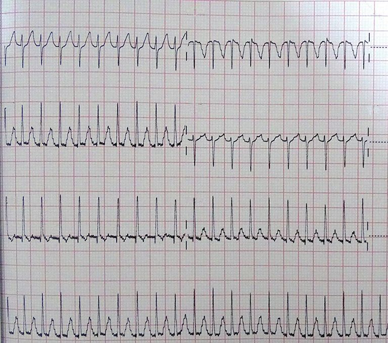
Annals of Palliative Medicine, Vol 10, No 7 July 2021                                                                                8323

         I                         aVR                  V1             no abnormal mass in the left ventricle. Cranial magnetic
                                                                       resonance imaging (MRI) revealed scattered lacunar
                                                                       cerebral infarctions in the right lateral ventricle. Limb
                                                                       twitching was still present after symptomatic treatment, as
         II                        aVL                  V2
                                                                       were tonic-clonic seizures and muscular hypertonia of the
                                                                       left limb, along with recurrent episodes of PSVT.
                                                                           The girl was transferred to our hospital for further
         III                       aVF                  V3             treatment. The results of physical examination were as
                                                                       follows: blood pressure: left upper extremity 98/45 mmHg,
                                                                       right upper extremity 92/55 mmHg, left lower extremity
         II                                                            125/68 mmHg, right lower extremity 110/60 mmHg;
                                                                       the patient was alert and active, with smooth respiration,
                                                                       and heart rate 105 bpm with sinus rhythm. The nervous
Figure 1 Electrocardiogram showing supraventricular tachycardia.       system examination results were as follows: left upper limb
                                                                       tone 3/5, left leg tone 3/5, right arm tone 5/5, right leg
                                                                       tone 5/5. The left Babinski signs (+), the right Babinski
of PSVT in infants. We present the following case in                   signs (−), and Brudzinski’s sign (−). The cranial MRI
accordance with the CARE reporting checklist (available at             (Figure 4A) was verified as showing the formation of
http://dx.doi.org/10.21037/apm-20-1797).                               local cerebral softening lesions and multiple abnormal
                                                                       intracranial signal shadows. During the hospitalization,
Case presentation                                                      recurrent episodes of PSVT were observed eight times.
                                                                       Sinus rhythm was maintained by the combination of
A 23-month-old girl was admitted to the hospital with the              antiarrhythmic agents, including propranolol, esmolol,
complaint of recurrent episodes of PSVT over a year, and               and amiodarone, and she was given anti-infective therapy,
intermittent seizures for eight days. The periodic episodes            intracranial pressure decreasing treatment (mannitol),
of PSVT were of 1–2 hours duration each, at a frequency                heart and brain cell nutritional therapy, and anticoagulant
of 3–4 times per week. The attacks occurred during activity            therapy. The girl’s symptoms of infection improved, and
or sleep, and were mainly spontaneously terminated. When               anti-infective therapy was stopped accordingly.
episodes were of long duration, the girl cried, lost appetite,             Meanwhile, we gradually reduced the treatment of
and became pale. These signs disappeared with termination              decreasing intracranial conventional pressure until the
of PSVT and recovery of sinus rhythm. The girl was given               medication was completely withdrawn. The girl was
amiodarone and digoxin without good effect. Two weeks                  discharged from our hospital after 22 days and continued
before admission, the girl was admitted to the local hospital          with oral propranolol (5 mg/kg q6h) and amiodarone
on continuous crying and pallor. Electrocardiogram                     (3–5 mg/kg/d). Cranial MRI (Figure 4B) was checked
(ECG) revealed a ventricular rate of 233 bpm (Figure 1),               3 weeks after admission, and it revealed that there were
and echocardiography showed that the motion amplitude                  liquefaction lesions in the right sub-region of the nucleus,
of the left ventricular wall was weakened and left cardiac             the right frontal lobe, temporal lobe, and occipital lobe;
function was decreased [ejection fraction (EF) 44%]. An                lesions in the left parietal cortex were enhanced, and all
abnormal mass, suspected to be a thrombus, was detected                lesions had disappeared in diffusion-weighted imaging.
in the left ventricle with a small amount of pericardial               Echocardiography (Figure 5A,B) revealed that atrial and
effusion (Figure 2A,B). In the process of cardioversion, the           ventricular structure was normal, left ventricle diastolic
girl suffered systemic convulsions and loss of consciousness,          diameter was 29 mm, and EF was 66%. In the present case,
and she was subsequently transferred to another hospital.              it is unknown whether the thrombosis was developed by
Upon admission to the second hospital, her heart rate was              the long-term PSVT alone or atrial fibrillation. During
105 bpm with sinus rhythm (Figure 3). Echocardiography                 hospitalization, we continuously monitored the patients’
was performed, revealing enlargement of the left ventricle             ECG, and we did not detect any of the usual characteristics
chamber, decreased movement of the ventricular wall (left              of atrial fibrillation. These physical examinations, clinical
ventricle diastolic diameter was 29 mm, EF 43%), and                   manifestations, and imaging results supported the diagnosis

© Annals of Palliative Medicine. All rights reserved.        Ann Palliat Med 2021;10(7):8322-8327 | http://dx.doi.org/10.21037/apm-20-1797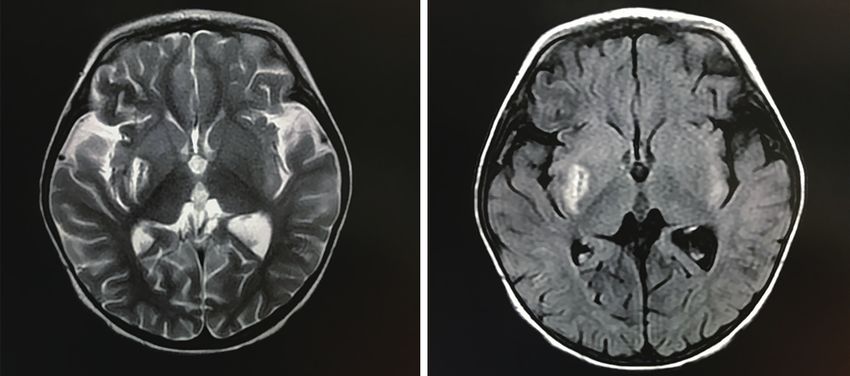
8324 Li et al. Left ventricular thrombosis and cerebral infarction after PSVT in an infant
A B
LV RV LV
LA RA
X
LA
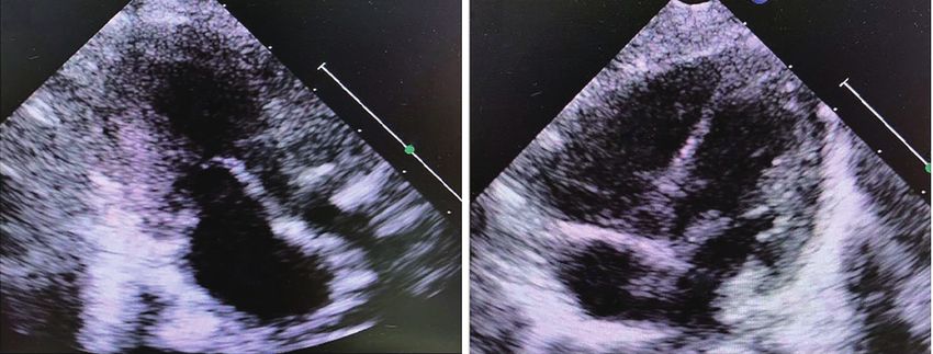
Figure 2 The abnormal echocardiography 2 weeks before admission. (A) Echocardiography showing an abnormal mass (arrow) suspected
thrombus in the left ventricle; (B) echocardiography showing an abnormal mass (arrow) suspected thrombus in the left ventricle with a small
amount of pericardial effusion. LV, left ventricular; LA, left atrial; RV, right ventricular; RA, right atrial.
I aVR V1 medication are currently gradually tapered. (All procedures
performed in studies involving human participants were in
accordance with the ethical standards of the institutional
II V2
and/or national research committee(s) and with the Helsinki
aVL
Declaration (as revised in 2013). This study was approved
by the ethical review committee of The Children’s Hospital
Capital Institute of Pediatrics (registration number:
III
aVF V3 SHERLL2018020). Written informed consent was obtained
from the patient for publication of this manuscript and any
accompanying images).
II
Discussion
Although PSVT is the most common tachyarrhythmia in
Figure 3 Electrocardiogram showing sinus rhythm.
children, few reports of thrombosis result from PSVT in
children. So far, there have only been three reported cases
of PSVT causing left atrial appendage and left ventricular
of left ventricular thrombosis and cerebral infarction.
thrombosis (3-5) in pediatric patients.
We reported the case of a 23-month-old girl with
Follow-up and outcomes echocardiography revealing a thrombus in the left ventricle
with heart failure and signs of peripheral embolization
The patient was followed-up by our cardiology and
(cerebral infarction) following episodic PSVT. When her
neurology department, focusing on the liver, kidney, and
sinus rhythm was restored, neurological sequelae resulting
thyroid function. We performed ECG, echocardiography,
from cerebral infarction remained, and it was characterized
thyroid ultrasound, and her left limb and general limb
by the increased tone and impaired movement of the left
activity were assessed. She was followed-up for 18 months
side.
with ECG, echocardiography, and 24-hour dynamic ECG
The defining characteristic of PSVT is its abrupt onset
every 3 months. There have been no abnormalities of
and termination, with varying durations (6). Clinical
cardiac size or function and no further attacks of PSVT on
manifestations include anxiety, tachypnea, pallor, cyanosis,
drug treatment. No obvious abnormalities were found at
and poor appetite. An ECG examination is required for
the follow-up physical examination. During follow-up, we
confirmation of PSVT (7-9). Diagnosis and treatment may
did not record any adverse or unanticipated events. Doses of
© Annals of Palliative Medicine. All rights reserved. Ann Palliat Med 2021;10(7):8322-8327 | http://dx.doi.org/10.21037/apm-20-1797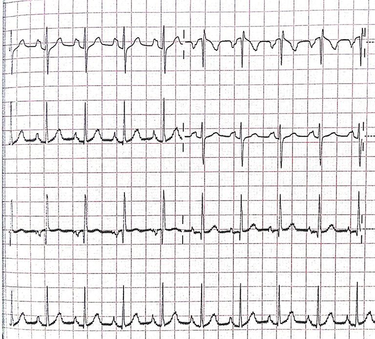
Annals of Palliative Medicine, Vol 10, No 7 July 2021                                                                                   8325

                    A                                                  B

                                           LV
                                                                                      RV

                                                               X
                                                                                                     LV

                                                 LA                                  RA         LA

Figure 4 Cranial MRI: the cranial MRI checked during the hospitalization and 3 weeks after admission. (A) The formation of local
cerebral softening lesions and multiple abnormal intracranial signal shadows; (B) there were liquefaction lesions in the right sub-region
of the nucleus, the right frontal lobe, temporal lobe, and occipital lobe; lesions in the left parietal cortex were enhanced, and all lesions
had disappeared in diffusion-weighted imaging. LV, left ventricular; LA, left atrial; RV, right ventricular; RA, right atrial; MRI, magnetic
resonance imaging.

                    A                                                  B

Figure 5 The echocardiography checked 3 weeks after admission. (A) Echocardiography showing the atrial and ventricular structures were
normal, no thrombus or pericardial effusion was observed; (B) echocardiography showing the atrial and ventricular structures were normal,
no thrombus or pericardial effusion was observed.

be delayed due to nonspecific symptoms in infants or young                 the medical history and performed related examinations,
children and result in severe sequelae. In the present case,               no other risk factors, except for PSVT, were found for
the child’s episodes of PSVT recurred until she developed                  thrombosis and neurological diseases leading to cerebral
serious complications, including ventricular thrombus and                  infarction in the girl, such as coagulation disorders, hepatic
cerebral infarction.                                                       function abnormality, septicemia, disseminated intravascular
   When the girl was admitted to our department, she                       coagulation (DIC), malignant tumor, hyperlipidemia,
remained in sinus rhythm with intermittent episodes                        diabetes mellitus, nephrotic syndrome, obstructive
of PSVT. There are multiple possible sources or causes                     sleep apnea syndrome (OSAS), coronary heart disease,
of cerebral infarction, and it becomes difficult to                        rheumatic heart disease, infectious endocarditis, dilated
differentially diagnose PSVT as the cause after an episode                 cardiomyopathy, hypertensive disease, congenital heart
has terminated. After our team had carefully reviewed                      disease, chronic obstructive pulmonary disease, pulmonary

© Annals of Palliative Medicine. All rights reserved.          Ann Palliat Med 2021;10(7):8322-8327 | http://dx.doi.org/10.21037/apm-20-1797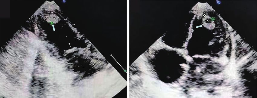
8326                                                    Li et al. Left ventricular thrombosis and cerebral infarction after PSVT in an infant

heart disease, or history of surgery (10,11). Therefore, we                  In order to prevent this rare but severe clinical
speculated that recurrent episodes of PSVT caused the left                complication, future research may concentrate on risk
ventricular thrombus.                                                     factors leading to thrombosis, including the duration and
    The mechanism of thrombus formation by PSVT is                        frequency of the PSVT attack, ventricular rate at the time
unknown, and several factors are potentially involved. Both               of the attack, as well as the platelet count and function,
the onset and termination of PSVT are characteristically                  thrombin, fibrinolytic enzymes, and so on. More attention
abrupt. The sudden attack and cessation of PSVT may                       should be paid to children with high risk factors, and
result in the loss of effective contraction and relaxation in             preventive measures must be taken swiftly against severe
the atrium and atrioventricular node, leading to the decline              complications.
or loss of pumping function. The sudden termination of
longer duration PSVT attacks may lead to a decline of atrial
                                                                          Acknowledgments
and atrioventricular nodes’ systolic function and then cause
turbulent blood flow in the ventricle. This would, in turn,               The authors thank the patient’s guardian for their
increase the contact between platelets and endothelium;                   cooperation.
increased platelet activation results in an increased risk of             Funding: This paper was supported by the Beijing Hospital
thrombus formation (12-14).                                               Administration “Peak Climbing” Talents Development
    Atrial fibrillation is the most common cause of blood                 Program (DFL20181301) and the Key Project of
clots in adult arrhythmias; thus, anticoagulant therapy is                Capital Clinical Characteristic Application Research
the routine treatment for such patients. In atrial fibrillation,          (Z181100001718189).
local blood stasis and turbulent flow give rise to thrombosis
formation (14). Atrial fibrillation in PSVT patients is
                                                                          Footnote
significantly higher than that of the general population
and may reach 12–44% (15,16). In the present case, it is                  Reporting Checklist: The authors have completed the CARE
unknown whether the patient developed thrombosis due to                   reporting checklist. Available at http://dx.doi.org/10.21037/
the long-term PSVT alone or atrial fibrillation.                          apm-20-1797
    In conclusion, this was a rare case of left ventricular
thrombus, and cerebral infarction after sustained episodes                Conflicts of Interest: All authors have completed the ICMJE
of PSVT in an infant diagnosed based on cross-sectional                   uniform disclosure form (available at http://dx.doi.
echocardiography. We recommend echocardiographic                          org/10.21037/apm-20-1797). Dr. DL reports grants
evaluation of the left ventricular function, including                    from Beijing Hospital Administration “Peak Climbing”
the screening for thrombi, and that the possibility of a                  Talents Development Program, grants from Key Project
thrombus must be considered when an abnormal structure                    of Capital Clinical Characteristic Application Research,
is identified in the left ventricle.                                      during the conduct of the study. Dr. MMZ reports grants
    The clinical significance of this case report highlights              from Beijing Hospital Administration “Peak Climbing”
the importance of health education for general community                  Talents Development Program, grants from Key Project
populations. Additionally, the attending parent or                        of Capital Clinical Characteristic Application Research,
guardian should pay adequate attention to abnormal                        during the conduct of the study. Dr. XHL reports grants
clinical manifestations such as tachypnea, pallor, cyanosis,              from Beijing Hospital Administration “Peak Climbing”
and poor appetite from the patients’ side. If the above-                  Talents Development Program, grants from Key Project
mentioned abnormal symptoms occur, the parent or                          of Capital Clinical Characteristic Application Research,
guardian should promptly take their baby to the hospital.                 during the conduct of the study. Dr. LS reports grants from
From the pediatricians’ side, they should be trained for                  Key Project of Capital Clinical Characteristic Application
early recognition of the nonspecific clinical manifestations              Research, during the conduct of the study. Dr. Yao L reports
of PSVT, and for making diagnosis by ECG, evaluation                      grants from Key Project of Capital Clinical Characteristic
of cardiac function and thrombosis by echocardiography,                   Application Research, during the conduct of the study. Dr.
and termination PSVT as rapidly as possible.                              Yang L reports grants from Key Project of Capital Clinical
Thromboprophylactic therapy should be provided in                         Characteristic Application Research, during the conduct of
instances of recurrent attacks that are not easily terminated.            the study. The authors have no other conflicts of interest to

© Annals of Palliative Medicine. All rights reserved.           Ann Palliat Med 2021;10(7):8322-8327 | http://dx.doi.org/10.21037/apm-20-1797
Annals of Palliative Medicine, Vol 10, No 7 July 2021 8327
declare. after sustained supraventricular tachycardia. Int J Cardiol
2008;131:e17-9.
Ethical Statement: The authors are accountable for all 5. Hanséus K, Björkhem G. Left ventricular thrombosis
aspects of the work in ensuring that questions related during infancy: report of two cases. Pediatr Cardiol
to the accuracy or integrity of any part of the work are 1995;16:182-5.
appropriately investigated and resolved. All procedures 6. Salerno JC, Seslar SP. Supraventricular tachycardia. Arch
performed in studies involving human participants were in Pediatr Adolesc Med 2009;163:268-74.
accordance with the ethical standards of the institutional 7. Tavera MC, Bassareo PP, Neroni P, et al. Supraventricular
and/or national research committee(s) and with the Helsinki tachycardia in neonates: antiarrhythmic drug choice
Declaration (as revised in 2013). This study was approved dilemma. J Matern Fetal Neonatal Med 2010;23 Suppl
by the ethical review committee of The Children’s Hospital 3:30-3.
Capital Institute of Pediatrics (registration number: 8. Lupoglazoff JM, Denjoy I. Practical attitude toward
SHERLL2018020). Written informed consent was obtained arrhythmia in the neonate and infant. Arch Pediatr
from the patient for publication of this manuscript and any 2004;11:1268-73.
accompanying images. 9. Kantoch MJ. Supraventricular tachycardia in children.
Indian J Pediatr 2005;72:609-19.
Open Access Statement: This is an Open Access article 10. Blann AD, Steele C, McCollum CN. The influence of
distributed in accordance with the Creative Commons smoking and of oral and transdermal nicotine on blood
Attribution-NonCommercial-NoDerivs 4.0 International pressure, and haematology and coagulation indices.
License (CC BY-NC-ND 4.0), which permits the non- Thromb Haemost 1997;78:1093-6.
commercial replication and distribution of the article with 11. Nobili L, Schiavi G, Bozano E, et al. Morning increase of
the strict proviso that no changes or edits are made and the whole blood viscosity in obstructive sleep apnea syndrome.
original work is properly cited (including links to both the Clin Hemorheol Microcirc 2000;22:21-7.
formal publication through the relevant DOI and the license). 12. Wu N, Tong S, Xiang Y, et al. Association of hemostatic
See: https://creativecommons.org/licenses/by-nc-nd/4.0/. markers with atrial fibrillation: a meta-analysis and meta-
regression. PLoS One 2015;10:e0124716.
13. Lim HS, Willoughby SR, Schultz C, et al. Effect of atrial
References
fibrillation on atrial thrombogenesis in humans: impact of
1. Joung B, Lee M, Sung JH, et al. Pediatric radiofrequency rate and rhythm. J Am Coll Cardiol 2013;61:852-60.
catheter ablation: sedation methods and success, 14. Khoo CW, Krishnamoorthy S, Lim HS, et al. Atrial
complication and recurrence rates. Circ J 2006;70:278-84. fibrillation, arrhythmia burden and thrombogenesis. Int J
2. Paul T, Bertram H, Bökenkamp R, et al. Supraventricular Cardiol 2012;157:318-23.
tachycardia in infants, children and adolescents: diagnosis, 15. Campbell RW, Smith RA, Gallagher JJ, et al. Atrial
and pharmacological and interventional therapy. Paediatr fibrillation in the preexcitation syndrome. Am J Cardiol
Drugs 2000;2:171-81. 1977;40:514-20.
3. B AG, Mathew RC. Left atrial thrombus in a neonate with 16. Bauernfeind RA, Wyndham CR, Swiryn SP, et al.
normal heart after sustained supraventricular tachycardia. J Paroxysmal atrial fibrillation in the Wolff-Parkinson-
Clin Diagn Res 2014;8:PD01-2. White syndrome. Am J Cardiol 1981;47:562-9.
4. Russo G, Trappan A, Benettoni A. Unusual left atrial
appendage thrombus in a neonate with normal heart (English Language Editors: J. Jones and J. Chapnick)
Cite this article as: Li D, Guo J, Li XH, Liao Y, Zhang
MM, Shi L, Lin Y, Liu Y. A case report of an infant after
episodes of paroxysmal supraventricular tachycardia with left
ventricular thrombosis and cerebral infarction. Ann Palliat Med
2021;10(7):8322-8327. doi: 10.21037/apm-20-1797
© Annals of Palliative Medicine. All rights reserved. Ann Palliat Med 2021;10(7):8322-8327 | http://dx.doi.org/10.21037/apm-20-1797You can also read