Obesity alters the ovarian proteomic response to zearalenone exposure
←
→
Page content transcription
If your browser does not render page correctly, please read the page content below
Biology of Reproduction, 2021, 105(1), 278–289
                                                                                                https://doi.org/10.1093/biolre/ioab069
                                                                                                                     Research Article
                                                                                     Advance Access Publication Date: 14 April 2021

Research Article

Obesity alters the ovarian proteomic response
to zearalenone exposure†

                                                                                                                                                                          Downloaded from https://academic.oup.com/biolreprod/article/105/1/278/6225347 by guest on 07 December 2021
M. Estefanía González-Alvarez, Bailey C. McGuire and Aileen F. Keating*
Department of Animal Science and Interdepartmental Toxicology Graduate Program, Iowa State University, Ames
IA, USA
*Correspondence: Department of Animal Science and Interdepartmental Toxicology Graduate Program, Iowa State
University, 2356H Kildee Hall, 806 Stange Rd, Ames IA 50011, USA. Tel: 515-294-3849; E-mail: akeating@iastate.edu
† Grant Support:This study was funded by the Iowa State University Martin Fund from the Nutritional Science Council
and the Bailey Career Development Award to AFK.

Received 20 November 2020; Revised 10 March 2021; Accepted 7 April 2021

Abstract
Zearalenone (ZEN), a nonsteroidal estrogenic mycotoxin, is detrimental to female reproduction.
Altered chemical biotransformation, depleted primordial follicles and a blunted genotoxicant
response have been discovered in obese female ovaries, thus, this study investigated the hypothe-
sis that obesity would enhance ovarian sensitivity to ZEN exposure. Seven-week-old female wild-
type nonagouti KK.Cg-a/a mice (lean) and agouti lethal yellow KK.Cg-Ay/J mice (obese) received
food and water ad libitum, and either saline or ZEN (40 μg/kg) per os for 15 days. Body and
organ weights, and estrous cyclicity were recorded, and ovaries collected posteuthanasia for
protein analysis. Body and liver weights were increased (P < 0.05) in the obese mice, but obesity
did not affect (P > 0.05) heart, kidney, spleen, uterus, or ovary weight and there was no impact
(P > 0.05) of ZEN exposure on body or organ weight in lean or obese mice. Obese mice had shorter
proestrus (P < 0.05) and a tendency (P = 0.055) for longer metestrus/diestrus. ZEN exposure in
obese mice increased estrus but shortened metestrus/diestrus length. Neither obesity nor ZEN
exposure impacted (P > 0.05) circulating progesterone, or ovarian abundance of EPHX1, GSTP1,
CYP2E1, ATM, BRCA1, DNMT1, HDAC1, H4K16ac, or H3K9me3. Lean mice exposed to ZEN had
a minor increase in γ H2AX abundance (P < 0.05). In lean and obese mice, LC–MS/MS identified
alterations to proteins involved in chemical metabolism, DNA repair and reproduction. These data
identify ZEN-induced adverse ovarian modes of action and suggest that obesity is additive to ZEN-
induced ovotoxicity.

Key words: zearalenone, obesity, ovary, chemical metabolism, DNA damage repair, ovarian pro-
teome.

Introduction                                                                          Zearalenone (ZEN) is a nonsteroidal mycotoxin with estrogenic
The ovary performs two major roles: development of oocytes and                    activity produced by Fusarium species [14–18]. Occurring naturally
hormone production and secretion [1–3]. Proper functioning of the                 in warm temperatures [19], ZEN is a common contaminant in cereal
ovary can be affected by exposure to xenobiotics [4–6], which can                 crops like maize [20–23], rye [20], wheat [20, 24], barley [20, 23],
cause temporary or permanent infertility [3] or endocrine disruption              but can also be detected in other dietary sources like nuts [25], flour
[6]. Premature cessation of ovarian activity is associated with a                 [20], beer [20, 26], and milk [27]. Stable at high temperatures, ZEN
heightened risk for development of a number of diseases in women                  is difficult to degrade during common food processing, becoming a
[7–13].                                                                           public health problem [19, 28, 29]. The chemical structure of ZEN

                                                         © The Author(s) 2021. Published by Oxford University Press on behalf of Society for the Study of Reproduction.
                                              This is an Open Access article distributed under the terms of the Creative Commons Attribution Non-Commercial License
278                            (http://creativecommons.org/licenses/by-nc/4.0/), which permits non-commercial re-use, distribution, and reproduction in any medium,
                                                     provided the original work is properly cited. For commercial re-use, please contact journals.permissions@oup.com
The ovarian proteome is affected by zearalenone, 2021, Vol. 105, No. 1 279
is similar to 17β-estradiol and other natural estrogens, which allows (7076 s) secondary antibodies were purchased from Cell Signaling
it to bind to estrogen receptors, causing estrogenicity [16, 17], and Technology (Danvers, MA, USA). Progesterone and estradiol ELISA
thus, ZEN is considered an endocrine disruptor [18]. The most sen- kits were obtained from DRG International, Inc. (Springfield, NJ,
sitive ZEN target is the reproductive system, but estrogen receptor- USA). Pierce BCA (bicinchoninic acid assay) protein assay kit was
positive tissues can also be responsive [14, 16, 19, 30]. Reproductive obtained from Thermo Fisher Scientific (Rockfield, IL, USA).
phenotypic effects of ZEN exposure in swine include abnormal
lactation [14], vulvovaginitis [14], pseudopregnancy [31], abortion Animals
[14], stillbirths [14], altered follicle stimulating hormone [30, 32], All the experimental animal protocols for this study were approved
progesterone [33] and 17β-estradiol [33] and follicular impacts [34]. by the Iowa State University Animal Care Committee. Female
Rats and mice also have negative reproductive effects due to ZEN wild-type normal nonagouti KK.Cg-a/a, designated lean henceforth
exposure including anovulation [35], altered folliculogenesis [36], (n = 10 per group) and agouti lethal yellow KK.Cg-Ay/J, designated
persistent estrus [14], decreased fertility [37], and reduced litter size obese henceforth (n = 10 per group) were purchased from Jackson
[37]. Laboratories (Bar Harbor, Maine) at 5 weeks of age. The mice
Downloaded from https://academic.oup.com/biolreprod/article/105/1/278/6225347 by guest on 07 December 2021
In US adults, the prevalence of female obesity is approximately were housed in Innovive cages with 2 or 3 animals per cage under
40%, and 20% in females aged 19 years and younger. Higher rates identical controlled conditions; temperature between 21◦ C and
affect minority populations, especially non-Hispanic black and His- 22◦ C, humidity of 20–30% and a light cycle of 12 h light/12 h
panic women in whom 50% are obese [38]. Unsurprisingly, obesity darkness. The mice were given water and food (2014 Teklad Global
is also an issue in developing countries [39]. In women, obesity has 14% Protein Rodent Diet) ad libitum. At 7 weeks of age, the mice
negative reproductive effects [40–50] including polycystic ovarian were dosed with saline solution as vehicle control (CT) or a ZEN
syndrome [40], decreased fecundity [41, 42], poor oocyte quality solution of 40 μg/kg (0.04 ppm) per body weight for 15 days which
[43], increased risk of birth defects [44, 45], premature [46, 47] they drank from a pipette tip. The ZEN dose was chosen based upon
and stillbirth [48], and gestational diabetes [49]. Synthesis and documented ovarian effects [36] and on the level of human exposure
metabolism of ovarian steroid hormones are also altered by obesity [19, 57]. This age of mouse was chosen because the number of
[50]. Our previous studies discovered that obesity can induce low primordial follicles is decreased from 12 weeks onwards in the obese
level DNA damage [51, 52], accelerate oxidative DNA damage and mice [51, 55, 56]. Food intake (calculated as food disappearance per
oxidative stress [51], alter the phosphatidylinositol-3 kinase (PI3K) cage/number of mice per cage) and body weight gain were monitored
[53, 54] pathway, reduce and blunt the response of ovarian chemical twice per week.
metabolism proteins [53, 55] and reduce primordial follicle number
[51, 55, 56]. Thus, the ovary from an obese female has apparent Monitoring of estrous cycle
greater sensitivity to reproductive toxicants.
Vaginal cytology monitoring was performed for 14 days. Sterile
This study investigated ovarian mechanisms of ZEN-induced
plastic pipette tips were used to lavage the vagina with saline [58].
toxicity and also explored the hypothesis that obesity enhances
The saline solution was gently flushed 3–5 times and the final flush
ovarian sensitivity to ZEN by altering the ovarian abundance of
was collected and observed under a microscope [58]. During the
proteins involved in chemical metabolism, DNA damage sensing
proestrus stage, nucleated epithelial cells comprise the majority of
and repair, and impacting serum progesterone and 17β-estradiol in
cells though some cornified and leukocytes may appear [58, 59].
female mice.
Large cornified cells with irregular shape and no visible or degen-
erated nucleus denote the estrous stage [58, 59]. In the metestrus
stage, mostly leukocytes, some cornified epithelial cells, and a few
Materials and methods nucleated epithelial cells appear [58, 59]. Finally, at the diestrus
stage a predominance of polymorphonuclear leucocytes with few
Reagents
nucleated epithelial cells are identified [58, 59].
ZEN (CAS # 17924–92-4), 2-β-mercaptoethanol, phosphate-
buffered saline (PBS), tris-buffered saline (TBS), nonfat dry milk,
hematoxylin, eosin, tris–HCl were purchased from Sigma–Aldrich
Tissue collection
Inc (St. Louis, MO, USA). Tween 20 and glycine were purchased from Euthanasia and tissue collection were performed using CO2 asphyxi-
Fisher Bioreagents (Fair Lawn, NJ, USA). Precast 4–20% MINI- ation followed by cervical dislocation when mice were at the second
PROTEAN TGX gels were obtained from Bio-Rad Laboratories, day of the diestrus stage of the estrous cycle. Euthanasia was per-
Inc. (Hercules, CA, USA). SignalFire ECL reagent was purchased formed in 90% of the mice within 4 days after dosing completion.
from Cell Signaling Technology (Danvers, MA, USA). Ponceau Blood samples were collected via cardiac puncture. Heart, liver,
S was obtained from Thermo Fisher Scientific (Waltham MA, kidneys, spleen, uterus, and ovaries were collected, excess fat was
USA). Restore PLUS Western Blot Stripping buffer was purchased trimmed from all tissues and tissues were weighed. One ovary was
from Thermo Scientific (Rockford, IL, USA). Primary antibodies frozen and stored at −80◦ C for protein analysis.
against H4K16ac (NB21-2077), H3K9me3 (NB21-1073), HDAC1
(NB100-56340) and DNMT1 (NB100-56519) were purchased from Serum 17β-estradiol and progesterone hormone level
Novus Biologicals (Centennial, CO, USA). GSTP1 (ab8902) primary quantification
antibody was purchased from Millipore (Temecula, CA, USA). ATM Blood samples were centrifuged for 15 min at 10 621 rcf and
(ab199726), EPHX1 (ab96695) and CYP2E1 (ab28146) primary 4◦ C. Serum was separated from red blood cells which were dis-
antibodies were purchased from Abcam (Cambridge, MA, USA). carded. 17β-estradiol and progesterone in serum were quantified
BRCA1 ((D-9) SC-6954) primary antibody was purchased from using ELISA kits following the manufacturer’s instructions. For the
Santa Cruz Biotechnology, Inc. (Dallas, TX, USA). γ H2AX ((Ser139) progesterone (LC = 5; LZ = 5; OC = 5; OZ = 4) and 17β-estradiol
#2577) primary, goat antirabbit (7074 s) and goat antimouse (LC = 5; LZ = 5; OC = 5; OZ = 3) analyses, 19 and 18 serum samples280 M. Alvarez et al., 2021, Vol. 105, No. 1
were analyzed with the ELISA kit respectively with two technical
replicates per sample, as sufficient volume of blood was not obtained
from all animals. For the 17β-estradiol assay, several samples were
below the detectable range of the assay kit (LC = 2; LZ = 3; OC = 2;
OZ = 2) thus, the data are not reported herein. All samples were
within the analytical range of the progesterone ELISA kit.
Protein isolation
Ovaries were homogenized in lysis buffer (50 mM Tris–HCl and
1 mM EDTA (pH ∼8.5)) to isolate total ovarian protein. Samples
(LC = 5; LZ = 5; OC = 5; OZ = 5); were centrifugated at 10 621 rcf for
15 min twice and the supernatant was collected each time. Protein
Downloaded from https://academic.oup.com/biolreprod/article/105/1/278/6225347 by guest on 07 December 2021
concentration was measured using the BCA assay. Absorbance values
were detected at 560 nm by an Eon Microplate Spectrophotometer
(Bio-Tek Instruments Inc. Winooski, VT, USA).
Western blot analysis
Protein (7 μg) was separated on MINI-PROTEAN TGX gels for
15 min at 60 V followed by 1 h at 100 V. Protein (LC = 3; LZ = 3;
Figure 1. Impact of obesity and ZEN exposure on body weight. Mice
OC = 3; OZ = 3) was transferred from the gel to a nitrocellulose
were weighed prior to euthanasia. Bars represent body weight (g) ± SEM.
membrane using a semidry transfer (iBlot) or by wet transfer for 1 h Superscript letters indicate significant differences; P < 0.05; n = 5/treatment.
at 100 V in ice. Using Ponceau S staining, the total amount of proteins Lean control-treated mice = LC; lean zearalenone-exposed mice = LZ; obese
transferred in each lane was recorded. A solution of 5% nonfat dried control-treated mice = OC; obese zearalenone-exposed mice = OZ.
milk with 1× PBST (semidry transfer) or 1× TBST (wet transfer)
was used to incubate the membrane for 1 h at room temperature
to reduce nonspecific binding. Primary antibodies were added, biological, molecular and pathway information of each protein. For
and membranes were incubated overnight. Primary (H4K16ac— gene ontology (GO) analysis PANTHER version 15.0 software was
1:200, H3K9me3—1:200, HDAC1—1:100, DNMT1—1:200, used. All samples with P < 0.05 were compared to the Mus musculus
ATM—1:100, GSTP1—1:2500, EPHX1—1:500, CYP2E1—1:500, reference list to obtain pathways in which altered proteins are
BRCA1—1:100 and γ H2AX—1:100) and secondary (1:2000– involved.
1:5000) antibody solutions were prepared with 5% nonfat dried
milk with 1× PBST or 1× TBST. After this incubation, membranes Statistical analysis
were washed three times for 10 min each with 1× PBST or 1× TBST Statistical analyses were performed using GraphPad Prism 8.4.1
and incubated with secondary antibodies at room temperature for software. For comparisons of two treatments (i.e. LC vs. LZ; OC
1 h. After incubation with secondary antibodies, the membranes vs. OZ; LC vs. OC), unpaired t-tests without adjustments were
were washed repeating the steps above followed by a 7 min used. Two-way analysis of variance (two-way ANOVA) was per-
incubation period with ECL SignalFire reagent. X-ray film exposure formed to compare two independent variables (body composition
was performed in the darkroom. Densitometric analysis was and ZEN exposure) using Tukey’s multiple comparison test. A P
performed using Image J software (NCBI). All of the samples for value ≤0.05 was defined as a statistically different result between
each specific protein were run on the same gel. Specific protein levels treatments.
were normalized to Ponceau S staining of total protein to account
for any discrepancy in loading efficiency.
Results
LC–MS/MS proteome analysis and gene ontology Effects of ZEN exposure and obesity on food intake
analysis and body weight
Liquid chromatography–tandem mass spectrometry (LC–MS/MS) Lean and obese genotype mice were weighed before starting
analysis was performed as previously described [60]. Briefly, total oral administration of saline solution (CT) or ZEN. There was
protein samples (LC = 5; LZ = 5; OC = 5; OZ = 5) were digested with no difference in food intake (P > 0.05) due to ZEN exposure,
trypsin/Lys-C for 16 h, dried down and reconstituted in buffer A however, as anticipated, mean food intake for the obese was higher
(47.5 μL, 0.1% formic acid/water. Peptide Retention Time Calibra- compared to the lean mice (LC = 67.8 ± 0.7 g; LZ = 67.4 ± 1.7 g;
tion (PRTC) was spiked into each sample as an internal control. OC = 79.4 ± 0.25 g; OZ = 70.4 ± 5.3 g; data not shown). Body
Protein samples and PRTC were injected onto a LC column to weight was monitored twice per week for the 15 days of
be separated and analyzed with a mass spectrometer. Theoretical dosing duration and increased over time in all groups (data
fragmentation patterns from MASCOT or Sequest HT were used not shown). The obese genotype mice weighed more (P < 0.05)
to compare the fragmentation patterns and intact results to iden- than their lean genotype counterparts at the completion of the
tify peptides. The areas of the top three unique peptides were experiment (mean body weight: LC = 28.3 ± 0.5 g; LZ = 27.1 ± 1.3 g;
used to identify the proteins. Only proteins that had three peptide OC = 33.0 ± 1.0 g; OZ = 32.9 ± 1.2 g; Figure 1). ZEN exposure did
hits within samples and that were identified in all samples were not impact mean body weight gain in either lean or obese mice
considered for analysis. Uniprot identifiers were used to obtain (P > 0.05).The ovarian proteome is affected by zearalenone, 2021, Vol. 105, No. 1 281
Downloaded from https://academic.oup.com/biolreprod/article/105/1/278/6225347 by guest on 07 December 2021
Figure 2. Effect of obesity or ZEN exposure on organ weight. After euthanasia, (A) heart, (B) liver, (C) kidney, (D) spleen, (E) uterus, and (F) ovary were collected
and weighed (g). Bars represent mean weight ± SEM. Superscript letters indicate statistical difference; P < 0.05; n = 5/treatment. Lean control-treated mice = LC;
lean zearalenone-exposed mice = LZ; obese control-treated mice = OC; obese zearalenone-exposed mice = OZ.
were no impacts of obesity or ZEN exposure on mean weights
of heart (Figure 2A), kidney (Figure 2C), spleen (Figure 2D), uterus
(Figure 2E), or ovaries (Figure 2F).
Effects of obesity and ZEN exposure on estrous
cyclicity and ovarian steroid hormone levels
Vaginal cytology was performed on each of the mice for 14 con-
secutive days over the dosing period to determine the effects of
ZEN exposure, obesity and any additive impact of obesity and
ZEN exposure on time spent at stages of the estrous cycle. Obese
mice spent less time at proestrus (P < 0.05) and tended to spend
more time in the metestrus/diestrus (P = 0.055) stage than the lean
mice (Figure 3). The length of time spent at the estrus stage was
increased (P < 0.05) in the obese mice treated with ZEN compared
to the CT-treated obese group (Figure 3). There was also an additive
impact of obesity on ZEN-induced alteration to the time spent in
Figure 3. Estrous cyclicity impacts of obesity and ZEN exposure. The number
of days at each stage of the estrous cycle were calculated over a 14-day
metestrus/diestrus which was decreased (P < 0.05; Figure 3). There
period and presented as a percentage. Bars represent percentage of d at was no observable impact (P > 0.05) of ZEN exposure or obesity on
proestrus (black bars), estrus (dark gray bars), and metestrus + diestrus circulating progesterone (Figure 4). The level of circulating proges-
(light gray bars) ± SEM. Asterix indicates differences between treatments; terone as measured by ELISA was in the published range for mice in
P < 0.05; n = 5/treatment. Lean control-treated mice = LC; lean zearalenone- the diestrus phase of the estrous cycle using ELISA as a quantification
exposed mice = LZ; obese control-treated mice = OC; obese zearalenone-
method [61].
exposed mice = OZ.
Effects of obesity and ZEN exposure on abundance of
Effects of obesity and ZEN exposure on organ weight ovarian proteins involved in chemical
Mean hepatic weight was greater (P < 0.05) in the obese rela- biotransformation
tive to lean mice (Figure 2B; LC = 1.4 ± 0.05 g; LZ = 1.3 ± 0.04 g; Western blotting was performed to quantify the impact of ZEN
OC = 1.9 ± 0.1 g; OZ = 1.8 ± 0.13 g). The livers of the obese group exposure, obesity or an additive effect of obesity with ZEN exposure
also appeared visually paler compared to the lean group. There on ovarian protein abundance of proteins involved in chemical282 M. Alvarez et al., 2021, Vol. 105, No. 1
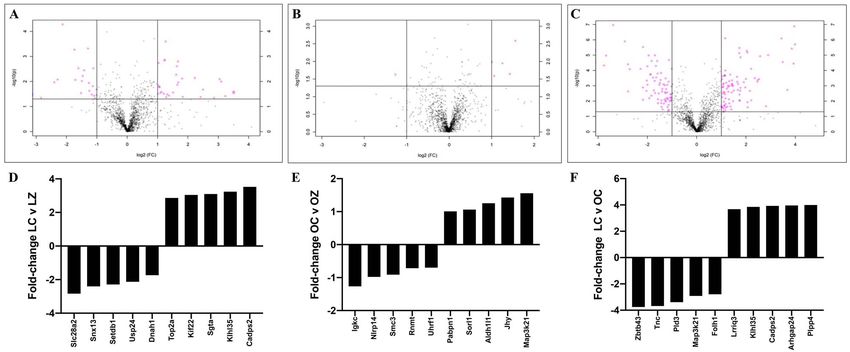
A total of 58 proteins were altered in the obese mice exposed
to ZEN (P < 0.05). Twenty-seven proteins were decreased, and
31 proteins increased (Figure 7B and Supplemental Table 2).
In the obese group, exposure to ZEN targeted the following
pathways “5-hydroxytryptamine degradation”, “alzheimer disease-
presenilin pathway”, “angiogenesis”, “angiotensin II-stimulated
signaling through G proteins and beta-arrestin”, “apoptosis
signaling pathway”, “asparagine and aspartate biosynthesis”, “axon
guidance mediated by semaphorins”, “CCKR signaling map”,
“cadherin signaling pathway”, “cytoskeletal regulation by Rho
GTPase”, “gonadotropin-releasing hormone receptor pathway”,
“heterotrimeric G-protein signaling pathway-Gq alpha and Go
alpha mediated pathway”, “huntington disease”, “inflammation
Downloaded from https://academic.oup.com/biolreprod/article/105/1/278/6225347 by guest on 07 December 2021
mediated by chemokine and cytokine signaling pathway”, “integrin
signaling pathway”, “parkinson disease”, “proline biosynthesis”,
“ras pathway”, and “vitamin D metabolism and pathway”.
Obesity alone altered 406 ovarian proteins (P < 0.05); 233 were
increased and 173 were decreased (Figure 7C and Supplemental
Table 3). In the obese group, pathways altered were: “5-
hydroxytryptamine degradation”, “DNA replication”, “angio-
genesis”, “gonadotropin-releasing hormone receptor pathway”,
Figure 4. Ovarian steroid hormone effects of obesity and ZEN exposure. Circu-
lating progesterone was measured by ELISA in lean control-treated mice = LC;
“CCKR signaling map”, “JAK/STAT signaling pathway”, “oxida-
lean zearalenone-exposed mice = LZ; obese control-treated mice = OC; obese tive stress response”, “p53 pathway”, “p53 pathway feedback
zearalenone-exposed mice = OZ. Bars represent mean concentration ± SEM. loops2”, “mRNA splicing”, “Wnt signaling pathway”, “vitamin D
metabolism and pathway”, “TCA cycle”, “ras pathway”, “pyruvate
metabolism”, and “inflammation mediated by chemokine and
metabolism. There was no difference (P > 0.05) between any of
cytokine signaling pathway”.
the treatments on ovarian protein abundance of EPHX1 (Figure 5A),
GSTP1 (Figure 5B) or CYP2E1 (Figure 5C).
Impact of obesity and ZEN exposure on ovarian Discussion
proteins involved in DNA repair Dietary exposure of humans and animals to ZEN is widespread, and
The ovarian protein abundance of ATM, γ H2AX, BRCA1, DNMT1, dependent on climate conditions since inadequate drying of grains
HDAC1, H4K16ac, and H3K9me3 were quantified via western can increase the incidence of ZEN contamination. Exposure to ZEN
blotting. There was no impact (P > 0.05) of ZEN exposure, obesity has reproductive effects in females [14, 16, 19] and this study had
or an additive effect of obesity and ZEN exposure on ovarian two major objectives: the first was to determine modes of action of
protein abundance of ATM (Figure 6A), BRCA1 (Figure 6C), ZEN on ovarian function and the second was to investigate if obesity
DNMT1(Figure 6D), HDAC1 (Figure 6E), H4K16ac (Figure 6F), influenced ZEN-induced ovarian toxicity.
and H3K9me3 (Figure 6G). However, ovarian γ H2AX protein The mean dietary intake of ZEN in the US has been estimated as
abundance was increased in lean mice treated with ZEN (P < 0.05) 30 ng/kg and 20 ng/kg for Canada, Denmark, and Norway [20]. In
but not in obese mice (Figure 6B). γ H2AX protein was also higher 2000, the Food and Agriculture Organization of the United States/-
(P < 0.05) in obese CT relative to lean CT mice. World Health Organization Expert Committee on Food Additives
established a provisional maximum tolerable daily intake (PMTDI)
Effects of ZEN and obesity on the global ovarian of 0.5 μg/kg [57]. The PMTDI was established based on ZEN and
proteome metabolites estrogenic activity in pigs, the most sensitive species
LC–MS/MS proteome analysis was performed to compare protein to ZEN [57]. The European Food Safety Authority Panel (EFSA)
abundance in CT and ZEN-exposed mouse ovaries and to determine on Contaminants in the Food Chain established a tolerable daily
if obesity altered the ovarian proteomic response to ZEN exposure. intake (TDI) for ZEN of 0.25 μg/kg after evaluating food samples
In lean mice, exposure to ZEN altered 177 proteins (P < 0.05). and grains from 19 European countries in the period between 2005
Of these, 72 were decreased and 105 were increased (Figure 7A and 2010 [19]. This study found that 0.4–17% of the total dietary
and Supplemental Table 1). Pathway analysis was performed exposure of ZEN in adults and 0.1–5.1% in toddlers resulted from
using PANTHER. Exposure to ZEN in the lean mice altered breakfast cereal consumption [19]. EFSA estimated the range of
“5-hydroxytryptamine degradation”, “angiogenesis”, “apoptosis dietary ZEN exposure as between 4 and 50 ng/kg body weight per
signaling pathway”, “arginine biosynthesis”, “DNA replication”, day and that chronic dietary exposure was higher among young
“de novo purine biosynthesis”, “dopamine receptor mediated people [19].
signaling pathway”, “glycolysis”, “gonadotropin-releasing hormone We chose a model of hyperphagia-induced obesity in which
receptor pathway”, “inflammation mediated by chemokine and mice overeat until they become corpulent. Their lean counterparts
cytokine signaling pathway”, “pentose phosphate pathway”, “ras consume the same food composition but eat fewer calories and are
pathway”, “toll receptor signaling pathway”, “ubiquitin proteasome of the same genetic background. This model has altered circulating
pathway”, “vitamin D metabolism and pathway”, “Wnt signaling blood glucose and insulin [62], reduced primordial follicle number
pathway”, “p53 pathway feedback loops 2”, and “p53 pathway”. [51], alterations to steroidogenesis [56], and a blunted response to aThe ovarian proteome is affected by zearalenone, 2021, Vol. 105, No. 1 283
Downloaded from https://academic.oup.com/biolreprod/article/105/1/278/6225347 by guest on 07 December 2021
Figure 5. Impact of obesity and ZEN exposure on ovarian proteins involved in chemical biotransformation. Total ovarian protein homogenates were analyzed for
(A) EPHX1, (B) GSTP, or (C) CYP2E1 protein abundance. Bars represent mean values relative to total protein staining ± SEM; n = 3/treatment. Lean control-treated
mice = LC; lean zearalenone-exposed mice = LZ; obese control-treated mice = OC; obese zearalenone-exposed mice = OZ.
Figure 6. Impact of obesity and ZEN exposure on ovarian proteins involved in DNA repair. Total ovarian protein homogenates were analyzed for (A) ATM, (B)
γ H2AX (C) BRCA1, (D) DNMT1, (E) HDAC1, (F) H4K16ac, and (G) H3K9me3. Bars represent mean values relative to total protein staining ± SEM. Asterix indicates
differences between treatment; P < 0.05; n = 3/treatment. Lean control-treated mice = LC; lean zearalenone-exposed mice = LZ; obese control-treated mice = OC;
obese zearalenone-exposed mice = OZ.
chemical challenge [51]. Exposure began when mice were 7 weeks of of different doses of ZEN (0.088–0.358 mg/kg) to gilts decreased
age, chosen so that there was not a difference in ovarian follicle com- mean daily weight gain [68], and three ZEN exposure studies in rats
position at the onset of dosing. Lean and obese mice were exposed documented decreased body weight gain [64, 65, 69], which was dose
to 40 μg/kg (0.04 ppm) body weight of ZEN for 15 days, and this (1–50 mg/kg) and developmental stage dependent (nonpregnant or
exposure was based upon a study in which exposure to ZEN affected pregnant) [64, 65, 69]. A chronic ZEN exposure (50 or 100 mg/kg)
meiotic progression and induced DNA double-strand breaks, altered study in mice (both male and female) for 103 weeks determined that
oocyte cyst breakdown and primordial follicle formation [36], albeit ZEN decreased mean body weight gain in both biological sexes [69].
in a different mouse strain. Further, another finding of decreased body weight in mice exposed
There are conflicting reports on the impact of ZEN exposure on to DON, was ameliorated by addition of dietary ZEN (10 mg/kg of
body weight gain. There was no effect of ZEN exposure on average ZEN + 5 mg/kg of DON) [67]. In contrast to all of these findings, is
daily body weight gain in prepubertal gilts [63]. There are studies, a study in which ZEN (1.8 mg/kg) exposure increased body weight
however, on ZEN exposure in which reduced body weight has been in exposed rats [70].
noted as a phenotypic impact. Two studies performed in rats noted Food intake and weight gain were monitored during the study, to
decreased food intake due to ZEN exposure [64, 65]. In mice exposed confirm that the obese strain reached a greater final body weight and
to either 10 ppm of ZEN plus 5 ppm deoxynivalenol (DON; another to determine if ZEN exposure altered food intake, potentially acting
mycotoxin produced by Fusarium species [66]), or 25 ppm DON as an obesogen. As per experimental design, the obese strain mice
decreased food intake was observed [67]. Administration via feeding had higher food intake and a subsequent higher final body weight.284 M. Alvarez et al., 2021, Vol. 105, No. 1
Downloaded from https://academic.oup.com/biolreprod/article/105/1/278/6225347 by guest on 07 December 2021
Figure 7. Ovarian protein identification and quantification via LC–MS/MS. Total ovarian protein homogenates were analyzed by LC–MS/MS and bioinformatic
comparison performed between peptides identified in (A) LC vs. LZ, (B) OC vs. OZ, and (C) LC vs. OC. Pink dots above the solid horizontal line indicate increased
(upper right corner) or decreased (upper left corner) proteins; n = 5/treatment; P < 0.05. The top-five increased and decreased proteins per comparison are
illustrated as fold-change in (D) LC vs, LZ, (E) OC vs. OZ, and (F) LC vs. OC treated mice.
Importantly, exposure to ZEN did not affect food intake or body an increase in the days spent at the diestrus stage [53]. Additionally,
weight of lean mice. These findings thus eliminate any confounding ZEN can cause changes in the estrous cyclicity depending on the
effect of reduced or increased food intake or body weight on the time of administration and dose [77]. Gilts provided 200 μg/kg body
molecular findings in this study and reduce the likelihood that ZEN weight of ZEN for 8 days had false estrus indications on day 4 of
influences obesity by stimulating higher food intake. treatment [78]. Longer interestrus periods were observed in gilts fed
In this study, there was no difference in the organ weight within with 5 and 10 ppm of ZEN during day 5–20 of the estrous cycle [79].
groups due to exposure to ZEN. Liver weight in the obese mice was Lengthened estrous cyclicity was noted in gilts exposed to 20 mg of
higher compared to the lean genotype, a finding that has been previ- ZEN on days 6–10 and days 11–15 of the estrous cycle [80]. Within
ously documented [51]. Similar to our findings, chickens exposed to 50 days after puberty, no behavioral estrus was detected in 45% of
a single oral dose of ZEN (15.0 g/kg) had no impact on liver, oviduct gilts fed with a diet containing 3.61 ppm of ZEN [31]. These findings
or comb weight [71]. On the other hand, organ weight impacts of indicate that exposure to ZEN alters estrous cyclicity in a variety of
ZEN have been noted in other species. Both male and female rats species including swine and mice.
exposed to 1.25 or 3.75 mg/kg body weight of ZEN for 8 or 10 weeks There was no impact of obesity or ZEN exposure on circulating
had enlarged livers and in male rats, the adrenal glands and spleens progesterone. Several samples were below the detectable range of the
were larger [72]. Female rats that were fed for 14 days with 250 μg/g 17β-estradiol assay kit, most likely due to blood being collected dur-
had enlarged kidney and liver but no effect on uterine weight [73]. ing diestrus. Progesterone was lower numerically in the obese mice
Chickens exposed to ZEN orally and intramuscularly (50, 200, 400, but the high level of variability in the lean mice precluded statistical
and 800 mg/kg) for 7 days had increased oviduct weight due to both difference being apparent. Thus, despite observable alterations to the
administration routes [71]. In addition, intramuscular ZEN exposure estrous cycle stage lengths due to both obesity and ZEN exposure in
caused increased liver and comb weight [71]. ZEN exposure caused obese mice, this was not reflected by a change in the major ovarian
uterine enlargement in Yorkshire gilts who received doses of 10, 20, hormone, progesterone, in circulation at the stage of the estrous cycle
or 40 μg/g for 4 weeks [73]. Furthermore, female pigs exposed to at which the ovaries were collected. Pregnant Sprague–Dawley rats
1.1 mg/kg of ZEN for 24 or 28 days had increased reproductive tract were exposed to a daily dose of 1–8 mg/kg ZEN from gestation day 6
weight [74, 75]. Gilts fed diets containing 1.1, 2.0, and 3.2 mg/kg to 19 [65] and 17β-estradiol and progesterone levels were decreased
of ZEN for 18 days had increased liver, kidney, and reproductive in a dose-dependent manner beginning at 2 mg/kg [65]. Prepubertal
tract (ovary, uteri, and vagina) weights whereas spleen relative weight pigs exposed to 238.5 μg/kg ZEN for 24 days and then to 20 or
was decreased in a dose-dependent manner [76]. The current study 40 μg/kg ZEN for 48 days had no impact on 17β-estradiol level,
did not note any ZEN-induced alterations to organ weight, again which could be attributable to their prepubertal status [81, 82]. In
emphasizing that overt toxicity was absent, and suggesting a dose- postpubertal pigs progesterone concentrations were increased due
and species-dependent impact of ZEN. to ZEN exposure (20 mg on days 6–10 or days 11–15 of the estrous
Days spent in the estrus stage were increased whereas days spent cycle) [80]. Further, pigs fed 60 or 90 mg/kg ZEN at 2, 3, and 6 weeks
at metestrus/diestrus stages decreased in the ZEN-exposed obese postbreeding had decreased circulating progesterone and at 4 weeks
but not lean mice, suggesting enhanced sensitivity of the obese postbreeding 17β-estradiol was also decreased [83]. Considering the
mice to ZEN exposure. Obesity also altered estrous cyclicity with low ZEN exposure level used in the current study, it is perhaps not
the obese mice having a shortened proestrus stage but lengthened surprising that endocrine disruption was not evident. It would also
metestrus/diestrus stage. It is known that obesity can impact the be a consideration for the future to collect ovaries at an estrous cycle
estrous cycle causing a decrease in the length of the estrus stage and stage more appropriate for quantification of 17β-estradiol effects.The ovarian proteome is affected by zearalenone, 2021, Vol. 105, No. 1 285
Total ovarian protein abundance of xenobiotic biotransforma- abundance due to obesity suggests that in this study, as in others from
tion enzymes (GSTP, CYP2E1, and EPHX1), as well as proteins our group [53–55], that obesity alters the basal ovarian capacity
involved in DNA damage repair (ATM, γ H2AX, BRCA1, DNMT1, for chemical biotransformation. In addition, these findings provide
HDAC1, H4K16ac, and H3K9me3) was investigated. Surprisingly, additional information on ovarian ZEN chemical biotransformation
there was no difference in the ovarian protein abundance of EPHX1, and the impact of obesity on xenobiotic metabolism.
CYP2E1, GSTP, ATM, BRCA1, DNMT1, HDAC1, H4K16ac, or Proteins that are involved in DNA damage repair were also iden-
H3K9me3 due to either obesity or ZEN exposure. Interestingly, the tified to be altered by either ZEN or obesity. Exposure to ZEN in lean
gold standard marker of DNA double-strand breaks, γ H2AX was mice reduced RPS27L and UBE2N, but increased CUL4A, USP47,
slightly increased in the lean mice treated with ZEN, but not in the TOP2A, KIF22. Ribosomal protein S27 like (RPS27L) regulates P53
obese group, supporting that ZEN may induce genotoxicity in the activity [94], which functions in cell survival and is activated by DNA
ovary as a mode of action. The level of γ H2AX increase is very minor damaging agents [95]. Ubiquitin conjugating enzyme E2 N (UBE2N)
and whether this is of biological significance remains unclear. Other is involved in regulation of innate immune signaling [96], glycolysis
studies have determined that ZEN exposure alters γ H2AX and [96], cell survival [96], RNA splicing [96], and the DNA damage
Downloaded from https://academic.oup.com/biolreprod/article/105/1/278/6225347 by guest on 07 December 2021
BRCA1 expression, and other proteins related to DNA double-strand response [96]. Kinesin family member 22 (KIF22) plays an important
breaks and repair [36, 84]. At the diplotene stage of meiosis γ H2AX role in mitosis and it is part of kinesin superfamily proteins that have
staining as well as mRNA abundance of Mlh1, Rad51, and Brca1 been associated with carcinogenesis and cancer progression [97].
were increased in mice treated with 20 μg/kg of ZEN [36] and mice Exposure to ZEN in lean mice reduced abundance of PPP1R12A,
treated with 10 and 30 μM of ZEN had increased γ H2AX positive CCT2, RHOA, and TUFM and increased HSPA5, GSTP1, AKR7A2,
ovarian cells [84]. Our finding of increased γ H2AX, therefore, albeit USP47, BLMH, SCFD1, and TOP2A level. Protein phosphatase 1
a small increase, recapitulates findings of other studies on the ovarian regulatory subunit 12A (PPP1R12A) is associated with alterations in
effects of ZEN exposure. Lack of γ H2AX increase in ovaries of obese the PI3K/AKT pathway and is a tumor suppressor with a proposed
mice could indicate alterations to ZEN-induced genotoxicity or an role in cancer chemoresistance [98]. DNA topoisomerase II alpha
altered protein response to DNA double-strand break in the ovary (TOP2A) has an important role in DNA replication [99], response to
of the obese mice. DNA damage stimulus [85], and drug binding [85] and is increased
Proteomic profiling can provide important insights into the by ZEN exposure.
impact of a toxic exposure on cellular function. Since the aim of Related to reproductive function, HSP90AA1, GPI, CA2,
the study was to identify modes of action of ZEN exposure in lean CTNNB1, and STRA6 were increased whereas NSD1, MGARP,
and obese mice, ovaries were collected at the same stage of the and OXCT1 were decreased by ZEN exposure. Nuclear receptor-
estrous cycle on the second day of diestrus. In choosing this strategy, binding SET domain protein 1 (NSD1) is part of histone lysine
it is acknowledged that a lag time from the cessation of dosing methyltransferases family [100, 101], with a molecular function
to tissue collection exists but minimizing hormonal milieu variation related to estrogen receptor-binding [101]. Signaling receptor and
was considered an important aspect of the data interpretation. Using transporter of retinol STRA6 (STRA6) regulates cellular uptake of
LC–MS/MS, alterations to the protein profile were identified in three vitamin A [102], is present in the reproductive organs [103] and
specific treatment comparisons: lean CT vs. lean ZEN, obese CT vs. placenta [103] and has been associated with activation of JAK–
obese ZEN, and lean CT vs. obese CT. Altered ovarian abundance of STAT pathway [104, 105] and insulin signaling [106]. Thus, ZEN
proteins involved in DNA damage and repair, chemical metabolism, exposure in lean mice altered ovarian proteins involved in xenobiotic
and reproduction were consistently discovered in the ovaries of metabolism, DNA repair and reproductive function.
ZEN-exposed mice, with treatment effects as expanded upon below. In obese mice exposed to ZEN, DNA damage repair proteins
Proteomics analysis identified that GSTP1 ovarian protein abun- increased were DDX5, RUVBL1, and ACTR2 whereas SMC3,
dance was increased in the lean mice exposed to ZEN and due UHRF1, and HSPA1A were decreased in abundance, again
to obesity. The biological and molecular functions of this protein supporting that ZEN induces ovarian DNA damage but that
are related to drug binding [85], xenobiotic metabolism process the ovarian response differs to that of the lean mice. Structural
[85], and response to toxic substances [85]. With this finding, it is maintenance of chromosomes protein 3 (SMC3) is part of a protein
important to keep in mind that it is known that LC–MS/MS is a more complex that is important for DNA repair [85, 107] and regulation
sensitive technique compared to western blot, and that could be the of gene expression [107]. Actin related protein 2 (ACTR2) is part
reason that altered abundance of GSTP1 was determined by LC– of the ARP2/3 complex that is required for cell migration [108],
MS/MS but not using western blotting. Ovarian protein abundance regulation of actin filament nucleation [109] and organization
of EPHX2 was also altered due to obesity, and due to ZEN exposure in the cytoplasm [110, 111], and double-strand breaks involved
in lean mice, but in opposing directions in both. EPHX2, as well in DNA damage and repair [85, 112]. RAN binding protein 1
as EPHX1, is an epoxide hydrolase involved in the metabolism of (RANBP1) can control mitotic microtubules function that can be
chemicals [86–88]. Epoxide hydrolases are divided into two groups: a target for chemotherapeutic drugs [113], and its biological process
microsomal epoxide hydrolase (EPHX1) and soluble cytosolic epox- is associated with cellular response to drugs [85]. The altered
ide hydrolase (EPHX2) [89–92]. Interestingly, ras homolog family ovarian protein abundance related to chemical metabolism were
member A (RHOA) is a protein whose ovarian abundance was RHOA, ACTR2, ANXA1, NAMPT, CCT7, and EPHX2 which
decreased by basal obesity and by ZEN exposure in lean mice, but were increased in abundance whereas RANBP1 and CCT3 were
was increased by ZEN exposure in obese mice. RHOA is part of the lower in ovaries of obese ZEN-exposed mice. Thus, there is strong
Rho GTPase family, and it is important for certain cell functions such evidence for the involvement of RHOA and EPHX2 in the ovarian
as migration and survival [93], gene expression [93], and cell division response to ZEN in both lean and obese mice. Finally, in the group
[93]. A response to drug [85], glucose [85], and ethanol [85] is also of proteins related to reproduction, two proteins with an identified
considered a biological function of RHOA. These proteins are all reproductive function were increased—ANXA1 and AKR1C18.
involved in the metabolism of chemicals, and changes in the ovarian Annexin A1 (ANXA1) is abundantly expressed in many tissues286 M. Alvarez et al., 2021, Vol. 105, No. 1
including testis [114], ovaries [115], and placenta [115, 116], and Data availability statement
regulates steroid hormone secretion [117], and ANXA1 is elevated
Data available on request.
during pregnancy [116, 118]. Other biological processes in which
ANXA1 is involved are estrous cyclicity [117], prolactin secretion
[85], response to estradiol [85], and response to drugs [85]. Taken References
together, these findings support that ZEN exposure also alters DNA
1. Hoyer PB. 11.16—Female reproductive toxicology. In: McQueen CA
repair, chemical metabolism, and proteins involved in reproduction (ed.), Comprehensive Toxicology, 2nd ed. Oxford: Elsevier; 2010:
in the ovary but there is differential protein abundance between the 339–345.
ovaries from lean and obese mice. 2. Senger PL. Pathways to pregnancy & parturition. Washington State
Obesity in the absence of ZEN exposure altered proteins with University Research and Technology Park, Pullman WA: Current Con-
potential important ovarian function. Uveal autoantigen with coiled- ceptions Inc. 2012.
coil domains and ankyrin repeats (UACA) regulates apoptotic path- 3. Hoyer PB, Keating AF. Xenobiotic effects in the ovary: temporary
ways and is associated with DNA damage [119]. SMC3 was con- versus permanent infertility. Expert Opin Drug Metab Toxicol 2014;
Downloaded from https://academic.oup.com/biolreprod/article/105/1/278/6225347 by guest on 07 December 2021
siderably increased by obesity. Tenascin C (TNC) and fibronectin 10:511–523.
4. Mattison DR. Clinical manifestations of ovarian toxicity. Reprod Toxi-
have involvement in the extracellular matrix (ECM) and bind to
col 1985; 109:697–724.
integrins at the cell surface [120]. These have importance as ECM
5. Keating AF, Hoyer PB. Mechanisms of reproductive toxicity. In: Drug
regulates synaptic transmission, and cell signaling, and its expres- Metabolism Handbook. Hoboken, NJ: John Wiley and sons, Inc. 2009:
sion and activity can be controlled by chemicals [120]. Gamma- 697–736.
aminobutyric acid type A receptor subunit alpha4 (GABRA4) is a 6. Hoyer PB. Damage to ovarian development and function. Cell Tissue
GABAA receptor that is expressed in glial cells and neurons and Res 2005; 322:99–106.
is involved in neurotransmission processes [121]. The alpha4 and 7. Krarup T. Oocyte destruction and ovarian tumorigenesis after direct
alpha6 subunits have extrasynaptic localization, and are altered by application of a chemical carcinogen (9:10-dimethyl-1:2-benzanthrene)
xenobiotic exposure including benzodiazepines [121], alcohol [121], to the mouse ovary. Int J Cancer 1969; 4:61–75.
and drugs of abuse [121]. Related to reproduction, hexosaminidase 8. Krarup T. Effect of 9,10-dimethyl-1,2-denzanthracene on the mouse
ovary. Ovarian tumorigenesis. Br J Cancer 1970; 24:168–186.
subunit beta (HEXB) has an important role in the central nervous
9. Beamer WG, Tennent BJ. Gonadotropin uptake in genetic and irra-
system and breaks down sphingolipids, oligosaccharides, and gly-
diation models of ovarian Tumorigenesis1. Biol Reprod 1986; 34:
coproteins [122]. It has been associated with reproduction [123], 761–770.
fertility rates [85, 123], and oogenesis [85]. Serpin family F mem- 10. Tennent BJ, Beamer WG. Ovarian tumors not induced by irradiation
ber 1 (SERPINF1) encodes the pigment epithelium-derived factor and gonadotropins in hypogonadal (HPG) mice. Biol Reprod 1986;
(PEDF) [124], an inhibitor of angiogenesis [125], and regulator 34:751–760.
of bone density [126]. Alterations in PEDF levels are associated 11. Maronpot R. Ovarian toxicity and carcinogenicity in eight recent
with cancer [127], ovarian hyperstimulation [127], endometriosis National Toxicology Program Studies. Environ Health Perspect 1987;
[127], and diabetes [127]. Thus, in the absence of chemical expo- 73:125–130.
sure, obesity alone alters ovarian protein abundance in manners 12. Hoyer PB, Sipes IG. Assessment of follicle destruction in chemical-
induced ovarian toxicity. Annu Rev Pharmacol Toxicol 1996;
that could contribute to altered fertility and sensitivity to chemical
36:307–331.
exposure.
13. Hoyer PB, Devine PJ, Xiaoming Hu, Thompson KE, Sipes IG. Ovarian
It is recognized that ZEN can act in an estrogenic manner by toxicity of 4-vinylcyclohexene diepoxide: a mechanistic model. Toxicol
binding the estrogen receptors [128]. Thus, we interrogated our Pathol 2001; 29:91–99.
proteomics findings manually by searching the literature for links 14. Kuiper-Goodman T, Scott PM, Watanabe H. Risk assessment of the
between estrogen signaling and proteins that were altered in abun- mycotoxin zearalenone. Regul Toxicol Pharmacol 1987; 7:253–306.
dance in our LC–MS/MS experiments. We determined that RHOA 15. Urry WH, Wehrmeister HL, Hodge EB, Hidy PH. The structure of
[129, 130], GSTP1 [131], NSD1 [101], SMC3 [132], ANXA1 [133], zearalenone. Tetrahedron Lett 1966; 7:3109–3114.
TCN [134], GABRA4 [135], PEDF [136], and TOP2A [137] are 16. Bennett JW, Klich M. Mycotoxins. Clin Microbiol Rev 2003;
associated with estrogen receptor activity, whereas STRA6 gene 16:497–516.
17. Caldwell RW, Tuite J, Stob M, Baldwin R. Zearalenone production by
expression is regulated by progesterone receptor [138]. Thus, the
fusarium species. Appl Microbiol 1970; 20:31–34.
exposure to ZEN in this study altered proteins that are linked with
18. Shier WT, Shier AC, Xie W, Mirocha CJ. Structure-activity relationships
estrogen signaling, supporting an estrogenic impact of ZEN. for human estrogenic activity in zearalenone mycotoxins. Toxicon 2001;
In conclusion, both obesity and/or ZEN exposure altered liver 39:1435–1438.
weight, affected the estrous cycle, increased a marker of DNA dam- 19. Chain EPoCitF. Scientific opinion on the risks for public health related
age and repair, and proteomics analysis revealed changes in ovarian to the presence of zearalenone in food. EFSA J 2011; 9:2197.
abundance of proteins related to DNA damage repair, chemical 20. Zinedine A, Soriano JM, Moltó JC, Mañes J. Review on the toxicity,
metabolism and reproduction. Taken together, the data identify occurrence, metabolism, detoxification, regulations and intake of zear-
ZEN-induced ovarian alterations and support that ovarian response alenone: an oestrogenic mycotoxin. Food Chem Toxicol 2007; 45:1–18.
to ZEN exposure is different in obese relative to lean mice. These 21. Caldwell RW, Tuite J. Zearalenone in freshly harvested corn. Phy-
topathology 1974; 64:752.
findings raise concerns about how an altered physiological status can
22. Shotwell OL. Mycotoxins in hot spots in grains. I. Aflatoxin and
influence the effects of ovarian xenobiotic exposure.
zearalenone occurrence in stored corn. Cereal chemistry 1975; 52: pp.
687–697.
23. Shotwell OL. Assay Methods for Zearalenone and Its Natural Occur-
rence. Park Forest South IL: Pathotox Publishers, Inc.; 1977.
Supplementary material
24. Goyarts T, Dänicke S, Valenta H, Ueberschär K-H. Carry-over of
Supplementary material is available at BIOLRE online. fusarium toxins (deoxynivalenol and zearalenone) from naturallyThe ovarian proteome is affected by zearalenone, 2021, Vol. 105, No. 1 287
contaminated wheat to pigs. Food Addit Contam 2007; 47. Smith GCS, Shah I, Pell JP, Crossley JA, Dobbie R. Maternal obesity in
24:369–380. early pregnancy and risk of spontaneous and elective preterm deliveries:
25. Poór M, Kunsági-Máté S, Bálint M, Hetényi C, Gerner Z, Lemli B. a retrospective cohort study. Am J Public Health 2007; 97:157–162.
Interaction of mycotoxin zearalenone with human serum albumin. J 48. Aune D, Saugstad OD, Henriksen T, Tonstad S. Maternal body mass
Photochem Photobiol B Biol 2017; 170:16–24. index and the risk of fetal death, stillbirth, and infant death: a systematic
26. Okoye ZSC. Stability of zearalenone in naturally contaminated corn dur- review and meta-analysis. JAMA 2014; 311:1536–1546.
ing Nigerian traditional brewing. Food Addit Contam 1987; 4:57–59. 49. Chu SY, Callaghan WM, Kim SY, Schmid CH, Lau J, England LJ, Dietz
27. Prelusky DB, Scott PM, Trenholm HL, Lawrence GA. Minimal transmis- PM. Maternal obesity and risk of gestational diabetes mellitus. Diabetes
sion of zearalenone to milk of dairy cows. J Environ Sci Health B 1990; Care 2007; 30:2070–2076.
25:87–103. 50. Pasquali R, Pelusi C, Genghini S, Cacciari M, Gambineri A. Obesity
28. Ryu D, Hanna MA, Bullerman LB. Stability of zearalenone during and reproductive disorders in women. Hum Reprod Update 2003;
extrusion of corn grits†. J Food Prot 1999; 62:1482–1484. 9:359–372.
29. Bennett GA, Shotwell OL, Hesseltine CW. Destruction of zearalenone in 51. Ganesan S, Nteeba J, Madden JA, Keating AF. Obesity alters phospho-
contaminated corn. J Am Oil Chem Soc 1980; 57:245–247. ramide mustard-induced ovarian DNA repair in mice. Biol Reprod 2017;
Downloaded from https://academic.oup.com/biolreprod/article/105/1/278/6225347 by guest on 07 December 2021
30. He J, Wei C, Li Y, Liu Y, Wang Y, Pan J, Liu J, Wu Y, Cui S. Zearalenone 96:491–501.
and alpha-zearalenol inhibit the synthesis and secretion of pig follicle 52. Ganesan S, Nteeba J, Keating AF. Enhanced susceptibility of ovaries
stimulating hormone via the non-classical estrogen membrane receptor from obese mice to 7,12-dimethylbenz[a]anthracene-induced DNA dam-
GPR30. Mol Cell Endocrinol 2018; 461:43–54. age. Toxicol Appl Pharmacol 2014; 281:203–210.
31. Etienne M, Jemmali M. Effects of zearalenone (F2) on estrous activity 53. Nteeba J, Ganesan S, Madden JA, Dickson MJ, Keating AF. Progres-
and reproduction in gilts2. J Anim Sci 1982; 55:1–10. sive obesity alters ovarian insulin, phosphatidylinositol-3 kinase, and
32. Diekman MA, Green ML, Malayer JR, Brandt KE, Long GG. Effect chemical metabolism signaling pathways and potentiates ovotoxicity
of zearalenone and estradiol benzoate on serum concentrations of induced by phosphoramide mustard in mice†. Biol Reprod 2017; 96:
LH, FSH and prolactin in ovariectomized gilts. Theriogenology 1989; 478–490.
31:1123–1130. 54. Nteeba J, Ross JW, Perfield II JW, Keating AF. High fat diet induced obe-
33. Long GG, Diekman M, Tuite JF, Shannon GM, Vesonder RF. Effect of sity alters ovarian phosphatidylinositol-3 kinase signaling gene expres-
fusarium roseum corn culture containing zearalenone on early pregnancy sion. Reprod Toxicol 2013; 42:68–77.
in swine. Am J Vet Res 1982; 43:1599–1603. 55. Nteeba J, Ganesan S, Keating AF. Impact of obesity on Ovotoxic-
34. Malekinejad H, Schoevers EJ, Daemen IJJM, Zijlstra C, Colenbrander ity induced by 7,12-dimethylbenz[a]anthracene in Mice1. Biol Reprod
B, Fink-Gremmels J, Roelen BAJ. Exposure of oocytes to the fusarium 2014; 90:68.
toxins zearalenone and deoxynivalenol causes aneuploidy and abnormal 56. Nteeba J, Ganesan S, Keating AF. Progressive obesity alters ovarian
embryo development in Pigs1. Biol Reprod 2007; 77:840–847. folliculogenesis with impacts on pro-inflammatory and steroidogenic
35. Kumagai S, Shimizu T. Neonatal exposure to zearalenone causes persis- signaling in female mice1. Biol Reprod 2014; 91:86.
tent anovulatory estrus in the rat. Arch Toxicol 1982; 50:279–286. 57. Joint FAOWHOECoFA, World Health O. Evaluation of Certain Food
36. Liu KH, Sun XF, Feng YZ, Cheng SF, Li B, Li YP, Shen W, Li L. Additives and Contaminants: Fifty-Third Report of the Joint FAO/WHO
The impact of zearalenone on the meiotic progression and primordial Expert Committee on Food Additives. Geneva: World Health Organiza-
follicle assembly during early oogenesis. Toxicol Appl Pharmacol 2017; tion; 2000.
329:9–17. 58. Caligioni CS. Assessing reproductive status/stages in mice. Curr Protoc
37. Becci PJ, Johnson WD, Hess FG, Gallo MA, Parent RA, Taylor JM. Com- Neurosci 2009; Appendix 4:Appendix 4I.
bined two-generation reproduction-teratogenesis study of zearalenone in 59. Byers SL, Wiles MV, Dunn SL, Taft RA. Mouse estrous cycle identifica-
the rat. J Appl Toxicol 1982; 2:201–206. tion tool and images. PLoS One 2012; 7:e35538.
38. Hales CM, Carroll MD, Fryar CD, Ogden CL. Prevalence of obesity 60. Clark KL, Talton OO, Ganesan S, Schulz LC, Keating AF. Develop-
among adults and youth: United States, 2015–2016. In: NCHS data mental origins of ovarian disorder: impact of maternal lean gestational
brief . Hyattsville, MD: National Center for Health Statistics; 2017: 8. diabetes on the offspring ovarian proteome in mice†. Biol Reprod 2019;
39. World Health O. Obesity and overweight. In: Fact sheets. Geneva, 101:771–781.
Switzerland: World Health Organization. vol. 2020. 2020. 61. Zenclussen ML, Casalis PA, Jensen F, Woidacki K, Zenclussen AC. Hor-
40. Pasquali R, Casimirri F. The impact of obesity on hyperandrogenism and monal fluctuations during the estrous cycle modulate heme oxygenase-1
polycystic ovary syndrome in premenopausal women. Clin Endocrinol expression in the uterus. Front Endocrinol 2014; 5:32.
(Oxf) 1993; 39:1–16. 62. Yang Z, Norwood KA, Smith JE, Kerl JG, Wood JR. Genes involved
41. Rich-Edwards JW, Goldman MB, Willett WC, Hunter DJ, Stampfer MJ, in the immediate early response and epithelial-mesenchymal transition
Colditz GA, Manson JE. Adolescent body mass index and infertility are regulated by adipocytokines in the female reproductive tract. Mol
caused by ovulatory disorder. Am J Obstet Gynecol 1994; 171:171–177. Reprod Dev 2012; 79:128–137.
42. Grodstein F, Goldman MB, Cramer DW. Body mass index and ovulatory 63. Green ML, Diekman MA, Malayer JR, Scheidt AB, Long GG. Effect
infertility. Epidemiology 1994; 5:247–250. of prepubertal consumption of zearalenone on puberty and subsequent
43. Jungheim ES, Schoeller EL, Marquard KL, Louden ED, Schaffer JE, reproduction of gilts. J Anim Sci 1990; 68:171–178.
Moley KH. Diet-induced obesity model: abnormal oocytes and per- 64. Hueza IM, Raspantini PCF, Raspantini LER, Latorre AO, Górniak SL.
sistent growth abnormalities in the offspring. Endocrinology 2010; Zearalenone, an estrogenic mycotoxin, is an immunotoxic compound.
151:4039–4046. Toxins 2014; 6:1080–1095.
44. Watkins ML, Rasmussen SA, Honein MA, Botto LD, Moore CA. Mater- 65. Collins TF, Sprando RL, Black TN, Olejnik N, Eppley RM, Alam HZ,
nal obesity and risk for birth defects. Pediatrics 2003; 111:1152. Rorie J, Ruggles DI. Effects of zearalenone on in utero development in
45. Stothard KJ, Tennant PW, Bell R, Rankin J. Maternal overweight and rats. Food Chem Toxicol 2006; 44:1455–1465.
obesity and the risk of congenital anomalies: a systematic review and 66. Sobrova P, Adam V, Vasatkova A, Beklova M, Zeman L, Kizek R.
meta-analysis. JAMA 2009; 301:636–650. Deoxynivalenol and its toxicity. Interdisciplinary toxicology 2010;
46. McDonald SD, Han Z, Mulla S, Beyene J, on behalf of the Knowledge 3:94–99.
Synthesis Group. Overweight and obesity in mothers and risk of preterm 67. Forsell JH, Witt MF, Tai JH, Jensen R, Pestka JJ. Effects of 8-week
birth and low birth weight infants: systematic review and meta-analyses. exposure of the B6C3F1 mouse to dietary deoxynivalenol (vomitoxin)
BMJ 2010; 341:c3428. and zearalenone. Food Chem Toxicol 1986; 24:213–219.You can also read