Cellular Models and Assays to Study NLRP3 Inflammasome Biology - MDPI
←
→
Page content transcription
If your browser does not render page correctly, please read the page content below
International Journal of
            Molecular Sciences

Review
Cellular Models and Assays to Study NLRP3
Inflammasome Biology
Giovanni Zito 1 , Marco Buscetta 1,† , Maura Cimino 1,† , Paola Dino 2 , Fabio Bucchieri 2,3 and
Chiara Cipollina 1,3, *
 1    Fondazione Ri.MED, via Bandiera 11, 90133 Palermo, Italy; gzito@fondazionerimed.com (G.Z.);
      mbuscetta@fondazionerimed.com (M.B.); mcimino@fondazionerimed.com (M.C.)
 2    Dipartimento di Biomedicina Sperimentale, Neuroscenze e Diagnostica Avanzata (Bi.N.D.),
      University of Palermo, via del Vespro 129, 90127 Palermo, Italy; paola.dino@unipa.it (P.D.);
      fabio.bucchieri@unipa.it (F.B.)
 3    Istituto per la Ricerca e l’Innovazione Biomedica-Consiglio Nazionale delle Ricerche, via Ugo la Malfa 153,
      90146 Palermo, Italy
 *    Correspondence: ccipollina@fondazionerimed.com; Tel.: +39-091-6809191; Fax: +39-091-6809122
 †    These authors contributed equally to this work.

 Received: 19 May 2020; Accepted: 12 June 2020; Published: 16 June 2020

 Abstract: The NLRP3 inflammasome is a multi-protein complex that initiates innate immunity
 responses when exposed to a wide range of stimuli, including pathogen-associated molecular patterns
 (PAMPs) and danger-associated molecular patterns (DAMPs). Inflammasome activation leads to the
 release of the pro-inflammatory cytokines interleukin (IL)-1β and IL-18 and to pyroptotic cell death.
 Over-activation of NLRP3 inflammasome has been associated with several chronic inflammatory
 diseases. A deep knowledge of NLRP3 inflammasome biology is required to better exploit its potential
 as therapeutic target and for the development of new selective drugs. To this purpose, in the past
 few years, several tools have been developed for the biological characterization of the multimeric
 inflammasome complex, the identification of the upstream signaling cascade leading to inflammasome
 activation, and the downstream effects triggered by NLRP3 activation. In this review, we will report
 cellular models and cellular, biochemical, and biophysical assays that are currently available for
 studying inflammasome biology. A special focus will be on those models/assays that have been used
 to identify NLRP3 inhibitors and their mechanism of action.

 Keywords: NLRP3; inflammasome; NLRP3 inhibitors; cell models; biochemical assays; biophysical
 assays; read-outs

1. Introduction
     Innate immunity represents the first line of defense against invading pathogens or endogenous
stress signals. Innate immune responses are mediated by a series of biological processes that have the
common aim of restoring tissue homeostasis. The inflammatory cascade is triggered by the recognition
of pathogen-associated molecular pattern (PAMP) and danger-associated molecular pattern (DAMP) by
pattern recognition receptors (PRRs) that are mainly expressed by immune cells, such as macrophages.
Among PRR, the nucleotide-binding domain and leucine-rich repeat containing receptors (NLRs) family
is able to recognize cytosolic DAMPs/PAMPs. NLRs are expressed in the cytosol of myeloid-derived
immune cells as well as in other cell types, such as epithelial and endothelial cells. Among NLRs,
NLRP3 is one of the most studied and represents an attractive therapeutic target for several chronic
diseases. Upon activation, NLRP3 assembles into a multimeric inflammasome complex comprising a
core unit containing the adaptor apoptosis-associated speck-like protein containing a CARD (ASC) and

Int. J. Mol. Sci. 2020, 21, 4294; doi:10.3390/ijms21124294                             www.mdpi.com/journal/ijms
Int. J. Mol. Sci. 2020, 21, 4294 2 of 19
Int. J. Mol. Sci. 2020, 21, x FOR PEER REVIEW 2 of 19
cases, NIMA-related
the effector kinaseIn7 specific
pro-caspase-1. (NEK7) cases,
binding to NLRP3 appears
NIMA-related kinase to be required
7 (NEK7) bindingfor NLRP3
to NLRP3 activation
appears
[1]. Following
to be required forinflammasome
NLRP3 activation assembly, autocatalytic
[1]. Following activation of
inflammasome caspase-1
assembly, takes place,activation
autocatalytic triggering of
the cleavage and release of the pro-inflammatory cytokines IL-1β and IL-18,
caspase-1 takes place, triggering the cleavage and release of the pro-inflammatory cytokines IL-1β and the processing of
gasdermin-D
and IL-18, and (GSDMD), whichofleads
the processing to a form(GSDMD),
gasdermin-D of programmed inflammatory
which leads to a formcell death called
of programmed
pyroptosis.
inflammatory cell death called pyroptosis.
Gain
Gain ofof function
function mutations
mutations of of NLRP3
NLRP3 genesgenes cause
cause cryopyrin
cryopyrin associated
associated periodic
periodic syndromes
syndromes
(CAPS)
(CAPS) [2].
[2]. Over-activation
Over-activationof ofNLRP3
NLRP3has hasbeen
beenassociated
associatedwith withmany
manychronic
chronicinflammatory
inflammatorydiseasesdiseases
such as Alzheimer’s disease [3], Parkinson’s disease [4], multiple sclerosis [5], metabolic
such Alzheimer’s disease [3], Parkinson’s disease [4], multiple sclerosis [5], metabolic disease and type disease and
type 2 diabetes
2 diabetes mellitus
mellitus (T2D),(T2D), atherosclerosis
atherosclerosis [6], [7],
[6], gout gout [7], osteoarthritis
osteoarthritis [8], and[8],rheumatoid
and rheumatoid arthritis
arthritis [6,7,9].
[6,7,9]. These evidences,
These evidences, combined combined with proofs
with genetic geneticthat
proofs that knocking
knocking out NLRP3 out NLRP3
restores restores
healthy healthy
phenotype
phenotype in several
in several disease modelsdisease
and models andthat
the finding the downregulation
finding that downregulation
of NLRP3 has of NLRP3
minor has on
impact minor
host
impact
defenseon host defense
mechanisms [10]mechanisms
make NLRP3[10] make NLRP3
an attractive an attractive
therapeutic therapeutic
target. Although target. Although
numerous factors of
numerous
NLRP3 biologyfactors of NLRP3
have biology have
been extensively been extensively
described, many aspects described, many aspects
remain subject remain
of debate. Thissubject
review
of debate. This review aims to summarize recent findings in NLRP3 inflammasome
aims to summarize recent findings in NLRP3 inflammasome biology with a special focus on the tools biology with a
special
(cellularfocus
models on the
and tools
assays)(cellular models
developed andtoassays)
so far developed so far
study inflammasome to studyand
activation inflammasome
the action of
activation and the action
small molecule inhibitors. of small molecule inhibitors.
1.1.Mechanisms
1.1. MechanismsofofNLRP3
NLRP3Activation
Activation
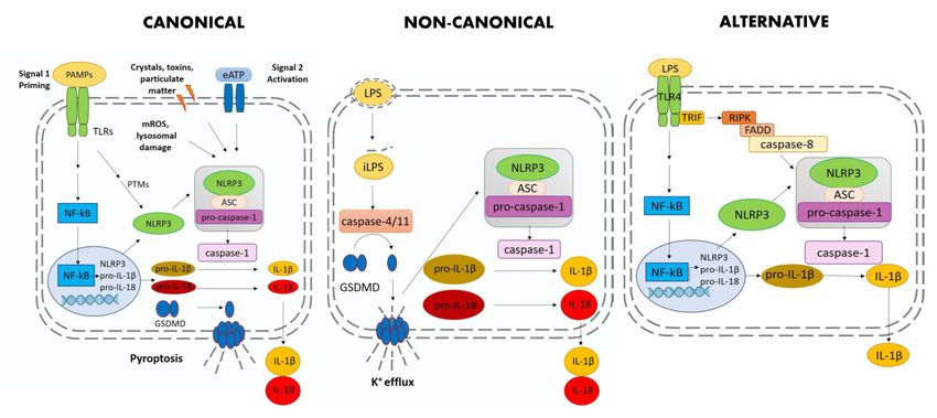
Threepathways
Three pathwaysofofNLRP3
NLRP3 inflammasome
inflammasome activation
activation have
have so been
so far far been described:
described: canonical,
canonical, non-
non-canonical
canonical and alternative
and alternative pathway
pathway (Figure
(Figure 1). 1).
Figure 1. Schematic representation of the mechanisms regulating inflammasome activation in
canonical,
Figure 1. non-canonical and alternative
Schematic representation of pathway. Abbreviations:
the mechanisms PAMP,inflammasome
regulating pathogen-associate molecular
activation in
pattern; TLRs,
canonical, toll-like receptors;
non-canonical NF-kB, nuclear
and alternative pathway.factor kappa-light-chain-enhancer
Abbreviations: of activated
PAMP, pathogen-associate
B cells; PTM,
molecular post-translational
pattern; TLRs, toll-likemodifications; eATP,nuclear
receptors; NF-kB, extracellular
factoradenosine triphosphate; mROS,
kappa-light-chain-enhancer of
mitochondrial
activated B cells;reactive oxygen species; ASC,
PTM, post-translational apoptosis-associated
modifications; speck-like
eATP, extracellular proteintriphosphate;
adenosine containing a
CARD; IL,
mROS, interleukin; GSDMD,
mitochondrial reactive gasdermin D; iLPS,ASC,
oxygen species; intracellular LPS; TRIF, TIR-domain-containing
apoptosis-associated speck-like protein
adapter-inducing interferon-β; RIPK, receptor-interacting serine/threonine-protein
containing a CARD; IL, interleukin; GSDMD, gasdermin D; iLPS, intracellular LPS; kinase 1; FADD,
TRIF, TIR-
Fas-associated protein
domain-containing with death domain;
adapter-inducing NLRP3, NLR
interferon-β; family
RIPK, pyrin domain containing
receptor-interacting 3.
serine/threonine-
protein kinase 1; FADD, Fas-associated protein with death domain; NLRP3, NLR family pyrin domain
Canonical activation is the classical two-step model where two signals are required for optimal
containing 3.
activation of the NLRP3 inflammasome. Signal 1, or priming, requires binding of toll-like receptors
(TLRs) with pathogen-associated
Canonical molecular
activation is the classical patterns
two-step (PAMPs)
model such
where two assignals
lipopolysaccharide
are required(LPS). Signal
for optimal
1 induces the
activation transcriptional
of the up-regulation
NLRP3 inflammasome. of NLRP3,
Signal pro-IL-1β,
1, or priming, and pro-IL-18
requires bindingvia Nuclear receptors
of toll-like Factor-kB
(NF-kB) activation [11,12]. Growing evidence indicates that Signal 1 promotes more
(TLRs) with pathogen-associated molecular patterns (PAMPs) such as lipopolysaccharide (LPS). than transcriptional
up-regulation,
Signal 1 inducesastheit induces a number
transcriptional of post-translational
up-regulation of NLRP3, modifications
pro-IL-1β, and(PTMs) that allow
pro-IL-18 NLRP3
via Nuclear
to switch into
Factor-kB its active
(NF-kB) conformation
activation [13]. Signal
[11,12]. Growing 2 is triggered
evidence bythat
indicates diverse stimuli
Signal including
1 promotes PAMPs,
more than
transcriptional up-regulation, as it induces a number of post-translational modifications (PTMs) thatInt. J. Mol. Sci. 2020, 21, 4294 3 of 19
DAMPs, and particulate matter which NLRP3 “senses” via yet undefined mechanisms. Signal 2 leads
to the formation of the active inflammasome complex and the auto-proteolytic cleavage of caspase-1.
A typical feature of the NLRP3 inflammasome is the ability to respond to a wide range of signals such as
extracellular adenosine triphosphate (ATP), microbial toxins, crystals, particulate matter, and viral
proteins. The exact molecular mechanism that triggers NLRP3 activation in response to such a diverse
set of signals is still under investigation. Many NLRP3 activators induce K+ efflux. The consequent
drop in intracellular K+ has been at first identified as a common trigger for NLRP3 inflammasome
activation [14,15]. However, growing evidence has shown that, along with K+ efflux, other mechanisms
may contribute to NLRP3 activation such as Cl− efflux, Ca2+ signaling, reactive oxygen species (ROS)
mitochondrial dysfunction, and lysosomal rupture [16,17]. Given the diversity of these signals, it is
likely that NLRP3 “senses” a common pathway induced in the cytosolic environment by intracellular
processes rather than directly interacting with all these molecules. In all cases, optimal activation of
NLRP3 inflammasome requires multiple PTMs such as de-sumoylation [18], de-ubiquitination [19,20],
phosphorylation [21,22], de-phosphorylation, acetylation [23], and alternative splicing [17,24]. Different
PTMs have been accurately described in the literature and are reviewed in [13,17].
Non-canonical activation is triggered by caspase-4 in humans and caspase-11 in mice and occurs
in response to intracellular infection by Gram-Negative bacteria (e.g., Escherichia coli) [25]. It has been
reported that caspase-11 [26] and caspase-4 [27] are activated by intracellular LPS through direct binding
of LPS with their CARD domain. Furthermore, it has been recently shown that other components of
Gram-negative bacteria, as well as exogenous drugs, can activate caspase-4 and caspase-11 [28,29].
Several works, nicely summarized in Yi, 2020 [30], have shown that the non-canonical pathway in
mice cooperates with the NLPR3 inflammasome in order to provide a robust inflammatory response.
In fact, caspase-11/4 activation mediated by iLPS can promote K+ efflux, either by GSDMD cleavage
and consequent pyroptosis or by currently unknown mechanisms leading to membrane rupture. As a
consequence of K+ efflux, NLRP3 inflammasome becomes activated [27,31].
Alternative inflammasome activation is a new species-specific NLRP3 inflammasome pathway
that was first reported in 2016. It exists in human and porcine peripheral blood mononuclear
cells (PBMCs), but it is absent in murine ones [32]. In this pathway, LPS per se is sufficient to
induce activation of the NLRP3 inflammasome with consequent activation of caspase-1 and IL-1β
processing and secretion. Inflammasome assembly occurs upon activation of the TLR4 by LPS
triggering the TIR-domain-containing adapter-inducing interferon-β (TRIF)—receptor-interacting
serine/threonine-protein kinase 1 (RIPK1)—Fas-associated protein with death domain (FADD) caspase-8
signaling cascade, which in turns leads to the activation of the NLRP3 inflammasome. This pathway is
not dependent on K+ efflux. No pyroptosis occurs, thus IL-1β is released gradually, as opposed to the
all-or-nothing response of the canonical activation [32].
1.2. Role of Domains
NLRP3 has a N-terminal effector pyrin domain (PYD), which interacts with ASC via PYD–PYD
interaction, a central NACHT domain carrying the ATPase activity, and a C-terminal leucine-rich
repeats (LRR) domain.
The NLRP3-PYD domain recruits ASC via PYD–PYD interaction and it is therefore required for
the formation of the active inflammasome complex [33]. It consists of a six-helical bundle structural
fold containing several conserved residues as compared to other PYD domains interacting with
ASC and with a possible homodimeric interface [34]. Due to its relevance for the activation of the
NLRP3 inflammasome, the PYD domain represents an attractive target for the development of NLRP3
inhibitors, as recently reported [35].
The central NACHT domain provides the ATPase activity that is required for NLRP3 activation
and inflammasome formation. The NACHT domain contains a walker A motif responsible for ATP
binding and a Walker B motif that is necessary for ATPase activity [36]. An intact and functional
NACHT domain is required for interaction with ASC, activation of caspase-1, and IL-1β release inInt. J. Mol. Sci. 2020, 21, 4294 4 of 19
THP-1 cells [37]. Of note, mutations of the NACHT domain are associated with spontaneous NLRP3
activation observed in CAPS [37]. Finally, it has recently been reported that the NACHT domain is
involved in NLPR3 activation in response to viral infection through its binding with viral DexD/H-box
helicase (DHX) proteins [38]. Current knowledge supports the hypothesis that the NACHT domain is
a primary druggable site for the development of selective inhibitors of NLRP3.
The LRR domain is evolutionarily conserved in several different proteins that serve as pattern
recognition receptors and typically harbors the sensing domain. Structurally, the LRR domain is a large
β-helical array with horseshoe or arc shape [36,39]. The role of NLRP3-LRR is still under investigation.
NLRP3-LRR has been proposed to be involved in auto-regulation, protein–protein interaction, and signal
sensing. LRR appears to be dispensable for canonical NLRP3 activation. In fact, a truncated
form of NLRP3 (residues 1–686, lacking the LRR domain) can be fully activated by the canonical
pathway, indicating that LRR is not necessary for sensing and assembling of the inflammasome [40].
Nonetheless, LRR domain is involved in the recognition of microbial ligands through direct binding.
For example, it has been reported that viral 3D RNA polymerase of Enterovirus 71 (EV71) associates
with LRR domain, forming a “3D-NLRP3-ASC” ring-like structure [41]. Very recently, it has been
shown that SARS-CoV open reading frame-8b (ORF-8b) binds the LRR domain and localizes with
NLRP3 and ASC in cytosolic dot-like structures, suggesting that this interaction is functionally relevant
for IL-1β release in response to the virus [42]. Finally, a possible inflammasome-independent function
for the LRR domain has been described referring to the binding of LRR to the transcription factor IRF4,
thus promoting the activity of CD4+ TH 2 cells via IL-4 transcription [43].
1.3. Inhibition of NLRP3 for the Treatment of Inflammatory Diseases
Growing pre-clinical evidence indicates that inhibition of NLRP3 displays therapeutics benefits in
several disease models while causing minimal impairment of host immune responses [10]. Along the
same line, a number of clinical studies have shown that agents blocking IL-1β are effective for the
treatment of several conditions including rheumatic diseases and autoinflammatory syndromes, and
decrease the incidence of atherosclerotic disease in at risk patients with an inflammatory signature [44].
However, blockade of IL-1β signaling is associated with an increase of infections and sepsis [45].
Therefore, therapies selectively targeting the NLRP3 inflammasome, rather than downstream cytokine
effectors, would display improved safety by preserving host immune defenses. Furthermore, they
would provide increased therapeutic potency due to the simultaneous inhibition of IL-1β, IL-18, and
pyroptosis. As a consequence, NLRP3 appears to be an ideal target for drug discovery. In recent
years, a wide range of molecules has been developed as NLRP3 inhibitors (reviewed in [46]). To date,
MCC950 is the most potent and selective inhibitor of NLRP3. MCC950 directly binds to NLRP3
NACHT domain affecting the ATPase function, leading to an inactive NLRP3 conformation [47–49].
Other NLRP3 inhibitors have been developed that directly target the NACHT domain, including
CY-09, Oridonin, Tranilast, OLT1177 and MNS [50–53]. Other compounds inhibit the activation of
the NLRP3 inflammasome via indirect mechanisms, either by blocking other components of the
inflammasome complex or by inhibiting the signaling cascade leading to NLRP3 activation [54–58].
Very recently, it has been reported that targeting the PYD domain may represent an effective strategy for
NLRP3 inhibition [35]. The efficacy of selective NLRP3 inhibitors for the treatment of NLRP3-related
inflammatory diseases, such as neurodegenerative diseases, gouty arthritis, CAPS and metabolic disease,
has been widely demonstrated in several preclinical models and ex-vivo systems [17,50,51,59,60].
However, despite great effort, none of the newly developed inhibitors of NLRP3 have so far been
approved by the Food and Drug Administration (FDA). Therefore, current research should aim either
at the improvement of the pharmacokinetic properties of the available molecules or at the discovery of
novel classes of NLRP3 inhibitors, to reach a selective, potent and cost-effective remedy for a wide
range of human diseases.Int. J. Mol. Sci. 2020, 21, 4294 5 of 19
2. Cell Models to Study NLRP3 Inflammasome Biology
In this section, we will highlight the main cell models that are currently in place for inflammasome
studies as summarized in Table 1. In addition, we will bring up new cell models that are currently
under development and that may be useful for the creation of novel ex-vivo models of NLRP3-driven
diseases. Finally, we will discuss about potential pitfalls and challenges when it comes to the choice
of the proper model. Although inflammasomes are expressed both in myeloid and non-myeloid cell
types, myeloid-derived cells and in particular macrophages are the cell type expressing the highest
levels of NLRP3 and releasing the highest amount of cytokines. Therefore, these are normally used as
model system for the study of inflammasome activation.
Table 1. Cell models used in NLRP3 inflammasome research.
Cell Model Description Source Example of Applications References
Primary Canonical and
BMDMs bone-marrow-derived Mouse non-canonical activation; [1,14,15,18,19,22,24,26,54,61–64]
macrophages (wt and KO) identification of inhibitors
Immortalized primary Canonical and
iBMDM bone-marrow derived Mouse non-canonical activation; [1,18,21,22,40,49,52,65–70]
macrophages (wt and KO) identification of inhibitors
Raw264.7 Macrophage-like cell line Mouse Priming events studies [61,71–73]
Monocyte/macrophage cell Canonical activation;
J774A.1 Mouse [53,69–71,74–79]
line identification of inhibitors
Primary human Canonical and
hMDMs monocyte-derived Human non-canonical activation; [11,12,22,24,48,52,54,80–85]
macrophages identification of inhibitors
Monocyte-like cell line, Canonical and
THP-1 (from Acute Monocytic Human non-canonical activation; [22,37,42,52,86–94]
Leukemia) identification of inhibitors
Monocyte-like cell line (from
Canonical activation;
U937 pro-monocytic Myeloid Human [22,53,95]
identification of inhibitors
Leukemia)
Human
BlaER1 monocytes/macrophages Human Alternative activation [32,86,96–98]
(from immortalized B cells)
HEK293T Human embryonic cell line Human Mechanistic studies [22,37,40,42,48,49,52,99–103]
Human auto-inflammatory
iPS-DM iPS-derived macrophages Human [86,104–111]
disease studies
2.1. Murine Cell Models
2.1.1. Primary Murine Bone-Marrow-Derived Macrophages (BMDMs)
Primary murine bone-marrow-derived macrophages (BMDMs) have been a very useful tool
for the study of NLRP3 inflammasome biology. Established protocols exist to obtain BMDMs
from femurs and tibia of C57BL/6 mice. An advantage of using BMDMs is represented by the
fact that knock-out BMDMs can be obtained from mice genetically deficient in inflammasome
components such as Caspase 1-/- , ASC-/- , and NLRP3-/- [14,26,61]. For this reason, BMDMs have
been of primary importance for the elucidation of the molecular mechanisms underlying NLRP3
inflammasome activation [1,14,15,18,19,22,24,26,54]. For example, BMDMs have been used to define
the precise signals that activate canonical NLRP3 inflammasome pathway, including K+ efflux and Ca2+
mobilization [15,62]. Furthermore, BMDMs were used to evaluate NLRP3 activation in non-canonical
inflammasome pathway to demonstrate that caspase-11 plays a role in modulation of K+ efflux and to
prove that GSDMD is essential for caspase-11-dependent pyroptosis and for IL-1β maturation [26,63,64].
Given the high costs of animal housing and breeding, and considering that it is always better to
work with fresh primary cells, in the past few years, several protocols for BMDM immortalization
have been developed [65–67]. Briefly, the approach uses the Cre-J2 retroviral method of infection usingInt. J. Mol. Sci. 2020, 21, 4294 6 of 19
a J2 retrovirus carrying v-raf and v-myc oncogenes. This method, developed in the 1980s and recently
improved, allows the generation of iBMDMs phenotypically comparable to their primary counterparts,
displaying many of the trademark functions of macrophages [68]. iBMDMs have been widely used to
study NLRP3 PTMs and identify NLRP3 inhibitors [1,18,21,22,40,49,52,69,70].
2.1.2. Murine Macrophage Cell Lines
Currently, two mouse cell lines are mostly used for the study of NLRP3 inflammasome
biology—RAW264.7 and J774A.1. They are both macrophage-like adherent cell lines, very easy
to grow and manipulate, and commonly used to study the innate immune responses. Despite their
use in inflammasome research, they differ for a very important molecular aspect: RAW264.7 cells do
not express the ASC protein, while J774A.1 cells do express it [61,71]. As a consequence, RAW264.7
cells can be used to study the transcriptional events that regulate priming, but not the downstream
cascade, as they cannot activate the NLRP3 inflammasome complex. In addition, for this specific
feature, the RAW264.7 cell line has been used to identify a novel mechanism of NLRP3-independent
bacterial killing mediated by K+ efflux [72]. Interestingly, stable transfection of RAW264.7 cells with
plasmids containing the full length sequence for ASC can restore the whole NLRP3 inflammasome
machinery [73].
J774A.1 is a macrophage-like cell line able to form a complete functional NLRP3 inflammasome
system [71]. These cells represent a reliable model to study inflammasome biology as they are easy to
manipulate and grow [69,70,74]. For example, J774A.1 cells have been used by Yaron and collaborators
to demonstrate that K+ efflux is upstream of Ca2+ influx in the production of mtROS, thus beginning
the cascade of events leading to NLRP3 inflammasome activation [75]. J774A.1 cells have been widely
used to test the efficacy of potentially novel inhibitors of NLRP3 [53,76–78]. For instance, Hu et al.
nicely demonstrated that the antimicrobial cathelicidin peptide LL-37 inhibits LPS/ATP-mediated
pyroptosis, thus providing new insights into modulation of sepsis [79].
2.2. Human Cell Models
While the murine cell models set the ground primarily for the characterization of the molecular
mechanisms regulating NLPR3 inflammasome activation, the use of human models has been necessary
to define the mechanisms of NLRP3-driven human diseases.
2.2.1. Human Monocyte-Derived Macrophages (hMDMs)
Human monocytes can be obtained from freshly isolated PBMCs according to established
protocols [12,80] and differentiated into human monocyte-derived macrophages (hMDMs). To this
purpose, different approaches have been developed where different stimuli can be used in order to
obtain a specific macrophage polarization [12,81,82]. For example, M-CSF-differentiated hMDMs
treated with LPS/IFNγ or IL-4, become polarized toward the M1 or M2 phenotype, respectively [83,84].
The high plasticity of this cellular model can be used to recapitulate the different aspects of the
immune response in vitro. hMDMs were used primarily to study NLRP3 canonical activation and
inhibition [11,22,24,48,52,54] and became crucial for the identification of the non-canonical pathway
of NLRP3 inflammasome activation mediated by caspase-4 [85]. hMDMs can be cultured for two to
three weeks in vitro for experimental purposes. It is always recommended to work with fresh hMDMs.
Nevertheless, they can be frozen in specific freezing culture for long-term storage.
2.2.2. Human Monocyte/Macrophage Cell Lines
Considering the difficulties to obtain primary monocytes, given the high variability among
donors and the fact that they are not amenable to genetic manipulation, the use of hMDMs has
been limited in time. In the last few decades, a variety of human cell lines have been tested for
their potential capability to activate NLRP3 inflammasome. Among others, the THP-1 and the U937
cell lines have been mostly used. The THP-1 monocyte-like cell line, derived from acute monocyticInt. J. Mol. Sci. 2020, 21, 4294 7 of 19
leukemia, has been extensively used in the field, despite its tumoral derivation and consequent
genomic instability [86]. This cell line can be differentiated into macrophages by treatment with
phorbol-12-myristate-13-acetate (PMA) [87,88]. Differentiated THP-1 cells display several features
of primary hMDMs, as shown by macrophage marker expression, morphology, phagocytic activity,
and cytokine release. When used for the study of inflammasome activation, THP-1 cells are usually
differentiated with PMA into macrophages for a time ranging of 24–72h, primed with LPS, and then
subjected to different second signals necessary for NLRP3 inflammasome activation [22,37,42,52].
For instance, Petrilli and collaborators in 2007 treated THP-1-derived macrophages with ionophores
such as nigericin, gramicidin and valinomycin to demonstrate the key role of K+ efflux in triggering
NLRP3 inflammasome activation [89]. THP-1 cells were also used to demonstrate that DAMPs and
exogenous signals, including monosodium urate (MSU) crystals, asbestos, silica, and mitochondrial
ROS, activate the NLRP3 inflammasome [90–92]. Furthermore, Iyer et al. used THP-1 cells to
demonstrate that the treatment with the antibiotic Linezolid led to mitochondrial disruption, cardiolipin
release and NLRP3 inflammasome activation in a ROS-independent fashion [93]. THP-1 cells can also
be easily manipulated in vitro. Stable THP-1 knock-out cell lines targeting specific genes of the NLRP3
inflammasome cascade have been developed [27,94]. This has been instrumental for demonstrating
that not only non-canonical NLRP3 inflammasome activation is mediated by caspase-4-mediated LPS
intracellular sensing, but also that caspase-1 and -4 cleaves GSDMD, thus leading to pore formation
and pyroptosis.
The U937 cell line is a pro-monocytic myeloid leukemia cell line that, similarly to THP-1 cells,
has been used for inflammasome studies. U937 cells can be differentiated into macrophages with PMA,
and inflammasome activation can be achieved by LPS stimulation followed by different second signals.
U937-derived macrophages display similarities with hMDMs; thus, they have been used to study
mechanisms of inflammasome activation and for the identification of novel NLRP3 inhibitors [22,53,95].
2.2.3. BlaER1 Human Cell Model
THP-1 cells do not fully recapitulate the behavior of primary human monocytes, as they totally or
partially lack several signaling cascades that are present in primary immune cells and are characterized
by karyotypic abnormalities [86,96]. Thus, to fill the gap between cell lines and primary human
myeloid cells, a new human cell model has been established [32,97]. This human cell model,
called BlaER1, employed the stable expression of C/EBPα transcription factor in immortalized
immune B cells [97,98]. Activation of C/EBPα induces trans-differentiation, causing BlaER1 cells
to switch from their proliferative B-cell stage to a post-mitotic, monocytic status, in which they
become moderately adherent, highly phagocytic, and competent for multiple innate immune signaling
pathways. BlaER1 cells have been instrumental for the discovery of the alternative pathway of NLRP3
inflammasome activation mediated by the TLR4/TRIF/caspase-8 axis.
2.2.4. HEK293T Cell Line
A very useful tool for in vitro studies of the molecular mechanisms of inflammasome activation
is represented by the reconstitution of the NLRP3 inflammasome into the HEK293T human cell
line. This cell line can be easily transfected, as it is commonly used for protein expression and
production of recombinant retro/lentiviruses. Several research groups have used HEK293T cells
to study NLRP3 inflammasome biology [37,40,42,48,49,52]. These cells do not express any of the
inflammasome-related proteins, so it is necessary to transfect them with specific plasmids or retroviral
constructs carrying the gene of interest in order to express the proteins to study. HEK293T cells have
been used to reconstitute the entire NLRP3 mouse inflammasome system [99,100]. Different studies
used HEK293T cells to identify NLRP3 post-translational modifications occurring during inflammasome
activation and to study protein–protein interaction by evaluating co-localization or by performing
co-immunoprecipitation assays [52,59,101]. Among others, Song et al. used transfected HEK293T
cells to demonstrate that NLRP3 phosphorylation mediated by JNK1 is an essential priming event forInt. J. Mol. Sci. 2020, 21, 4294 8 of 19
inflammasome activation [22]. Wang et al. recently utilized HEK293T cells to prove that the stimulator
of interferon genes (STING) binds to NLRP3 thus mediating its localization into the ER and determining
its de-ubiquitination required for inflammasome activation [102]. Finally, Mao et al. used HEK293T
cells to demonstrate that Bruton tyrosine kinase (BTK) binds to NLRP3 to regulate its activation,
therefore suggesting that BTK deficiency is associated with several inflammatory NLRP3-mediated
diseases [103].
2.2.5. Induced Pluripotent Stem-Cells-Derived Macrophages (iPS-DM)
Despite the advances in cell models for the study of NLRP3 inflammasome biology, we still lack the
“perfect” system able to recapitulate the features of primary macrophages while at the same time being
able to replicate and being amenable to genetic manipulation. For example, with the tools we have in
place, it is difficult to genetically manipulate primary hMDMs and, on the other side, the use of THP-1
cell line is limited, among others, by the fact that they are karyotypically abnormal [86,104]. For these
reasons, several research groups have begun to establish macrophages from induced-pluripotent stem
cells (iPS). Currently, different protocols have been tested to efficiently reprogram iPS into mature
macrophages, and some of them nicely demonstrated how the iPS-DM showed similarities with
hMDMs, including morphology, expression of surface markers, transcriptional and cytokine release
profiles, and functional abilities, such as phagocytosis [105–110]. These studies opened the possibility
of using iPS-DMs derived from healthy subjects as well as from patients for drug discovery purposes.
For instance, in 2012, Tanaka et al. obtained iPS-DMs from patients affected by chronic infantile
neurologic cutaneous and articular syndrome (CINCA), an IL-1β-driven inflammatory disease caused
mainly by NLRP3 mutations leading to its constitutive activation. This study clearly showed the
impact of NLRP3 mutation on the development of the pathology, and began to define new potential
therapeutic approaches for this type of disease [111].
Despite the attempts and the different protocols in place to obtain iPS-DMs, few limitations need
to be considered, including the differentiation efficiency (still far from acceptable) and the choice of the
target genes to study (their expression or behavior have to be similar to hMDMs).
2.3. The Importance of Choosing the Right Model
Considering the abundance of cellular models available and their diversity (Table 1), the choice
of the right tool becomes extremely important. As mentioned, mouse models represent a very
useful tool to understand inflammasome biology, and murine cell lines are diverse regarding the
expression of NLRP3 inflammasome components. Therefore, it is very important to use the right
model according to the questions to address. Even when human cell models are used, the choice has
to be well thought as for example alternative NLRP3 activation up to now has been reported only in
BlaER1 cells. Finally, while some mouse models recapitulate key features of NLRP3-related human
disease (i.e., CAPS), it is always recommended, when possible, to compare the data obtained with
human in vitro/ex-vivo models or, even better, with samples derived from patients. In that sense,
the establishment of iPS-DMs models will be extremely useful for in vitro studies of NLRP3-related
auto-inflammatory diseases.
3. Cellular, Biochemical, and Biophysical Assays to Evaluate Activation and Function of the
NLRP3 Inflammasome
Activation of the NLRP3 inflammasome is a complex event regulated at multiple levels. It requires
the formation of a multimeric protein complex and leads to a cascade of events that can be monitored
by evaluating multiple read-out. Over the years, several assays have been developed that allowed to
study NLRP3 inflammasome biology (Table 2). In this section, we extensively review the different types
of assays that are currently used. Whenever cell-based assays are in place, independent of whether
murine or human macrophages, the canonical two-step model of activation is normally applied to
activate the NLRP3 inflammasome. Briefly, cells are stimulated with LPS (usually for 3–5 h) to induceInt. J. Mol. Sci. 2020, 21, 4294 9 of 19
the priming followed by treatment with nigericin or extracellular ATP (for 1–2 h) to license the active
NLRP3 inflammasome complex. Of note, if the experiments are aimed to assess the actions of selective
NLRP3 inhibitors, these are generally added after the priming step and before the second stimulation
with ATP or nigericin.
3.1. Immuno-Based Assays
Immune assays are based on the utilization of antibodies that specifically recognize and bind to a
given protein of interest. Recognition of a target protein by a specific antibody is exploited in large
number of assays; Enzyme-Linked Immunosorbent Assay (ELISA) is one of the most widely used assays
that allows the user to monitor soluble proteins and cytokine release in cell supernatants. When applied
to IL-1β and IL-18 quantification, this approach involves the use of antibodies that recognize the mature
form of the cytokines. ELISA assay provides quantitative information on the concentration of cytokine
present in cell supernatants. However, it is a long and multi-step procedure. Recently, different
homogeneous assays have been developed for quantitative detection of soluble proteins, including
IL-1β and IL-18, in a very rapid and sensitive procedure amenable to miniaturization [112,113]. They are
based on time-resolved fluorescence resonance energy transfer (TR-FRET), as is the case of HTRF and
Lance technologies (provided by PerkinElmer), and on the more sensitive bead-based luminescent
amplification assay, as is the case of AlphaLISA (provided by PerkinElmer) [114,115]. Western blot on
supernatant precipitates is the preferred approach to evaluate the processing of pro-IL-1β, pro-IL-18,
GSDMD, and caspases. In this respect, the choice of antibodies will determine which one among the
pro-form and the cleaved forms can be observed. Despite providing unique information regarding the
extent of protein cleavage and the different subunits generated upon processing, this techniques is
qualitative, as opposed to ELISA, TR-FRET-, and AlphaLISA-based assays [14,116,117]. Detection of
ASC oligomers is very often used to assess NLRP3 activation. Formation of ASC oligomers reflects
inflammasome activation. By applying an established cross-linking protocol followed by western
blot it is possible to detect and discriminate between ASC monomers or oligomers using a specific
anti-ASC antibody [52,118,119]. Another powerful approach to observe ASC oligomerization is the
detection of ASC specks, which reflect massive activation of the NLRP3 inflammasome that precedes
pyroptosis [22,116,120,121]. Traditional immunofluorescence staining techniques have been used to
detect ASC specks using a specific anti-ASC antibody. As an alternative, human and murine cell
lines have been developed that stably express a construct containing ASC-mCerulean, ASC-mCherry,
or ASC fused with other fluorescent tags [122–124]. By using these cell lines it is possible to monitor
the formation of ASC specks in live mode, with no need for staining [59]. Very recently, a new flow
cytometry-based approach has been reported for the detection of ASC specks in activated human
PBMCs [125]. Briefly, PBMCs are stained for ASC and CD14 (monocyte marker) and analyzed by flow
cytometry. The analysis consists of gating ASC+ cells in the monocyte population and analyzing the
distribution of ASC-FITC width vs ASC-FITC area in CD14+ monocytes in order to determine and
quantify the percentage of ASC specking monocytes. This method can also be applied in other cell
types including THP-1, J774A.1, and BMDMs [125]. Immunofluorescence can be used to study protein
expression and localization. For example, it has been reported that NLRP3 localization on mitochondria
membranes under certain circumstances is required for optimal inflammasome activation [91,102,126].
Co-immunoprecipitation (Co-IP) is a technique used to study protein–protein interactions. It can
be performed on endogenous proteins or in recombinant systems, such as HEK293T, where the proteins
of interest are co-transfected. Co-IP assays use antibodies specific to a target protein to indirectly
capture proteins that are bound to the target one. Pull-down of antibody-bound proteins is normally
performed using agarose or magnetic beads. Further downstream analysis, such as Western Blot,
is usually used in order to check whether the protein of interest has been pulled down together with the
target protein [127]. Furthermore, Co-IP experiments are used to assess the effect of a given molecule
on protein-protein interaction. For example, Co-IP experiments in HEK293T cells have been performed
to assess the impact of CY-09 and MCC950 on NLRP3-NLRP3 and NLRP3-ASC interaction [52,59].Int. J. Mol. Sci. 2020, 21, 4294 10 of 19
3.2. Probe-Based Assays
Probe-based assays include different approaches that use colorimetric, fluorimetric, or luminescent
probes to directly monitor a specific event in cell-based or cell-free assays. For instance, fluorescent
carboxyfluorescein-labeled inhibitor of caspases (FAM-FLICA) probes are widely used to detect
active caspases in cells using a fluorescence plate reader, flow-cytometry or fluorescence microscopy.
Briefly, FAM-FLICA reagents are cell permeable and irreversibly react with active caspases inside
the cell, releasing a fluorescent signal only when bound to caspases. Specificity is conferred by a
tetra-peptide incorporated in the substrate that can be recognized by a given caspase. For example,
the FLICA reagent FAM-YVAD-FMK specifically detects active caspase-1. Upon covalent reaction with
active caspase-1, the fluorescent probe is retained within the cell, while any unbound FAM-YVAD-FMK
diffuses out of the cell and it is washed away. The remaining green fluorescent signal is a direct measure
of active caspase-1 present inside the cell [12,128].
Recently, a bioluminescence assay has been developed to monitor caspase enzymatic activity
in cell-free extracts or cell supernatants [129]. As for the FAM-FLICA probes, specificity for a given
caspase is conferred by the peptide sequence of the bioluminescent substrate. Assay specificity can
be further enhanced by the addition of proper controls [12]. For example, to measure the activity of
caspase-1, the substrate Z-WEHD-aminoluciferin, which incorporates the optimal caspase-1 recognition
tetrapeptide, has been created. Active caspase-1 cleaves the substrate leading to aminoluciferin release,
thus resulting in the luciferase reaction and light production that can be measured with a luminescence
microplate reader [129].
3.3. Cell Death Assays
As above mentioned, massive activation of the NLRP3 inflammasome leads to pyroptosis,
a mechanism of cell death different from the well-known apoptosis. Pyroptosis is triggered by the
formation of GSDMD pores on the cell membrane and the consequent release of cytosolic proteins.
Quantification of extracellular lactate dehydrogenase (LDH) release is a typical read-out commonly
used for evaluating any form of cell death associated with rupture of cell membrane [116,130].
Several commercial kits are available that allow the quantification of extracellular LDH using
luminescent, fluorescent, or colorimetric readout. The procedure is normally very quick and can be
performed in two steps. Due to its simplicity and effectiveness, this assay has been widely used to test
the efficacy of selective NLRP3 inhibitors [59,116,120,130].
3.4. Surface Plasmon Resonance (SPR)
Surface plasmon resonance (SPR) is often used to determine the dissociation constant (“binding
constant”, KD ) between a protein and its ligand. Normally, a bait ligand is immobilized on a sensor chip.
Through a microfluidic system, a solution with the prey analyte is injected on the bait layer. From the
association (“on rate,” ka ) and dissociation rates (“off rate” kd ) of the bait ligand and the prey analyte, it is
possible to calculate the equilibrium dissociation constant (“binding constant” KD ) as a ratio. By using
this approach Hu and collaborators showed that, in Muckle–Wells Syndrome, the NLRP3-D31V
mutation enhances the binding of NLRP3 with ASC resulting in an over-production of IL-1β and
excessive immune responses including periodic fever, arthralgia and occasional conjunctivitis [131].
Using the same approach, Lee et al. demonstrated that caffeic acid phenethyl ester (CAPE) can
bind directly ASC, resulting in blockade of NLRP3-ASC interaction induced by MSU crystals [132].
Finally, Coll et al. found, by SPR analysis, that MCC950 directly interacts with the Walker B motif
within the NLRP3 NACHT domain, thereby blocking ATP hydrolysis and inhibiting NLRP3 activation
and inflammasome formation [48].Int. J. Mol. Sci. 2020, 21, 4294 11 of 19
Table 2. Assays and their applications in inflammasome biology.
What Can be Quantitative
Type of Assay Sample Type Readout Refs
Detected (Y/N)
ELISA Supernatants Y Absorbance [14,116,117]
Soluble proteins TR-FRET-based assays Supernatants Y Fluorescence [112,114]
(i.e., IL-1β, IL-18) AlphaLISA-based
Supernatants Y Luminescence [113,115]
assays
Chemi/
Western blot Cell Lysates N [14,52,118,119]
Fluorescence
Protein expression
Living/fixed cells or
(Immuno)fluorescence N Fluorescence [22,59,102,116,120–125]
Tissue
Protein processing Chemi/
Western blot Supernatants N [12,116,117]
(i.e., pro- IL-1β, Fluorescence
pro-caspase-1) BRET-based probes Living cells Y Bioluminescence [69,70]
Chemi/
ASC Oligomers Western blot Cell Lysates N [50,118,119]
Fluorescence
Living/fixed cells or
(Immuno)fluorescence Y* Fluorescence [22,116,120,121]
ASC Specks Tissue
Flow cytometry Living/fixed cells Y Fluorescence [125]
Fluorescence Living/fixed cells or
N Fluorescence [12]
Active caspases (probe-based) Tissue
Flow cytometry Living/fixed cells Y Fluorescence [128]
Cell free
Enzymatic assay
Caspase activity extracts/supernatants/ Y Luminescence [12,129]
(probe-based)
recombinant enzyme
Response/
Cell-free extract/
SPR Y Resonance [48,131,132]
Recombinant proteins
Protein-protein Units
Interaction Chemi/
Co-IP Cell Lysates N [52,59,127]
Fluorescence
BRET-based probes Living cells Y Luminescence [47,133]
Conformational
BRET-based probes Living cells Y Luminescence [133]
changes
Cell death LDH release Supernatants Y Absorbance [59,116,120,130]
* when ASC specks are expressed as % of total counted cells. Abbreviations: ELISA, enzyme-linked immunosorbent
assay; TR-FRET, time-resolved fluorescence resonance energy transfer; IL-1β, Interleukin-1 beta; IL-18, Interleukin-18;
BRET, bioluminescence resonance energy transfer; ASC, apoptosis-associated speck-like protein containing a CARD;
SPR, surface plasmon resonance; Co-IP, co-immunoprecipitation; LDH, lactate dehydrogenase.
3.5. Bioluminescence Resonance Energy Transfer (BRET)-Based Assays
Bioluminescence resonance energy transfer (BRET) technology allows the user to monitor
protein–protein interaction, protein cleavage, and conformational changes in living cells and in
cell-free systems. In the context of inflammasome research, several examples of BRET-based assays
have been reported. For example, a BRET-based approach was proposed for the study of the interaction
between NLRP3 proteins in living cells [133]. Another example reported by Pelegrin et al. developed
a BRET-based biosensor to monitor pro-IL-1β processing in living cells using a plate reader or a
microscope. Specifically, in this example, pro- IL-1β was fused at its terminals with a donor (Rluc8)
and an acceptor (Venus). The proximity of the two molecules leads to energy transfer for the detection
of the protein. However, when pro-IL-1β is cleaved, donor and acceptor become distant and the
BRET signal is reduced [69,70]. More recently, BRET-based approaches have been used to study the
molecular conformation of NLRP3. For example, Hafner-Bratkovic et al. reported a construct were
the donor (luciferase) and the acceptor (YFP fluorescent protein) probes were added intramolecularly
(N-terminus and C-terminus of the same NLRP3 protein) to study NLRP3 change of conformation
upon activation and intermolecularly (donor and acceptor probes in different NLRP3 proteins) to
follow NLRP3 oligomerization upon different stimuli [40]. Similarly, Tapia-Abellan et al. used aInt. J. Mol. Sci. 2020, 21, 4294 12 of 19
similar approach to demonstrate that the MCC950 inhibits NLRP3 activation by closing the active
conformation into an inactive one [47].
4. Concluding Remarks
Recent advances in inflammasome research have provided new information on host defense
mechanisms and unveiled a key role for NLRP3 in the development of several chronic-inflammatory
and age-related diseases. The growing interest on NLRP3 biology calls for a parallel increase in the
development of methods that are necessary for molecular and biochemical studies. As herein
reported, several established cellular models are available and used throughout the world to
investigate inflammasome biology. However, they cannot always recapitulate the behavior of diseased
macrophages in inflammatory NLRP3-mediated pathologies. For this reason, major efforts have to be
made toward the establishment of patient-derived iPS-DM in vitro. The development of these models
will speed up the process of defining new patient-specific therapies for NLRP3-related disease.
Furthermore, several established cellular, biochemical, and biophysical assays exist for the study
of NLRP3 inflammasome biology and continuous efforts are done toward the development of novel,
quantitative, and specific approaches. An area that will require special attention in the near future will
be the study of NLRP3 from a biochemical and structural point of view. In fact, a high-resolution 3D
structure of NLRP3 is still missing, and the production of the recombinant NLRP3 protein still poses
technical challenges. Therefore, future research efforts should be directed toward filling these gaps.
The knowledge that will be generated will surely represent a breakthrough for the study of NLRP3
biology and the development of selective drugs.
Author Contributions: Conceptualization, G.Z. and C.C.; Writing—original draft preparation, G.Z., M.B., M.C.,
and P.D.; Writing—review and editing, G.Z., F.B., and C.C.; Visualization, M.C. and C.C.; Supervision, C.C.;
Funding Acquisition, C.C. All authors have read and agreed to the published version of the manuscript.
Funding: This work was supported by the Sicilian Region under the Programme Cohesion Development Fund
2014/2020-Grant “Patto per il Sud-Project CheMiST-CUP G77B17000110001 and by Fondazione Ri.MED.
Conflicts of Interest: The authors declare no conflict of interest.
Abbreviations
ASC Apoptosis-associated speck-like protein containing a CARD
BRET Bioluminescence resonance energy transfer
CoIP Co-immunoprecipitation
eATP Extracellular adenosine tri-phosphate
ELISA Enzyme-linked immunosorbent assay
FADD Fas-associated protein with Death Domain
GSDMD Gasdermin D
IL Interleukin
iLPS Intracellular LPS
iPS Induced pluripotent Stem cells
KO Knock-out
LDH Lactate dehydrogenase
mROS Mitochondrial reactive oxygen species
NFkB Nuclear factor kappa-light chain enhancer of activated B cells
NLRP3 NLR family pyrin domain containing 3
PAMPI Pathogen-associated molecular pattern
PTM Post-translational modifications
RIPK Receptor-interacting seriine-threonine-protein kinase 1
SRP Surface plasmon resonance
TLR Toll-like receptor
TR-FRET Time-resolved fluorescence resonance energy transfer
TRIF TIR domain-containing adapter inducing interferon-β
Wt Wild-typeInt. J. Mol. Sci. 2020, 21, 4294 13 of 19
References
1. He, Y.; Zeng, M.Y.; Yang, D.; Motro, B.; Núñez, G. NEK7 is an essential mediator of NLRP3 activation
downstream of potassium efflux. Nature 2016, 530, 354–357. [CrossRef]
2. Quartier, P.; Rodrigues, F.; Georgin-Lavialle, S. Cryopyrin-Associated periodic syndromes. Rev. Med. Interne
2018, 39, 287–296. [CrossRef] [PubMed]
3. Heneka, M.T.; Kummer, M.P.; Stutz, A.; Delekate, A.; Schwartz, S.; Vieira-Saecker, A.; Griep, A.; Axt, D.;
Remus, A.; Tzeng, T.C.; et al. NLRP3 is activated in Alzheimer’s disease and contributes to pathology in
APP/PS1 mice. Nature 2013, 493, 674–678. [CrossRef] [PubMed]
4. Wang, X.; Chi, J.; Huang, D.; Ding, L.; Zhao, X.; Jiang, L.; Yu, Y.; Gao, F. Alpha-Synuclein promotes progression
of Parkinson’s disease by upregulating autophagy signaling pathway to activate NLRP3 inflammasome.
Exp. Med 2020, 19, 931–938.
5. Olcum, M.; Tastan, B.; Kiser, C.; Genc, S.; Genc, K. Microglial NLRP3 inflammasome activation in multiple
sclerosis. Adv. Protein Chem. Struct. Biol. 2020, 119, 247–308. [PubMed]
6. Jin, Y.; Fu, J. Novel insights INTO the NLRP 3 inflammasome in atherosclerosis. J. Am. Heart Assoc. 2019, 8,
e012219. [CrossRef] [PubMed]
7. Pirzada, R.H.; Javaid, N.; Choi, S. The roles of the NLRP3 inflammasome in neurodegenerative and metabolic
diseases and in relevant advanced therapeutic interventions. Genes 2020, 11, 131. [CrossRef]
8. McAllister, M.J.; Chemaly, M.; Eakin, A.J.; Gibson, D.S.; McGilligan, V.E. NLRP3 as a potentially novel
biomarker for the management of osteoarthritis. Osteoarthr. Cart. 2018, 26, 612–619. [CrossRef]
9. Guo, C.; Fu, R.; Wang, S.; Huang, Y.; Li, X.; Zhou, M.; Zhao, J.; Yang, N. NLRP3 inflammasome activation
contributes to the pathogenesis of rheumatoid arthritis. Clin. Exp. Immunol. 2018, 194, 231–243. [CrossRef]
10. Coll, R.C.; O’Neill, L.; Schroder, K. Questions and controversies in innate immune research: What is the
physiological role of NLRP3? Cell Death Discov. 2016, 2, 16019. [CrossRef]
11. Bauernfeind, F.G.; Horvath, G.; Stutz, A.; Alnemri, E.S.; MacDonald, K.; Speert, D.; Fernandes-Alnemri, T.;
Wu, J.; Monks, B.G.; Fitzgerald, K.A.; et al. Cutting edge: NF-kappaB activating pattern recognition and
cytokine receptors license NLRP3 inflammasome activation by regulating NLRP3 expression. J. Immunol.
2009, 183, 787–791. [CrossRef]
12. Buscetta, M.; Di Vincenzo, S.; Miele, M.; Badami, E.; Pace, E.; Cipollina, C. Cigarette smoke inhibits the NLRP3
inflammasome and leads to Caspase-1 activation via the TLR4-TRIF-Caspase-8 axis in human macrophages.
FASEB J. 2020, 34, 1819–1832. [CrossRef] [PubMed]
13. Yang, J.; Liu, Z.; Xiao, T.S. Post-Translational regulation of inflammasomes. Cell Mol. Immunol. 2017, 14,
65–79. [CrossRef] [PubMed]
14. Mariathasan, S.; Weiss, D.S.; Newton, K.; McBride, J.; O’Rourke, K.; Roose-Girma, M.; Lee, W.P.; Weinrauch, Y.;
Monack, D.M.; Dixit, V.M. Cryopyrin activates the inflammasome in response to toxins and ATP. Nature
2006, 440, 228–232. [CrossRef] [PubMed]
15. Munoz-Planillo, R.; Kuffa, P.; Martinez-Colon, G.; Smith, B.L.; Rajendiran, T.M.; Nunez, G. K(+) efflux is the
common trigger of NLRP3 inflammasome activation by bacterial toxins and particulate matter. Immunity
2013, 38, 1142–1153. [CrossRef]
16. Mangan, M.S.J.; Olhava, E.J.; Roush, W.R.; Seidel, H.M.; Glick, G.D.; Latz, E. Targeting the NLRP3
inflammasome in inflammatory diseases. Nat. Rev. Drug Discov. 2018, 17, 688. [CrossRef] [PubMed]
17. Yang, Y.; Wang, H.; Kouadir, M.; Song, H.; Shi, F. Recent advances in the mechanisms of NLRP3 inflammasome
activation and its inhibitors. Cell Death Discov. 2019, 10, 128. [CrossRef] [PubMed]
18. Barry, R.; John, S.W.; Liccardi, G.; Tenev, T.; Jaco, I.; Chen, C.H.; Choi, J.; Kasperkiewicz, P.;
Fernandes-Alnemri, T.; Alnemri, E.; et al. SUMO-Mediated regulation of NLRP3 modulates inflammasome
activity. Nat. Commun. 2018, 9, 3001. [CrossRef]
19. Kawashima, A.; Karasawa, T.; Tago, K.; Kimura, H.; Kamata, R.; Usui-Kawanishi, F.; Watanabe, S.; Ohta, S.;
Funakoshi-Tago, M.; Yanagisawa, K.; et al. ARIH2 ubiquitinates NLRP3 and negatively regulates NLRP3
Inflammasome activation in macrophages. J. Immunol. 2017, 199, 3614–3622. [CrossRef]
20. Lopez-Castejon, G. Control of the inflammasome by the ubiquitin system. FEBS J. 2020, 287, 11–26. [CrossRef]
21. Stutz, A.; Kolbe, C.C.; Stahl, R.; Horvath, G.L.; Franklin, B.S.; van Ray, O.; Brinkschulte, R.; Geyer, M.;
Meissner, F.; Latz, E. NLRP3 inflammasome assembly is regulated by phosphorylation of the pyrin domain.
J. Exp. Med. 2017, 214, 1725–1736. [CrossRef] [PubMed]Int. J. Mol. Sci. 2020, 21, 4294 14 of 19
22. Song, N.; Liu, Z.S.; Xue, W.; Bai, Z.F.; Wang, Q.Y.; Dai, J.; Liu, X.; Huang, Y.J.; Cai, H.; Zhan, X.Y.; et al. NLRP3
phosphorylation is an essential priming event for inflammasome activation. Mol. Cell 2017, 68, 185–197.e6.
[CrossRef] [PubMed]
23. He, M.; Chiang, H.H.; Luo, H.; Zheng, Z.; Qiao, Q.; Wang, L.; Tan, M.; Ohkubo, R.; Mu, W.C.; Zhao, S.; et al.
An acetylation switch of the NLRP3 Inflammasome regulates Aging-Associated chronic inflammation and
insulin resistance. Cell Metab. 2020, 31, 580–591.e5. [CrossRef] [PubMed]
24. Hoss, F.; Mueller, J.L.; Rojas Ringeling, F.; Rodriguez-Alcazar, J.F.; Brinkschulte, R.; Seifert, G.; Stahl, R.;
Broderick, L.; Putnam, C.D.; Kolodner, R.D.; et al. Alternative splicing regulates stochastic NLRP3 activity.
Nat. Commun. 2019, 10, 3238. [CrossRef]
25. Casson, C.N.; Copenhaver, A.M.; Zwack, E.E.; Nguyen, H.T.; Strowig, T.; Javdan, B.; Bradley, W.P.; Fung, T.C.;
Flavell, R.A.; Brodsky, I.E.; et al. Caspase-11 activation in response to bacterial secretion systems that access
the host cytosol. PLoS Pathog. 2013, 9, e1003400. [CrossRef]
26. Kayagaki, N.; Stowe, I.B.; Lee, B.L.; O’Rourke, K.; Anderson, K.; Warming, S.; Cuellar, T.; Haley, B.;
Roose-Girma, M.; Phung, Q.T.; et al. Caspase-11 cleaves gasdermin D for Non-Canonical inflammasome
signalling. Nature 2015, 526, 666–671. [CrossRef]
27. Schmid-Burgk, J.L.; Gaidt, M.M.; Schmidt, T.; Ebert, T.S.; Bartok, E.; Hornung, V. Caspase-4 Mediates
Non-Canonical activation of the NLRP3 inflammasome in human myeloid cells. Eur. J. Immunol. 2015, 45,
2911–2917. [CrossRef]
28. Chu, L.H.; Indramohan, M.; Ratsimandresy, R.A.; Gangopadhyay, A.; Morris, E.P.; Monack, D.M.;
Dorfleutner, A.; Stehlik, C. The oxidized phospholipid oxPAPC protects from septic shock by targeting the
Non-Canonical inflammasome in macrophages. Nat. Commun. 2018, 9, 996. [CrossRef]
29. Du, S.H.; Qiao, D.F.; Chen, C.X.; Chen, S.; Liu, C.; Lin, Z.; Wang, H.; Xie, W.B. Toll-Like receptor 4 mediates
Methamphetamine-Induced neuroinflammation through Caspase-11 signaling pathway in Astrocytes. Front.
Mol. Neurosci. 2017, 10, 409. [CrossRef]
30. Yi, Y.S. Functional crosstalk between Non-Canonical Caspase-11 and canonical NLRP3 inflammasomes
during Infection-Mediated inflammation. Immunology 2020, 159, 142–155. [CrossRef]
31. Shi, J.; Zhao, Y.; Wang, Y.; Gao, W.; Ding, J.; Li, P.; Hu, L.; Shao, F. Inflammatory caspases are innate immune
receptors for intracellular LPS. Nature 2014, 514, 187–192. [CrossRef] [PubMed]
32. Gaidt, M.M.; Ebert, T.S.; Chauhan, D.; Schmidt, T.; Schmid-Burgk, J.L.; Rapino, F.; Robertson, A.A.;
Cooper, M.A.; Graf, T.; Hornung, V. Human monocytes engage an alternative inflammasome pathway.
Immunity 2016, 44, 833–846. [CrossRef] [PubMed]
33. Oroz, J.; Barrera-Vilarmau, S.; Alfonso, C.; Rivas, G.; de Alba, E. ASC pyrin domain Self-Associates and
binds NLRP3 protein using equivalent binding interfaces. J. Biol. Chem. 2016, 291, 19487–19501. [CrossRef]
[PubMed]
34. Bae, J.Y.; Park, H.H. Crystal structure of NALP3 protein pyrin domain (PYD) and its implications in
inflammasome assembly. J. Biol. Chem. 2011, 286, 39528–39536. [CrossRef] [PubMed]
35. Yang, G.; Lee, H.E.; Moon, S.J.; Ko, K.M.; Koh, J.H.; Seok, J.K.; Min, J.K.; Heo, T.H.; Kang, H.C.; Cho, Y.Y.;
et al. Direct binding to NLRP3 pyrin domain is a novel strategy to prevent NLRP3-Driven inflammation and
gouty arthritis. Arthritis Rheumatol. 2020. [CrossRef]
36. MacDonald, J.A.; Wijekoon, C.P.; Liao, K.C.; Muruve, D.A. Biochemical and structural aspects of the
ATP-Binding domain in Inflammasome-Forming human NLRP proteins. Iubmb. Life 2013, 65, 851–862.
[CrossRef]
37. Duncan, J.A.; Bergstralh, D.T.; Wang, Y.; Willingham, S.B.; Ye, Z.; Zimmermann, A.G.; Ting, J.P.
Cryopyrin/NALP3 binds ATP/dATP, is an ATPase, and requires ATP binding to mediate inflammatory
signaling. Proc. Natl. Acad. Sci. USA 2007, 104, 8041–8046. [CrossRef]
38. Yu, J.; Wu, Y.; Wang, J. Activation and role of NACHT, LRR, and PYD Domains-Containing Protein 3
inflammasome in RNA viral infection. Front. Immunol. 2017, 8, 1420. [CrossRef]
39. Ng, A.; Xavier, R.J. Leucine-Rich repeat (LRR) proteins: Integrators of pattern recognition and signaling in
immunity. Autophagy 2011, 7, 1082–1084. [CrossRef]
40. Hafner-Bratkovič, I.; Sušjan, P.; Lainšček, D.; Tapia-Abellán, A.; Cerović, K.; Kadunc, L.; Angosto-Bazarra, D.;
Pelegrin, P.; Jerala, R. NLRP3 lacking the leucine-Rich repeat domain can be fully activated via the canonical
inflammasome pathway. Nat. Commun. 2018, 9, 5182. [CrossRef]You can also read