X2Teeth: 3D Teeth Reconstruction from a Single Panoramic Radiograph
←
→
Page content transcription
If your browser does not render page correctly, please read the page content below
X2Teeth: 3D Teeth Reconstruction from a Single
                                                    Panoramic Radiograph

                                             Yuan Liang, Weinan Song, Jiawei Yang, Liang Qiu, Kun Wang, and Lei He

                                                         University of California, Los Angeles, CA 90095, USA
                                                                            lhe@ee.ucla.edu
arXiv:2108.13004v1 [cs.CV] 30 Aug 2021

                                                 Abstract. 3D teeth reconstruction from X-ray is important for den-
                                                 tal diagnosis and many clinical operations. However, no existing work
                                                 has explored the reconstruction of teeth for a whole cavity from a sin-
                                                 gle panoramic radiograph. Different from single object reconstruction
                                                 from photos, this task has the unique challenge of constructing multi-
                                                 ple objects at high resolutions. To conquer this task, we develop a novel
                                                 ConvNet X2Teeth that decomposes the task into teeth localization and
                                                 single-shape estimation. We also introduce a patch-based training strat-
                                                 egy, such that X2Teeth can be end-to-end trained for optimal perfor-
                                                 mance. Extensive experiments show that our method can successfully
                                                 estimate the 3D structure of the cavity and reflect the details for each
                                                 tooth. Moreover, X2Teeth achieves a reconstruction IoU of 0.681, which
                                                 significantly outperforms the encoder-decoder method by 1.71× and the
                                                 retrieval-based method by 1.52×. Our method can also be promising for
                                                 other multi-anatomy 3D reconstruction tasks.

                                                 Keywords: Teeth reconstruction · Convolutional Neural Network · Panoramic
                                                 radiograph.

                                         1     Introduction

                                         X-ray is an important clinical imaging modality for dental diagnosis and surgical
                                         operations. Compared to Cone Beam Computed Tomography (CBCT), X-ray ex-
                                         ceeds in lower cost and less absorbed dose of radiation. However, X-ray imagery
                                         cannot provide 3D information about teeth volumes or their spatial localization,
                                         which is useful for many dental applications [4,12], e.g., micro-screws planning,
                                         root alignment assessment, and treatment simulations. Moreover, with volumet-
                                         ric radiation transport, the understanding and interpretation of X-ray imagery
                                         can be only apparent to experienced experts [8]. As such, the 3D visualization
                                         of X-rays can also be beneficial for applications related to patient education and
                                         physician training.
                                             There have been several researches on the 3D reconstruction of a single tooth
                                         from its 2D scanning. For example, [10] models the volume of a tooth from X-
                                         rays by deforming the corresponding tooth atlas according to landmark align-
                                         ing. [1,2] reconstruct a tooth from its crown photo by utilizing the surface re-
                                         flectance model with shape priors. Despite those work, no one has explored the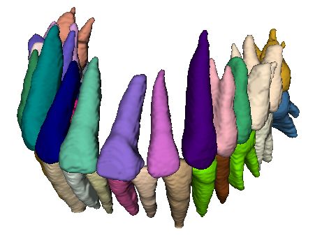
2       Yuan Liang, Weinan Song, Jiawei Yang, Liang Qiu, Kun Wang, and Lei He

3D teeth reconstruction of a whole cavity from a single panoramic radiograph.
This task is more challenging than the single tooth reconstruction, since not only
tooth shapes but also spatial localization of teeth should be estimated from their
2D representation. Moreover, all the existing methods of tooth reconstruction
[1,2,10] utilize ad-hoc image processing steps and handcrafted shape features.
Currently, Convolutional Neural Networks (ConvNet) provide an accurate so-
lution for single-view 3D reconstruction by discriminative learning, and have
become the state-of-the-art for many photo-based benchmarks [6,8,14]. How-
ever, the application of ConvNet on the teeth reconstruction has not yet been
explored.
    In this work, we pioneer the study of 3D teeth reconstruction of the whole
cavity from a single panoramic radiograph with ConvNet. Different from most
3D reconstruction benchmarks [5,13], which target at estimating a single vol-
ume per low-resolution photo, our task has the unique challenge to estimate
the shapes and localization of multiple objects at high resolutions. As such, we
propose X2Teeth, an end-to-end trainable ConvNet that is compact for multi-
object 3D reconstruction. Specifically, X2Teeth decomposes the reconstruction
of teeth for a whole cavity into two sub-tasks of teeth localization and patch-
wise tooth reconstruction. Moreover, we employ the random sampling of tooth
patches during training guided by teeth localization to reduce the computational
cost, which enables the end-to-end optimization of the whole network. Accord-
ing to experiments, our method can successfully reconstruct the 3D structure of
the cavity, as well as restore the teeth with details at high resolutions. More-
over, we show X2Teeth achieves the reconstruction Intersection over Union (IoU)
of 0.6817, outperforming the state-of-the-art encoder-decoder method by 1.71×
and retrieval-based method by 1.52×, which demonstrates the effectiveness of
our method. To the best of our knowledge, this is the first work that explores
3D teeth reconstruction of the whole cavity from a single panoramic radiograph.

2   Methodologies

Fig. 1 shows the overall architecture of our X2Teeth. We define the input of
X2Teeth as a 2D panoramic radiograph (Fig.1(1)), and the output as a 3D
occupancy grid (Fig.1(5)) of multiple categories for indicating different teeth.
Different from the existing single-shape estimations [6,14] that mostly employ
a single encoder-decoder structure for mapping the input image to one recon-
structed object, X2Teeth decomposes the task into object localization (Fig.1(b))
and patch-wise single tooth reconstruction (Fig.1(c)). As such, the reconstruc-
tion can be carried out at high resolutions for giving more 3D details under the
computational constraint, since tensor dimensions within the network can be
largely reduced compared to directly reconstructing the whole cavity volume.
Moreover, both sub-tasks share a feature extraction subnet (Fig.1(a)), and the
whole model can be end-to-end optimized by employing a sampling-based train-
ing strategy for the optimal performance. With the derived teeth localization
and tooth volumes, the final reconstruction of the cavity is derived by assem-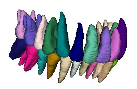
X2Teeth: 3D Teeth Reconstruction from a Single Panoramic Radiograph                                                                  3

 (a) ExtNet                                             (c)   RecoNet

                                                                                                                     depth

                                                        Sampling
                                                         Patch
                                                                                                                        Width
                                                                                                               (4) Estimated Arch Curve

 (1) Panoramic Radiograph                                                            (3) Reconstructed Tooth
                                                        (b) SegNet
    2D Conv     2D Conv        2D Trans     3D Cov
     3x3 +     2x2 stride 2   Cov 2x2 +     3x3x3 +
     ReLu +     + ReLu +        ReLu +      ReLu +

                                                                   Denosing
                                                                   Cropping
                                                                   Padding
   BatchNorm   BatchNorm      BatchNorm    BatchNorm

    3D Trans     Fully
   Cov 2x2x2   Connected       Sigmoid      Softmax
    + ReLu +    Layer +       Activation   Activation
   BatchNorm     ReLu                                                                                            (5) Reconstructed Teeth
                                                                              (2) Teeth Localization Map

Fig. 1. Overall architecture of X2Teeth. X2Teeth consists of three subnets: (a) ExtNet,
(b) SegNet and (c) ReconNet. ExtNet captures deep representations of teeth from the
input panoramic radiograph. Based on the representations, SegNet performs pixel-wise
classification followed by segmentation map denoising for localizing teeth. ReconNet
samples tooth patches from the derived feature map and performs single-shape re-
construction. The final reconstruction of the whole cavity is the assembling of the
reconstructed teeth according to the teeth localization and arch curve that estimated
via β function model. The whole model can be end-to-end trained.

bling different objects along the dental arch that is estimated via a β function
model.

2.1      Model Architecture

Given the panoramic radiograph, our X2Teeth consists of three components: (1)
a feature extracting subnet ExtNet for capturing teeth representations, (2) a
segmentation subnet SegNet for estimating teeth localization, and (3) a patch-
wise reconstruction subnet ReconNet for estimating the volume of a single tooth
from the corresponding feature map patch. The detailed model configuration can
be seen from the Fig.1.

ExtNet As shown in Fig.1(a), ExtNet has an encoder-decoder structure con-
sisting of 2D convolutions for capturing contexture features from the input
panoramic radiograph (Fig.1(1)). The extracted features are at high resolutions
as the input image, and are trained to be discriminative for both SegNet and
ReconNet to increase the compactness of the network. ExtNet utilizes strided
convolutions for down-sampling and transpose convolutions for up-sampling, as
well as channel concatenations between different layers for feature fusion.

SegNet Given the feature map of ExtNet, SegNet maps it into a categorical
mask Yseg ∈ ZH×W ×C , where H and W are image height and width, while
C denotes the number of categories of teeth. Especially, a categorical vector
y ∈ Ymask is multi-hot encoded, since nearby teeth can overlap in a panoramic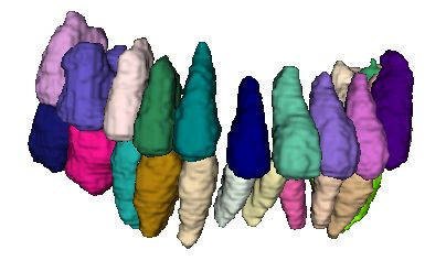
4       Yuan Liang, Weinan Song, Jiawei Yang, Liang Qiu, Kun Wang, and Lei He

radiograph because of the 2D projecting. With the categorical mask, SegNet
further performs denoising by keeping the largest island of segmentation per
tooth type, and localizes teeth by deriving their bounding boxes as shown in
Fig.1(2). As indicated in Fig.1(b), SegNet consists of 2D convolutional layers
followed by a Sigmoid transfer in order to perform the multi-label prediction. In
our experiments, we set C = 32 for modeling the full set of teeth of an adult,
including the four wisdom teeth that possibly exist for some individuals.

ReconNet ReconNet samples the feature patch of a tooth, and maps the 2D
patch into the 3D occupancy probability map Yrecon ∈ RHp ×Wp ×Dp ×2 of that
tooth, where Hp , Wp , Dp are patch height, width and depth, respectively. The
2D feature patch is cropped from the feature map derived from ExtNet, while
the cropping is guided by the tooth localization derived from SegNet. Similar to
[6,8], ReconNet has an encoder-decoder structure consisting of both 2D and 3D
convolutions. The encoder employs 2D convolutions, and its output is flattened
into a 1D feature vector for the fully connected operation; while the decoder
employs 3D convolutions to map this feature vector into the target dimension.
Since our ReconNet operates on small image patches rather than the whole x-
ray, the reconstruction can be done at high resolutions for restoring the details
of teeth. In this work, we set Hp = 120, Wp = 60, Dp = 60 since all teeth fit
into this dimension.

Teeth Assembling By assembling the predicted tooth volumes according to
their estimated localization from x-ray segmentation, we can achieve the 3D re-
construction of the cavity as a flat plane without the depth information about the
cavity. This reconstruction is an estimation for the real cavity that is projected
along the dental arch. Many previous work has investigated the modeling and
prediction of the dental arch curve [11]. In this work, we employ the β function
model introduced by [3], which estimates the curve by fitting the measurements
of cavity depth and width (Fig.1(4)). As the final step, our prediction of teeth
for the whole cavity (Fig.1(5)) can be simply achieved by bending the assembled
flat reconstruction along the estimated arch curve.

2.2   Training Strategy

The loss function of X2Teeth is composed of two parts: segmentation loss Lseg
and patch-wise reconstruction loss Lrecon . For Lseg , considering that a pixel
can be of multiple categories because of teeth overlaps on X-rays, we define the
segmentation loss as the average of dice loss across all categories. Denote the
segmentation output Yseg at a pixel (i, j) to be a vector Yseg (i, j) of length C,
where C is the number of possible categories, then
                                          P
                                     1 X         2Yseg (i, j)Ygt (i, j)
            Lseg (Yseg , Ygt ) = 1 −     P i,j                            ,    (1)
                                     C     i,j (Yseg (i, j) + Ygt (i, j))
                                      C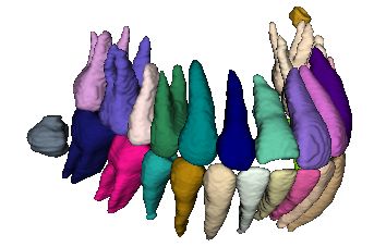
X2Teeth: 3D Teeth Reconstruction from a Single Panoramic Radiograph               5

where Ygt is the multi-hot encoded segmentation ground-truth. For Lrecon , we
employ the 3D dice loss for defining the difference between the target and the
predicted volumes. Let the reconstruction output Yrecon at a pixel (i, j, k) be a
Bernoulli distribution Yrecon (i, j, k), then
                                     P2 P
                                        c=1     i,j,k Yrecon (i, j, k)Ygt (i, j, k)
    Lrecon (Yrecon , Ygt ) = 1 − 2 P2 P                                              , (2)
                                     c=1    i,j,k (Yrecon (i, j, k) + Ygt (i, j, k))

where Ygt is the reconstruction ground-truth.
   We employ a two-stage training paradigm. In the first stage, we train ExtNet
and SegNet for the teeth localization by optimizing Lseg , such that the model
can achieve an acceptable tooth patch sampling accuracy. In the second stage,
we train the whole X2Teeth including ReconNet by optimizing the loss sum
L = Lseg +Lrecon for both localization and reconstruction. Note that Adam opti-
mizer is used for optimization. For each GPU, we set the batch size of panoramic
radiograph as 1, and the batch size of tooth patches as 10. Besides, standard aug-
mentations are employed for images, including random shifting, scaling, rotating
and adding Gaussian noise. Finally, we implement our framework in Pytorch,
and trained for the experiments on three NVidia Titan Xp GPUs.

3     Experiments
In this section, we validate and demonstrate the capability of our method for
the teeth reconstruction from the panoramic radiograph. First, we introduce
our in-house dataset of X-ray and panoramic radiograph pairs with teeth an-
notations from experts. Second, we validate X2Teeth by comparing with two
state-of-the-art single view 3D reconstruction methods. Finally, we look into the
performance of X2Teeth on the two sub-tasks of teeth localization and single
tooth reconstruction.

3.1    Dataset
Ideally, we need paired data of the panoramic radiographs and CBCT scans cap-
tured from the same subject to train and validate X2Teeth. However, in order
to control the radiation absorbed by subjects, such data pairs can rarely be col-
lected in clinical settings. Therefore, we take an alternative approach by collect-
ing high resolution CBCT scans and synthesize their corresponding panoramic
radiographs. Such synthesis is valid since CBCT scans contain full 3D infor-
mation of cavity, while panoramic radiographs are the 2D projections of them.
Several previous work has demonstrated promising results for high quality syn-
thesis, and in our work, we employ the method of Yun et al. [16] for building
our dataset. Our in-house dataset contains 23 pairs of 3D CBCT scans and
panoramic radiographs, each with a resolution ranging form 0.250 mm to 0.434
mm. All CBCT scans and panoramic radiographs are first labeled with pixel-
wise tooth masks by 3 annotators, and then reviewed by 2 board-certificated
dentists. Finally, we randomly split the dataset into 15 pairs for training, 1 pair
for validation, and 7 pairs for testing.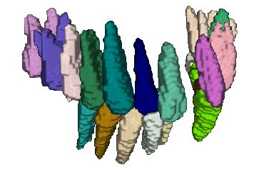
6       Yuan Liang, Weinan Song, Jiawei Yang, Liang Qiu, Kun Wang, and Lei He

3.2   Overall Evaluation of Teeth Reconstruction

Table 1. Comparison of reconstruction accuracy between X2Teeth and general purpose
reconstruction methods in terms of IoU, detection accuracy (DA) and identification
accuracy (FA). We report each metric in the format of mean ± std.

         Method               IoU           DA            DF
         3D-R2N2         0.398 ± 0.183 0.498 ± 0.101 0.592 ± 0.257
         DeepRetrieval   0.448 ± 0.116 0.594 ± 0.088 0.503 ± 0.119
         X2Teeth (ours) 0.682 ± 0.030 0.702 ± 0.042 0.747 ± 0.038

                                                                       X2Teeth (ours)
                                                                         3D-R2N2
                                                                       DeepRetriveral

Fig. 2. IoU comparison of different tooth types between X2Teeth, 3D-R2N2, and Deep-
Retrieval.

    We compare our X2Teeth with two general purpose reconstruction methods
that have achieved state-of-the-art performance as baselines: 3D-R2N2 [6] and
DeepRetrieval [15]. 3D-R2N2 employs an encoder-decoder network to map the
input image to a latent representation, and reasons about the 3D structure upon
it. To adapt 3D-R2N2 for high resolution X-rays in our tasks, we follow [15] by
designing the output of the model to be 1283 voxel grids, and up-sampling the
prediction to the original resolution for evaluation. DeepRetrieval is a retrieval-
based method that reconstructs images by deep feature recognition. Specifically,
2D images are embedded into a discriminative descriptor by using a ConvNet
[9] as its representation. The corresponding 3D shape of a known image that
shares the smallest Euclidean distance with the query image according to the
representation is then retrieved as the prediction.
    Quantitative Comparison. We evaluate the performance of models with
intersection over union (IoU) between the predicted and the ground-truth voxels,
as well as detection accuracy (DA) and identification accuracy (FA) [7]. The
formulations of the metrics are:
                    |D ∩ G|          |D|             |D ∩ G|
            IoU =           , DA =         and F A =         ,                    (3)
                    |D ∪ G|        |D ∩ G|             |D|
X2Teeth: 3D Teeth Reconstruction from a Single Panoramic Radiograph 7
a c d
b e f
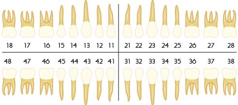
18 17 16 15 1413 1211 212223 2425 26 27 28
48 47 46 45 4443 4241 313233 3435 36 37 38
X8: Wisdom Tooth; X7,X6: Molar;
X4,X5: Bicusipid; X3: Cuspid; X1,X2: Incisor
Fig. 3. Comparison of the reconstruction between (d) X2Teeth (ours), (e) 3D-R2N2,
and (f) DeepRetrieval. (a) shows the input panoramic radiograph from the testing set,
(c) shows the ground-truth of reconstruction, and (b) is the teeth numbering rule.
where G is the set of all teeth in ground-truth data, and D is the set of pre-
dicted teeth. As shown in Table 1, X2Teeth outperforms both baseline models
significantly in terms of all three metrics. Specifically, X2Teeth achieves a mean
IoU of 0.682, which outperforms 3D-R2N2 by 1.71×, and DeepRetrieval 1.52×.
Similarly, Fig.2 reveals IoUs for all the 32 types of tooth among the three meth-
ods, where our method has the highest median and the smallest likely range
of variation (IQR) for all tooth types, which shows the consistent accuracy of
X2Teeth. Yet, we also find that all algorithms have a lower accuracy for wisdom
teeth (numbering 18, 28, 38, and 48) than the other teeth, indicating that the
wisdom teeth are more subject-dependent, and thus difficult to predict.
Qualitative Comparison. Fig.3 visualizes the 3D reconstructions of a
panoramic radiograph (Fig.3(a)) from the testing set, which clearly shows our
X2Teeth can achieve more appealing results than the other two methods. As for
3D-R2N2, its reconstruction (Fig.3(e)) misses several teeth in the prediction as
circled with green boxes, possibly because spatially small teeth can lose their rep-
resentations within the deep feature map during the deep encoding process. The
similar issue of missing tooth in predictions has also been previously reported
in some teeth segmentation work [7]. Moreover, the reconstruction of 3D-R2N2
has coarse object surfaces that lack details about each tooth. This is because
3D-R2N2 is not compact enough and can only operate at the compressed reso-
lution. As for DeepRetrieval, although the construction (Fig.3(f)) has adequate
details of teeth since its retrieved from high-resolution dataset, it fails to reflect
the unique structure of individual cavity. The red boxes in Fig.3(f) point out
the significant differences in wisdom teeth, tooth root shapes, and teeth occlu-
sion between the retrieved teeth and the ground-truth. Comparing to these two
methods, X2Teeth has achieved a reconstruction (Fig.3(d)) that can reflects both
the unique structure of cavity and the details of each tooth, by formulating the8 Yuan Liang, Weinan Song, Jiawei Yang, Liang Qiu, Kun Wang, and Lei He
task as the optimization of two sub-tasks for teeth localization and single tooth
reconstruction.
3.3 Sub-task Evaluations
a
X2Teeth (ours)
b
X2Teeth (ours)
Fig. 4. (a) Segmentation IoUs of various teeth for the teeth localization sub-task. (b)
Reconstruction IoUs of various teeth for the single tooth reconstruction sub-task.
For better understanding the performance of X2Teeth, we evaluate its ac-
curacy on the two sub-tasks of teeth localization and single tooth reconstruc-
tion. Fig.4(a) shows the IoUs of different teeth for the 2D segmentation, where
our method achieves an average IoU of 0.847±0.071. The results validate that
X2Teeth can accurately localize teeth, which enables the further sampling of
tooth patches for the patch-based reconstruction. We also observe that the mean
segmentation IoU for the 4 wisdom teeth (numbering X8) is 0.705±0.056, which
is lower than the other teeth. This is possibly because they have lower contrasts
with surrounded bone structures, such that are more challenging to segment.
Fig. 4(b) demonstrates the IoUs of different types of teeth for the single tooth
reconstruction, where our method achieves a mean IoU of 0.707±0.044. Still,
wisdom teeth have the significantly lower mean IoU of 0.668±0.050, which can
be contributed by the lower contrast with surroundings, less accurate localiza-
tion, and the subject-dependent nature of their shapes. Moreover, incisor teeth
(numbering X1 and X2) are observed to have less accurate reconstructions with
the mean IoU of 0.661±0.031. We argue the reason can be their feature vanishing
in the deep feature maps considering their small spatial size.X2Teeth: 3D Teeth Reconstruction from a Single Panoramic Radiograph 9
4 Conclusion
In this paper, we initialize the study of 3D teeth reconstruction of the whole
cavity from a single panoramic radiograph. In order to solve the challenges
posed by the high resolution of images and multi-object reconstruction, we pro-
pose X2Teeth to decompose the task into teeth localization and single tooth
reconstruction. Our X2Teeth is compact and employs sampling-based training
strategy, which enables the end-to-end optimization of the whole model. Our ex-
periments qualitatively and quantitatively demonstrate that X2Teeth achieves
accurate reconstruction with tooth details. Moreover, our method can also be
promising for other multi-anatomy 3D reconstruction tasks.
References
1. Abdelrahim, A.S., El-Melegy, M.T., Farag, A.A.: Realistic 3d reconstruction of the
human teeth using shape from shading with shape priors. In: 2012 IEEE Computer
Society Conference on Computer Vision and Pattern Recognition Workshops. pp.
64–69. IEEE (2012)
2. Abdelrehim, A.S., Farag, A.A., Shalaby, A.M., El-Melegy, M.T.: 2d-pca shape mod-
els: Application to 3d reconstruction of the human teeth from a single image. In:
International MICCAI Workshop on Medical Computer Vision. pp. 44–52. Springer
(2013)
3. Braun, S., Hnat, W.P., Fender, D.E., Legan, H.L.: The form of the human dental
arch. The Angle Orthodontist 68(1), 29–36 (1998)
4. Buchaillard, S., Ong, S.H., Payan, Y., Foong, K.W.: Reconstruction of 3d tooth
images. In: 2004 International Conference on Image Processing, 2004. ICIP’04.
vol. 2, pp. 1077–1080. IEEE (2004)
5. Chang, A.X., Funkhouser, T., Guibas, L., Hanrahan, P., Huang, Q., Li, Z.,
Savarese, S., Savva, M., Song, S., Su, H., et al.: Shapenet: An information-rich
3d model repository. arXiv preprint arXiv:1512.03012 (2015)
6. Choy, C.B., Xu, D., Gwak, J., Chen, K., Savarese, S.: 3d-r2n2: A unified approach
for single and multi-view 3d object reconstruction. In: European conference on
computer vision. pp. 628–644. Springer (2016)
7. Cui, Z., Li, C., Wang, W.: Toothnet: automatic tooth instance segmentation and
identification from cone beam ct images. In: Proceedings of the IEEE Conference
on Computer Vision and Pattern Recognition. pp. 6368–6377 (2019)
8. Henzler, P., Rasche, V., Ropinski, T., Ritschel, T.: Single-image tomography: 3d
volumes from 2d cranial x-rays. In: Computer Graphics Forum. vol. 37, pp. 377–
388. Wiley Online Library (2018)
9. Krizhevsky, A., Sutskever, I., Hinton, G.E.: Imagenet classification with deep con-
volutional neural networks. In: Advances in neural information processing systems.
pp. 1097–1105 (2012)
10. Mazzotta, L., Cozzani, M., Razionale, A., Mutinelli, S., Castaldo, A., Silvestrini-
Biavati, A.: From 2d to 3d: Construction of a 3d parametric model for detection
of dental roots shape and position from a panoramic radiograph—a preliminary
report. International journal of dentistry 2013 (2013)
11. Noroozi, H., Hosseinzadeh Nik, T., Saeeda, R.: The dental arch form revisited. The
Angle Orthodontist 71(5), 386–389 (2001)10 Yuan Liang, Weinan Song, Jiawei Yang, Liang Qiu, Kun Wang, and Lei He
12. Rahimi, A., Keilig, L., Bendels, G., Klein, R., Buzug, T.M., Abdelgader, I., Ab-
boud, M., Bourauel, C.: 3d reconstruction of dental specimens from 2d histological
images and µct-scans. Computer Methods in Biomechanics and Biomedical Engi-
neering 8(3), 167–176 (2005)
13. Sun, X., Wu, J., Zhang, X., Zhang, Z., Zhang, C., Xue, T., Tenenbaum, J.B., Free-
man, W.T.: Pix3d: Dataset and methods for single-image 3d shape modeling. In:
Proceedings of the IEEE Conference on Computer Vision and Pattern Recognition.
pp. 2974–2983 (2018)
14. Tatarchenko, M., Dosovitskiy, A., Brox, T.: Octree generating networks: Efficient
convolutional architectures for high-resolution 3d outputs. In: Proceedings of the
IEEE International Conference on Computer Vision. pp. 2088–2096 (2017)
15. Tatarchenko, M., Richter, S.R., Ranftl, R., Li, Z., Koltun, V., Brox, T.: What do
single-view 3d reconstruction networks learn? In: Proceedings of the IEEE Confer-
ence on Computer Vision and Pattern Recognition. pp. 3405–3414 (2019)
16. Yun, Z., Yang, S., Huang, E., Zhao, L., Yang, W., Feng, Q.: Automatic reconstruc-
tion method for high-contrast panoramic image from dental cone-beam ct data.
Computer methods and programs in biomedicine 175, 205–214 (2019)You can also read


