When the Locus Coeruleus Speaks Up in Sleep: Recent Insights, Emerging Perspectives
←
→
Page content transcription
If your browser does not render page correctly, please read the page content below
International Journal of
               Molecular Sciences

Review
When the Locus Coeruleus Speaks Up in Sleep: Recent Insights,
Emerging Perspectives
Alejandro Osorio-Forero                   , Najma Cherrad        , Lila Banterle      , Laura M. J. Fernandez          and Anita Lüthi *

                                           Department of Fundamental Neurosciences, University of Lausanne, CH-1005 Lausanne, Switzerland;
                                           alejandro.osorioforero@unil.ch (A.O.-F.); najma.cherrad@unil.ch (N.C.); lila.banterle@unil.ch (L.B.);
                                           laura.fernandez@unil.ch (L.M.J.F.)
                                           * Correspondence: anita.luthi@unil.ch

                                           Abstract: For decades, numerous seminal studies have built our understanding of the locus coeruleus
                                           (LC), the vertebrate brain’s principal noradrenergic system. Containing a numerically small but
                                           broadly efferent cell population, the LC provides brain-wide noradrenergic modulation that optimizes
                                           network function in the context of attentive and flexible interaction with the sensory environment.
                                           This review turns attention to the LC’s roles during sleep. We show that these roles go beyond
                                           down-scaled versions of the ones in wakefulness. Novel dynamic assessments of noradrenaline
                                           signaling and LC activity uncover a rich diversity of activity patterns that establish the LC as an
                                           integral portion of sleep regulation and function. The LC could be involved in beneficial functions
                                           for the sleeping brain, and even minute alterations in its functionality may prove quintessential in
                                           sleep disorders.

                                           Keywords: NREM sleep; REM sleep; monoamine; noradrenaline; arousability; sleep architecture;

                                           infraslow time scale; microvasculature; sleep disorder; Alzheimer’s disease

Citation: Osorio-Forero, A.; Cherrad,
N.; Banterle, L.; Fernandez, L.M.J.;
Lüthi, A. When the Locus Coeruleus         1. Introduction
Speaks Up in Sleep: Recent Insights,
                                                 Noradrenaline (NA) is a monoamine neurotransmitter that acts in the brain and
Emerging Perspectives. Int. J. Mol.
                                           body to induce and coordinate states of wakefulness, and to facilitate adaptive behaviors
Sci. 2022, 23, 5028. https://doi.org/
                                           in response to environmental novelty. The mammalian brainstem contains a cluster of
10.3390/ijms23095028
                                           up to seven NA-synthetizing nuclei (A1–A7) that have been anatomically identified in
Academic Editors: Michael Lazarus          >80 mammals [1], from rat [2], to cat [3], to human [4]. The tightly appositioned A4 and
and Wang Yi-Qun                            A6 nuclei stand out as the largest, often densest, and predominant forebrain-projecting
Received: 28 February 2022
                                           nuclei that share a common embryonic origin [5] and in which activity levels correlate with
Accepted: 19 April 2022
                                           the degree of wakefulness (for review, see [6–9]). In tissue sections, these nuclei appear
Published: 30 April 2022
                                           sky-blue because of their pigmentation with neuromelanin, a by-product of catecholamine
                                           metabolism, which gave it the name locus coeruleus (LC, Latin for “sky-blue spot”). The LC
Publisher’s Note: MDPI stays neutral
                                           lies in the pontine brainstem as an anteroposteriorly extended tube with a central ventral
with regard to jurisdictional claims in
                                           extension along the fourth ventricle (for review, see [1,8]) and it is part of the ascending
published maps and institutional affil-
                                           arousal systems, together with other monoaminergic and cholinergic nuclei (for review,
iations.
                                           see [10,11]). The LC provides brain-wide axonal arborizations and fine meshworks of
                                           varicose fibers that arise from a comparatively small number of NA-synthetizing neurons
                                           (thousands in rodents [12,13], tens of thousands in humans [14]). The axons from LC cells
Copyright: © 2022 by the authors.
                                           span the neuraxis from the spinal cord to the cerebellum, midbrain, thalamus, and cortex
Licensee MDPI, Basel, Switzerland.         and are thought to release NA through both synaptic and non-synaptic release mechanisms
This article is an open access article     (for review, see [15,16]) to regulate neurons, glial cells, and fine microvessels (for review,
distributed under the terms and            see [17–19]), stimulating wakefulness and attentional orienting (for review, see [8]), sensory
conditions of the Creative Commons         processing (for review, see [20]), muscle tone (for review, see [21]), and breathing (for
Attribution (CC BY) license (https://      review, see [22]), while inhibiting sleep-promoting brain areas (for review, see [10,23]).
creativecommons.org/licenses/by/           The LC also plays prominent roles in pathological forms of arousals, commonly linked to
4.0/).                                     acute stress (for review, see [24]), post-traumatic stress disorder (for review, see [25]), pain

Int. J. Mol. Sci. 2022, 23, 5028. https://doi.org/10.3390/ijms23095028                                             https://www.mdpi.com/journal/ijms
Int. J. Mol. Sci. 2022, 23, 5028 2 of 23
and analgesia [26], motivation and relapse (for review, see [27]), hypercapnia (for review,
see [28]), and hypotension (for review, see [29]), many of which are accompanied by sleep
disturbances.
Novel anatomical and physiological technologies, together with advanced behavioral
measures, are about to bring fundamentally renewed insights into the LC’s functions. The
LC shows a genetic and/or functional heterogeneity at multiple levels from its embryonic
and evolutionary origins (for review, see [1,5]), its synaptic interactions with the peri-
coerulear area (for review, see [30]), its input–output connectivity (for review, see [31]),
to its cellular identities and neurotransmitter release (for review, see [6,30]), neuronal
ensemble formation (for review, see [32]), regulation of whole-brain states [33], brain-state-
dependent firing patterns (for review, see [7,30]), and behavioral roles (for review, see [34]).
The LC emerges as a dynamic and plastic assembly of functionally specialized LC neuronal
subgroups that act locally or globally according to recently lived experiences, ongoing
demands, and future challenges (for review, see [30,35]).
Time is also ready to complement the prevalent LC-wakefulness association with
the appreciation that the LC is important for sleep. The central message of this review
is that LC’s role in sleep has remained underestimated. Novel real-time monitoring and
interferential approaches now start to indicate that LC contributes to sleep in fundamental
ways—to its cellular functions, its micro- and macroarchitectural organization and regula-
tion, associated behaviors, and possible roles in disease. These insights are at their very
beginnings, yet they indicate that the LC could become an important factor in profiles of
perturbed sleep that arise from diverse conditions. In this review, we discuss these exciting
developments primarily based on animal experimentation, but we include human studies
whenever they help complement available evidence. For a more human sleep-oriented
recent review on the LC’s role in sleep, we refer to Van Egroo et al. [36].
2. The Activity of the LC in Sleep: Pioneering Studies
This chapter reviews studies from the last decades that provided evidence for a
maintained activity of the LC in sleep. Quantitatively, these studies revealed that the LC
unit activity was clearly lower compared to wakefulness, yet remained distinctly elevated
during non-rapid-eye-movement (NREM) sleep compared to REM sleep. NREM and
REM sleep are the two major mammalian sleep states, also referred to as “quiescent” and
“active” sleep, respectively. These two states show distinct spectral characteristics and
functions for sleep (for review, see [37]). Qualitatively, the studies summarized in this
chapter suggest that the noradrenergic system appeared to be involved in the alternation of
NREM and REM sleep. For these results, diverse techniques in animals and humans were
used that span from electrophysiology and pharmacology to microdialysis and functional
imaging. A summary of the traditional view that has emerged from these studies is shown
in Figure 1 (left).
2.1. Animal Studies
Rat [38,39], cat [40–42], and monkey [38,43] recordings showed that the action poten-
tial discharge rates of LC units during NREM and REM sleep were minor compared to
wakefulness. Unit activity was low during NREM sleep, but remained detectable, while
it ceased during REM sleep [38,39]. However, researchers also noted that not all putative
LC units reduced activity during NREM and/or REM sleep [40,41]. LC activity was also
low in quiet—as opposed to active—wakefulness [39,42]. More recent studies indicate
that some LC units may even be as active in NREM sleep as in quiet wakefulness and
occasionally fire in bursts [44–46]. Furthermore, although activity during sleep states was
overall low, it nevertheless was not random. For example, LC unit activity has been related
to the organization [39] and termination [46] of sleep spindles, an essential NREM sleep
rhythm in the 10–15 Hz frequency range originating from the thalamo-cortical loop [47].
Additionally, LC unit activity during NREM sleep preceded the cortical up-state periodsInt. J. Mol. Sci. 2022, 23, 5028 3 of 23
from another important NREM sleep-associated slow rhythm, the cortical slow oscillation
(~1 Hz) [45], and was increased during a post-learning NREM-sleep period [44].
Figure 1. Summary of traditional and revised views on the neuromodulatory profiles of wakefulness
and sleep, with a focus on noradrenergic signaling. Traditional (left) and revised (right) views derived
from animal experimentation are summarized and complemented with data-derived schematic represen-
tations of NA dynamics and LC unit activity. From top to bottom: mean levels of major neuromodulators
(blue up and gray down arrows symbolize high and low levels in the traditional view), a representative
hypnogram of mouse sleep–wake behavior, free NA levels, and representative discharges of a LC
unit. Novel insights central to the revised view are highlighted with the red arrow, whereas unaltered
neuromodulatory levels are shown with light grey arrows. NA, noradrenaline; 5HT, serotonin; ACh,
acetylcholine; NREM, NREM sleep; REM, REM sleep.
Microdialysis allows sampling of mean concentrations of neurochemicals present in
the extracellular fluid surrounding neural tissue. Microdialysis for NA revealed its levels
to be lower for states of sleep compared to wakefulness across rodents, cats, and seals,
yet values for NREM sleep consistently were intermediate with respect to the ones for
wakefulness and REM sleep in various brain areas (for review, see [48]). This suggested
that even low LC unit activity leads to detectable NA release. However, no or minor
increases in cortical NA levels in response to electrical or chemical stimulation of the LC
were observed at low (1–3 Hz) compared to higher (>5 Hz) stimulation frequencies [49–51].
The fast-scan cyclic electrochemical voltammetry technique allows millisecond-resolution
assessments of NA levels with nanomolar sensitivity, but it has so far been mostly applied
for high-frequency stimulation of the LC [52]. Therefore, the relation between LC unit
activity and real-time NA output has remained undefined.
Jouvet’s monoaminergic theory of sleep–wake control [53] prompted examinations
of the spontaneous sleep–wake cycle after lesion or pharmacological manipulation of LC
and NA signaling, or after constitutive removal of genes encoding proteins involved in
NA turnover. These approaches made it clear that noradrenergic activity sustains wake-
fulness at the expense of sleep (for review, see [7–9,23]). At the same time, they provided
the first hints that NA signaling remained relevant for sleep. For example, neurotoxic
lesions of noradrenergic LC neurons or genetic elimination of the NA-synthetizing en-
zyme dopamine-β-hydroxylase (DBH) altered the relative times spent in NREM and REM
sleep [54–56]. These approaches lacked the necessary specificity in time and in the site of
action to conclude about the LC’s role in regulating the timing of NREM and REM sleep.
Furthermore, noradrenergic receptors are expressed both centrally and peripherally, and
LC projections target both sympathetic and parasympathetic autonomic pathways (for
review, see [29]). Therefore, systemic drug administration may affect sleep–wake statesInt. J. Mol. Sci. 2022, 23, 5028 4 of 23
by acting on the autonomic nervous system. Nevertheless, these studies are part of initial
evidence that monoaminergic systems, including NA, could remain active in sleep.
Particularly noteworthy are the effects of pharmacological α2-adrenergic receptor
activation. These receptors are Gi -protein-coupled receptors activated by NA in the cen-
tral nervous system and periphery. In the brain, they act as both presynaptic negative
autoreceptors within the LC and in sleep–wake regulatory centers to suppress NA release
and attenuate postsynaptic excitability [57] (for review, see [58]). These receptors are also
the target of powerful sedatives used in clinics, such as dexmedetomidine (for review
see [58]). The α2 agonist clonidine suppresses the activity of the LC [59,60] but also targets
pre- and postsynaptic receptors in sleep-regulatory areas (for review, see [23]). α2 agonists
such as clonidine or detomidine, when applied locally in cat pontine brainstem [61] or
peri-coerulear areas [62], or systemically in rat [63], suppressed REM sleep while increasing
the depth of NREM sleep. The use of clonidine in humans was also found to attenuate
REM sleep (see Section 2.2). These effects on sleep macroarchitecture are in line with an
active LC during sleep.
2.2. Human Studies
The functional activity within dorsal brainstem areas, including the LC, was examined
through functional magnetic resonance imaging (MRI) in sleeping healthy individuals [64].
This imaging technique uses magnetic resonance signals to detect changes in brain activity
based on increases in the flow of oxygenated over non-oxygenated blood. Signal increases
involving the LC were particularly prominent during NREM sleep-associated slow (Int. J. Mol. Sci. 2022, 23, 5028 5 of 23
renewed versions keep being developed, expanding the range of sensitivity and kinetics
with which measures can be taken (see http://www.yulonglilab.org/faq.html, accessed
on 1 February 2022). Furthermore, a mutant version of the sensor that is not responsive
to NA should be used to control for potential non-specific alterations of the fluorescence
signal that can limit its interpretation. For example, local alterations in neuronal environ-
ments, such as in brain temperature or blood pressure accompany transitions between
NREM and REM sleep. These could alter light scattering or biosensor properties in vivo.
Two studies in mouse, one published [72], one yet to be peer-reviewed [73], have now
used these GRAB NE sensors to describe the real-time dynamics of free NA levels in the
medial prefrontal cortex [73] and in the primary sensory thalamus [72] during the natural
sleep–wake cycle. These studies report unexpectedly high levels of NA during NREM sleep
compared to wakefulness. Furthermore, they observe a dynamically varying signal during
states of sleep. This chapter presents the most important findings derived from these two
studies. A summary of the resulting revised view on NA signaling in sleep is shown in
Figure 1 (right).
3.1. Mean NA Levels Differ across the Sleep–Wake Cycle
The signals provided by the genetic sensor showed characteristic alterations across
wakefulness, NREM sleep, and REM sleep. In the prefrontal cortex, mean NA levels during
wakefulness were high but variable [73], which is consistent with the large variations in
LC activity in wakefulness (see Section 2.1). During NREM sleep, the mean free NA levels
became lower but still overlapped with the ones of wakefulness. During REM sleep, the
levels of NA were consistently low. In the sensory thalamus, similar measures of NA even
revealed that mean levels were significantly higher during NREM sleep when compared
specifically to quiet wakefulness (Figure 2) [72]. Again, values were low during REM sleep
in this area. These findings provide the first evidence that NA levels remain more elevated
in NREM sleep in forebrain areas than what was expected based on unit measures (see
Section 2.1). The expected low NA levels during REM sleep appear as a common feature
across the recorded areas. The considerable discrepancy between the numerically sparse LC
unit activity (see Section 2.1) and high free NA levels generated during NREM sleep shows
that much remains to be learned about the mode of operation of LC neuronal ensembles in
different states of vigilance.
3.2. NA Levels and LC Activity Fluctuate During NREM Sleep
The next notable observation found in both the thalamus and prefrontal cortex is that
NA levels were not steady during NREM sleep (Figure 2A–C). Instead, these fluctuated on
an infraslow timescale of tens of seconds, with an average cycle length of 30–50 s [72,73].
These fluctuations in NA levels were linked to phasic bouts of LC neuronal activity over
the same intervals, as evident by correlated Ca2+ transients in LC somata [73]. This activity
pattern points to a periodic synchronization of LC population activity on an infraslow time
scale during NREM sleep [74].
The role of these recently identified fluctuations is a current topic of investigation [72,75,76]
(for review, see [77]). Optogenetic modulation of noradrenergic LC neuronal activity evoked
variations in the appearance of sleep rhythms and heart rate, suggesting that infraslow NA
fluctuations are relevant for NREM sleep’s physiological correlates. Thus, NA released
by the LC lead to a periodic clustering of sleep spindles, such that they appeared at high
density when NA levels were low and they were scarce when these levels were high
(Figure 2A) [72,73]. Mechanistically, sleep spindle clustering relied on the α1- and β-
adrenergic receptor-mediated modulation of membrane potentials in the thalamic circuits,
in which sleep spindles are generated [72]. Sleep spindles are involved in the sleeping
brain’s elaboration of sensory input (for review, see [78]), which implies the LC in NREM
sleep-related sensory processing (see Section 4.1).Int. J. Mol. Sci. 2022, 23, 5028 6 of 23
Figure 2. Real-time dynamics of NA levels in somatosensory thalamus, forebrain sleep spindle power,
and heart rate during NREM sleep. Representative simultaneous recordings in a freely behaving
mouse combining (from top to bottom): hypnogram (gray), free NA levels in somatosensory thalamus
obtained through fiberphotometry imaging (red, A1), local field potential sigma power (10–15 Hz)
in somatosensory cortex (dark blue, A2) and heart rate (light blue, A3), with labeled portions (A,
B, C) shown expanded on the right. The variations in sigma power reflect the clustering of sleep
spindle density ([72]). Insets on the right expand portions of the traces highlighted with letters in the
hypnogram to show (A) NREM sleep (double-headed arrow marks the 50 s periodicity); (B) NREM-
to-REM sleep transitions (double-headed arrow marks the decay time of NA levels prior to REM
sleep onset); (C) NREM-to-wake transitions. Portions of two of these traces have been published
previously [72]. NA, noradrenaline; W, wakefulness; NR, NREM sleep; R, REM sleep; ∆F/F, relative
fluorescence changes; AU, arbitrary unit; bpm, beats per minute.
NA fluctuations also correlated with infraslow variations in heart rate during NREM
sleep (Figure 2A). The LC thus acts bidirectionally to coordinate forebrain sleep spindle
rhythms with heart rate variations. Indeed, optogenetic activation of LC noradrenergic
neurons disrupted the heart rate variations during NREM sleep and their anticorrelation
with the spindle clustering [72,76]. Mechanistically, the coupling of LC activity to the heart
rate depended on parasympathetic signaling. Likewise, parasympathetic signaling also
underlies coordinated infraslow fluctuations between pupil diameter and sigma power
during NREM sleep [79].
3.3. NA Levels Decay to Low Levels during REM Sleep
The NA levels declined in both the prefrontal cortex [73] and the thalamus [72] during
REM sleep, in line with the quiescence of LC units in this behavioral state (Figure 2B). As a
result, NA levels reached a level that lay below that of wakefulness and NREM sleep. This
result directly and strikingly supports the proposition that REM sleep periods are relatively
NA-free (see Section 5.2). The quantification of the extent and time course of this decline
will now allow us to refine this proposition, in particular in terms of the relation to REM
sleep bout duration.
3.4. NA Levels Show Characteristic Dynamics at Behavioral State Transitions
The dynamics of NA levels at moments of transition from NREMS to REMS or wake-
fulness showed characteristic properties. At NREM-to-REM transitions, a decrease in NA
levels began ∼40 s before the onset of REM sleep (Figure 2B). This time period recalls a tran-
sitional moment of sleep that has been referred to as “intermediate sleep” in rodents [80],
cats [81] and humans [82,83]. Intermediate sleep shows a mixed spectral profile combiningInt. J. Mol. Sci. 2022, 23, 5028 7 of 23
an increase in sigma power and the density of fast spindles, while hippocampal theta
rhythms appear (for review, see [78]). On the time scale of intermediate sleep, there is
a cessation of LC unit activity [39,73] and the appearance of cholinergic activity in REM
sleep-promoting tegmental nuclei [84,85]. The coincidence of declining NA levels with
unit and spectral correlates of intermediate sleep suggests that the activity levels of the LC
during NREM sleep may determine the timing of NREM-to-REM sleep transitions.
Transition periods from NREM sleep to both sustained wakefulness and to microarousals
were both associated with an increase in NA levels in the prefrontal cortex that appeared
to start before the transition (∼10 s) [73]. On the same time scale, there was an increase in
Ca2+ activity of noradrenergic LC neurons that was higher for transitions to consolidated
wakefulness compared to microarousals. This appeared also to be the case for NA levels at
NREM sleep-to-wake transitions (Figure 2C). These alterations are in line with unit activity
measures around moments of wake-up (see Sections 2.1 and 4.1).
3.5. Emerging Dynamics of Other Monoamines and Wake-Promoting Neurotransmitters
In vivo measures using genetically encoded sensors showed that, in addition to NA,
other monoamines and wake-promoting neurotransmitters remain high during NREM
sleep. In Ca2+ -based fiber photometric measures of spontaneous activity in the dorsal
raphe, fluctuations were observed in phase relation to spontaneous brief arousals [86].
Furthermore, measures with a genetically encoded sensor for free serotonin levels revealed
slow fluctuations in both the orbital frontal cortex and the bed nucleus of the stria termi-
nalis during NREM sleep, and declines during REM sleep [87]. The time course of these
fluctuations, and their consistent appearance at two distant brain sites, are reminiscent of
the findings with NA described in this chapter. Given the rapid advance in the availability
of novel sensors for dopamine [88,89] but also for other neuromodulatory transmitters
involved in sleep–wake control (such as acetylcholine, [90] or hypocretin [91]), more de-
tails on the spatiotemporal map of neurotransmitter dynamics during states of sleep will
soon become available. Intriguingly, transient free dopamine increases in the basolateral
amygdala were just discovered as triggers for NREM-to-REM sleep transitions [92].
4. The Role of the LC in the Regulation of Sleep and Sleep Functions
This chapter builds on the newly revealed real-time dynamics of NA levels described
in Section 3. It aims to review how these findings advance insight and motivate experimen-
tation in the quest for the functional roles of the LC during sleep.
4.1. LC as Part of Sensory Arousal Circuits during NREM Sleep
Pioneering recordings from LC units found that these respond with a short latency to
stimuli from different sensory modalities [38,42,43,93,94]. Increases in LC unit discharge
rates also preceded spontaneous, unsolicited awakenings from NREM sleep [38,39,94].
Moreover, activation of LC through electrical, opto-, or chemogenetic stimulation elicited
transitions from sleep to wakefulness [46,95,96] and recruited whole-brain networks in-
volved in salience processing [33]. Acute knockdown of DBH specifically in LC neurons
disrupted sleep-to-wake transitions elicited by optogenetic LC stimulation, confirming
the importance of NA signaling for wake-ups [96]. Given LC’s powerful capacity to
drive sleep-to-wake transitions, LC activity might be involved in sensory-induced sleep–
wake transitions.
Indeed, Hayat et al. [63] showed a causal link between the levels of ongoing LC activity
during NREM sleep and the probability of sensory stimulus-evoked awakenings. Mild
optogenetic LC stimulation lowered the auditory arousal threshold, whereas inhibiting LC
heightened it. In line with this, the natural infraslow fluctuations of LC activity during
undisturbed NREM sleep coincided with variations of auditory and somatosensory arous-
ability [75,76]. Furthermore, spontaneous brief arousals from NREM sleep in mice were
most frequent at moments of low spindle density, when LC activity is high [72,75].Int. J. Mol. Sci. 2022, 23, 5028 8 of 23
The exact roles of the LC in the cognitive, motor, and autonomic aspects of arousal
remain to be determined. As LC neurons are activated by sensory input (Figure 3A), NA
release is promoted by the sensory stimulus itself. It is also noteworthy that even low-
frequency LC discharge (1–2 Hz) sharpened sensory responsiveness and receptive fields
at the level of the thalamus and cortex [97–99]. Through depolarizing thalamic neurons,
the LC also suppresses the appearance of sleep spindles that limit sensory throughput
in thalamocortical areas [78]. The LC could hence promote sequential arousal-promoting
actions that are graded with its activity levels as the transition from sleep to wakefulness
takes place.
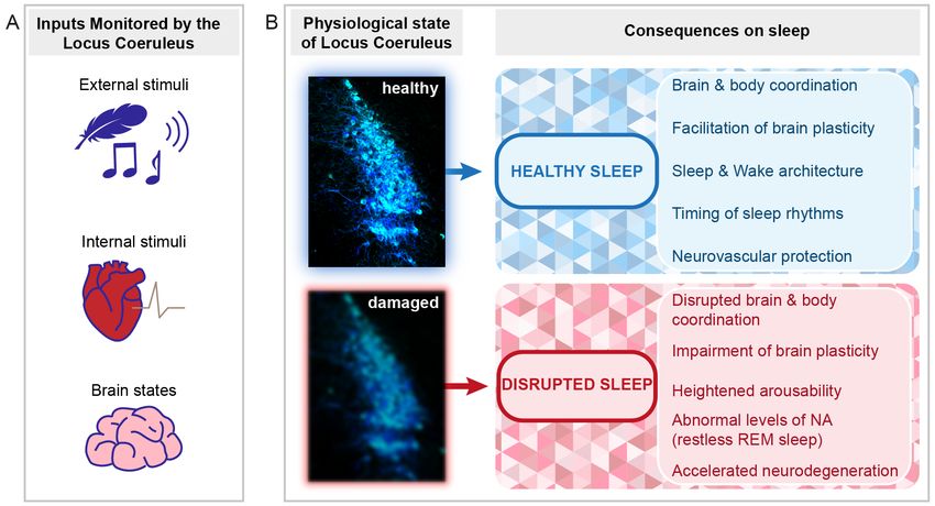
Figure 3. Perspectives for the implication of the LC in healthy and disrupted sleep. Schematic indicat-
ing the types of signals monitored by the LC and the implications of LC function and dysfunction for
sleep. (A) The LC monitors external stimuli (e.g., sensory stimuli such as touch or sound, symbolized
by a feather and a musical note, see Section 4.1), internal stimuli (symbolized by the heart, see
Section 4.5), and internal brain states important for the regulation of NREM-to-REM sleep transitions
(symbolized by the brain, see Sections 3.4 and 4.3). (B) Depending on the LC status (healthy or
damaged), beneficial or adverse consequences on sleep can arise. Several outcomes are listed on the
right. The LC micrograph was obtained from an immunohistochemically stained brain section of one
of the mice used for the data published in [72]. The color choice of cell labeling was made deliberately
to mark it as the sky-blue spot. The blurring of the blue color in the bottom micrograph symbolizes
both structural and functional alterations that can lead to LC dysfunction.
4.2. The LC as Part of the Regulatory Mechanisms of NREM Sleep
The real-time dynamics of NA for the first few hours of the light phase, the predom-
inant resting phase of rodents, underscore the importance of the LC in the regulation of
sleep architecture [72]. The elucidation of these dynamics across the light–dark cycle and
across major sleep–wake control areas will reveal the full impact of NA on sleep’s brain
states and associated sleep–wake behaviors. The LC is part of arousal circuits that are under
circadian control [100] and it receives afferents from hypothalamic preoptic areas involved
in NREM sleep homeostasis [31]. Therefore, beyond its regulation of sleep architecture and
spectral composition, the LC could also contribute to circadian and homeostatic regulation
of NREM sleep (Figure 3B).
4.3. The LC in REM Sleep Control
In spite of much pioneering work (see Sections 2.1 and 2.2), how LC regulates REM
sleep remains an open question (for review, see [101]). Recent research has focused on glu-
tamatergic and GABA-ergic circuits involved in REM sleep regulation, whereas monoamin-
ergic systems were attributed mostly a modulatory role (for review, see [102]). Measures of
real-time NA dynamics, together with NREM sleep-specific optogenetic manipulation ofInt. J. Mol. Sci. 2022, 23, 5028 9 of 23
the LC in rodents, instead indicate important changes in LC activity at moments of REM
sleep onset. These recent data revive the questions about the LC’s role in REM sleep that
we outline here in three aspects that could be important in future studies.
First, as described in Section 3, forebrain NA levels remained high during NREM
sleep (Figure 2A) and LC neurons continue to be active [73]. This elevated activity in nora-
drenergic signaling suppresses REM sleep, as suggested by electrical or pharmacological
LC stimulation in the rat [103,104] and by NREM sleep-specific optogenetic activation of
noradrenergic LC neurons at low frequency [72]. A noradrenergic inhibition of REM sleep-
promoting brain areas is a likely underlying mechanism for this suppression [105–107].
Importantly, the spontaneous activity of LC during natural undisturbed sleep seems even
sufficient to antagonize NREM-to-REM sleep transitions. This was concluded from NREM
sleep-specific optogenetic inhibition of the LC in freely sleeping mice, which increased the
time spent in REM sleep [72]. This indicates that the LC is a powerful target to manip-
ulate the balance between NREM and REM sleep in response to various regulatory and
experience-dependent processes (see below and Section 5.2).
Second, NA levels declined in both the thalamus and cortex in REM sleep (Figure 2B).
This decline took tens of seconds to complete once REM sleep began, raising the question
of which are the determinants of this time course. The LC is inhibited by GABAergic
mechanisms [108,109], of which several have been tested for their role in REM sleep control.
Monosynaptic inhibitory afferents arise from the local and pericoerulear interneurons [110],
ventrolateral periaqueductal gray [111], and from nucleus prepositus hypoglossi and dorsal
paragigantocellular reticular nucleus [107,112,113]. Acetylcholine release from cholinergic
REM sleep-promoting areas could also act through GABAergic mechanisms [109]. Cholin-
ergic areas are likely initiating the LC inhibition as their discharge onset precedes REM
sleep [84], but auto-inhibitory mechanisms within the LC could also play in at this mo-
ment [114]. At least one of the dorsal medullar inhibitory afferents increases activity exactly
at REM sleep onset [113], suggesting that NA decline could become strengthened due to
additional sources of inhibition. NA uptake mechanisms lagging behind synaptic inhibition
of the LC could instead retard the decline of free NA levels. How the strength and the
efficiency of synaptic inhibition regulate LC silencing and NA decline and/or interact with
other excitatory and/or modulatory synaptic mechanisms of LC inhibition (see e.g., [110])
is currently unexplored. The determinants of NA dynamics at NREM-to-REM sleep transi-
tions are critical to understanding how REM sleep evolves into an NA-free state because of
its likely role in the regulation of emotional memory (see Section 5.2).
Third, the fluctuating levels of NA during NREM sleep indicate that chances for a
NREM-to-REM sleep transition increase at moments of relatively low NA levels. Inter-
estingly, the probability to enter REM sleep was indeed found to be phase-locked to the
infraslow fluctuation of sigma power measured at the level of the EEG [113]. This lends
support to the idea that fluctuating LC activity during NREM sleep generates brain states
that are permissive for transitions, such as the ones to REM sleep (Figure 3A) [75,76]. The
LC activity oscillating between high and low levels might suppress REM sleep on the
one hand, but also open moments where transitions are favored. The LC is, therefore,
positioned as a brain area capable of autonomously regulating the timing of REM sleep
in a bidirectional manner during NREM sleep, yet how it is integrated into REM sleep
regulatory mechanisms will require further research.
Alterations in REM sleep propensity, duration, and hippocampal-related theta activity
are ubiquitous after stress- and fear-related experiences. These are part of the acute physio-
logical responses to the hormonal and autonomic changes accompanying stress [115], but
they also contribute to the consolidation of fear- [116] and extinction-related memories [115].
Increases in REM sleep are part of an adaptive process to mild stress exposure [117]. Given
the LC’s high reciprocal connectivity with areas implied in fear, such as the amygdala
(for review, see [31]), it is a strong candidate for linking stress-related experiences during
the day to the timing of REM sleep. Indeed, acute decreases in REM sleep in response to
inescapable footshock could be alleviated by optogenetic inhibition of excitatory neuronsInt. J. Mol. Sci. 2022, 23, 5028 10 of 23
in the basolateral amygdala [118] or by dual hypocretin receptor antagonism in the LC
or the dorsal raphe [119]. In case of such or even more traumatic experience, states of
hyperarousal associated with elevated monoaminergic activity may arise, to which the LC
contributes (for review, see [120]) (see Section 5.2).
4.4. The LC in Hippocampus-Dependent and Independent Memory Consolidation
LC activity, in part due to its implication in novelty detection, has been found to
actively contribute to online memory consolidation. A series of studies found that acti-
vation of the LC favors different types of learning such as spatial learning [121–124], fear
learning and reconsolidation [124–126], and perceptual learning [127,128]. Optogenetically
activating LC tyrosine hydroxylase-positive neurons shortly after memory encoding of
food rewards in a navigation task promoted memory retention in mice, which persisted
until the next experimental assessment [121]. Such LC stimulation mimicked the effects of
environmental novelty on memory encoding. Intriguingly, local pharmacological inhibition
of dopaminergic but not noradrenergic receptors in the hippocampus implied a role of LC
fiber-dependent dopamine release in novelty enhancement of hippocampus-dependent
memory. Optogenetic stimulation of the LC during a spatial object recognition task lead
to similar results [122]. Inhibition of the LC had, on the contrary, a detrimental effect on
hippocampal place cell formation in goal-directed spatial learning [123]. The LC’s role in
cued fear conditioning concerned both, memory acquisition of the pairing between the
conditioned and the unconditioned stimulus, and later extinction [126]. Here, a dual role
for LC afferent projections to the amygdala and to the medial prefrontal cortex could be
identified, with the former implied in the acquisition, and the latter in the extinction of
fear memory, demonstrating a modular functionality of LC subgroups depending on their
projection targets. Pairing LC activation with stimulus presentation could also accelerate
the learning of a new target sound in a perceptual learning paradigm [127] in rats and
electrical/optogenetic stimulation of the LC during sound presentation promoted NA-
dependent long-term plastic strengthening in auditory tuning curves of primary auditory
cortex neurons [128].
The LC’s role as a regulator of memory acquisition likely relies on manifold actions
of NA on neuronal excitability, in particular in hippocampal circuits, and on enduring
changes in synaptic strength (for review, see [129]). One principal action of endogenously
released NA, identified through optogenetic stimulation of LC fibers, appears to be a
suppression of postsynaptic potassium currents, which enhanced the excitability of CA1
pyramidal neurons in response to Schaffer collateral stimulation [130]. This effect was
blocked by β adrenoceptor antagonists, with no apparent implication of dopamine release.
It is noteworthy that this action was already present when fibers were stimulated at low
frequency (1 Hz), suggesting that such neuromodulation could be effective during NREM
sleep, when the LC discharges at low frequencies (see Section 2.1).
In contrast to the strong evidence for the LC’s involvement in the memory acquisition
phase, evidence that it plays a role during offline processing, including during sleep, is
currently scarce. Pioneering pharmacological studies found that rats trained in an olfactory
reward association task performed less well when they were injected with adrenergic
antagonists intracerebroventricularly [131] or within prefrontal cortex [132] 2 h after train-
ing, but not at shorter or longer time intervals. These authors also provided evidence
for a transient increase in NA levels during the time window in which these antagonists
were effective. This pointed to a delayed re-activation of the LC that facilitated offline
processing and memory consolidation. Follow-up studies suggest that such re-activation of
the LC may indeed occur during post-learning sleep stages, as LC unit activity transiently
doubled within the presumed re-activation window, without apparent alteration in sleep
architecture [44]. The activity of LC units was further observed to be time-locked to slow
waves in both rat [45] and human [64] and to hippocampal sleep spindles [46], suggesting
that enhanced NA release is linked to the sleep rhythms that enable active systems con-
solidation. Finally, high-frequency stimulation of the LC disrupted the coupling of sleepInt. J. Mol. Sci. 2022, 23, 5028 11 of 23
spindles with hippocampal ripples that are high-frequency oscillatory patterns critical for
memory consolidation [133]. This adds to evidence that the degree of LC activity might be
critical in coordinating sleep rhythms relevant for offline processing (for review, see [134]).
4.5. The LC as Mediator of Vagal Afferent Information
Among the innervations that the LC receives, one is of particular interest as a gate
for interoceptive signals, the Nucleus Tractus Solitarius [135,136] (for review, see [137]).
This brainstem nucleus is part of the dorsal vagal complex (nucleus tractus solitarius,
area postrema and dorsal motor nucleus of the vagus) which is the first recipient for vagal
afferents (for review, see [138]). The vagus nerve is part of the parasympathetic system and
it is a mixed nerve containing both motor and sensory fibers. Sensory information arising
from the vagus nerve is important for autonomic feedback reflexes, such as the baroreceptor
reflex and the Hering–Breuer reflex that serves to control breathing (for review, see [139]),
and it reaches the LC via the dorsal vagal complex [140]. Vagus nerve stimulation is well-
known for its beneficial role in clinical conditions, as evident from the highly diversified
effects of vagus nerve stimulation (VNS). Indeed, this technique has been proposed to
facilitate brain plasticity [141] (for review, see [142]) and memory formation (for review,
see [143]). Some important domains of clinical application for VNS include drug-resistant
epilepsy [144] (for review, see [145,146]), depression [147] (for review, see [148]), eating
disorders [149], and neurodegenerative disorders [150].
Several animal studies support the LC as a major target of vagal afferent nerve stimu-
lation. VNS caused an increase in the expression of the immediate-early gene c-fos in LC
neurons in conscious unanesthetized rabbits [151] and in anesthetized rats [152]. Moreover,
lesioning of the LC led to a suppression of the anticonvulsant effects of VNS in epileptic
rats, supporting the idea that the LC is involved in this circuitry [153]. This implication of
the LC was further supported by directly recording LC unit activity during VNS [154–157].
Using in vivo Ca2+ imaging in head-fixed awake mice, a recent study showed an increase in
the noradrenergic neuromodulatory system in response to VNS [158]. Furthermore, in vivo
microdialysis showed an increase in NA extracellular levels in the hippocampus and cortex
during chronic VNS in anesthetized rats [159,160] and an increase in dopamine in extracellu-
lar levels in the prefrontal cortex and nucleus accumbens [161]. Additionally, vagal afferent
electrical stimulation has been related to pupil dilation in animals and humans [162–165],
consistent with the correlation between pupil diameter and firing of noradrenergic LC cells
(for review, see [7]). Together, these results indicate that monoaminergic systems, including
the LC, act as monitors of internal stimuli conveyed by vagal afferents (Figure 3A).
Given the role of the LC in the regulation of sleep, stimulation of vagal afferents may
contribute to LC-dependent sleep regulatory effects. Animals studies suggest that VNS can
promote REM sleep [166,167] and/or increase NREM sleep quantity as well as power in the
delta and sigma bands [168] in freely moving cats. Several clinical studies also investigated
the effects of VNS on sleep regulation. In epileptic and depressive patients, VNS treatment
improved daytime alertness [169], increased the mean sleep latency [170], decreased awake
time and stage 2 sleep and increased stage 1 sleep [171], increased delta power during
NREM sleep and reduced REM sleep quantity [172,173], increased time spent in NREM
sleep and decreased sleep latency and stage 1 sleep [174], and increased wakefulness and
decreased light sleep and REM sleep [175]. These differences in the outcome could be
related to the variability of the VNS parameters and/or the use of antiepileptic drugs which
are known to affect sleep architecture (for review, see [176]).
So far, the contributions of sensory and motor components of VNS to sleep have
not been determined. In a first step in this direction, a chemogenetic stimulation of the
sensory afferents of the vagus nerve showed an alteration of sleep architecture and spectral
composition, and a strong increase in the latency to REM sleep onset [177].Int. J. Mol. Sci. 2022, 23, 5028 12 of 23
4.6. The Role of the LC in the Regulation of Brain Vascular Activity
DBH-positive LC terminals are tightly apposed on the fine arborizations of the neu-
rovascular tree, notably the intraparenchymal capillaries. There is also evidence that
released NA regulates cerebral blood flow, neurovascular coupling, and the maintenance of
the blood–brain barrier (for review, see [178]). For example, the localized increase in blood
supply to the somatosensory cortex, in response to paw stimulation depended on an intact
LC [179,180].
As NA levels remain high in the forebrain during NREM sleep, it is likely that its
actions on the microvasculature continue (Figure 3B). The LC innervates several compo-
nents of the neurovascular unit, including astrocytic endfeet, as well as peri- and endocytes,
which control different aspects of glial and capillary function (for review, see [181]) that
are regulated differentially between sleep and wake [73]. One of the most important
insights in this field was obtained for the brain’s glymphatic system that regulates the
entry of cerebrospinal fluid along the perivascular space of small capillaries (for review,
see [182]). Fluid exchange via the glymphatic system is enhanced during NREM sleep
and cleanses the brain from toxic products such as amyloid-β-protein [183]. The fluctuat-
ing NA levels during NREM sleep could hence contribute to the pulsatile nature of this
exchange process, perhaps through acting on vasomotor activity that is thought to be
critical for the paravascular clearance of solutes, in particular when occurring at infraslow
frequencies [184]. Interestingly, a recent study indicated a temporal correlation between
cerebrospinal fluid exchange and the occurrence of slow and infraslow electrical activity in
the EEG [185]. In view of these most exciting developments, we speculate that the LC’s
dual capability of modulating neural oscillation control and arteriolar vasoconstriction
makes it a master regulator of the sleeping brain’s functions because it could potentially
play a role in coordinating the timing of sleep architecture, sleep electrical rhythms, and
brain waste clearance.
An implication of the LC in gross cerebral blood flow arises from functional MRI stud-
ies. These have repeatedly reported the presence of spontaneous slow signal fluctuations
during rest and sleep, including during N2 sleep in humans. Frequencies involved are
in the infraslow range, close to values found for infraslow activity fluctuations of the LC
during NREM sleep in rodent [186–188]. Furthermore, chemogenetic activation of the LC
in lightly anesthetized mice generates a functional activation pattern [33] that overlaps with
some of the areas found in early sleep stages [188]. The infraslow activity of the LC during
NREM sleep could conceivably impose a time frame for resting-state network activity,
which remains a question for future work.
5. The LC and Sleep Function in Pathology
As the LC has been known primarily as a wake- and attention-promoting brain area,
the idea that LC dysfunctions could play a role in sleep (rather than wake) problems has
been less considered. Moreover, the idea that a dysfunctional LC could be involved in a
decrement of some major neuroprotective roles of sleep is so far underexplored. As the LC’s
profound implication in sleep architecture and sleep function is increasingly recognized,
these possibilities come to center stage and open novel inroads for preventive strategies
(Figure 3B).
5.1. Aging and Neurodegenerative Disorders
Many aspects of sleep, from its timing and initiation to its maintenance and depth
deteriorate with aging (for review, see [189]), and this process is aggravated in the case
of neurodegenerative dementias, of which Alzheimer’s disease (AD) is the most com-
mon form (for review, see [190]). In healthy aging mice, hypothalamic orexin neurons
undergo increases in intrinsic excitability that cause sleep fragmentation [191]. In aging
accompanied by neurodegeneration, much interest has recently focused on the LC that
appears to be afflicted at early stages of AD [192]. Ample evidence further indicates that
disturbed sleep adversely affects the progression of AD pathology (for review, see [193]).Int. J. Mol. Sci. 2022, 23, 5028 13 of 23
Therefore, addressing whether early LC pathology links to sleep disruptions bears potential
to identify early stages of disease. This potential is strengthened by newest evidence that
structural measures of LC integrity in vivo can be related to the initial stages of AD-related
neurodegeneration and cognitive decline [194].
It is currently open how exactly LC neuronal activity and NA signaling are altered
with aging and pathologically aggravated with the progression of AD. Chemogenetically
stimulating LC in a rat model of AD recovered spatial learning capacities, but how much
and in which brain areas NA signaling was restored remained an open question [195]. As
free NA dynamics have become accessible through biosensors (see Ch. 3), it is now possible
to determine when and how these are affected by the neurodegenerative processes and to
which types of sleep disruptions they might be linked. Amongst the diverse alterations
in sleep in patients with neurodegenerative disorders (for review, see [190]), recent focus
has been on alterations in sleep’s microarchitecture [66,196] and possible links to LC
dysfunction, which make altered NA signaling during NREM sleep as a reasonable path
to be pursued. On top of this, evidence for the LC’s implication in the vascular pathology
and decline of glymphatic activity in AD pathogenesis has attracted enormous interest
(for review, see [178]). At this stage, deepening the causal links between LC dysfunction
and altered NA signaling is a very promising path to the LC’s broad implication in sleep
disorders linked to neurodegenerative diseases (for review, see [36,182].
5.2. Stress-Related Disorders
Increased noradrenergic LC activity is a common observation after stressful or trau-
matic life experiences (for review, see [25]). This increase persists beyond the momentary
insult and may continue during sleep. Even comparatively mild stress in rats, such as a
simple cage exchange, activates major wake-promoting areas, including the LC, and leads
to sleep fragmentation [197]. Both mild and excessive stress, such as the one inflicted by
traumatic events, have been related to a maintained hyperactivity of the LC noradrenergic
system (for review, see [25]). As stress and various sleep disruptions are tightly linked, it
is likely that the NA signaling profile during NREM and REM sleep becomes altered at
various levels and adversely affects sleep physiology.
First, elevated LC activity and NA signaling is arousal-promoting through its desyn-
chronizing effect on EEG that favors high- over low-frequency oscillatory activity, as demon-
strated by pharmacologic [198], electrical [199], chemogenetic [200], or optogenetic [63]
activation of LC neurons. Alteration in the LC noradrenergic system may thus contribute
to cortical hyperarousal states during sleep. Interestingly, cortical hyperarousal states are a
common trait of sleep disruptions arising from neuropsychiatric conditions, but also from
pain (for review, see [201]) and primary insomnia (for review, see [202]).
Second, elevated LC activity promotes arousability to external stimuli (see Section 4.1),
facilitating sleep disruptions. It is well accepted that lightened NREM sleep and more
frequent awakenings are part of the disease profile in post-traumatic stress disorder (for
review, see [25,203]).
Third, elevated LC activity may compromise the decline of NA levels during REM
sleep. While this possibility awaits a direct demonstration, the idea that insufficient decline
of NA levels during REM sleep has been put forward as a mechanism inhibiting extinction
of emotional memory (for review, see [25,202]). Mechanistically, it is thought that the
quiescence of LC neurons during REM sleep allows a depotentiation of synaptic strength
in anxiety-related networks, including the amygdala. Therefore, during NA-enriched REM
sleep, also referred to as “restless REM sleep”, behavioral reactions to emotional stress do
not decline overnight [204].
More generally, high and fluctuating levels of NA in NREM sleep may support synap-
tic plasticity while the low levels during REM sleep could promote synaptic depotentiation
and downscaling. As a consequence, aberrant noradrenergic activity during REM sleep
may contribute to the maladaptive recall of complex experiences in which emotional as-
pects remain highly salient. The real-time dynamics of NA during NREM and REM sleepInt. J. Mol. Sci. 2022, 23, 5028 14 of 23
will be essential in refining the proposed picture of the LC as an important coordinator of
memory consolidation processes during sleep.
5.3. Sleep and Cardiovascular Regulation
The cardiovascular correlates of NREM and REM sleep arise from the interplay of
autonomic reflex arcs and central commands that regulate the balance between sympa-
thetic and parasympathetic activity (for review, see [205]). Both circadian and sleep-driven
mechanisms contribute to the central control of the cardiovascular system (for review,
see [206]). NREM sleep is dominated by parasympathetic influences, whereas sympathetic
ones prevail in REM sleep (for review, see [206,207]). LC efferents target both preganglionic
sympathetic and parasympathetic output areas, activating the former while inhibiting the
latter. Further cardiovascular impact may arise through the LC’s connections with stress-
and attention-responsive brain areas (for review, see [29]). However, the LC’s role in the
central autonomic commands for cardiovascular control in sleep is not clarified, although
brainstem mechanisms are particularly prevalent in cardiovascular control during NREM
sleep (for review, see [207]). In mice, infraslow variations in heart rate during NREM
sleep were mediated by the parasympathetic system [72]. Furthermore, continuous and
global optogenetic stimulation of LC noradrenergic neurons during NREM sleep disrupted
previously observed anticorrelations between spindle clustering and heart rate, whereas
LC stimulation at infraslow frequencies strengthened this anticorrelation [72]. The LC is
thus positioned to regulate central and autonomic activity during NREM sleep. Given the
numerous sleep-related cardiovascular alterations in neuropsychiatric and neurodegenera-
tive diseases, it will be of great interest to examine the LC’s and other monoaminergic’s
contributions to the pathophysiological manifestations of these conditions [207].
6. Closing Remarks and Future Directions
We outlined novel evidence showing that the noradrenergic LC plays important and
previously underestimated roles in sleep. We reviewed and contrasted existing literature
with recent findings that unraveled the real-time dynamics of the LC and its NA output
during sleep.
A central step forward is the recognition that NA signals span an unexpectedly
high dynamic range, from high and comparable levels between wakefulness and NREM
sleep to low levels in REM sleep, at least in the two forebrain areas measured so far.
This dynamic currently is not congruent with what we know about variations in LC
unit activity across sleep and wakefulness. Clearly, much is still unknown about how
LC neuronal activity determines NA release, possible target-specific presynaptic release
properties, and variations in local uptake mechanisms, all of which shape NA dynamics. It
is furthermore going to be important to determine whether these fluctuations arise as part
of the LC’s spontaneous activity and/or secondarily from its integration into large-scale
sleep-regulatory networks within the central and autonomic nervous systems. In this
review, we outlined that recognizing NA as a neuromodulator during sleep opens novel
mechanistic ideas on how sleep architecture and spectral dynamics are organized to the
benefit of sleep functions. Future studies will undoubtedly reveal that fluctuations in other
neuromodulators, such as the ones already reported for serotonine [86] and dopamine [92],
work conjointly with NA in these processes.
An additional unique observation is the infraslow fluctuations in NA levels that
characterize NREM sleep. These dynamics bring, for the first time, a neural in vivo
foundation to a time scale of brain oscillatory activity that has long revolved in whole-brain
measures and behavioral output, but that has not been a systematic part in the check-box
list of sleep rhythms that are important for sleep functions [77]. Now, times become ready
for speculations about its origins in the coordination of sleep and offline brain functions
that are central to brain and bodily health.
As they currently stand, these new observations will have manifold implications for the
LC’s role in healthy and disordered sleep. Some of these implications have been proposedInt. J. Mol. Sci. 2022, 23, 5028 15 of 23
but not pursued for years, yet they are now accessible with unprecedented spatiotemporal
control. Most intriguingly, we may soon come to realize that the high NA levels are integral
to enabling restorative NREM sleep and generating its unique benefits for health. Some
other implications, however, are newly emerging. The NA, and perhaps other monoamines,
present a profile of sleep as a behavioral state that integrates neuromodulation to monitor
environmental, bodily, and brain states to enable adaptive behaviors. We propose that NA
could show us the way to the neural foundation of a vigilance system for sleep, based on
which novel insights into sleep’s benefits and in-roads for therapeutic treatments of sleep
disorders arise.
Author Contributions: A.L. and A.O.-F. conceptualized this review; A.O.-F., N.C., L.B., L.M.J.F. and
A.L. contributed portions of the main text; L.M.J.F. designed the figure layout. Original data presented
in Figure 2 were obtained by A.O.-F. All authors have read and agreed to the published version of
the manuscript.
Funding: This article was funded by SNF grant number 310030-184759 to AL and Etat de Vaud. AOF
was supported by a LifeSciences PhD Fellowship from the University of Lausanne.
Acknowledgments: The authors are grateful for the critical and constructive reading of earlier
versions of the manuscript, in particular S. Astori, M. Bandarabadi, G. Foustoukos, F. Siclari and
M. Tafti. We also acknowledge the many inspiring discussions by colleagues within the DNF and at
the University of Lausanne.
Conflicts of Interest: The authors declare no conflict of interest.
Abbreviations
The following abbreviations are used in this manuscript:
LC Locus Coeruleus
NA Noradrenaline
NREM Non-Rapid-Eye-Movement
REM Rapid-Eye-Movement
DBH Dopamine-β-Hydroxylase
MRI Magnetic Resonance Imaging
5HT 5-hydroxytryptamine (or serotonin)
ACh Acetylcholine
GRAB G-Protein-Coupled-Receptor-Activation-Based
EEG Electroencephalogram
VNS Vagus Nerve Stimulation
AD Alzheimer’s Disease
References
1. Manger, P.R.; Eschenko, O. The Mammalian Locus Coeruleus Complex—Consistencies and Variances in Nuclear Organization.
Brain Sci. 2021, 11, 1486. [CrossRef] [PubMed]
2. Fuxe, D. Evidence for existence of monoamine-containing neurons in central nervous system. I. Demonstration of monoamines
in the cell bodies of brain stem neurons. Acta Physiol. Scand. 1964, 62, 1–55.
3. Maeda, T.; Pin, C.; Salvert, D.; Ligier, M.; Jouvet, M. Les neurones contenant des catecholamines du tegmentum pontique et leurs
voies de projection chez le chat. Brain Res. 1973, 57, 119–152. [CrossRef]
4. Bogerts, B. A brainstem atlas of catecholaminergic neurons in man, using melanin as a natural marker. J. Comp. Neurol. 1981,
197, 63–80. [CrossRef]
5. Robertson, S.D.; Plummer, N.W.; De Marchena, J.; Jensen, P. Developmental origins of central norepinephrine neuron diversity.
Nat. Neurosci. 2013, 16, 1016–1023. [CrossRef]
6. Aston-Jones, G. Locus Coeruleus, A5 and A7 Noradrenergic Cell Groups. In The Rat Nervous System, 3rd ed.; Elsevier: Amsterdam,
The Netherlands, 2004; pp. 259–294. [CrossRef]
7. Aston-Jones, G.; Cohen, J.D. Adaptive gain and the role of the locus coeruleus–norepinephrine system in optimal performance.
J. Comp. Neurol. 2005, 493, 99–110. [CrossRef]
8. Sara, S.J.; Bouret, S. Orienting and reorienting: The locus coeruleus mediates cognition through arousal. Neuron 2012, 76, 130–141.
[CrossRef]You can also read