Immunoglobulin G N-glycan Biomarkers for Autoimmune Diseases: Current State and a Glycoinformatics Perspective
←
→
Page content transcription
If your browser does not render page correctly, please read the page content below
International Journal of
             Molecular Sciences

Review
Immunoglobulin G N-glycan Biomarkers for Autoimmune
Diseases: Current State and a Glycoinformatics Perspective
Konstantinos Flevaris *                   and Cleo Kontoravdi *

                                            Department of Chemical Engineering, Imperial College London, London SW7 2AZ, UK
                                            * Correspondence: k.flevaris21@imperial.ac.uk (K.F.); cleo.kontoravdi@imperial.ac.uk (C.K.)

                                            Abstract: The effective treatment of autoimmune disorders can greatly benefit from disease-specific
                                            biomarkers that are functionally involved in immune system regulation and can be collected through
                                            minimally invasive procedures. In this regard, human serum IgG N-glycans are promising for
                                            uncovering disease predisposition and monitoring progression, and for the identification of specific
                                            molecular targets for advanced therapies. In particular, the IgG N-glycome in diseased tissues
                                            is considered to be disease-dependent; thus, specific glycan structures may be involved in the
                                            pathophysiology of autoimmune diseases. This study provides a critical overview of the literature
                                            on human IgG N-glycomics, with a focus on the identification of disease-specific glycan alterations.
                                            In order to expedite the establishment of clinically-relevant N-glycan biomarkers, the employment
                                            of advanced computational tools for the interpretation of clinical data and their relationship with
                                            the underlying molecular mechanisms may be critical. Glycoinformatics tools, including artificial
                                            intelligence and systems glycobiology approaches, are reviewed for their potential to provide insight
                                            into patient stratification and disease etiology. Challenges in the integration of such glycoinformatics
                                            approaches in N-glycan biomarker research are critically discussed.

Citation: Flevaris, K.; Kontoravdi, C.
                                            Keywords: autoimmune disorders; glycosylation; glycoinformatics; artificial intelligence; systems
Immunoglobulin G N-glycan                   biology; precision medicine
Biomarkers for Autoimmune
Diseases: Current State and a
Glycoinformatics Perspective. Int. J.
Mol. Sci. 2022, 23, 5180. https://          1. Introduction
doi.org/10.3390/ijms23095180                      The discrimination between self-antigens and non-self-antigens lies at the core of im-
Academic Editor: Alexander                  munology and is imperative for well-regulated innate and adaptive immunity. The aberrant
O. Chizhov                                  adaptive immune response targeting self-antigens—also referred to as “autoantigens”—is
                                            termed “autoimmunity”, and the associated disorders are called autoimmune diseases [1].
Received: 28 February 2022
                                            The United States Autoimmune Association (https://autoimmune.org/ (accessed on
Accepted: 4 May 2022
                                            17 February 2022)) has reported the existence of more than 100 autoimmune diseases col-
Published: 6 May 2022
                                            lectively affecting approximately 4–5% of the world’s population [2]. The etiology of these
Publisher’s Note: MDPI stays neutral        diseases, characterised by both genetic predisposition and environmental triggers, has
with regard to jurisdictional claims in     yet to be completely elucidated, and effective treatment hinges on timely diagnosis and
published maps and institutional affil-     monitoring [1,3,4]. However, the identification of the onset of an autoimmune disease in a
iations.                                    patient is hampered by the fact that there is no single highly specific diagnostic test that
                                            can confirm the presence of a particular autoimmune disorder; rather, multiple laboratory
                                            tests are needed, including a complete blood count, serologies, cytokine analysis, and
                                            acute phase reactants [3]. Furthermore, in multiple sclerosis (MS), which is a neurodegen-
Copyright: © 2022 by the authors.
                                            erative autoimmune disease, cerebrospinal fluid (CSF) is routinely collected by lumbar
Licensee MDPI, Basel, Switzerland.
                                            puncture [5], which, although safe, is an invasive procedure that is uncomfortable for the
This article is an open access article
distributed under the terms and
                                            patient. Thus, the diagnosis and monitoring of autoimmune diseases would greatly benefit
conditions of the Creative Commons
                                            from disease-specific biomarkers, preferably collected by minimally invasive means, which
Attribution (CC BY) license (https://       could be used to identify the disease development compared to healthy controls, stratify
creativecommons.org/licenses/by/            patients based on disease severity, and quantify the patient response to therapy [6,7].
4.0/).

Int. J. Mol. Sci. 2022, 23, 5180. https://doi.org/10.3390/ijms23095180                                             https://www.mdpi.com/journal/ijms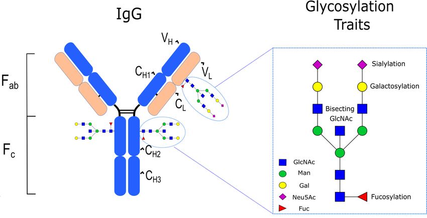
Int. J. Mol. Sci. 2022, 23, 5180                                                                                           2 of 23

                                         The emergent field of glycomics holds great promise for the advancement of biomarker
                                   research in the context of autoimmune diseases, with the goals to uncover disease predis-
                                   position and development, and to assist in the identification of specific molecular targets
                                   for advanced therapies [8–11]. Glycomics refer to the study of the glycome, which consti-
                                   tutes the entire set of glycans present in an organism, tissue, cell, or protein [12]. Glycans
                                   are natural biopolymers that are highly diverse in structure, with profound biological
                                   and immunological significance [13–15], which are known to be explicitly involved in
                                   every major disease pathophysiology [16]. Their functional role in the latter remains to
                                   be fully characterised, partly due to the complexity of glycan biosynthesis, which is a
                                   multi-step enzyme-mediated biochemical process and the most abundant and complex
                                   post-translational modification in eukaryotic cells [17]. Additionally, glycoform distribution
                                   is a product of the interplay between the cell’s genome, transcriptome, and metabolome, as
                                   the subset of the glycan structures synthesised by a particular tissue or cell under different
                                   environmental conditions and physiological states is time-specific and dependent on the
                                   expression levels and activity of glycan-processing enzymes, as well as the availability of
                                   enzyme-related co-substrates, which is affected by metabolic function [18]. Although pro-
                                   tein glycosylation takes place in both healthy and diseased tissues, glycoform distribution
                                   is considered to be disease-dependent [15]. It has been postulated that in autoimmunity,
                                   each autoimmune disorder could be characterised by a distinct glycan signature of immune
                                   cells and serum proteins [9]. These glycan signatures would be site-specific and quantified
                                   by the relative abundance of different glycan structures in these proteins [9].
                                         The relative abundance of N-glycans decorating human plasma proteins has been
                                   found to remain surprisingly stable in healthy individuals, with notable variation majorly
                                   emerging due to aging, pathology, and/or lifestyle changes [19]. This, in addition to
                                   the ease of sample collection, makes human plasma glycoproteins excellent candidates
                                   for the discovery of biomarkers. Of particular interest are the N-glycans found on im-
                                   munoglobulins, particularly immunoglobulin G (IgG), which is a glycoprotein which is
                                   abundantly present in human plasma/serum [20]. In general, immunoglobulins are the
                                   cornerstone of adaptive immunity, and mediate a number of effector immune responses,
                                   including antibody-dependent cellular phagocytosis (ADCP), antibody-dependent cellular
                                   cytotoxicity (ADCC), and complement-dependent cytotoxicity (CDC) [21]. Among their
                                   other roles—including securing protein solubility and conformation, as well as intracellular
                                   transport and clearance—immunoglobulin glycans are responsible for the regulation of
                                   these effector functions [21]. Interestingly, the presence of certain glycan structures in
                                   immunoglobulins has been associated with pro-inflammatory antibody activity [9,22], thus
                                   intensifying the need for a systematic study of immunoglobulin N-glycan profiles and their
                                   role in disease [23,24].
                                         Significant developments in the elucidation of the role of glycosylation in cancer
                                   have been realised in previous decades [25–28], with the consensus that aberrant protein
                                   glycosylation, including that of IgG, is explicitly associated with known hallmarks of
                                   cancer [29,30]. The importance of this association is also reflected in the fact that the
                                   majority of FDA-approved diagnostic biomarkers for tumours used in clinical practice
                                   are glycoproteins or glycan-related [8,29,30]. Thus, N-glycan biomarker discovery for
                                   cancer diagnostics is a relatively mature field, with current research efforts largely being
                                   directed at the design of glycosylation-targeted immunotherapies [31–35]. In contrast, while
                                   some clinically-relevant protein-based biomarkers—including self-reactive IgG antibodies,
                                   also known as IgG autoantibodies—have been identified and used for the diagnosis of
                                   autoimmune diseases [36–38], the study of their glycosylation profiles is less developed.
                                         The aim of this article is to review human N-glycomics studies in the context of
                                   autoimmune diseases. Particular emphasis is placed on the identification of specific glyco-
                                   sylation traits for each autoimmune disease among the reviewed studies based on altered
                                   N-glycan profiles between healthy individuals and patients, and the presentation of their
                                   correlation with other relevant clinical parameters. Owing to the increased complexity
                                   of N-glycosylation, glycoinformatics approaches, including artificial intelligence and sys-
Int. J. Mol. Sci. 2022, 23, 5180 3 of 23
tems glycobiology, are also discussed due to their potential to be applied in N-glycan
biomarker research as a means of enabling patient stratification through the interpretation
of clinical data, as well as providing insight into the link between disease-specific N-linked
glycosylation aberrations and their underlying molecular mechanisms, thus expediting the
translation of such biomarkers into clinical practice.
2. IgG Structure and N-Linked Glycosylation in Healthy Individuals
IgG is one of the five immunoglobulin isotypes in vertebrates (the others being IgA,
IgD, IgE, and IgM), and is the most abundant in terms of its concentration in human serum,
accounting for approximately 10–20% of the total plasma proteome [39]. Similarly to the
other immunoglobulin isotypes, the structure of IgG is characterized by four polypeptide
chains, covalently bound by disulphide bridges, consisting of two identical γ heavy (H)
chains and two identical κ or λ light (L) chains. Each heavy chain is compartmentalised into
four domains—one variable domain (VH) and three constant domains (CH1 , CH2 , CH3 )—
while each light chain comprises just two domains: one variable (VL ) and one constant
(CL ). A further structural subdivision of IgG that is important for its function includes
the Fab, representing the antigen-binding fragment of the IgG which is formed by the VH
and CH1 domains, and the Fc, representing the crystallizable fragment of the IgG which is
responsible for the modulation of its effector functions through the binding to dedicated Fc
receptors (FcγRs) [40,41]. With regard to its glycosylation, IgG has a conserved glycan at
the N297 position (i.e., Asn-297) in each of its CH2 domains of the Fc region. This glycan
typically corresponds to a core structure consisting of N-acetylglucosamine (GlcNAc) and
mannose residues (Man) that can be further embellished with fucose (Fuc), galactose
(Gal), sialic acid (NeuAc), and bisecting GlcNAc (Figure 1), with large population studies
indicating that 30 main N-glycan structures are found in human serum IgG [42]. In healthy
adults, the total serum IgG Fc is highly fucosylated (>90%), contains 35% agalactosylated
(IgG-G0), 35% monoglycosylated (IgG-G1), 15% digalactosylated (IgG-G2), and 10–15%
mono- and disialylated structures (IgG-S) [23]. Approximately 10% of circulating IgG also
contains bisecting Fc glycans [23]. In contrast to IgG Fc N-linked glycosylation, there is
no conserved N-linked glycosylation position in IgG Fab; rather, N-glycosylation sites
can emerge during the somatic hypermutation of the variable domain [43]. In fact, it has
been estimated that approximately 15–25% of serum IgG contains Fab N-glycans [44],
Int. J. Mol. Sci. 2022, 23, x FOR PEER REVIEW
which—compared to Fc N-glycans—are typically biantennary complex-type structures 4 of 25
with a significantly higher extent of sialylation [45].
Figure
Figure 1.
1. IgG
IgG structure
structure and
and glycosylation
glycosylation traits.
traits.
3. Differential Serum IgG N-Linked Glycosylation in Autoimmune Diseases
The presence of autoantigen-specific antibodies of the IgG isotype in human serum
is considered a hallmark of various autoimmune diseases. However, the levels of serum
autoantibodies alone do not seem to be associated with autoimmunity, as natural IgG au-Int. J. Mol. Sci. 2022, 23, 5180 4 of 23
3. Differential Serum IgG N-Linked Glycosylation in Autoimmune Diseases
The presence of autoantigen-specific antibodies of the IgG isotype in human serum
is considered a hallmark of various autoimmune diseases. However, the levels of serum
autoantibodies alone do not seem to be associated with autoimmunity, as natural IgG
autoantibodies can be found in abundance in serum from healthy individuals deprived of
a potential antigen [46,47]. Their glycosylation, as quantified by different traits including
galactosylation, sialylation, fucosylation, and bisecting GlcNAc, is likely a critical aspect of
their role in the pathophysiology of autoimmune diseases. Indeed, experimental evidence
corroborates the importance of glycosylation in the function of IgG autoantibodies, as
their enzymatic deglycosylation leads to a loss of function in vivo [48]. Glycosylation,
and particularly the extent of sialylation, confers a similar functional role in intravenous
immunoglobulin (IVIg) preparations, which are IgG antibodies collected from donor sera,
and are a common treatment option in autoimmune diseases [49]. In particular, the removal
of terminal sialic acid moieties from IVIg abrogates its anti-inflammatory activity [50,51].
Furthermore, the link between glycosylation and autoimmunity may be promising as a
therapeutic target, as the treatment with IVIg, which leads to reduced disease severity in
patients with a neurological autoimmune disease, is also accompanied by a normalisation
of specific glycosylation traits to the respective levels in healthy individuals [52]. Thus, the
identification of differences in the N-linked glycosylation of total serum IgG, as well as
autoantigen-specific antibodies, between healthy individuals and patients would not only
enable accurate diagnosis but also guide the rational design of appropriate therapies.
In order to facilitate the analysis presented herein, autoimmune diseases are grouped into
two categories: rheumatic autoimmune diseases and autoimmune diseases with other patho-
physiological features. Rheumatic autoimmune diseases are grouped according to the classifi-
cation provided by the Harvard Medical School (https://www.health.harvard.edu/diseases-
and-conditions/whats-the-deal-with-autoimmune-disease (accessed on 17 February 2022)).
The altered glycosylation traits reported for each autoimmune disease in different studies
comparing the serum IgG N-glycan profile between healthy individuals and patients are
summarised in Table 1.
Table 1. Comparative serum IgG N-linked glycosylation studies between healthy individuals and
patients with autoimmune diseases.
Altered IgG Glycosylation Traits in Patients
Disease
Total IgG Autoantigen-Specific Antibodies
Galactosylation ↓ [53–80]
Galactosylation ↓ [75,79,84,85]
Sialylation ↓ [55,68,72,78,81,82]
Rheumatoid arthritis Sialylation ↓ [86]
Fucosylation ↑ [75,78,83]
Fucosylation ↑ [84]
Bisecting GlcNAc ↑ [63,68,73,76]
Galactosylation ↓ [53,60,62,63,73,74,87,88]
Sialylation ↓ [88]
Juvenile idiopathic arthritis
Fucosylation ↑ [60]
Bisecting GlcNAc ↑ [63,73]
Galactosylation ↓ [71,73,82]
Osteoarthritis
Sialylation ↓ [82]
Spondyloarthropathies Galactosylation ↓ [73,89,90]
Galactosylation ↓ [56,64,73,91,92]
Sialylation ↓ [92,93]
Systemic lupus erythematosus
Fucosylation ↓ [92]
Bisecting GlcNAc ↑ [92]
Neonatal lupus Galactosylation ↓ [94]
Galactosylation ↓ [95]
Lupus nephritis
Bisecting GlcNAc ↑ [95]Int. J. Mol. Sci. 2022, 23, 5180 5 of 23
Table 1. Cont.
Altered IgG Glycosylation Traits in Patients
Disease
Total IgG Autoantigen-Specific Antibodies
Galactosylation ↓ [63,73]
Sjogren’s syndrome Sialylation ↓ [96]
Bisecting GlcNAc ↑ [63]
Galactosylation ↓ [97,98]
ANCA-associated systemic vasculitis Sialylation ↓ [99–101]
Bisecting GlcNAc ↓ [102]
Galactosylation ↓ [59,64,73,103–109]
Sialylation ↓ [106]
Crohn’s disease
Fucosylation ↑ [109]
Bisecting GlcNAc ↑ [73]
Galactosylation ↓ [59,73,103–109]
Ulcerative colitis Fucosylation ↓ [109]
Bisecting GlcNAc ↑ [73]
Galactosylation ↑ [111]
Hashimoto’s thyroiditis Fucosylation ↓ [110] Sialylation ↑ [111,112]
Fucosylation ↑ [111,112]
Multiple sclerosis Fucosylation ↓ [113]
Galactosylation ↓ [52]
Guillain-Barre syndrome
Sialylation ↓ [52]
Chronic inflammatory demyelinating Galactosylation ↓ [114]
polyneuropathy Sialylation ↓ [114]
Myasthenia gravis Galactosylation ↓ [115]
Galactosylation ↓ [115]
Lambert-Eaton myasthenic syndrome Fucosylation ↓ [115]
Bisecting GlcNAc ↑ [115]
Coeliac disease Galactosylation ↓ [116]
Type 1 diabetes Galactosylation ↓ [117]
Myositis Galactosylation ↓ [118]
Galactosylation ↓ [119]
Autoimmune hemolytic anemia Galactosylation ↓ [119] Sialylation ↑ [119]
Bisecting GlcNAc ↓ [119]
Antiphospholipid syndrome Sialylation ↓ [120]
↓ = decreased; ↑ = increased.
3.1. Rheumatic Autoimmune Diseases
The largest number of differential studies of IgG N-linked glycosylation in biomarker
research published in the last 40 years is focused on rheumatic autoimmune diseases
and, in particular, on rheumatoid arthritis [53–59,61–72,74,75,77,79,80,82,88,121–123], sys-
temic lupus erythematosus [56,59,62,64,73,74,91–93,124], and, to a lesser extent, on Sjogren
syndrome [59,63,73,74] and ANCA-associated systemic vasculitis [97–102,125].
3.1.1. Rheumatoid Arthritis
Rheumatoid arthritis (RA) is the most common rheumatic autoimmune disease, and
is characterised by systemic synovial inflammation, leading to joint destruction and bone
erosion [126]. The impact of RA on patients is not limited to articular joint damage, and
additional clinical manifestations include chronic inflammation affecting other organs, such
as the blood vessels, eyes, skin, lungs, and heart [127].Int. J. Mol. Sci. 2022, 23, 5180 6 of 23
The first study identifying the presence of aberrant IgG N-linked glycosylation be-
tween healthy individuals and patients with RA was carried out by Parekh et al. [82].
After evaluating approximately 1400 oligosaccharide structures from 46 IgG samples, this
seminal work demonstrated that RA patients had significantly reduced galactosylation
compared to healthy individuals, resulting in an increased relative abundance of complex
biantennary N-glycans with terminal GlcNAc residues in one or both antennae. This work
was not only a milestone in the identification of altered glycosylation traits in autoimmune
diseases but was also one of the first demonstrations of glycan-based diagnostics. Sub-
sequent studies have corroborated the prevalence of agalactosylated IgG glycoforms in
RA patients, making reduced IgG N-linked galactosylation a well-established feature of
this disease [53–80]. In addition to aberrant galactosylation, additional RA traits include
reduced sialylation [55,68,72,78,81,82] and increased fucosylation [75,78,83]. The majority
of these studies were carried out in the last 10 years, indicating that the concomitant ad-
vancement in analytical techniques has enabled the discernment of additional N-glycan
features that can be used in tandem with aberrant galactosylation for biomarker discovery.
An appreciable subset of the reviewed studies focused on the identification of correlations
between IgG galactosylation levels and various biological and clinical parameters. It is
important to note that because N-linked IgG galactosylation levels are reported to be age-
related in healthy individuals [128], sample analysis results must first be age-corrected in
order to enable statistically valid comparisons [53,65]. Using linear regression analysis for
results from a 38-patient cohort, Tomana et al. [64] demonstrated that IgG galactose content
and patient age were negatively correlated; however, this finding was not replicated by
the same authors in a study 6 years later [66], with the authors pointing to the relatively
small cohort size (i.e., 11 patients) as a plausible explanation. The patient cohort size may,
nevertheless, not be the deciding factor in this instance because, in a study accounting for a
larger number of patients (i.e., 50), Gindzienska-Sieskiewicz et al. [77] reported no corre-
lation between IgG galactose content and patient age. Although earlier studies found no
correlation between IgG galactose content and several clinical parameters, such as sex, race,
the volume of packed red blood cells, radiographic grade, disability index, extra-articular
manifestations, erosions, corticosteroid use, and RA-specific autoantibody levels [62,64],
Bodman-Smith et al. found a high degree of correlation between IgG galactosylation in RA
patients and their probable outcome [70]. In particular, the probable clinical outcome of
two cohorts (A: 40 patients and B: 24 patients) was successfully predicted (with accuracies
of 95% and 78%, respectively) through discriminant functional analysis based on agalac-
tosylated IgG levels at the onset of the disease, combined with patient age, sex and grip
strength. These findings highlight the importance of incorporating multiple clinical and/or
serological parameters in the statistical analysis of such studies in order to draw clinically
relevant conclusions.
A different approach to the interpretation of aberrant IgG galactosylation was followed
by Axford et al. [69], whose study aimed to investigate the relationship between the extent
of IgG galactosylation in RA patients with the activity of lymphocytic galactosyltransferase
(GalT), which is responsible for the catalysis of galactose transfer from the UDP-galactose
donor to an GlcNAc acceptor within the Golgi. Interestingly, GalT activity was found to
have a negative linear correlation with agalactosylated IgG levels. Based on this finding,
Bodman et al. [58] aimed to distinguish whether the reduced levels of IgG galactosylation
were explicitly associated with the reduced GalT activity, or whether this galactosylation
deficiency was caused by IgG modification by hydrolytic enzymes after secretion. Using
IgGs produced by B-cells in vitro as a reference, it was shown that the latter hypothesis
was probably not the case [58].
Among the different correlations discussed here, altered galactosylation may also be
useful in facilitating patient stratification, as galactosylated IgG levels have been shown
to be negatively correlated with disease activity in RA [53,65,77,79]. A similar negative
correlation appears to exist for sialylation as well [78]. Importantly, this negative correlation
for both glycosylation traits was particularly evident when monitoring galactosylationInt. J. Mol. Sci. 2022, 23, 5180 7 of 23
and sialylation levels in RA patients in order to assess their response to therapy. After
treatment with various pharmacological agents, including monoclonal antibodies, it was
found that RA improvement coincided with an increase in the galactosylation [78,129,130]
and sialylation [129] levels. Finally, the prognostic potential of aberrant galactosylation
as a biomarker in RA has also been investigated [55,79,131]. In a 10-year follow-up study
considering large population cohorts, Gudelj et al. [55] observed that alterations in IgG
galactosylation preceded the onset of RA-related symptoms by a median time of 4.31 years,
which corresponded to the time that had elapsed between blood sampling at the beginning
of the study and the time that a part of the cohort was diagnosed with RA within the
follow-up period. Interestingly though, the authors reported that there appeared to be
no significant statistical correlation between IgG galactosylation and the time preceding
symptom manifestation, suggesting that aberrant galactosylation could be a pre-existing
risk factor in RA.
In RA, the majority of disease incidence is represented by women, with a female to male
ratio of 3:1 [132]. This imbalance is also reflected in the sex ratio of the patient cohort that
was investigated in most of the studies reviewed herein [53–55,64,65,69–71,74,75,79,121].
Generally, within female RA patients, a large percentage (approximately 75–90%) shows an
improvement in disease activity during pregnancy and a subsequent exacerbation, referred
to as ‘flare’, after delivery [133]. Despite this observation, relatively few research efforts
have focused on providing more insight into this phenomenon through IgG N-linked gly-
cosylation studies of pregnant RA patients [57,68,72,123]. Earlier studies demonstrated that
this pregnancy-induced amelioration of RA disease activity was associated with an increase
in IgG galactosylation [57,123]. Despite this important finding, which is consistent with
the independent observations mentioned above reporting a negative correlation between
galactosylated IgG levels and disease activity, the patient cohort size in both studies was
relatively small (i.e., 7 and 23, respectively) [57,123]. Additionally, no information was
provided regarding disease exacerbation taking place postpartum. The comprehensive
study of van de Geijn et al. [72], based on a much larger cohort of 148 patients, accounted
for galactosylation levels of IgG1 and IgG2 both during pregnancy and postpartum, and
corroborated the incidence of increased galactosylation during the first, which reached a
maximum for both IgG1 and IgG2 during the third trimester. The subsequent reduction of
galactosylation levels in the postpartum period could potentially explain the flare experi-
enced by RA patients. A similar trend to that of galactosylation was observed for sialylation
as well, which could be expected because these glycosylation traits are associated [72]. In
a subsequent highly detailed study from the same group, Bondt et al. [68] investigated
several glycosylation traits in pregnant RA patients, particularly galactosylation, fucosyla-
tion, sialylation, and bisecting GlcNAc in the four IgG subclasses. This work accounted
for the largest patient cohort found in pregnancy-focused RA studies, with 219 RA par-
ticipants. The maximum value for both galactosylation and sialylation was also found in
the third trimester of RA patients for all IgG subclasses while, for bisecting GlcNAc and
fucosylation, a minimum was generally observed for all IgG subclasses in the second and
third trimesters, respectively. Importantly, this study underlined that the increased disease
activity in RA was associated with reduced galactosylation independently of sialylation,
thus providing more insight into the causative role of individual IgG glycosylation traits in
the pathophysiology of RA.
Important developments in N-glycan biomarker discovery in RA have also been
made while studying alterations in the glycosylation patterns of RA patients with re-
spect to autoantigen-specific antibodies, particularly anti-citrullinated protein autoanti-
bodies (ACPA). The N-linked glycosylation of ACPA has also been shown to be charac-
terised by reduced galactosylation and sialylation compared to total IgG [84–86]. More re-
cently, ACPA Fab N-glycans have been implicated in the pathophysiology of RA [134–136].
Rombouts et al. [134] showed that the majority of ACPA-IgG are glycosylated in their vari-
able domains and, in a follow-up study, that these glycans are highly sialylated [135]. In
their latest publication, Kissel et al. [136] aimed to elucidate the functional roles of glycansInt. J. Mol. Sci. 2022, 23, 5180 8 of 23
found in the variable domain of ACPA, suggesting that these glycans may mediate the
activation of autoreactive B cells, and thus are, at least in part, involved in the dysregulation
of the adaptive immune response in RA. These important findings pave the way for future
investigations regarding the functional role of autoantigen-specific antibodies in rheumatic
autoimmune diseases, further highlighting the potential of N-glycan biomarker discovery,
combined with its diagnostic and prognostic value, in shedding light on the etiology of
autoimmune diseases.
3.1.2. Systemic Lupus Erythematosus and Sjogren’s Syndrome
Systemic lupus erythematosus (SLE) is a chronic rheumatic autoimmune disease that
affects multiple organs, including the joints, skin, central nervous system, and kidneys [137].
Clinical manifestations can vary significantly, and high incidence rates are found in people
of non-Caucasian ethnicities and women of childbearing age, with a female to male ratio
of up to 13:1 [138,139]. Sjogren’s syndrome (SS) is also a chronic rheumatic autoimmune
disease that is commonly manifested with mouth and eye dryness owing to the inflam-
mation of the salivary and lacrimal glands [140]. This disease is associated with other
rheumatic autoimmune diseases, including RA and SLE, but can also manifest itself alone
(i.e., primary SS).
Compared to RA, the number of comparative serum IgG N-glycomics studies on SLE
and SS is considerably smaller and, especially in earlier years, these studies were a subset of
larger studies that focused primarily on RA [56,59,62,64,73,74,91]. Similarly to RA, reduced
galactosylation has also been generally reported in patients with SLE [56,64,73,91,92] and
SS [63,73]. Contrary to this finding, in an early comparative study to discern disease-
specific aberration in IgG glycosylation in several autoimmune diseases, Parekh et al. [59]
found that serum IgG was normally galactosylated in patients with SLE and primary
SS, excluding, however, a seropositive patient subgroup showing higher levels of agalac-
tosylated IgG. Interestingly, this subgroup met the diagnostic criteria for both SLE and
primary SS [59]. A follow-up study from the same group a few years later [56] also reported
reduced galactosylation in patients with SLE complicated by SS, reinforcing the notion
that stratifying patients between closely-related diseases solely based on aberrant IgG
galactosylation is likely impossible. The most comprehensive study regarding serum IgG
N-linked glycosylation aberration in SLE patients was carried out by Vuckovic et al. [92].
This study was based on a large cohort comprising a total of 475 SLE patients, and was
not limited to the identification of changes in IgG galactosylation, but extended to sialy-
lation, fucosylation, and bisecting GlcNAc. While corroborating the findings of earlier
studies with regard to reduced galactosylation, Vuckovic et al. [92] also reported reduced
sialylation and increased bisecting GlcNAc. The general trend of downregulated galacto-
sylation, sialylation combined with increased bisecting GlcNAc is also discerned in RA
patients (Table 1); however, contrary to RA, it was shown in this particular study that, in
SLE patients, fucosylation was notably reduced. This development shows promise for
finding disease-specific biomarkers within rheumatic autoimmune diseases. An additional
contribution of this work by Vuckovic et al. [92] is the indication that altered IgG glycans
are associated with disease status, disease risk, and symptom severity in SLE patients,
thus potentially opening up new avenues for the exploration of personalised treatments
predicated upon aberrant IgG glycosylation. Regarding SS, fewer developments outside
of downregulated IgG galactosylation have been identified, and the pertinent studies are
more than 20 years old [63,73,96]. Due to the manifestation of SS in patients with RA and
SLE, additional studies employing the improved analytical technologies available today
would be beneficial for the discovery of more disease-specific IgG glycosylation traits in
patients with SS in order to successfully predict and/or differentiate patients from other
rheumatic autoimmune diseases through altered IgG glycosylation.Int. J. Mol. Sci. 2022, 23, 5180 9 of 23
3.1.3. ANCA-Associated Vasculitis
Antineutrophil cytoplasmic antibody (ANCA)-associated vasculitis (AAV) comprises
a group of rheumatic autoimmune diseases, including polyangiitis (GPA), microscopic
polyangiitis (MPA), and eosinophilic granulomatosis with polyangiitis (EGPA) [141]. AAV
is characterised by the inflammation and eventual necrosis of blood vessels with heteroge-
neous clinical manifestations [141,142]. As suggested by the name given to this collection
of diseases, circulating ANCA IgG is considered central to its pathogenesis, primarily
targeting two autoantigens: proteinase-3 (PR3) and myeloperoxidase (MPO) [143,144].
Similarly to the other rheumatic autoimmune diseases presented here, early studies in-
dicated that serum IgGs from ANCA-positive AAV patients show reduced galactosylation
compared to healthy controls [97,98]. Subsequent studies have extended this research to ad-
ditional glycosylation traits, including sialylation, which also appears to be downregulated
in ANCA-positive AAV patients [99–101]. Focusing on GPA, Wuhrer et al. [102] investi-
gated glycosylation aberration with respect to IgG1 and IgG2 between healthy individuals
and patients. Significantly reduced total IgG galactosylation was found for both subclasses
in GPA patients compared to controls of the same age. The total sialylation of IgG1 and
IgG2 was also reported to be correlated with galactosylation and thus reduced in patients
with GPA. Surprisingly, the presence of bisecting GlcNAc residues in the total IgG in GPA
patients was shown to be decreased compared to healthy controls [102]. This appears to
be a deviation from the differential glycosylation traits reported for the other rheumatic
autoimmune diseases presented herein, which may indicate that aberrant bisecting GlcNAc
could be monitored for differentiation between GPA and other rheumatic diseases, such
as RA, SLE, and SS. Finally, the glycosylation profiles corresponding to anti-PR3 specific
IgG1 and total IgG1 in patients with GPA were found to be similar; however, the total
galactosylation and sialylation of IgG1 were found to be weakly or not correlated with their
anti-PR3 specific IgG1 counterparts, respectively.
Aberrant glycosylation profiles in patients with AAV have also been reported to
correlate with disease activity. Lardinois et al. [99] observed that there was a negative
correlation between total IgG1 galactosylation and disease activity, quantified using the
Birmingham Vasculitis Activity Score (BVAS), in samples from PR3-ANCA patients. This
correlation appears to be autoantigen-specific, as MPO-ANCA samples did not show a
correlation. This finding could provide a basis for the differentiation of PR3-ANCA- and
MPO-ANCA-associated AAV diseases. With respect to other glycosylation traits, such as
sialylation, fucosylation, and bisecting GlcNAc, no statistically significant association was
observed with respect to disease activity, regardless of the autoantigen patient sample. In
this study, IgG galactosylation levels were also shown to be reliable indicators of active
PR3-ANCA patients, and were distinguishable from remission and healthy individuals [99].
Regarding autoantigen-specific IgG glycosylation, Espy et al. [101] reported a negative
correlation between anti-PR3 IgG sialylation and BVAS in patients with GPA. However,
this finding was not corroborated by the subsequent study of Wuhrer et al. [102], which
indicated that the bisecting GlcNAc of anti-PR3 IgG was the only glycosylation trait
negatively correlated with BVAS. This discrepancy could not necessarily be attributed to
the size of the patient cohort, as it was comparable in both studies; rather, it could be the
result of the different analytical methods used.
3.2. Autoimmune Diseases with Non-Rheumatic Pathophysiology
3.2.1. Inflammatory Bowel Disease
Inflammatory bowel disease (IBD) refers to a constellation of disorders related to
chronic inflammation in the gastrointestinal tract, including Crohn’s disease (CD) and ulcer-
ative colitis (UC), and is associated with several symptoms, including fatigue, abdominal
pain, and chronic diarrhea [145,146].
Similarly to all of the autoimmune diseases reviewed herein, reduced IgG N-linked
galactosylation in patients with IBD has also been widely reported [59,64,73,103–109]. Within
these studies, the earliest ones—being significant in terms of the introduction of antibodyInt. J. Mol. Sci. 2022, 23, 5180 10 of 23
glycomic analysis concerning IBD—were limited regarding the patient cohort size used, which
did not exceed 60 patients [59,64,73,103,104]. The study by Trbojevic-Akmacic et al. [106]
investigated a markedly larger cohort of patients, namely 287 CD and 507 UC patients, and
corroborated the findings of previous studies on downregulated IgG galactosylation in
both CD and UC. Furthermore, a significant decrease in sialylation was found exclusively
in CD. In a subsequent study from the same consortium, Simurina et al. [109] extended
their analysis to 1065 CD patients and 1009 UC patients, now investigating the aberration
of IgG subclass-specific glycosylation. The findings from their previous study, namely
the downregulation of galactosylation in both CD and UC patients compared to healthy
controls, as well as the downregulation of sialylation seemingly exclusive to CD, were
confirmed in this study as well. Interestingly, a subclass- and disease-specific aberration
in fucosylation was also observed. In particular, IgG1 fucosylation was increased in CD
patients, but IgG2/3 fucosylation was decreased in UC patients. This result could be
promising as a biomarker, which, along with the disease-specific clinical manifestation of
IBD, could potentially facilitate the stratification of patients with CD or UC.
Throughout the last 40 years, research efforts have been undertaken to elucidate
the potential correlation between the changes in IgG glycosylation in IBD patients and
other parameters of clinical importance, such as C-reactive protein (CRP) levels and disease
activity, with ambiguous results [73,103,104,108]. As early as 1990, Dube et al. [103] reported
that galactosylated IgG levels were negatively correlated with CRP levels in CD patients.
This correlation was not statistically significant in UC patients. However, in a subsequent
study, Shinzaki et al. [108] found no such correlation, either for CD patients or UC patients.
Regarding disease activity, the early study by Go et al. [104] reported that IgG galactose
deficiency was positively correlated with clinical activity in CD patients but not in UC
patients. In particular, the molar ratio of mannose to galactose in serum IgG was used as
the monitored glycosylation trait. However, in a subsequent study, Bond et al. [73] found
no association between the relative abundance of galactosylated glycan structures and
disease activity for either CD or UC patients. In turn, Shinzaki et al. [108] showed that
the ratio between the agalactosylated and digalactosylated fraction of fucosylated glycans
(i.e., G0F/G2F) could be a promising biomarker, as it was positively correlated with active
disease both in CD and UC patients. The ambiguity in these results could be an impetus
for further investigation, particularly with regard to the selected glycosylation traits used
to identify potential associations with important clinical parameters.
3.2.2. Autoimmune Thyroid Diseases
Autoimmune thyroid diseases (AITD) represent a group of organ-specific disorders
that dysregulate the function of the thyroid gland, with the most frequent forms being
Hashimoto’s thyroiditis (HT) and Graves’ disease (GD) [147,148]. AITDs are characterised
by the production of autoantibodies targeted at three main autoantigens, namely thy-
roid peroxidase (TPO), thyroglobulin (Tg), and thyroid-stimulating hormone receptor
(TSHR) [148]. The prevalence of anti-TPO and anti-Tg autoantibodies is a known hallmark
in HT, while GD is characterised by the presence of anti-TSHR autoantibodies [147,148].
Studies regarding IgG N-glycome analysis in AITD patients are centered on the glycosy-
lation of autoantigen-specific IgGs and, particularly, anti-Tgs [111,112,149]. Yuan et al. [112]
studied 32 patients with HT, and found that sialylation and fucosylation were increased
compared to healthy controls. More recently, Li et al. [111] investigated the subclass-
specific glycosylation profiles of anti-Tg IgG in patients with HT and GD. With respect
to anti-Tg IgG1, it was shown that sialylation and fucosylation were also increased in HT
patients compared to healthy individuals, a finding which is in agreement with the study
of Yuan et al. [112], which, however, did not account for subclass-specific glycosylation
changes. Interestingly, increased IgG1 galactosylation was reported for HT patients. Re-
garding patients with GD, no aberration was found in anti-Tg IgG1 glycosylation. However,
this was also the case for both HT and GD patients with respect to anti-Tg IgG4 glycosy-
lation, for whom it was shown that no differences could be discerned between healthyInt. J. Mol. Sci. 2022, 23, 5180 11 of 23
controls and patients. These results suggest that autoantigen-specific IgG biomarkers might
not be well-suited for GD diagnosis; however, more studies are required in order to draw
any reliable conclusions. Nonetheless, when it comes to distinguishing between patients
with HT and GD, Zhao et al. [149] showed that fucosylation was significantly decreased in
the former.
Regarding total serum IgG N-glycomic studies, Martin et al. [110] conducted a large
investigation incorporating three independent patient cohorts looking for correlations of
total IgG glycosylation with AITD and autoantibody levels. Fucosylation was shown to be
decreased in AITD patients, and negatively correlated with anti-TPO autoantibody levels.
This finding highlights the fact that aberrant glycosylation compared to healthy controls
can manifest itself differentially in total serum IgG and autoantigen-specific IgG, indicating
potential differences in the underlying regulatory mechanisms of glycosylation.
3.2.3. Neurological Autoimmune Diseases
While a large subset of autoimmune diseases involves disorders that affect the ner-
vous system [150], limited developments have been made regarding serum IgG N-glycan-
based biomarkers compared to the other autoimmune diseases reviewed in this article.
Relevant studies have reported altered glycosylation in patients with multiple sclerosis
(MS) [113,151], Guillain–Barre syndrome (GBS) [52], chronic inflammatory demyelinating
polyneuropathy (CIDP) [114], and myasthenic syndromes, including myasthenia gravis
(MG) [56,115] and Lambert-Eaton myasthenic syndrome (LEMS) [115].
MS is a chronic neuroinflammatory autoimmune disorder affecting the brain and the
spinal cord. Its clinical manifestation is associated with lesions within the central nervous
system (CNS) that promote demyelination and neurodegeneration, with the eventual
disruption of neuronal signalling [152]. Wuhrer et al. [151] focused on changes in IgG1
glycosylation, as intrathecal IgG synthesis is a hallmark of MS, both in the CSF and serum
of a cohort of 48 MS patients. Interestingly, compared to healthy controls, the glycosylation
profiles obtained from serum-derived IgG1 were not significantly altered. However, in a
more recent independent study with a larger patient cohort (i.e., 83 MS patients), serum
IgG core fucosylation was found to be significantly reduced, while a higher prevalence
of high mannose glycans was also observed [113]. Interestingly, antennary fucosylation
in patients’ total plasma proteins was found to be upregulated, thus indicating the multi-
faceted role of the same glycan structures in disease, further complicating the identification
of disease-specific N-glycan changes that can be promptly translated into clinical practice.
GBS and CIDP are also neuroinflammatory demyelinating autoimmune disorders
which, contrary to MS, involve the peripheral nervous system (PNS) [153,154]. Although
rare, the incidence of GBS has recently been reported to be a very rare side-effect after vac-
cination with particular vaccines against COVID-19 [155,156]. Regarding IgG glycosylation
studies, Fokkink et al. [52] investigated subclass-specific N-linked glycosylation patterns in
174 patients with GBS. In particular, the N-linked glycosylation changes in IgG1 and IgG2
were monitored in patients at the onset of GBS and after treatment with IVIg. Before the
administration of IVIg, and compared to healthy individuals, galactosylation was downreg-
ulated in serum IgG1 and IgG2, while sialylation was only downregulated in serum IgG2.
Interestingly, this pro-inflammatory state was shown to partially normalise in patients after
treatment, which could potentially be attributed to elevated levels of galactosylation and
sialylation in IVIg-contained IgG. These findings demonstrate the potential of monitoring
these glycosylation traits in order to assess disease activity and the response to therapy in
GBS. The study of IgG sialylation and its correlation with disease activity has also been a
topic of interest in patients with CIDP. Wong et al. [114] reported that IgG sialylation was
reduced in CIDP patients compared to healthy individuals, and found that IVIg treatment
increased sialylation levels. This study suggested the use of IgG sialylation combined with
the ratio of sialylated to agalactosylated IgG as measures to monitor disease activity and
the response to therapy in CIDP.Int. J. Mol. Sci. 2022, 23, 5180 12 of 23
Myasthenic syndromes, such as MG and LEMS, are neurological autoimmune dis-
orders that involve defects in neuromuscular transmissions, leading to muscle weak-
ness [157,158]. Similarly to MS, GBS, and CIDP, limited research efforts have focused on
IgG N-glycan biomarker discovery in MG and LEMS. Notably, Selman et al. [115] stud-
ied the N-linked glycosylation profiles associated with serum IgG1 and IgG2 in patients
with both MG and LEMS. Importantly, the reported glycosylation changes were found
to be subclass- and disease-specific. With respect to galactosylation, patients with both
diseases exhibited reduced levels; however, in MG this reduction appears to be related
solely to serum IgG1. The sialylation of IgG1 and IgG2 did not differ in MG and LEMS
patients compared to healthy individuals, with fucosylation following the same trend in
MG. Nonetheless, in LEMS patients, downregulated fucosylation was found for serum
IgG2. Finally, the bisecting GlcNAc level in both IgG subclasses was unchanged in patients
with MG, while, contrastingly, in patients with LEMS, this trait was upregulated for serum
IgG1 and IgG2.
It is important to note that the diagnosis of neurological autoimmune diseases, particu-
larly those characterised by demyelination, is difficult due to the similarities in their clinical
manifestation [159]. Thus, the discovery of disease-specific IgG N-glycan biomarkers
would greatly benefit the handling of diseases with such pathophysiology. Despite existing
research efforts, more studies in this direction are required in order to draw informative
conclusions in this regard.
4. Integration of Systems Glycobiology and Artificial Intelligence Approaches in
N-glycomics Biomarker Discovery in Autoimmune Diseases
The discovery of N-glycan biomarkers for autoimmune diseases based on differential
serum IgG glycosylation is a laborious and time-consuming endeavor requiring multi-
disciplinary collaborations. As shown in the studies reviewed in this article, despite the
identification of notable differences in the N-linked glycosylation profiles of IgGs in various
autoimmune diseases (Table 1), significant ambiguity is still present with regard to disease-
specific changes, hampering the establishment of biomarkers that could be promptly trans-
lated into clinical practice. Furthermore, the complexity associated with the mechanisms of
glycan biosynthesis and N-linked glycosylation regulation further complicates the explicit
mapping of serum glycosylation profiles to aberrant cellular functions. Computational
approaches based on systems glycobiology and artificial intelligence/machine learning
Int. J. Mol. Sci. 2022, 23, x FOR PEER REVIEW 13 of 25
could facilitate the incorporation of N-glycan-based biomarkers into diagnosis, prognosis,
the mechanisms of glycan biosynthesis and N-linked glycosylation regulation further
and monitoring (Figure
complicates 2).
the explicit They
mapping could
of serum further
glycosylation profiles to suggest
aberrant cellulartreatment
func- avenues for autoimmune
tions. Computational approaches based on systems glycobiology and artificial intelli-
diseases by providing insight
gence/machine learning intothethe
could facilitate mechanistic
incorporation of N-glycan-based relationships
biomarkers between biological param-
into diagnosis, prognosis, and monitoring (Figure 2). They could further suggest treat-
eters and the resulting
ment avenues for IgG glycoforms,
autoimmune diseases by providing helping tomechanistic
insight into the infer glycanrela- motifs that allow for patient
tionships between biological parameters and the resulting IgG glycoforms, helping to in-
stratification with
fer glycanrespect toforspecific
motifs that allow disease
patient stratification subtypes
with respect and/or
to specific disease sub- disease progression.
types and/or disease progression.
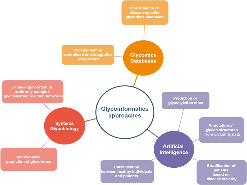
Figure 2. Current trends and developments in glycoinformatics within biomarker discovery.
Figure 2. Current trends and developments in glycoinformatics within biomarker discovery.
4.1. Disease-Specific Glycomics Databases
The need to formalise the analysis of glycans and integrate the glycomic data pro-
duced from a multitude of research efforts into dedicated databases and repositories that
would facilitate their visual interpretation and the extraction of meaningful biological in-
formation is by no means a new idea [160]. In order to this end, significant progress has
been made in recent decades in integrating not only glycomic data but also other omic
data in curated databases and repositories [161–164]. These resources are based on infor-
mation for numerous glycan structures, glycoproteins, glycosylation-relevant genes, and
even glycan–protein interactions [162]. In order to facilitate pertinent research efforts, cen-
tralised and integrative web portals—incorporating multiple bioinformatic resources rel-Int. J. Mol. Sci. 2022, 23, 5180 13 of 23
4.1. Disease-Specific Glycomics Databases
The need to formalise the analysis of glycans and integrate the glycomic data produced
from a multitude of research efforts into dedicated databases and repositories that would
facilitate their visual interpretation and the extraction of meaningful biological information
is by no means a new idea [160]. In order to this end, significant progress has been made in
recent decades in integrating not only glycomic data but also other omic data in curated
databases and repositories [161–164]. These resources are based on information for numer-
ous glycan structures, glycoproteins, glycosylation-relevant genes, and even glycan–protein
interactions [162]. In order to facilitate pertinent research efforts, centralised and integrative
web portals—incorporating multiple bioinformatic resources relevant to glycobiology—
have been developed recently and are available to users. These portals are GlyGen [165]
in the United States (https://www.glygen.org/ (accessed on 17 February 2022)), Gly-
comics@ExPASy [166] in Europe (https://www.expasy.org/search/glycomics (accessed
on 17 February 2022)), and GlyCosmos [167] in Japan (https://glycosmos.org/ (accessed
on 17 February 2022)). The systematisation of such information is a decisive step towards
establishing the study of glycans as an integral part of biology and immunology. However,
distinguishing which pieces of information need to be extracted from various databases and
repositories in order to gain insight into glycan-based biomarker research might be cum-
bersome, and would inevitably require a strong background in glycobiology. This would
potentially hamper the immediate adoption of such biomarkers by clinicians. This issue is
exacerbated by the minimal existence of disease-specific glycomic databases. An important
exemption is GlyConnect (https://glyconnect.expasy.org/ (accessed on 17 February 2022)),
which is a Glycomics@ExPASy integrated platform that allows the collection, monitoring,
and visualisation of glycobiological data, with a particular focus on the characterisation of
the molecular components associated with protein glycosylation [168]. Different data types
are available in GlyConnect, and can be accessed under specific categories, one of which
is ‘Diseases’. Information about glycan types (e.g., N-linked, O-linked, free), as reported
in pertinent scientific references, can be found for several diseases regarding different
glycoproteins of interest, with the user also being given the option to focus on and explore
glycan types regarding human IgG alone. Currently, with regard to the types of disease,
great attention is being paid to different types of cancer, with N-linked glycans associated
with different autoimmune diseases, mainly related to RA, SLE, and SS. The expansion of
GlyConnect to include more references on already-existing entries for autoimmune diseases
(e.g., RA, SLE, and SS), and to gather more data on other autoimmune diseases, would be
an important step in the systematisation of glycobiological data for biomarker discovery
in autoimmune diseases, while at the same time expediting the clinical translation of this
field by helping clinicians without a relevant glycobiological background develop a better
overview of the existing knowledge.
4.2. Artificial Intelligence for Glycomic Analysis and N-glycan Biomarker Discovery
In recent years, artificial intelligence (AI) has found many applications in healthcare,
with machine learning (ML) techniques being particularly useful in clinical research aiming
to improve disease management and enable precision medicine [169,170]. ML can facilitate
disease diagnosis, prognosis, and the stratification of patients with similar diseases and/or
disease types by leveraging diverse datasets, including clinical imaging, electronic health
records, and omic data [171,172]. Several AI and ML applications have focused on diag-
nosis, prognosis, the identification of disease subtypes, and even drug development with
regard to autoimmune diseases, including RA, SLE, IBD, and MS [173,174]. However, the
wealth of data types used in these applications glaringly misses a very important piece of
the puzzle: glycomic data. Indeed, AI approaches integrating glycosylation-related infor-
mation within the context of personalised medicine, and especially towards autoimmune
disorders, are scarce. Most advancements in this regard can be found in ML-powered
glycomic analysis, with artificial neural networks (ANN) and deep learning (DL) being
applied to annotate glycan structures from data [175,176], and to predict glycosylationInt. J. Mol. Sci. 2022, 23, 5180 14 of 23
sites [177–179]. Cancer-related advancements have been realised as well, including the
employment of kernel classifiers, such as support vector machines (SVM), to identify glycan
biomarkers in leukemic cells [180–183]. Regarding autoimmune diseases, several studies
have built logistic regression-based classifiers to infer associations between disease status
and predictors such as age, sex, and IgG glycosylation traits in juvenile-onset RA, SLE, AAV,
IBD, autoimmune cholestatic liver diseases (ACLD), and MS [88,92,99,106,109,113,125,184].
The models have had appreciable success in distinguishing patients from controls, and in
the classification of patients based on disease severity, with their predictive power being
highly improved by the inclusion of glycosylation traits as predictors. Data-driven predic-
tive modelling, such as that demonstrated in these studies, has the potential to expedite
the clinical adoption of N-glycan biomarkers by providing an assessment of the impact
of differential glycosylation in autoimmune disease diagnostics based on quantitative
metrics, such as sensitivity and specificity. These metrics provide a concrete foundation to
evaluate false/true positive/negative rates that can be directly used by clinicians without
any background in glycobiology.
4.3. Lessons Learned from Biotherapeutics Manufacturing: Systems Glycobiology Approaches
Systems glycobiology strives to develop, simulate, and analyse glycobiological sys-
tems at the molecular and cellular level through the integration of multiple omic data
types [35,185]. In the last few decades, several research efforts have been undertaken to
mathematically model N-linked protein glycosylation, with the aim to predict the glycosy-
lation profiles of biotherapeutic products, such as monoclonal antibodies, and to provide
insight into the glycosylation machinery itself [186]. Recently, such mathematical models,
varying in complexity and modelling approaches, have had appreciable success in the pre-
diction of glycoforms [187–190], as well as in the successful reconstruction of the secretory
pathway [191] in Chinese Hamster Ovary (CHO) cell cultures. In these mathematical mod-
els, the prediction of the N-linked glycosylation profiles for a given protein by a specific cell
line was based on a given glycosylation reaction network that associated the intracellular
glycosylation mechanisms with the final N-glycan structures. Such glycosylation reaction
networks are essential for the quantitative analysis of biological data in the field of gly-
comics. In this regard, the early work of Krambeck et al. [192] was a critical step towards the
automated in silico construction of glycosylation reaction networks. Their model, referred
to as “KB2005”, considered a very large network of 22,871 reactions that were catalysed by
11 enzymes, leading to the prediction of 7565 distinct glycan structures with variable extents
of different glycosylation traits, such as galactosylation and fucosylation. By adjusting the
concentrations of the enzymes involved in this particular glycosylation reaction network,
the experimental distributions of the glycoforms of a recombinant protein in CHO cells were
successfully predicted. The same group generalised KB2005 to a more flexible modelling
platform, with the updated name “KB2009”, which incorporated 19 glycosylation enzymes
and enzyme reaction rules to define enzyme specificities [193]. Based on these specificities
and given enzyme concentrations and kinetic parameters, a glycosylation reaction network
could be automatically generated, and could predict the relative abundances of N-linked
glycans. More interestingly, and closely related to the discovery of N-glycan biomarkers,
KB2009 was able to discern differences in enzyme activities between normal and malignant
human monocytes through the analysis of pertinent N-glycan mass spectroscopic data.
Through the integration of transcriptomic data related to enzyme expression levels, the
KB2009 modelling platform was employed to infer enzyme activity changes using mass
spectroscopic data from human prostate cancer cells [194]. KB2009 has also influenced the
independent development of similar N-linked glycosylation modelling platforms, such
as the K2014 platform [195]. Although such system glycobiology approaches can find
immediate application in the identification of glycoengineering targets to improve the
quality and efficacy of biotherapeutic products, as well as in inferring differences in the un-
derlying glycosylation mechanisms among different cell lines [196], they could potentially
benefit N-glycan biomarker research in autoimmune diseases by quantitatively analysingYou can also read