Acquired Glucocorticoid Resistance Due to Homologous Glucocorticoid Receptor Downregulation: A Modern Look at an Age-Old Problem
←
→
Page content transcription
If your browser does not render page correctly, please read the page content below
cells
Review
Acquired Glucocorticoid Resistance Due to Homologous
Glucocorticoid Receptor Downregulation: A Modern Look
at an Age-Old Problem
Lee-Maine L. Spies              , Nicolette J. D. Verhoog and Ann Louw *

                                          Department of Biochemistry, Stellenbosch University, Van de Byl Street, Stellenbosch 7200, South Africa;
                                          18244017@sun.ac.za (L.-M.L.S.); nverhoog@sun.ac.za (N.J.D.V.)
                                          * Correspondence: al@sun.ac.za

                                          Abstract: For over 70 years, the unique anti-inflammatory properties of glucocorticoids (GCs), which
                                          mediate their effects via the ligand-activated transcription factor, the glucocorticoid receptor alpha
                                          (GRα), have allowed for the use of these steroid hormones in the treatment of various autoimmune
                                          and inflammatory-linked diseases. However, aside from the onset of severe side-effects, chronic GC
                                          therapy often leads to the ligand-mediated downregulation of the GRα which, in turn, leads to a
                                          decrease in GC sensitivity, and effectively, the development of acquired GC resistance. Although the
                                          ligand-mediated downregulation of GRα is well documented, the precise factors which influence this
                                          process are not well understood and, thus, the development of an acquired GC resistance presents an
                                          ever-increasing challenge to the pharmaceutical industry. Recently, however, studies have correlated

                                   the dimerization status of the GRα with its ligand-mediated downregulation. Therefore, the current
                                          review will be discussing the major role-players in the homologous downregulation of the GRα pool,
Citation: Spies, L.-M.L.; Verhoog,
N.J.D.; Louw, A. Acquired
                                          with a specific focus on previously reported GC-mediated reductions in GRα mRNA and protein
Glucocorticoid Resistance Due to          levels, the molecular mechanisms through which the GRα functional pool is maintained and the
Homologous Glucocorticoid Receptor        possible impact of receptor conformation on GC-mediated GRα downregulation.
Downregulation: A Modern Look at
an Age-Old Problem. Cells 2021, 10,       Keywords: glucocorticoid receptor alpha; glucocorticoids; glucocorticoid receptor dimerization;
2529. https://doi.org/10.3390/            acquired glucocorticoid resistance; ubiquitin-proteasome system
cells10102529

Academic Editors: Marcel J.
M. Schaaf and Onno C. Meijer              1. Introduction
                                               The pharmacological use of glucocorticoids (GCs) can be traced back over a century
Received: 27 August 2021
                                          to the work of the clinical physician, Solomon Solis-Cohen, who reported on the therapeu-
Accepted: 21 September 2021
Published: 24 September 2021
                                          tic benefits of orally administered adrenal gland extracts in the treatment of asthma [1].
                                          However, due to the previously reported bronchodilator effect of adrenaline, these benefits
Publisher’s Note: MDPI stays neutral
                                          were assumed to be a result of adrenaline from the adrenal medulla [2,3]. Later, however,
with regard to jurisdictional claims in
                                          Thomas Addison would also report on the therapeutic benefits of adrenal extracts; this time,
published maps and institutional affil-
                                          in the treatment of chronic fatigue, muscular degeneration, weight loss and darkening of
iations.                                  the skin, in what would later be called Addison’s disease [4,5]. As the therapeutic potential
                                          of adrenal extracts became apparent, the search for its active factor became internationally
                                          competitive. In 1946, Edward Calvin Kendall isolated four steroidal compounds from
                                          adrenal extracts, one of them, cortisol [6]. Later that year, cortisol was synthesized by
                                          Sarett [7] and, three years later, rheumatologist Phillip Hench proved cortisol to be ex-
Copyright: © 2021 by the authors.
Licensee MDPI, Basel, Switzerland.
                                          tremely beneficial in the treatment of rheumatoid arthritis [2]. Merck and company then
This article is an open access article
                                          began the commercial production of the synthetic GC, cortisone and, in 1950, Kendall
distributed under the terms and
                                          and Hench, together with Swiss biochemist, Tadeus Reichard, received the Nobel Prize in
conditions of the Creative Commons        Physiology and Medicine for their work regarding adrenal hormones [2]. This catapulted
Attribution (CC BY) license (https://     GCs into the limelight and these synthetic steroids quickly became the mainstay therapeutic
creativecommons.org/licenses/by/          choice in the treatment of auto-immune and inflammatory-linked conditions [8,9].
4.0/).

Cells 2021, 10, 2529. https://doi.org/10.3390/cells10102529                                                      https://www.mdpi.com/journal/cells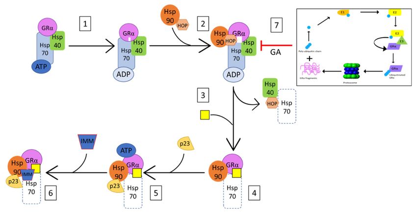
Cells 2021, 10, 2529                                                                                            2 of 27

                             The excitement surrounding this wonder drug would, however, quickly die-down
                       as reports of adverse side-effects [10,11] and GC insensitivity [12,13] arose, specifically in
                       cases were high doses of GCs were used for long periods of time. As GC centered research
                       progressed, it was discovered that cortisol (F) and corticosterone, the main endogenous
                       GC in humans and rodents, respectively, are produced and secreted in response to circa-
                       dian/ultradian and stress cues, and that, in addition to their anti-inflammatory properties,
                       these steroids also regulate life-sustaining processes, such as growth, development, and
                       metabolism [8,14–16]. GCs were found to predominantly regulate these processes via the
                       binding and activation of the cytoplasmic ligand-activated transcription factor (TF), the
                       glucocorticoid receptor alpha (GRα) [17–19]. Once bound, the GC-GRα complex regulates
                       the transcription of genes through various activation and repression mechanisms. Gener-
                       ally, direct binding of the GC-GRα complex, as a homodimer, at glucocorticoid response
                       elements (GREs) in the promoter of GC-responsive genes, is associated with transcrip-
                       tional activation, while transcriptional regulation via the monomeric GC-GRα complex
                       is associated with gene transrepression. The latter occurs via both DNA-dependent and
                       independent mechanisms, with DNA-dependent monomeric GC-GRα signaling occurring
                       via the binding of the complex to negative glucocorticoid response elements (nGREs), and
                       DNA-independent monomeric signaling occurring via tethering of the complex to DNA-
                       bound TFs [20,21]. Additional investigation of GC-GR signaling found that dimer-mediated
                       GRα signaling resulted in the transactivation of genes involved in glucose synthesis and
                       fat metabolism, which would explain the many undesirable side effects that often accom-
                       pany GC therapy. In contrast, monomeric GRα signaling results in the transrepression of
                       pro-inflammatory genes by disrupting the action of TFs, such as nuclear factor kappa B
                       (NFκB) and activator protein 1 (AP-1) [5,8,22]. Since only the immunosuppressive prop-
                       erties of GCs are clinically desired, GC-centered research quickly shifted to the possible
                       functional separation of monomeric and dimeric GRα signaling. While it soon became
                       apparent that this approach was a gross over-simplification of GRα signaling, the strategy
                       would give rise to the development of selective glucocorticoid receptor agonists (SEGRAs)
                       and selective glucocorticoid receptor modulators (SEGRMs). These ligands, which are
                       collectively referred to as SEGRAMS, were essentially designed to preferentially induce
                       monomeric signaling of the GC-GRα complex, over dimeric signaling, in an effort to retain
                       the potent anti-inflammatory properties of GCs while limiting the many undesirable side
                       effects [8,15,23,24]. While some of these ligands have been somewhat successful in ad-
                       dressing the adverse side-effects of GC therapy [25–27], the development of GC-mediated
                       resistance still remains a threat and is under-researched.
                             Currently, approximately 1% of the population of the United Kingdom and the United
                       States undergo long-term GC treatment, of which 30% experience some degree of GC
                       insensitivity [28]. More specifically, up to 10% of asthma patients, ~30% of rheumatoid
                       arthritis and Crohn’s disease patients, and 90% of chronic obstructive pulmonary disease
                       (COPD) and sepsis patients experience GC resistance [28–30]. Various regulatory processes,
                       which modulate either GRα function or GRα expression, are responsible for a decrease in
                       GC sensitivity and, ultimately, GC resistance. Based on the underlying cause, GC resistance
                       may be categorized into two major groups: generalized GC resistance and acquired GC
                       resistance [31,32]. Primary generalized GC resistance or hereditary GC resistance mostly
                       affect all tissue types and is characterized by disruptions in GRα functionality, affecting the
                       subcellular localization, ligand binding, and transactivation ability of the receptor, amongst
                       others. Alterations in GRα function largely stem from mutations and/or polymorphisms
                       of the GR gene (NR3C1 in humans, and Nr3c1 in mice), an increase in non-transcriptionally
                       active GR isoforms (e.g., GRβ, GRγ, GR-A) due to alternative splicing events [33,34], and
                       changes in GC sensitivity due to a pro-inflammatory environment [16,35,36]. Unlike gener-
                       alized GC resistance, acquired GC resistance or secondary GC resistance, mainly affects
                       specific tissues and/or cells in a biological system and is hallmarked by the homologous
                       down-regulation of the GRα [30,37], rather than disruptions in receptor function. This form
                       of GC resistance occurs more frequently and has been linked to a plethora of psychological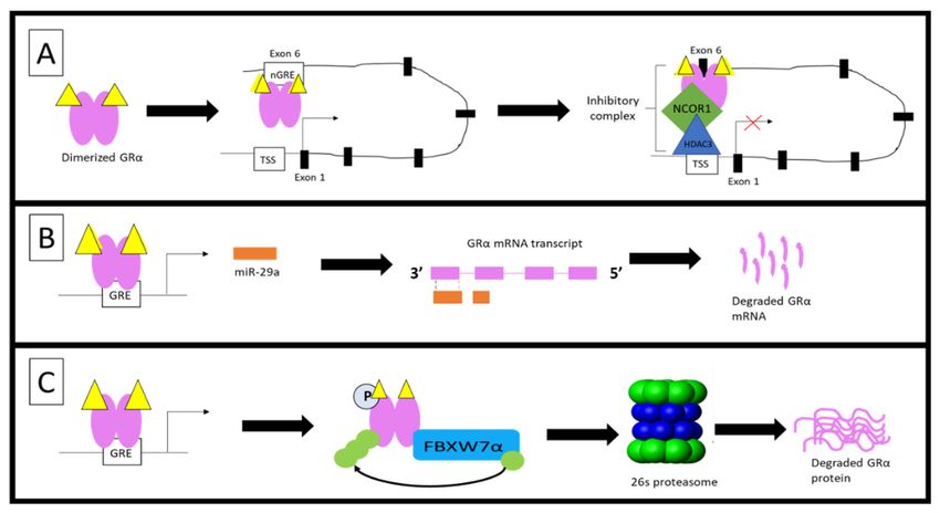
Cells 2021, 10, 2529                                                                                             3 of 27

                       and pathological conditions/diseases [38–42]. The molecular mechanisms behind general-
                       ized GC resistance are better understood, allowing for more precise diagnostic tools [43].
                       In contrast, however, the molecular mechanism underlying acquired GC resistance, is rela-
                       tively unknown and thus, diagnosis of this condition requires more exhaustive diagnostic
                       approaches in order to determine GC sensitivity of specific tissue and/or cells [30,43]. The
                       current review will thus be discussing the major role-players in the homologous down-
                       regulation of the GRα pool, with a specific focus on previously reported GC-mediated
                       reductions in GRα mRNA and protein levels, the molecular mechanisms through which
                       the GRα functional pool is maintained and the possible impact of receptor conformation
                       on GC mediated GRα downregulation.

                       2. Acquired GC Resistance
                             Acquired GC resistance is often brought on by the homologous downregulation of
                       GRα mRNA and protein as a result of an increase in circulating GCs, as a consequence
                       of either, chronic GC therapy or as a side-effect of the pathophysiological processes that
                       accompany the inflammatory disease state, or both. This section briefly discusses the
                       interrelatedness of disease- and treatment associated reductions in GRα mRNA and protein
                       levels, before a more in-depth discussion of the known molecular mechanisms which
                       govern GC-mediated GRα downregulation.
                             Stress-related reductions in GRα mRNA and protein levels are among the most stud-
                       ied disease-associated modulations of the GRα pool, with a reduction in GRα mRNA
                       and/or protein being noted in various human and rodent studies, in response to pre-/post-
                       natal, physical and psychological stress [39,44–47]. Factors which influence the extent
                       of the stress-related reduction in the GRα pool include, length of exposure to the stres-
                       sor, the environment in which the stress occurs and how sensitive the individual is to
                       stress [39,44–46]. Many, but not all, stress-related modulations of the GRα pool have been
                       linked to an increase in circulating endogenous GCs [39,47,48], subsequently resulting in
                       the homologous downregulation of GRα as a protective mechanism against the detrimental
                       effects of prolonged stress-related activation of GRα signaling. In addition to a decrease
                       in GC sensitivity, a reduction in the GRα pool has been implicated in the increased sus-
                       ceptibility to various psychological and pathological conditions, such as depression and
                       schizophrenia [49,50]. While GRα expression is extremely diverse in patients suffering from
                       psychological conditions, many display increased activation of inflammatory processes
                       and HPA-axis signaling, resulting in hypercortisolaemia and a decrease in the GRα pool in
                       peripheral tissues [51].
                             Likewise, a significant number of patients who suffer from auto-immune and
                       inflammatory-linked diseases, such as atopic dermatitis (AD) [41], systemic lupus erythe-
                       matosus (SLE) [52], steroid resistant Type II asthma [53,54], chronic obstructive pulmonary
                       disease (COPD) [55,56], and adult immune thrombocytopenia (ITP) [42], have reduced GRα
                       mRNA expression, and thus, reduced GC sensitivity within peripheral blood mononuclear
                       cells (PBMCs). Additionally, many patients suffering from auto-immune or inflammatory-
                       linked conditions or cancer [57–59], receive GC treatment, thus, it is difficult to distinguish
                       between disease-associated and treatment-associated reductions in the GRα pool, within
                       these groups [60]. It should be noted, however, that GC resistance within these disease
                       groups may occur via factors other than the reduction of GR.
                             Many studies have noted a reduction in GRα mRNA and protein levels in response to
                       exogenous GC-treatment. More specifically, Dexamethasone (Dex), a potent GRα agonist, has
                       been shown to cause up to a 90% reduction in GRα mRNA and/or protein levels [25,61–69],
                       with one study demonstrating the complete downregulation of GRα [64]. Similarly, in vivo
                       and ex vivo studies demonstrate a Dex-induced decrease in the GRα mRNA and/or protein
                       pool in various mouse and rat tissues, with many of these studies correlating with the reduction
                       through an increase in GC-insensitivity [25,65,68,70–72]. In addition to Dex, other exogenous
                       GCs (e.g., hydrocortisone [73], triamcinolone acetonide [74] and various prednisolone-based
                       steroids [60,75–79] have also been linked to a decrease in the GRα mRNA and/or protein pool.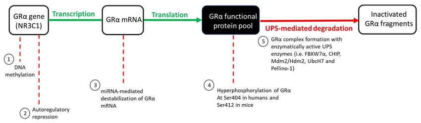
Cells 2021, 10, 2529                                                                                                           4 of 27

                                      In summary, both disease- and treatment-associated reductions in the GRα pool
                                 contribute to GC insensitivity and the development of acquired GC resistance (Figure 1).
                                 With GC resistance remaining one of the two major drawbacks in GC treatment, it has
                                 never been more important to identify and understand the molecular mechanisms involved
                                 in the homologous downregulation of GRα.

      Figure 1. Disease- and treatment-associated effects lead to acquired GC resistance. Disease-associated (orange boxes)
      triggering of the stress-signaling pathway often results in the dysregulation and subsequent hyperactivation of HPA
      axis signaling, which, in turn, leads to the overproduction and secretion of endogenous GCs, GRα downregulation and,
      ultimately, acquired GC resistance. Similarly, the GC-mediated treatment of autoimmune and inflammatory conditions
      (green boxes), results in an increase in circulating exogenous GCs, which mediates the downregulation of GRα and,
      subsequently, acquired GC resistance. Complicating matters further, patients suffering from a disease-associated reduction
      in GRα levels often receive GC-treatment, further compounding the development of acquired GC-resistance. Although
      the development of acquired GC resistance generally occurs due to a reduction in the GRα functional protein pool, it is
      important to note that other cellular processes may also be involved in the development of this condition.

                                 3. Molecular Mechanisms Involved in the Homologous Downregulation of GRα
                                      The GRα protein pool, like many other proteins, is regulated at several levels by vari-
                                 ous molecular mechanisms within living cells. Wilkinson et al. [30,69] eloquently describe
                                 the regulation of the GRα pool as a ‘push (synthesis) vs. pull (degradation)’ mechanism.
                                 When unperturbed, GRα synthesis and degradation exists in a state of dynamic equilib-
                                 rium, with these processes occurring at roughly the same rate, thus ensuring that the GRα
                                 pool remains constant. GR gene transcription, mRNA translation, and protein synthesis
                                 are defined as the ‘push’ of the mechanism, while the ‘pull’ refers to GRα degradation
                                 through the ubiquitin proteasome system (UPS) (Figure 2). The dynamic equilibrium state,
                                 in which the GRα pool exists under basal physiological conditions, may be perturbed by
                                 an increase in circulating GCs (endogenous or exogenous), subsequently increasing the
                                 GC-induced downregulation of the GRα functional pool. The GC-mediated regulation
                                 of the GRα functional pool includes the epigenetic, transcriptional, post-transcriptional
                                 and post-translational regulation of the receptor and occurs in an intricate and specific
                                 manner to either stabilize or destabilize the GRα [31,80], with the destabilization of GRα as
                                 a potential contributor to acquired GC resistance. While the ligand-independent regulation
                                 of GRα levels also occurs [31,32,80], the current review specifically focuses on the molecular
                                 mechanism through which GCs mediate the downregulation of the GRα.
Cells 2021, 10, 2529 5 of 27
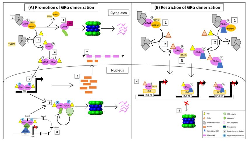
Figure 2. GC-mediated regulation of the GRα functional protein pool. Under basal physiological conditions, GRα synthesis
(indicated in green) and degradation (indicated in red) occur at roughly the same rate. An increase in circulating GCs,
however, disrupts the equilibrium of these processes, subsequently allowing for an increase in GRα downregulation via
various mechanisms: (1) DNA methylation of the GR gene promoter inhibits the initiation of GRα gene transcription, (2) the
formation of a repressive autoregulatory loop through which GC-bound GRα decreases the expression of its own nascent
mRNA, by forming a long-range interaction with a NCOR1-deacetylase 3-containing repression complex, (3) the binding of
miRNAs to AU-rich regions of the mRNA transcript, destabilizes mature GRα mRNA, (4) hyperphosphorylation of the
receptor at Ser404 in humans, and Ser412 in mice, serves as a signal for the UPS-mediated degradation of GRα protein
which occurs via (5) complex formation between GRα and several UPS enzymes. Hyperphosphorylation of the GRα refers
to the phosphorylation of the receptor above that of its basal phosphorylation.
3.1. GRα mRNA Regulation
Although the GRα is ubiquitously and constitutively expressed, vast heterogeneity
exists in the expression of the receptor in different cells and tissues. While this is mostly due
to the differential regulation of the GR gene within cells, GCs have been shown to impact the
regulation of GRα mRNA at various levels. The following section will thus highlight these
GC-mediated effects on GRα epigenetic, transcriptional and post-transcriptional regulation.
3.1.1. Epigenetic Regulation
DNA methylation of the GR gene promoter is one of the mechanisms through which
an increase in circulating GCs mediate the downregulation of GRα mRNA and protein
levels [81–83]. Several psychological and behavioural studies have linked an increase in
GR gene methylation to a decrease in GRα mRNA and/or protein expression, in response
to increases in circulating GCs [39,46,48,84,85]. Specifically, McCormick et al. [85] identified
the promoter region of the exon variant 17 as the region of the Nr3c1 gene that undergoes the
most significant methylation when rat pups experience ‘stressful’ conditions, with studies
by Zhu et al. [46] and Niknazar et al. [39] demonstrating that this GC-mediated increase in
the methylation status of exon 17 promoter results in a decrease in GRα mRNA expression
in adult Wistar rats. Supporting these findings, Mifsud et al. [84] demonstrated that an
increase in circulating GCs, caused by acute stress, results in an increase in the methylation
status of the exon 17 promoter, which in turn, leads to a decrease in GRα mRNA expression
of up to 75% in male Wistar rats. However, in contrast, Witzmann et al. [86] indicate that
chronic, rather than acute stress, resulted in increased methylation of the promoter of
exon 17 in peripheral tissues. Moreover, exon 17 methylation has also been shown to be
important for regulation of the mouse Nr3c1 gene, as an increase in the methylation status
of this exon results in a reduction of GRα protein expression in mice [87].
Similarly, human studies demonstrate a negative correlation between DNA methy-
lation of the NR3C1 gene and GRα mRNA and protein expression [44,45,47,58,87]. For
example, studies by McGowan et al. [45] and Wang et al. [47] correlate DNA methylation
of exon 1F of the human NR3C1 gene with a decrease in GRα mRNA expression in the
brain hippocampus and PBMCs, respectively. Wang et al. [47] further relates this decrease
in GRα mRNA expression to a decrease in GC sensitivity within PBMCs.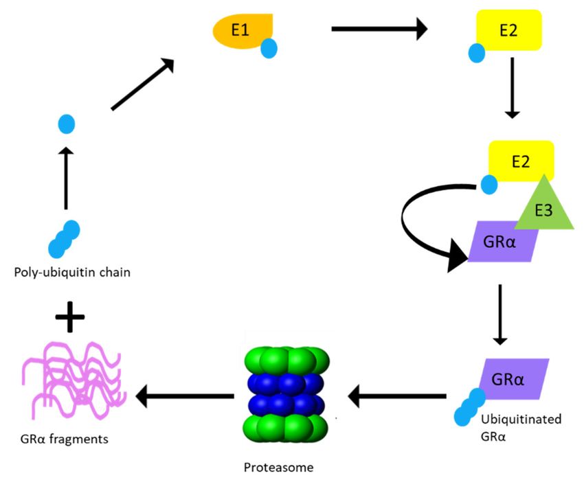
Cells 2021, 10, 2529                                                                                          6 of 27

                            Although the studies discussed, focus on the methylation of the promoter of the
                       GR exon variant, 17 , in rats, or its human homolog, exon 1F , it should be noted that the
                       human GR exon 1F (or 17 in rats) was found to account for only 1–5% of total GR mRNA
                       transcription within the brain [88] and peripheral tissues [89]. As such, the contribution of
                       GR gene methylation at these sites to total GC-mediated downregulation of GRα may be
                       negligible. Although other exon promoters, such as exon 1B , 1C and 1H , have been shown
                       to undergo GC-mediated methylation within the NR3C1 gene, with an increase in the
                       methylation status of these exon promoters relating to a reduction in GRα mRNA and/or
                       protein expression [44,58,87], generally these other exons have been less investigated.

                       3.1.2. Transcriptional Regulation
                            As previously described, the monomeric GC-GRα complex can transrepress genes
                       through both an indirect tethering mechanism and by directly binding to a nGRE within
                       the promoter of genes. Over 28 years ago, Burnstein et al. [90–92] postulated that the
                       GR gene contains sequences which allow for the GC-mediated downregulation of GRα
                       mRNA. In line with this postulation, Ramamoorthy et al. [68] identified the presence of
                       an intragenic nGRE in exon 6 of the GR gene and subsequently demonstrated the GC-
                       mediated regulation of GRα through a direct interaction of the receptor with this nGRE.
                       Ramamoorthy et al. [68] went on to describe a GC-mediated autoregulatory loop that
                       functions by inhibiting transcriptional initiation of the GR gene, consequently result-
                       ing in a decrease in nascent GRα mRNA expression of up to 90% in some cells such as
                       A459 lung carcinoma cells. This transrepression of the GR gene is mediated via a long-
                       range interaction between the GC-GRα complex at the nGRE and a NCOR1-deacetylase
                       3 (HDAC3)-containing repression complex, assembled at the transcriptional start site of
                       the gene. The GC-dependent ability of GRα to regulate its own transcription was found to
                       be consistent across human, mouse and rat cell lines, as well as in mouse tissues [68]. An
                       increase in circulating GCs (whether endogenous or exogenous) is likely to augment this
                       constitutive repression of the GR gene.
                            Interestingly, studies by Hua et al. [93,94] identify the sumoylation of GRα at lysine
                       293 (K293) in humans and K310 in mice (Figure 3), as a prerequisite for Dex-mediated
                       GRα transrepression. The group illustrates the association of sumoylated and Dex-bound
                       GRα with SMRT/NCOR1-HDAC3-containing repression complexes at nGRE sites within
                       the promoters of genes, confirming that sumoylation of the GRα is indispensable in the
                       above-described autoregulation of GRα transcription. Sumoylation is one of the many
                       post-translational modifications (PTMs) which GRα undergoes and is described in more
                       detail in an upcoming section.

                       3.1.3. Post-Transcriptional Regulation
                             Post-transcriptional regulation results in the destabilization of the mature GRα mRNA
                       transcript, rather than modulation of the levels of nascent GRα mRNA as is the case in
                       transcriptional regulation of the GR gene. Destabilization of the mature GRα mRNA transcript
                       is mediated by the incomplete base pairing of micro-RNAs (miRNAs) to adenylate uridylate
                       (AU)-rich elements within the 30 -untranslated region (UTR) of the GRα mRNA transcript.
                       The binding of these small (20–25 nucleotides), single-stranded non-coding RNAs, generally
                       prevent the translation of the mRNA transcript and protein expression; however, similar to
                       small interference RNAs (siRNAs), the binding of miRNAs could also result in the degradation
                       of transcripts [95–98]. The downregulation of GRα mRNA mediated by miRNAs has been
                       shown to occur across species [80,84,99–101] in response to an increase in circulating GCs,
                       whether endogenous (disease-associated) or exogenous (treatment-associated), and is thus
                       implicated in the development of acquired GC resistance.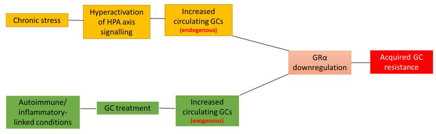
Cells 2021, 10, 2529                                                                                                           7 of 27

      Figure 3. Post-translational modification sites for human (top) and mouse (bottom) GRα pertaining to phosphorylation,
      ubiquitination and sumoylation. The human GRα protein consists of 777 and the mouse GRα of 784 amino-acid residues,
      with both of these proteins undergoing PTMs at various sites. Phosphorylation (light blue) occurs primarily at serine
      residues while, sumoylation (yellow) and ubiquitination (purple) occur at lysine residues for both receptors. Moreover,
      phosphorylation at S404 in humans and S412 in mice (dark blue), as well as ubiquitination of K419 in humans and K426
      in mice, is known to modulate receptor levels. The precise site of symolation which affect GRα stability, has not yet been
      determined. Indicated in red are the alanine to threonine point mutations which yield the dimerization deficient GR
      mutant, GRdim.
                                       As it pertains to rodent studies, four miRNAs (miR-96, miR-101a, miR-142-3p and
                                 miR-433p) have been shown to reduce GRα mRNA expression by up to 40% in mice ex-
                                 posed to adrenocorticotropic hormone (ACTH), which resulted in increased GC levels [99].
                                 In addition, Mifsud et al. [84] demonstrated a stress related time-dependent increase in
                                 miR-124a that led to a reduction in GRα mRNA expression in male Wistar rats. Addition-
                                 ally, elevated levels of neuronally expressed miRNA-124-3p, was found to result in GRα
                                 downregulation, decreased GC sensitivity and an increase in depression-like symptoms in
                                 several rat and mouse studies [100,102].
                                       Similarly, human studies negatively correlated miRNA-124-3p expression to GC
                                 sensitivity and GRα protein expression in ALL patients who display decreased sensitivity
                                 to GC therapy [101]. Additionally, the addition of miRNA-124-3p to a GC-sensitive cell
                                 line in vitro decreased the anti-proliferative and proapoptotic effect of GC therapy [101].
                                 Similarly, elevated expression of miRNA-142-3p in ALL patients was also correlated with a
                                 decreased sensitivity to GC therapy [103].
                                       Because miRNAs mainly function by blocking the translation of GRα mRNA tran-
                                 scripts, their effects are often only reflected at GRα protein level. This is demonstrated in
                                 studies by Vreugendenhil et al. [100] and Shimizu et al. [104] who investigated the effects
                                 of miR-18 and miR-124a on GRα mRNA expression in rats and mice, respectively. Both
                                 studies found that these miRNAs did not significantly affect GRα mRNA expression, but
                                 that GRα protein expression decreased.

                                 3.2. GRα Protein Regulation
                                      While GC-mediated regulation of GRα synthesis occurs, it is well established that
                                 GC-mediated regulation of the GRα pool occurs primarily via receptor protein degradation.
                                 This section will, thus, focus on GC-induced post-translational modifications of GRα,
                                 which induces receptor protein degradation, and the molecular mechanism through which
Cells 2021, 10, 2529 8 of 27
degradation is mediated. While this section primarily reports on GRα protein degradation
via the ubiquitin–proteasome system (UPS), it is important to note that GRα protein
degradation also occurs via a lysosomal-dependent pathway. However, because there is
no evidence to support a GC-mediated increase in the downregulation of GRα protein
levels via lysosomal activity [105,106], this mode of receptor protein degradation has been
omitted from the current review.
3.2.1. Post-Translational Regulation
Once synthesized, GRα undergoes a number of PTMs which impact GC-responsiveness
in selective tissues and may contribute to acquired GC resistance. The most important
PTMs, which are directly involved in GRα protein turnover via the proteasome, are ubiqui-
tination, phosphorylation and, less frequently, sumoylation (Figure 3) [16,107–109].
A number of amino acid sites are hyperphosphorylated in a GC-dependent manner
during the PTM of the GRα [80]. Hyperphosphorylation of these sites is known to modulate
GRα function as well as expression, however, for the purpose of this review only phos-
phorylation sites which affect GRα protein expression will be discussed. Specifically, the
GC-mediated hyperphosphorylation of human GRα at Serine 404 mediates the receptor’s
turnover, resulting in a decrease in GRα protein levels [110] and, therefore, plays an impor-
tant role in GC sensitivity. Studies by Galliher-Beckley et al. [110] provides evidence that
Dex-induced hyperphosphorylation, mediated by glycogen synthase kinase 3β (GSK3β), at
Ser404 for the human GRα (and Ser412 for mouse GRα) (Figure 3), enhances the turnover
of the receptor. Moreover, use of a mutant incapable of being phosphorylated at Ser404
or inhibition of phosphorylation at this site by a GSK3β inhibitor, 6-bromoindirubin-30 -
oxime (BIO), confirmed that restricting Dex-induced Ser404 hyperphosphorylation of GRα
resulted in an increase in receptor protein stability. Additionally, unliganded human GRα
was found to undergo constant phosphorylation and dephosphorylation at Ser-203 and
Ser-211, suggesting that, in the absence of ligand, different populations of phosphorylated
GRα are present in vivo [111].
In order for the degradation of the receptor to take place following phosphorylation,
GRα undergoes ubiquitination [61,66]. Previous studies have identified the single site
for ubiquitination to be within the PEST (a peptide sequence that is rich in proline (P),
glutamic acid (E), serine (S), and threonine (T) residues) degradation motif at lysine 426
(K426) in mice and lysine 419 (K419) in humans [61,66]. Furthermore, mutations at these
specific residues restore GRα protein expression by restricting the downregulation of the
receptor via the proteasome [61,66] (Figure 3). Once targeted for proteasomal degradation
through phosphorylation, GRα is covalently linked at the specific lysine residue to a
chain of ubiquitin molecules (polyubiquitin chain), which serves as a recognition motif for
specific ubiquitin-binding subunits of the proteasome. The attachment of the 76-amino
acid ubiquitin peptide to GRα requires the action of at least three sets of enzymes. Single
ubiquitin peptides are first linked to E1 ubiquitin-activating enzymes before they are
transferred to E2 ubiquitin-conjugating enzymes. The specificity of the final transfer
of ubiquitin from the E2 enzyme is dependent on E3 ubiquitin-protein ligases, which
provides specific substrate recognition. Two different classes of E3 ligases that use distinct
mechanisms to bring about substrate-specific transfer of ubiquitin to the target proteins
are involved. E3 ligases of the really interesting new gene (RING) finger family facilitate
the transfer of ubiquitin from an E2 enzyme to the target protein, whereas homologous to
the E6-AP C-terminus (HECT) domain, E3 ligases transfer a covalently linked ubiquitin
that was acquired from an associating E2 enzyme [112]. Although the involvement of both
RING finger and HECT domain E3 ligases have been described in the general proteasomal
pathway, to date only RING finger E3 ligases have been shown to interact with the GRα
(Figure 4).Cells 2021, 10, 2529 9 of 27
Figure 4. Proteasomal degradation of GRα via the ubiquitin–proteasomal system (UPS). Ubiquitina-
tion of the GRα requires multiple rounds of enzymatic processing before proteasomal degradation
occurs. An activating enzyme, E1, activates ubiquitin and transfers it to a conjugating enzyme, E2.
Hereafter, an E3 ligase from the RING finger family binds the GRα and recruits it to E2, which
transfers the activated ubiquitin to K419 of the human GRα or K426 of the mouse GRα, until a
poly-ubiquitin chain is formed. Once the poly-ubiquitin chain is complete, GRα is delivered to
the proteasome and degraded, resulting in the release of GRα protein fragments and the poly-
ubiquitin chain.
In addition to phosphorylation and ubiquitination, GRα also undergoes sumoyla-
tion [93,107,109,113–116]. Sumoylation involves the covalent attachment of small ubiquitin-
related modifiers (SUMO) to target proteins (i.e., GRα). While the sumoylation pathway
shares many similarities with the UPS, there are distinct differences between the enzymes
of these pathways. Specifically, the activation of SUMO by E1 activating enzymes is an
ATP-dependent reaction, which results in the transference of SUMO to an E2 conjugative
enzyme (in the case of GRα sumoylation the E2 enzyme is Ubc9), which subsequently
attaches it to various lysine residues of the target protein.
While sumoylation is known to regulate the protein–protein and protein–DNA inter-
actions of GRα, as well as receptor localization [93,107,109,113–116], one study specifically
implicates the overexpression of the symoylation protein, SUMO-1, in the Dex-induced
downregulation of GRα protein and provides evidence of proteasomal involvement in
this downregulation [109]. In addition to being the only study to link sumoylation to
GC-mediated GRα protein downregulation, the study also does not identify at which site
(i.e., lysine residue) sumoylation occurs (Figure 3).
3.2.2. UPS Enzymes That Modulate GRα Protein Levels
Various E2 conjugating enzymes and E3 ligases have been shown to interact with GRα
to facilitate both ligand-dependent and ligand-independent regulation of receptor protein
levels (Table 1). These UPS enzymes and their effect on GRα protein expression will be
reviewed in the following section. Note that while the E3 ligase, E6-AP, has been shown toCells 2021, 10, 2529 10 of 27
interact with GRα, its effect on GRα protein expression has not been determined; therefore,
it will not be discussed [117,118].
Table 1. UPS enzymes known to interact with the GRα.
Type of UPS GRα Ligand-Binding GRα Phosphorylation Effect on GRα
Name References
Enzyme Status Status Protein Level
E2 conjugating
UbcH7 Liganded Hyperphosphorylated Reduce [119]
enzyme
Inactive E2
TSG101 Unliganded Hypophosphorylated Stabilize [111,120,121]
conjugating enzyme
FBXW7α Liganded Hyperphosphorylated Reduce [69,121,124–126]
Phosphorylation- [112,124,127–
CHIP Ligand-independent Reduce
independent 131]
Ligand-independent but Phosphorylation-
Mdm2/Hdm2 Reduce [124,132–136]
E3 ligase requires p53 independent
(RING finger family)
Pellino-1 Liganded Hyperphosphorylated Reduce [137]
[122–124]
UBR1 Unliganded Hypophosphorylated Reduce [124,138,139]
Phosphorylation-
RNF6 Ligand-independent Stabilize [140]
independent
Siah2 Ligand-independent Hyperphosphorylated Reduce [141]
The E2 conjugating enzyme, Ubiquitin-conjugating enzyme (UbcH7) has been shown
to interact directly with the C-terminus of GRα in order to modulate the transactivation and
protein expression levels of the receptor in a hormone-dependent manner [119]. Specifically,
in the presence of Dex, an increase in the nuclear association of UbcH7 and GRα is observed,
which results in a decrease in GRα protein levels due to GC-mediated downregulation of the
receptor via the UPS [119]. Furthermore, Garside et al. [119] show that the overexpression
of a dominant negative UbcH7 mutant, which is unable to transfer ubiquitin molecules,
resulted in the increased stability of GRα and restricted the GC-mediated downregulation
of the receptor via the UPS. The latter highlights the importance of UbcH7 in the ligand-
induced downregulation of the GRα protein.
Tumor susceptibility gene 101 (TSG101) is an inactive E2 conjugating enzyme of the
UPS that binds to unliganded, hypo-phosphorylated GRα. TSG101 resides in the cytoplasm
and consists of a C-terminal coil domain, a central proline-rich segment and a N-terminal
ubiquitin-conjugating (E2) variant domain [111,120]. The latter is, however, inactive due
to the lack of a cysteine residue required to form a thioester bond with ubiquitin [111],
while the coil domain interacts with the N-terminal AF-1 region of the GRα, which pos-
sess intrinsic transcriptional activation potential [111,120]. Experiments in which TSG101
was targeted for knockdown demonstrated a decrease in the stability of unliganded,
hypophosphorylated GRα, thus revealing that TSG101 protects unliganded GRα from
receptor downregulation [111]. Additionally, the study demonstrates the phosphorylation-
dependent nature of the interaction between TSG101 and unliganded GRα, through the
use of a human GRα phosphorylation mutant, GR(S203A/S211A). TSG101 was shown to
have an increased association with GR(S203A/S211A), compared to its interaction with
the unliganded wild type receptor. This was thought to be due to the lack of basal phos-
phorylation at S203/S211 of the mutant receptor. This is in stark contrast to the wild-type
receptor, which constantly undergoes phosphorylation and de-phosphorylation cycles,
even in its unliganded state. More recently, Wilkinson [121] reported on the importance of
GRα phosphorylation status and its association with TSG101, by investigating the effect
of the dimerization abrogating, non-steroidal GRα ligand, 2-(4-acetoxyphenyl)-2-chloro-
N-methyl-ethylammonium chloride (CpdA), on the phosphorylation of human GRα at
S404. As previously stated, the ligand-mediated hyperphosphorylation of S404 is associ-
ated with an increase in GRα protein downregulation. Wilkinson illustrates that CpdA
treatment prevents the hyperphosphorylation of S404 which, in turn, resulted in the partialCells 2021, 10, 2529 11 of 27
stabilization of the liganded receptor. Furthermore, considering the previously reported
inhibition of S203 phosphorylation by CpdA [142] and the association and stabilization
of hypophosphorylated GRα by TSG101, Wilkinson postulated that TSG101 association
is important for the ability of CpdA to maintain GRα protein levels. Investigation into
the possible link between CpdA-binding and TSG101 association in the stabilization of
GRα revealed an increase in GRα protein expression following CpdA treatment in the
presence of endogenous TSG101, but not following TSG101 knockdown experiments [121],
suggesting that TSG101 is indeed required for the stabilization of GRα levels by CpdA.
F-box and WD repeat domain containing 7 alpha (FBXW7α) forms an SCF (Skp1/Cul1/
F-box) type of E3 ubiquitin ligase complex that is known to target various proteins, includ-
ing the GRα, for ubiquitination and proteasomal degradation [69,125,126]. The binding
of this catalytically active E3 ligase requires the GC-mediated phosphorylation of the
CDC4 phosphodegron motif of GRα, in order to mediate ubiquitination and proteasomal
degradation [126,143]. Specifically, FBXW7α binding to GRα is dependent on the GSK3β-
mediated phosphorylation of human GRα at Ser404, upon ligand binding [110]. Thus,
ligand binding and the subsequent phosphorylation of Ser404, serves as a signal for ubiq-
uitination and proteasomal degradation of GRα [125]. FBXW7α knockdown experiments,
resulting in a significant increase in GRα protein levels in both HeLa and HEK293 cells,
in a manner similar to the UPS inhibitor, MG132 [125], provides evidence for the role of
FBXW7α in the GC-mediated downregulation of GRα. Furthermore, it was shown that
a GRα phosphorylation mutant (Ser404A) was unable to undergo GC-mediated ubiquiti-
nation, which restricted the proteasomal degradation of the receptor [125]. Additionally,
Wilkinson et al. [69] confirms, through the use of co-immunoprecipitation experiments,
which were validated by proximity ligation assays (PLAs), that treatment with Dex and
cortisol (F) resulted in an increase in the association of human GRα (hGRα) with FBXW7α.
In contrast, unliganded or monomeric hGRα did not induce an interaction between hGRα
and FBXW7α.
Another E3 ligase, the carboxy-terminus of Hsc70 interacting protein (CHIP), is a
known modulator of basal nuclear receptor (NR) expression and availability, prior to ligand
binding [144]. This E3 ligase binds to and decreases the ATPase activity of heat shock
proteins (Hsps), the binding of which is known to stabilize and mediate the appropriate
folding of NR proteins [145,146]. Thus, the CHIP-mediated disruption of the NR-Hsp
interaction results in the misfolding and subsequent degradation of these NRs [127,131].
As it pertains to GRα, CHIP has been shown to ubiquitinate the receptor and directly
target the GRα for degradation via the UPS [131]. Additionally, CHIP has been shown to
bind to not only unliganded, but also liganded, GRα, thus acting independently of the
phosphorylation status of the receptor [112,127]. Wang et al. [112] confirms a role for CHIP
in mediating the GRα turnover of liganded GRα by showing enhanced GRα ubiquitination
and subsequent degradation when CHIP is overexpressed. Moreover, Wilkinson [121]
observed some GRα ubiquitination following treatment with dimerization abrogating
ligand, CpdA, suggesting that monomeric GRα is capable of being ubiquitinated, however,
it may not efficiently engage with the proteasome, thus evading degradation. Taking
into account that monomeric GRα restricts the interaction of the receptor with the E3
ligase, FBXW7α, this demonstration of GRα ubiquitination following treatment with CpdA
supports the notion of monomeric GRα, stabilizing an interaction with the other E3 ligases,
such as CHIP. In support of this, Tateishi et al. [147] provides evidence that unliganded
ER degradation is inhibited in cells where CHIP is absent, however, ligand-induced ER
turnover in these cells still occurs. Therefore, Wilkinson [121] suggests that predominantly
monomeric GRα associates with a complex involving CHIP and the catalytically inactive E2-
conjugating enzyme, TSG101, whereas dimeric GRα, following treatment with dimerization
promoting ligands such as Dex or F, preferentially associates with FBXW7α, rather than
CHIP or TSG101.
In contrast to the other UPS enzymes, the E3 ligase, murine double minute 2 (Mdm2
or Hdm2, the human homologue) depends on the presence of p53 to form a trimericCells 2021, 10, 2529 12 of 27
complex with GRα, in order to mediate GRα protein turnover via the UPS. The GRα-
p53-Mdm2/Hdm2 complex mediates GRα protein turnover independent of ligand bind-
ing [134]. More specifically, the interaction of GRα and p53 requires the ligase activity of
Mdm2 [136]/Hdm2 [132] for the successful ubiquitination and proteasomal degradation of
GRα. Sengupta et al. [132] demonstrated that Dex treatment enhances the association of
GRα and p53, subsequently resulting in the increased downregulation of GRα. Addition-
ally, the disruption of the interaction between p53 and Hdm2 inhibited the Dex-mediated
ubiquitination of GRα and p53 within human umbilical endothelial cells, suggesting that
the Dex-mediated downregulation of GRα requires the association of GRα with p53, the as-
sociation of p53 with Hdm2 and the ligase activity of Hdm2. These results were confirmed
by Kinyamu et al. [136], who found that both p53 and Mdm2 were required for estrogen-
mediated GRα protein down-regulation via the proteasomal degradation pathway in MCF
7 M cells (MCF-7 cells which were stably transfected with mouse mammary tumor virus
promoter-luciferase (MMTV-LUC) reporter and GRα expression constructs).
Pellino homolog-1 (Pellino-1) is a 47 kDa long protein which, due to its modulation
of inflammatory signaling pathways, is considered a key component in inflammation,
autoimmunity and tumorigenesis, with various studies indicating a positive correlation
between its expression and grade of inflammation [148–150]. Due to the aforementioned,
Petrillo et al. [137] investigated the role of Pellino-1 in the Dex-mediated reduction in
GRα protein expression following β-arrestin-1 knockdown. The study noted that GRα
associates with β-arrestin-1 in the cytoplasm in the absence of ligand and this complex
formation persists after Dex treatment and translocation into the nucleus. Subsequent β-
arrestin-1 knockdown experiments, however, revealed a time dependent reduction in GRα
protein expression following Dex treatment in both MEF and human lung adenocarcinoma
(A549) cells. The study then went on to confirm UPS involvement through PLA using
antibodies directed at GRα and ubiquitin, respectively. PLA signals confirmed an increase
in GRα ubiquitination in the absence of β-arrestin-1, following GRα activation by Dex [137].
An in vivo ubiquitination assay confirmed GRα ubiquitination to be poly-ubiquitination
and not mono-ubiquitination with the former known to target proteins for proteasomal
degradation, while the latter regulates protein trafficking [151]. Further qPCR results
identified PELI1 as a novel GC-responsive gene, which is upregulated in the absence of
β-arrestin-1. The PELI1 gene codes for the transcription of the Pellino-1 protein, which was
found to exclusively interact with GRα in the absence of β-arrestin-1 [137]. The association
of Pellino-1 with GRα significantly increased the Dex-mediated reduction in receptor
levels through the addition of a K48-linked ubiquitin chain to GRα. The study concluded
that Pellino-1 expression is upregulated in the absence of β-arrestin-1, allowing for the
association of Pellino-1 to ligand-activated GRα, which subsequently enhanced receptor
polyubiquitination and, thus, GRα proteasomal degradation [137].
Ubiquitin protein ligase E3 component N-recognin 1 (UBR1) belongs to a subset
of E3 ligases, which specifically recognizes degradation signals within the N-terminal
domain of proteins. These N-terminal signals are collectively referred to as N-degrons
and include, but are not limited to, the polyubiquitylation of N-terminal lysine residues,
which target the protein for proteasomal-dependent degradation [138,139,152]. Having
previously demonstrated the role of UBR1 in the degradation of misfolded kinases upon
Hsp90 inhibition [153], Sultana et al. [138] sought to determine the role of this E3 ligase in
the degradation of the known Hsp90 client protein, GRα, upon Hsp90 inhibition. Specif-
ically, the study illustrated the reduced rate of GRα protein degradation upon Hsp90
inhibition within UBR1 knockdown MEF cells, compared to wild type cells. Additionally,
co-transfection experiments with rat UBR1 (rUBR1) and HA-tagged GRα (HA-GRα) in-
dicated that rUBR1 overexpression resulted in a significant decrease in HA-GRα, even
in the absence of Hsp90 inhibition [138], suggesting that GRα protein levels are sensitive
to the expression of UBR1, independent of Hsp90 functionality. More recently, studies
by Vu et al. [139] show an increase in both GRα mRNA and protein expression upon
UBR1 knockdown, as well as the protein–protein association of UBR1 with a GRα isoformCells 2021, 10, 2529 13 of 27
(GR-D), which primarily consists of the receptor LBD. However, following Hsp90 inhi-
bition, GR-D underwent rapid degradation despite the total knockdown of UBR1 [139].
This suggests that UBR1 is not absolutely required for GRα downregulation upon loss
of Hsp90-mediated protection, which is in agreement with previous studies that have
demonstrated the modulation of NR expression by many proteolytical pathways [154].
Taken together, UBR1 facilitates the clearance of misfolded Hsp90 client proteins upon
inhibition of the protein chaperone; however, cellular quality control mechanisms are able
to function independently of its presence.
RING-type E3 ubiquitin transferase (RNF6) is known for its ubiquitination of vari-
ous substrates, including the androgen receptor (AR) [155], transducin-like enhancer of
split 3 (TLE3) [156] and the tyrosine phosphatase, SHP-1 [157], in order to mediate the
proliferation of various types of cancer cells. The overexpression of RNF6 has also been
linked to the development of doxorubicin resistance and, thus, leukemogenesis [158]. Since
many multiple myeloma (MM) patients experience some degree of Dex resistance, a novel
study by Ren et al. [140] sought to uncover a possible role for RNF6 in GC resistance
in MM patients. The study found that RNF6 is overexpressed in various MM cell lines
and that the presence of RNF6 increased the atypical K63-linked ubiquitination of GRα.
RNF6 expression was found to stabilize unliganded GRα, even when receptor synthesis is
inhibited by cycloheximide (CHX), increasing GRα half-life from 8 to 24 h, while RNF6
knockdown experiments resulted in a decrease in GRα protein levels [140]. Moreover,
the study provides evidence that RNF6 binds to the LBD of the GRα and that its binding
increases GRα transcriptional activity of prosurvival genes. MTT assays, furthermore, indi-
cated that the ectopic expression of RNF6 significantly increased the viability of MM cells
in the absence of Dex-treatment. Moreover, Dex treatment alone significantly decreased
cell viability, an effect that was partially overturned by RNF6 expression. This suggests
that the overexpression of RNF6 antagonizes the anti-proliferative action of Dex, with the
latter describing a possible mechanism for GC resistance in MM [140].
Lastly, a recent study by Burke et al. [141] identified the E3 ligase, seven-in-absentia-
mammalian homolog-2 (Siah2) as a modulator of GRα protein levels. Siah2 is a known
regulator of fat mass and inflammation within adipose tissue [159,160] and has been shown
to lead to the agonist-dependent activation of steroid receptors, such as GRα [156]. The
study, which made use of either wild type mice (SIAH2+/+) or mice with a global deletion
of SIAH2 (SIAH2-/-), found that corticosterone treatment led to an up to 42% decrease
in GRα protein levels in SIAH2+/+ mice. In contrast, SIAH2-/- mice showed elevated
levels of GRα protein, both in the absence and presence of corticosterone. Additionally,
the overexpression of wild type Siah2 led to a significant reduction in GRα protein levels,
an effect not evident in the presence of an enzymatically inactive mutant of the E3 ligase,
suggesting that the enzymatic function of Siah2 is required for its modulation of GRα
protein levels.
3.2.3. Hsp90 as a Modulator of GRα Stability
In addition to UPS enzymes, the protein chaperone, heat shock protein 90 (Hsp90), is
known to play a significant role in the stabilization of GRα [145,161–163]. The interaction
between unliganded-GRα and Hsp90 is highly ATP-dependent and initiated by Hsp70-
Hsp90 organizing protein (HOP), a Hsp90 co-chaperone, and a GRα bound Hsp70 assembly
complex (Figure 5). This heterocomplex facilitates the direct binding of Hsp90 to GRα,
upon which the receptor becomes competent for hormone binding [162]. Following this
transition, HOP, Hsp70 and several Hsp70 co-chaperones dissociate from the ligand-bound
GRα-Hsp90 complex, allowing for the association of p23 and one of many tetratricopeptide
repeat domain proteins (i.e., immunophilins (IMM)) (Figure 5). In the absence of Hsp90
machinery, the ligand binding clefts of GRα shifts between closed and open states, with
the open state exposing hydrophobic residues of the receptor to the surrounding solvent.
Prolonged exposure of these residues to the solvent may result in the unfolding and
destabilization of the GRα, which in turn, results in the Hsp70-dependent ubiquitinationCells 2021, 10, 2529 14 of 27
of the receptor [145,146,164]. Additionally, studies by Fang et al. [162] demonstrate the
interaction of ligand-bound GRα with Hsp90 within the nucleus, where Hsp90 binds
GRα by imitating the interaction of the receptor with transcriptional coactivators. These
studies establish Hsp90 as a stabilizer of both liganded and unliganded GRα. Furthermore,
inhibition of the direct association of Hsp90 with GRα, though the use of geldanamycin
(GA), has been shown to trigger the UPS-mediated degradation of GRα [138,165] (Figure 5).
Figure 5. Simplified mechanism of GRα-Hsp90 association as adapted from Pratt et al. (145). (1) Hsp70 binds GRα in a
Hsp40- and ATP-dependent manner, in order to facilitate the priming of the ligand-binding cleft (indicated in white) and
the subsequent association of Hsp90 and Hop (2) Association of Hsp90 and Hop to the GRα-Hsp70 complex, results in the
complete opening of the receptor’s ligand binding cleft (indicated in white), which allows for (3) the dissociation of Hsp40,
Hop, as well as some of the Hsp70, and (4) the subsequent ligand-binding of the receptor (indicated by yellow rectangle).
(5) Bound to Hsp90, GRα is now in its ATP-dependent conformation, allowing for the recruitment of p23, which stabilizes
and prevents dissociation of the GRα-Hsp90 complex. (6) The dissociation of Hop at step 3 allows for the association of
immunophilins (IMM) to the vacant tetratricopeptide repeat (TRP) acceptor site of bound Hsp90. Additionally, (7) Inhibition
of GRα-Hsp90 heterocomplex formation, by GA results in the UPS-mediated degradation of GRα.
4. GRα Downregulation and Receptor Conformation
While previous studies surrounding the impact of GRα dimerization on GRα signaling
were mainly aimed at addressing the number of adverse side-effects which accompany
GC therapy [166–168], rather than GC resistance, noteworthy reports have been made
regarding the role of the GRα dimerization state on receptor turnover and, thus, in effect,
the development of an acquired GC resistance.
Originally investigated for its role as a contraceptive [169,170], the SEGRAM CpdA
has become a useful tool in the investigation of dimerization dependent GRα signaling.
Not only was this GR modulator shown to bind and activate the GRα, but it was also
shown to retain the anti-inflammatory activity of GRα signaling, while preventing many of
the metabolic side effects which accompany GC therapy [25–27,171]. In vitro and in vivo
studies into the molecular mechanism, through which CpdA mediates these effects, showed
that, in contrast to the dimerization promoting, Dex, binding of the non-steroidal ligand
inhibits dimerization of the GRα, effectively ensuring monomeric signaling of the recep-
tor [25,172–174]. Additionally, unlike Dex, CpdA treatment was shown to maintain both
GRα mRNA and protein levels [67,142,171,175], suggesting that the dimerization status of
the receptor may influence the ligand-mediated regulation of the GRα. In agreement withCells 2021, 10, 2529 15 of 27
the latter, Wilkinson et al. [69,121] showed that treatment with dimerization promoting
GR agonists, Dex and F, resulted in an almost 50 h decrease in the half-life of GRα protein,
compared to the unliganded receptor, which was shown to have a half-life of 70-h. In stark
contrast, CpdA treatment did not affect the half-life of GRα protein. In fact, 72 h of CpdA
treatment significantly increased GRα protein levels. Further investigation into the molecu-
lar mechanism of CpdA revealed that this GRα modulator prevents the GSK3β-mediated
hyperphosphorylation of S404 of the human GRα. As previously stated, the hyperphos-
phorylation of this site by dimerization promoting ligands, such as Dex, has been shown to
lead to GRα instability and subsequent degradation. Moreover, Wilkinson et al. [69,121]
showed that the lack of S404 phosphorylation of the human GRα by CpdA restricts the
interaction of the receptor with the E3 ligase enzyme, FBXW7α, inhibiting the subsequent
degradation of the receptor via the proteasome [69,121].
As with CpdA, the restriction of GRα dimerization, through the use of the dimeriza-
tion deficient GRα mutant, GRdim, has recently proven beneficial in the study of acquired
GC-resistance [69,121,176]. Originally developed in 1994, this mutant receptor, which
contains an Alanine to Threonine substitution in the dimerization loop of the DBD of
the GR gene [177] (Figure 3), has been shown to have a restricted dimerization ability,
compared to wild type GRα [178,179]. While the majority of GRdim-centered research
focusses on improving the therapeutic index of GCs, with many illustrating this success-
fully [166,180,181], recent studies by Glantschnig et al. [176] and Wilkinson et al. [69,121]
highlight the dimerization dependent downregulation of both GRα mRNA and protein.
On a post-transcriptional level, Glantschnig et al. [176] found that Dex-activated GRα
increased the expression of miR-29a which, in turn, downregulated GRα levels through
the destabilization of GRα mRNA. Interestingly, the miR-29-mediated downregulation of
GRα mRNA was abolished in the GRα dimerization deficient mouse embryonic fibroblasts
(MEF-GRdim) cell line, suggesting, that the Dex-induced downregulation of GRα via
miR-29a is dimerization dependent [176]. On a post-translational level, Wilkinson [69,121]
shows that hGRwt protein undergoes significantly more Dex-induced downregulation,
compared to hGRdim. Moreover, hGRdim protein was found to be unaffected by up to
72 h of Dex treatment, in stark contrast to hGRwt protein, which was downregulated in a
time-dependent manner. Furthermore, Dex treatment was unable to induce the hyperphos-
phorylation of S404 in hGRdim; in fact, the receptor was completely void of even basal
phosphorylation at this site. The lack of receptor phosphorylation restricted the association
of GRdim with FBXW7α and inhibited the proteasomal degradation of the GRdim [69,121].
In addition to the abovementioned, restriction of GRα dimerization has also been
shown to inhibit the repression of receptor gene transcription. More specifically,
Ramamoorthy et al. [68], who reported on the Dex-mediated autoregulatory loop, through
which the GRα negatively regulates its own transcription, notes that treatment with the
GRα antagonist, mifepristone (RU486), does not induce the formation of the complex
involved in the downregulation of GRα mRNA. Moreover, the study demonstrates no
significant change in GRα mRNA levels following RU486 treatment, as well as restoration
of GRα mRNA levels upon Dex and RU486 co-treatment, relative to Dex treatment alone.
RU486, which has also been shown to be less effective in downregulating GRα protein
levels [182], significantly restricts GRα dimerization, compared to Dex [183].
Taken together, these studies identify three principal regulatory mechanisms through
which dimerization promoting GRα agonists control receptor expression (Figure 6). A
lack in our current understanding of the dimerization-dependent regulation of GRα levels,
however, is the collective effect of these processes, as, to the best of our knowledge, no pub-
lished study has investigated the combined transcriptional, translational and proteasomal
involvement in the GC-mediated downregulation of GRα within an endogenous system.
While the study by Wilkinson [69,121] accounts for translational and proteasomal regula-
tion of GRα, it was unable to address transcriptional regulation due to the predominant
use of transiently transfected receptors.You can also read


