Vasculitis associated with the use of an intrauterine device: A case report
←
→
Page content transcription
If your browser does not render page correctly, please read the page content below
EXPERIMENTAL AND THERAPEUTIC MEDICINE 22: 1277, 2021

                               Vasculitis associated with the use of
                               an intrauterine device: A case report
                    AURELIAN MIHAI GHITA1,2*, CRISTINA ALEXANDRESCU3, SANZIANA ISTRATE3*,
                                    ANCA EVSEI4 and ANA CRISTINA GHITA2*

    1
     Department of Physiology II, Faculty of Medicine, ‘Carol Davila’ University of Medicine and Pharmacy, 050474 Bucharest;
           2
            Department of Ophthalmology, Ocularcare Eye Clinic, 011204 Bucharest; 3Department of Ophthalmology,
                  Faculty of Dentistry, ‘Carol Davila’ University of Medicine and Pharmacy, 050474 Bucharest;
                      4
                        Department of Pathology, ‘Sf. Maria’ Clinical Hospital, 011172 Bucharest, Romania

                                         Received April 20, 2021; Accepted May 20, 2021

                                                    DOI: 10.3892/etm.2021.10712

Abstract. Retinal vasculitis is a complication associated with        Introduction
a local condition or it can be a retinal expression of a systemic
inflammatory disorder, which initially may go unnoticed.              Vasculitis is the inflammation of the blood vessel wall
Drug‑associated vasculitis is frequently difficult to identify,       secondary to an abnormal immune response (1,2). Systemic
because many patients follow treatments with more than one            vasculitis determines sufferance and fibrinoid necrosis of the
drug and the route of administration varies. A 35‑year‑old            endothelial cells, events followed by vascular leakage and blood
female patient presented with sudden hearing loss, headache           clot formation with secondary occlusion (1,3). Eventually, all
and blurred vision that had started two weeks earlier and had         these vascular events determine retinal ischemia and dysfunc‑
become progressively worse. Ophthalmological examination              tion of the organ (3).
revealed anterior uveitis, bilateral optic disc swelling and               Retinal vasculitis can be a complication of a local condition
retinal vasculitis. The orbito‑cerebral MRI, the CT scan and          or it can be a retinal expression of a systemic inflammatory
the serological tests were within normal limits. Unable to            disorder, which may initially be unnoticed (3‑5). The causes
identify the cause of the retinal vasculitis, the patient's medical   of retinal vasculitis are multiple and often overlap, making
history was reviewed. The patient had recently had a gyneco‑          both the diagnosis and the treatment options challenging (5).
logical procedure, where a 13.5 mg levonorgestrel intrauterine        Retinal vasculitis is frequently associated with inflammation
contraceptive device was implanted. After the device was              of the adjacent tissues, such as the choroid or the vitreous, but
removed and methylprednisolone treatment started, the patient         sometimes, remote ocular structures also appear to be caught
presented a visible remission of the symptoms and signs. To the       in the inflammatory process (6‑8).
best of our knowledge, there is no case of retinal disease and             Retinal vasculitis can be associated with infectious and
optic disc edema associated with auditory problems caused by          non‑infectious conditions (3‑5). The non‑infectious causes of
an intrauterine device. A proper examination, correlated with         vasculitis include ocular disorders and can be drug‑induced,
a very thorough medical history, could identify rare diseases         i.e., vasculitis associated with a systemic inflammatory
and associations, in order to provide adequate medical care.          disease and vasculitis associated with malignancies (9‑11).
                                                                      Drug‑associated vasculitis is frequently difficult to identify,
                                                                      because many patients follow treatments with more than one
                                                                      drug and the route of administration varies (12‑15). In addi‑
                                                                      tion, patients may forget, neglect or hide drug intake. For
                                                                      instance, an intrauterine device (IUD) is considered a medical
Correspondence to: Dr Aurelian Mihai Ghita, Department of             device. In terms of prevention of an undesired pregnancy,
Physiology II, Faculty of Medicine, ‘Carol Davila’ University of      IUDs are usually considered safe with rare side effects, some
Medicine and Pharmacy, 8 Eroii Sanitari Bvd., 050474 Bucharest,       of which can be severe (16). Currently, there are two types
Romania                                                               of IUDs available: The copper IUD, which releases copper
E‑mail: mihai.ghita@ocularcare.ro                                     ions, and the hormonal IUD, which releases a synthetic form
                                                                      of the progesterone hormone, named levonorgestrel (17). The
*
    Contributed equally
                                                                      most frequently cited side effects of hormonal IUDs comprise
Key words: vasculitis, hearing loss, immune response, hormonal        gynecological disorders, headaches, blood‑clotting issues,
intrauterine device, bilateral optic disc edema, methylprednisolone   developing acne and breast tenderness, which lead to a higher
pulse therapy                                                         rate of treatment discontinuation, more than 24% after 1 year
                                                                      and 33% after 2 years (5,17‑19). Currently, no previous reports
                                                                      of ocular vasculitis associated with the use of an intrauterine
                                                                      device are available in the literature.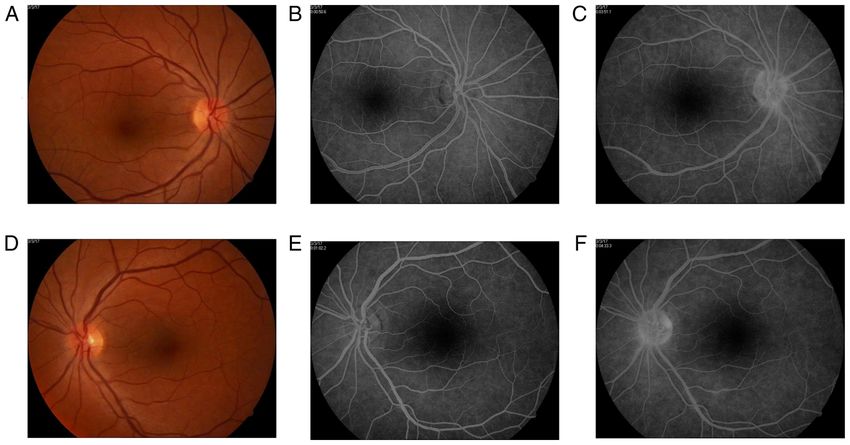
2                                GHITA et al: VASCULITIS ASSOCIATED WITH USE OF INTRAUTERINE DEVICE

Figure 1. Fundus photography and fluorescein angiography during clinical assessment. (A) The appearance of the posterior pole and (B) fundus angiography
in the right eye at 53.3 sec. (C) Fundus angiography in the right eye at 8 min and 3 sec. (D) The appearance of the posterior pole in the left eye on fundus
photography. (E) Fundus angiography in the left eye at 1 min and 54 sec and (F) at 8 min and 41 sec.

Case report                                                                     edema, retinal edema around the optic disc, normal aspect of
                                                                                arteries, but sinuous and dilated veins. The veins also presented
A 35‑year‑old female patient sought emergency care,                             a larger diameter than expected and some venous branches were
complaining of sudden hearing loss, headaches and blurred                       more dilated than others. Their diameter did not constantly
vision that had started two weeks previously and had gotten                     decrease towards the periphery, with some medium‑sized venous
progressively worse. The headaches were continuous and                          branches greater than the large‑sized venous branches (Fig. 1).
located in the occipital area, with episodes of increased pain,                     Therefore, a fundus fluorescein angiography was carried
accompanied by a decrease in hearing and sometimes dizzi‑                       out, with a delay of 2‑3 sec in the appearance of the laminar
ness. During the previous two weeks before presentation,                        venous flow and an unequal filling with dye of some temporal
the neurological exams had not identified any neurological                      medium‑sized venous branches. The late phase shows optic
signs. However, non‑steroidal anti‑inflammatory drugs were                      disc hyperfluorescence due to venous and capillary leakage.
prescribed, but without improvement. Furthermore, the patient                   Analyzing the venous branches, segmental diffuse hyperfluo‑
was examined by an ear, nose and throat (E.N.T.) doctor, but                    rescence in the early phases and staining in the later phases of
there were no clinical signs to explain the hearing loss.                       the angiogram were evident (Fig. 1). No additional retinal or
    Based on the patient's medical history, it was identified that              choroidal fluorescein lesions were identified.
she had a multinodular non‑toxic thyroid goiter, no drug aller‑                     In this context, the patient's diagnosis was bilateral
gies, she was a non‑smoker and had two natural child births.                    retinal vasculitis, papilledema and anterior uveitis, headache
From the Emergency Room of the University Emergency                             and hypoacusis. In order to identify the cause of the retinal
Hospital Bucharest, the patient was admitted into the                           vasculitis, various blood tests were performed based on the
neurology department, but still without any detectable clinical                 medical history, symptomatology and clinical assessment,
neurological signs. The dilated fundus examination revealed                     trying to avoid unnecessary and exhaustive investigations.
bilateral optic disc swelling. An orbito‑cerebral magnetic                      The purpose was to identify an immune or infectious cause
resonance imaging (MRI) and a computed tomography (CT)                          of the retinal vasculitis. All serological tests for syphilis,
scan were performed, but with no significant findings. In addi‑                 tuberculosis, HSV‑1, HSV‑2, HZV, CMV, HIV, and ELISA
tion, the blood pressure was normal. Therefore, the patient was                 for toxoplasmosis were negative. In addition, IgM and IgG
discharged from the neurological department and referred to                     anti‑β2‑ glycoprotein antibodies, IgM and IgG anti‑cardiolipin
the ophthalmologist.                                                            antibodies, lupus anticoagulant, homocysteine level, antibody
    The ophthalmological assessment revealed a slight decrease                  anti‑DNA double catenary, C‑ANCA, P‑ANCA, anti‑Ro/SSA
in the best corrected visual acuity of 20/30 in the right eye and               and anti‑La/SSB antibodies, ANA, and rheumatoid factor were
of 20/25 in the left eye. Slit‑lamp examination showed a 1+ faint               negative (Table I). Full blood count, biochemical blood profile
grade of flare in the anterior chamber, accompanied by a few                    and cerebrospinal fluid analysis were non‑contributory.
small corneal endothelium precipitates. Color vision, ocular                        Not being able to identify the cause of the retinal vasculitis,
motility and pupillary light reflex were within normal limits.                  the patient's medical history was reviewed, this time focusing
    Fundus examination reveals bilateral optic disc swelling,                   on rheumatological diseases, drug intake or other medical
hyperemia, loss of the optic cup, optic disc vessels drowned in                 procedures undergone in the recent period. As a result, it was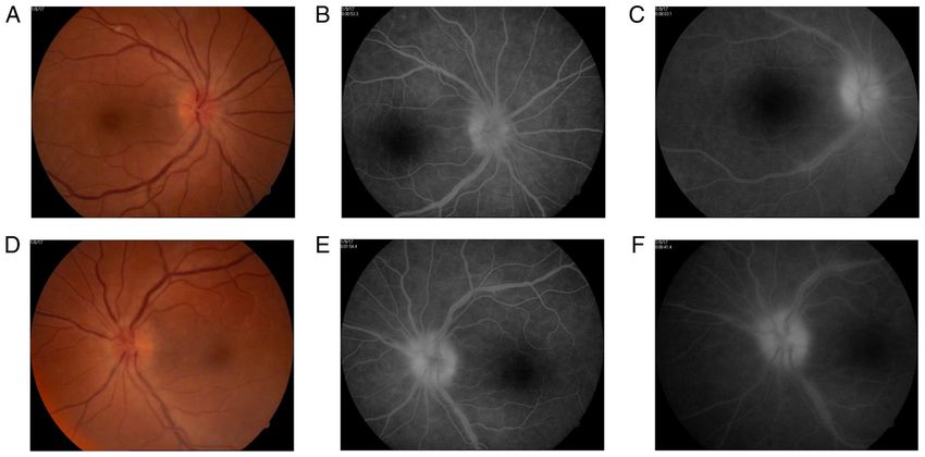
EXPERIMENTAL AND THERAPEUTIC MEDICINE 22: 1277, 2021                           3

Table I. Specific blood tests performed.

Immunology                                             Value         Normal range            Observation

Anti‑β2 glycoprotein antibodies (IgM) (U/ml)           3.3
4                               GHITA et al: VASCULITIS ASSOCIATED WITH USE OF INTRAUTERINE DEVICE

Table I. Contined.

Immunology                                                                 Value                     Normal range                          Observation

 Rapid plasma reagin (RPR)			                                                                                                            Non‑reactive
 Hepatitis B surface antigen (HBsAg)			                                                                                                  Negative
 Toxoplasmosis (IgM) Index                  0.1
EXPERIMENTAL AND THERAPEUTIC MEDICINE 22: 1277, 2021                                                   5

Table II. The Naranjo adverse drug reaction (ADR) probability scale for the intrauterine contraceptive device used.

Questions                                                                                                 Yes    No   Do not know Score

1. Are there previous conclusive reports on this reaction?                                    +1 0 0                                 1
2. Did the adverse event appear after the suspected drug was readministered?                  +2 0 0                                 2
3. Did the adverse reaction improve when the drug was discontinued or was a specific          +1 0 0                                 1
antagonist administered?
4. Did the adverse reaction reappear when the drug was readministered?                        0  0 0                                 0
5. Are there alternative causes (other than the drug) that, on their own, could have caused   0  2 0                                 2
the reaction?
6. Did the reaction reappear when a placebo was given?                                        0  0 0                                 0
7. Was the blood detected in the blood (or other fluids) in concentrations known to be toxic? 0  0 0                                 0
8. Was the reaction more severe when the dose was increased or less severe when the dose      0  0 0                                 0
was decreased?
9. Did the patient have a similar reaction to the same or similar drugs in any previous       +1 0 0                                 1
exposure?
10. Was the adverse event confirmed by any objective evidence?                                +1
Total				                                                                                                                            9

                              Figure 3. The OCT examination (A) prior to and (B) after one month of treatment.

and their branches (Fig. 2). The optical coherence tomography           Discussion
(OCT) confirmed the identified clinical aspects (Fig. 3). One
month later, fundus fluorescein angiography presented with a            First, the predominant optic disc edema accompanied by
decrease of the optic disc hyperfluorescence and fewer venous           very fine ocular signs suggested a neurological condition,
segments of slightly diffuse hyperfluorescence secondary to             misleading the clinicians. However, a careful examination
decreased vascular leakage (Fig. 2).                                    related to ancillary tests including fundus fluorescein angiog‑
   Ethics approval was obtained from the University                     raphy and OCT provides the information necessary for a proper
Emergency Hospital (Bucharest, Romania). The patient                    diagnosis (4,20). Rarely, retinal vasculitis may be isolated, and
provided written informed consent.                                      idiopathic without any other signs. Typical, retinal vasculitis
6 GHITA et al: VASCULITIS ASSOCIATED WITH USE OF INTRAUTERINE DEVICE
is a manifestation of a systemic disease or of a retinal inflam‑ In conclusion, to the best of our knowledge no previous case
matory condition. We followed up the guides in order to of retinal disease and optic disc edema associated with auditory
determine the cause of the disease; however, the tests did not problems (possible vascular) caused by an intrauterine device
offer the information required. A very thorough examination has been reported. A proper examination correlated with a
of the medical history supplied the missing piece of informa‑ very thorough medical history could identify rare diseases and
tion that was necessary to elucidate the cause. To the best of associations, in order to provide adequate medical care.
our knowledge, medical literature has not previously reported
any case of retinal vasculitis related to Jaydess® 13.5 mg Acknowledgements
levonorgestrel intrauterine contraceptive device (21). However,
some studies in literature indicate the occurrence of vasculitis Professional editing, linguistic and technical assistance were
related to oral contraceptive administration, but with mild performed by Irina Radu.
vascular involvement (16,18,22,23). Mosovich et al published
a case of necrotizing vasculitis caused by the Microgynon pills Funding
(levonorgestrel, 0.15 mg and ethinyl‑oestradiol, 0.03 mg) (13).
The diagnosis of generalized vasculitis with retinal No funding was received.
involvement is, similar to other drug‑induced vasculitis, based
on the temporal relationship between the drug administration Availability of data and materials
and the appearance of the clinical signs of disease (24). In
addition, the absence of other causes that could explain the Further information regarding the case is available from the
clinical picture, as systemic autoimmune diseases or infec‑ corresponding author upon reasonable request.
tious diseases (the ancillary test was negative or non‑reactive),
the remission of the disease after the device has been removed Authors' contributions
and the positive response to the systemic steroid therapy also
indicate a direct relationship between the IUD and disease. AMG had a substantial contribution to the conception and
In addition, we focused on thyroid disorders in order design of the work, analyzed and interpreted the data being
to exclude a secondary cause, being aware of the possible also the first investigator and coordinator of the article. AMG
relationship with primary or secondary ANCA vasculitis. also gave the final approval of the version to be published.
The test results excluded other retinal vasculitis associated CA collected all the data from the patient's medical history
with systemic inflammatory diseases, such as systemic lupus and produced the figures and tables. SI performed ocular
erythematosus, Wegener granulomatosis, microscopic poly‑ coherence tomography, fundus fluorescein angiography, and
angiitis, antiphospholipid syndrome and ANCA‑associated verified that the information and data were accurate. AE
vasculitis (25,26). performed the Naranjo adverse drug reaction (ADR) prob‑
As the patient was complaining of hearing dysfunction, ability scale and revised the paper. ACG was responsible for
and in the context of a mild anterior uveitis, Cogan syndrome literature research of the current data, drafted the study, and
was suspected (27). However, in the absence of any cardiac revised it critically for important intellectual content. AMG
signs, normal blood level of leukocytes, a slight increase of the and ACG assessed the authenticity of all the raw data. All
erythrocyte sedimentation rate, a normal C‑reactive protein, authors read and approved the final manuscript.
we excluded this syndrome (27,28).
Finally, the Naranjo adverse drug reaction (ADR) prob‑ Ethics approval and consent to participate
ability scale was performed and suggested an outcome to our
clinical evidence (29,30) (Table II). The last question is slightly Ethics approval was obtained from the University Emergency
contentious, since it was based on the patient's response. The Hospital, 050098 Bucharest, Romania. The patient provided
patient claimed that when she was previously under treatment written informed consent.
with similar drugs, such as oral contraceptive (similar to levo‑
norgestrel), she presented a low intensity similar response, but Patient consent for publication
could not provide medical evidence to support these aspects.
The Naranjo criteria classifies the probability that an Not applicable.
adverse event is related to a specific drug therapy, based on
a list of weighted questions, which examine factors such as Competing interests
the temporal association of drug administration and the
event, alternative causes that can explain the event, drug The authors declare that they have no competing interests.
levels, dose‑response relationship and the patient's previous
experience with that drug (29‑31). If ADR score is ≥9, the References
adverse drug reaction is considered as definite, if the score is
between 5 and 8 it is interpreted as probable, possible for a 1. Guillevin L and Dorner T: Vasculitis: Mechanisms involved and
score between 1 and 4, and doubtful if the score is 0 (31). The clinical manifestations. Arthritis Res Ther 9 (Suppl 2): S9, 2007.
2. Langford CA: Vasculitis. J Allergy Clin Immunol 125 (Suppl 2):
Naranjo criteria does not take into account drug interactions. S216‑S225, 2010.
Drugs are evaluated individually for causality, and points are 3. Trese MGJ, Yonekawa Y, Thomas BJ and Randhawa S: Vasculitic
deducted if another factor may have resulted in the adverse central retinal vein occlusion: The presenting sign of seronega‑
tive rheumatoid arthritis. Am J Ophthalmol Case Rep 2: 26‑29,
event, thereby weakening the causal association. 2016.EXPERIMENTAL AND THERAPEUTIC MEDICINE 22: 1277, 2021 7
4. Rosenbaum JT, Sibley CH and Lin P: Retinal vasculitis. Curr 18. Sturridge F and Guillebaud J: A risk‑benefit assessment of
Opin Rheumatol 28: 228‑235, 2016. the levonorgestrel‑releasing intrauterine system. Drug Saf 15:
5. Jennette JC: Overview of the 2012 revised International chapel 430‑440, 1996.
hill consensus conference nomenclature of vasculitides. Clin Exp 19. Vrettakos C and Bajaj T: Levonorgestrel. In: StatPearls [Internet]
Nephrol 17: 603‑606, 2013. Treasure Island (FL): StatPearls Publishing; 2021.
6. Apinyawasisuk S, Rothova A, Kunavisarut P and Pathanapitoon K: 20. Abu El‑Asrar AM, Herbort CP and Tabbara KF: Differential
Clinical features and etiology of retinal vasculitis in Northern diagnosis of retinal vasculitis. Middle East Afr J Ophthalmol 16:
Thailand. Indian J Ophthalmol 61: 739‑742, 2013. 202‑218, 2009.
7. Fukunaga H, Kaburaki T, Shirahama S, Tanaka R, Murata H, 21. Wildemeersch D, Andrade A, Goldstuck ND, Hasskamp T and
Sato T, Takeuchi M, Tozawa H, Urade Y, Katsura M, et al: Jackers G: Intrauterine levonorgestrel delivery with frameless
Analysis of inflammatory mediators in the vitreous humor of fibrous delivery system: Review of clinical experience. Int
eyes with pan‑uveitis according to aetiological classification. Sci J Womens Health 9: 49‑58, 2017.
Rep 10: 2783, 2020. 22. Fekete GL and Fekete L: Cutaneous leukocytoclastic vasculitis
8. Kaburaki T, Fukunaga H, Tanaka R, Nakahara H, Kawashima H, associated with erlotinib treatment: A case report and review of
Shirahama S, Izawa H, Komae K, Takamoto M, Soga H and the literature. Exp Ther Med 17: 1128‑1131, 2019.
Aihara M: Retinal vascular inflammatory and occlusive changes 23. Mansour D: The benefits and risks of using a levonorg‑
in infectious and non‑infectious uveitis. Jpn J Ophthalmol 64: estrel‑releasing intrauterine system for contraception.
150‑159, 2020. Contraception 85: 224‑234, 2012.
9. Agarwal A, Karkhur S, Aggarwal K, Invernizzi A, Singh R, 24. Radic M, Martinović Kaliterna D and Radic J: Drug‑induced
Dogra MR, Gupta V, Gupta A, Do DV and Nguyen QD: vasculitis: A clinical and pathological review. Neth J Med 70:
Epidemiology and clinical features of inflammatory retinal 12‑17, 2012.
vascular occlusions: Pooled data from two tertiary‑referral insti‑ 25. Papaliodis GN: Ophthalmologic manifestations of systemic
tutions. Clin Exp Ophthalmol 46: 62‑74, 2018. vasculitis. Curr Opin Ophthalmol 28: 613‑616, 2017.
10. Grau RG: Drug‑induced vasculitis: New insights and a changing 26. Tugal‑Tutkun I: Systemic vasculitis and the eye. Curr Opin
lineup of suspects. Curr Rheumatol Rep 17: 71, 2015. Rheumatol 29: 24‑32, 2017.
11. Iwahashi C, Ono H, Haruta M, Minami T, Mashimo H, 27. Iliescu DA, Timaru CM, Batras M, De Simone A and Stefan C:
Shimojo H and Ohguro N: New onset or exacerbation of uveitis Cogan's syndrome. Rom J Ophthalmol 59: 6‑13, 2015.
with infliximab: Paradoxical effects? BMJ Open Ophthalmol 4: 28. Vollertsen RS: Vasculitis and Cogan's syndrome. Rheum Dis
e000250, 2019. Clin North Am 16: 433‑439, 1990.
12. Agarwal A, Pilania RK, Anjani G, Choudhary H, Gupta A and 29. Behera SK, Das S, Xavier AS, Velupula S and Sandhiya S:
Gupta V: Retinal vasculitis with coats‑like response in a young Comparison of different methods for causality assessment of
girl with Parry‑Romberg syndrome. J Clin Rheumatol: May 21, adverse drug reactions. Int J Clin Pharm 40: 903‑910, 2018.
2019 (Epub ahead of print). 30. Murayama H, Sakuma M, Takahashi Y and Morimoto T:
13. Mosovich B, Biton A and Avinoach I: Vasculitis with cutaneous Improving the assessment of adverse drug reactions using the
necrosis induced by oral contraceptive. Harefuah 120: 451‑453, Naranjo Algorithm in daily practice: The Japan adverse drug
1991 (In Hebrew). events study. Pharmacol Res Perspect 6: e00373, 2018.
14. Trusau A and Brit ML: Propylthiouracil‑induced ANCA‑negative 31. Naranjo CA, Busto U, Sellers EM, Sandor P, Ruiz I, Roberts EA,
cutaneous small vessel vasculitis. J Community Hosp Intern Med Janecek E, Domecq C and Greenblatt DJ: A method for esti‑
Perspect 8: 35‑37, 2018. mating the probability of adverse drug reactions. Clin Pharmacol
15. Wiik A: Drug‑induced vasculitis. Curr Opin Rheumatol 20: Ther 30: 239‑245, 1981.
35‑39, 2008.
16. Kailasam C and Cahill D: Review of the safety, efficacy and
patient acceptability of the levonorgestrel‑releasing intrauterine This work is licensed under a Creative Commons
system. Patient Prefer Adherence 2: 293‑302, 2008. Attribution-NonCommercial-NoDerivatives 4.0
17. Andersson K, Odlind V and Rybo G: Levonorgestrel‑releasing International (CC BY-NC-ND 4.0) License.
and copper‑releasing (Nova T) IUDs during five years of use:
A randomized comparative trial. Contraception 49: 56‑72, 1994.You can also read