Surgical Treatment of Excessive Gingival Display Using Lip Repositioning Technique and Laser Gingivectomy as an Alternative
←
→
Page content transcription
If your browser does not render page correctly, please read the page content below
CRANIOMAXILLOFACIAL DEFORMITIES/COSMETIC SURGERY

     Surgical Treatment of Excessive Gingival
    Display Using Lip Repositioning Technique
     and Laser Gingivectomy as an Alternative
             to Orthognathic Surgery
                        Dragana Gabric Panduric, PhD, DMD,* Marko Blaskovic, DMD,y
                              Juraj Brozovic, DMD,z and Mato Susic, PhD, DMDx

     Excessive gingival display (EGD) is a condition in which an overexposure of the maxillary gingiva (>3 mm)
     is present during smiling. The proper diagnosis and determination of its etiology are essential for the se-
     lection of the right treatment modality. Different techniques have been used in cases of hyperactive upper
     lip: botulinum toxin injections, lip elongations with rhinoplasties, lip muscle detachments, myotomies,
     and lip repositions. This report presents a case of a young woman with an EGD larger than 10 mm during
     smiling caused by altered passive eruption, vertical maxillary excess, and a hyperactive upper lip that was
     treated with a modified lip repositioning technique and laser gingivectomy because she strongly refused
     orthognathic surgical treatment. A novel addition to the technique is proposed, a reversible trial accom-
     plished just by applying sutures on the borders of the future split-thickness flap, marked using diode laser,
     before starting the flap incision.
     Ó 2013 American Association of Oral and Maxillofacial Surgeons
     J Oral Maxillofac Surg -:e1-e11, 2013

Excessive gingival display (EGD), commonly termed                          tive upper lip, or a combination of these causes.
gummy smile, is a condition in which there is an over-                     Proper diagnosis of the etiologic factor is essential
exposure of the maxillary gingiva during smiling; in se-                   for the selection of the right treatment protocol. Pla-
vere cases, the overexposure is present in repositioning                   que- or drug-induced gingival enlargement and altered
of the mouth and lips.1 Although some gingival display                     or delayed passive eruption are treated with peri-
gives the impression of a youthful smile, a gingival                       odontal surgery. Depending on the classification of
display larger than 3 mm is considered unattractive.2                      the latter, bone surgery also may be required. Anterior
According to different investigators, a gummy smile is                     dentoalveolar extrusion is treated with orthodontic
considered a gingival display from 2 to 3 mm when smil-                    intrusion and vertical maxillary excess is treated
ing.3,4 It can affect about 10.5% of the population,5 with                 with orthognathic surgery.7 In the literature, different
a female predominance (2:1) and affecting persons 20                       techniques have been reported for the treatment of
to 30 years of age.6 The incidence of this condition de-                   the hyperactive upper lip: injections of botulinum
creases with age as a result of dropping of the upper and                  toxin,8 lip elongation associated with rhinoplasty,9
lower lips.2                                                               detachment of lip muscles,10 myotomy and partial
   The etiology of EGD is various: plaque- or drug-                        removal,11,12 and lip repositioning.13-15
induced gingival enlargement, altered or delayed                              The lip repositioning technique was first described
passive eruption, anterior dentoalveolar extrusion,                        1973 by Rubenstein and Kostianovsky16 as part of
vertical maxillary excess, short upper lip, a hyperac-                     medical plastic surgery. Later on, it was introduced

  *Assistant, Department of Oral Surgery, School of Dental                 versity of Zagreb, Gunduliceva 5, 10000 Zagreb, Croatia; e-mail:
Medicine, University of Zagreb, Zagreb, Croatia.                           dgabric@sfzg.hr
  yPrivate Dental Clinic, Rijeka, Croatia.                                 Received September 23 2013
  zPrivate Dental Office, Split, Croatia.                                  Accepted October 23 2013
  xProfessor, Department of Oral Surgery, School of Dental                 Ó 2013 American Association of Oral and Maxillofacial Surgeons
Medicine, University of Zagreb, Zagreb, Croatia.                           0278-2391/13/01328-1$36.00/0
  Address correspondence and reprint requests to Dr Gabric Pan-           http://dx.doi.org/10.1016/j.joms.2013.10.016
duric: Department of Oral Surgery, School of Dental Medicine, Uni-

                                                                      e1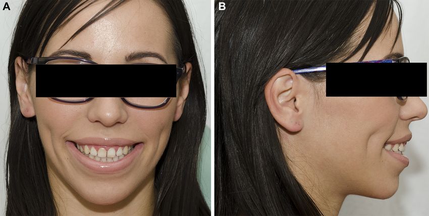
e2 TREATMENT OF EXCESSIVE GINGIVAL DISPLAY
smiling. The patient’s medical history disclosed heart
surgery for an aortic valve tumor at 10 years of age.
Otherwise, her history was unremarkable, with no
medication intake. She did not have active dental or
periodontal diseases. There were no contraindications
to surgical treatment. During clinical evaluation, it
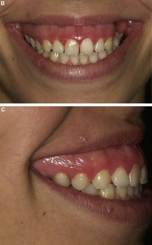
was verified that up to 10 mm of gingiva was displayed
during smiling (Fig 1A, B, C). With an exaggerated smile,
the patient’s teeth were visible from the maxillary right
first molar to the maxillary left first molar, with 10 mm
of excessive gingival tissue display in the medial line,
8.5 mm in the right canine line, 8 mm in the right first
molar line, 7 mm in the left canine line, and 5.5 mm
in the left first molar line. During clinical evaluation, a
normal upper lip length was found. During smiling,
there was 12 mm of lip raising, which led to a diagnosis
of a hyperactive upper lip. Tooth evaluation showed
discrete short clinical crowns in the maxillary anterior
region, and probing showed that the alveolar bone crest
was localized 2 to 3 mm apically to the cementoenamel
junction, leading to the diagnosis of altered passive
eruption. The final diagnosis was EGD from a combina-
tion of altered passive eruption, vertical maxillary
excess, and a hyperactive upper lip. After the patient
refused orthognathic surgical treatment, a modified lip
repositioning technique and concomitant gingivectomy
was proposed. The patient was counseled on manage-
ment options. The patient’s expectations were clarified
and a realistic outcome was presented, including the
FIGURE 1. A, Preoperative full face view with relaxed lips. (Fig 1 possibility of full or partial relapse. Pre-existing asymme-
continued on next page.)
try in the patient’s smile was pointed out to her, because
Gabric Panduric et al. Treatment of Excessive Gingival Display. J
Oral Maxillofac Surg 2013.
of the possibility that it would be more apparent with
the lip in closer proximity to the teeth. Written
informed consent was obtained after an explanation
of the risks, potential benefits, and treatment alterna-
in dentistry, after being modified in 2006 by Rosenblatt tives. Intra- and extraoral photographs were taken for
and Simon.13 planning and records.
This clinical report presents a case of a young female
patient with an EGD larger than 10 mm during smiling
caused by a combined etiology of a hyperactive upper SURGICAL PROCEDURE
lip and altered passive eruption of the frontal maxillary The treatment plan consisted of reversible lip repo-
teeth because she refused orthognathic surgery. The sitioning and definitive surgical repositioning. One
treatment plan consisted of a modified lip reposition- hour before surgery, the patient was given amoxicillin
ing technique with a reversible clinical trial17 and a 2 g for prophylaxis owing to her history of cardiac sur-
gingivectomy performed with a diode laser. gical treatment and preoperative analgesics (ibuprofen
600 mg) for pain management. Extraoral and intraoral
Report of Case antisepsis was performed with 2.0% chlorhexidine so-
lution and 0.12% chlorhexidine rinse for 1 minute.
PATIENT PROFILE, PRESURGICAL EVALUATION, AND Initial anesthesia consisted of bilateral infraorbital
CONSENT blocks (2% lidocaine with 1:200,000 epinephrine).
A 27-year-old woman reported to the Department of The infraorbital block was used to avoid thickening
Oral Surgery, School of Dental Medicine, University of of the lip and soft tissues with anesthetic fluid, allow-
Zagreb (Zagreb, Croatia) with the chief complaint of a ing the reversible trial to be a more realistic represen-
gummy smile. She reported dissatisfaction with the tation of the projected final result. To begin the
amount of gingiva exposed while smiling and her treat- reversible lip repositioning, the proposed surgical inci-
ment goal was to minimize the gingival display during sion lines were marked with a high-power diode laser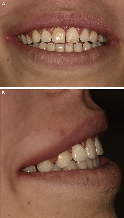
 PANDURIC
GABRIC         ET AL                                                                                                      e3

                           FIGURE 1 (cont’d). B, Preoperative enface view. C, Preoperative face profile view.
Gabric Panduric et al. Treatment of Excessive Gingival Display. J Oral Maxillofac Surg 2013.

(LaserHF, Hager & Werken, Duisburg, Germany) set to                      ing the next 2 days. Small dashed markings were
1.5 W using continuous-wave (CW) mode and fiber                          placed every 5 mm along the line of the proposed in-
with an active diameter (core) of 320 mm. When                           cisions. The inferior border was defined by the muco-
applied to the tissue, the laser beam does not cut the                   gingival junction from the mesial aspect of the first
tissue but leaves a dark mark that cannot be smeared                     molars bilaterally. The superior border was placed
or wiped away. Marks are not permanent and fade dur-                     slightly inferior in the area of the labial frenum,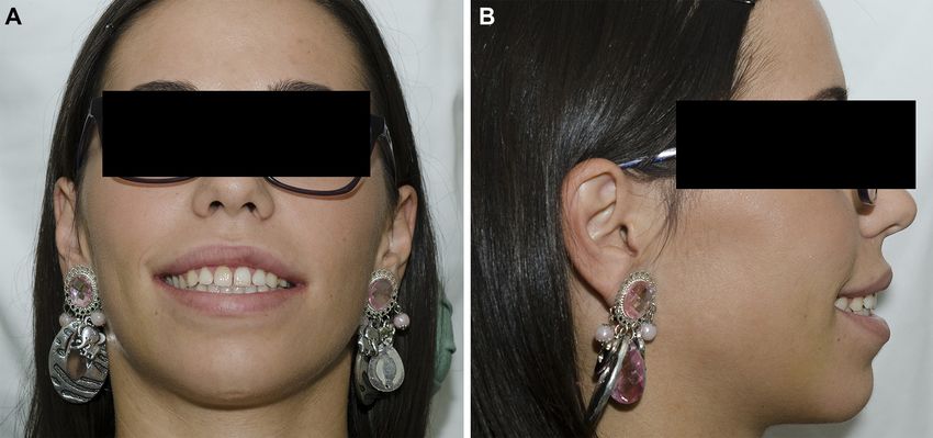
e4                                                                                   TREATMENT OF EXCESSIVE GINGIVAL DISPLAY

cresting in the area of the canine, and tapering toward                  exposed. The tissue thickness was approximately
the posterior area, forming moustache-like shape. The                    1 mm. Care was taken to avoid damage to any minor sali-
distance between the superior and inferior borders                       vary glands in the submucosa. High-frequency bipolar
was 1.5 the length of the repositioning desired in the                   forceps (LaserHF, Hager & Werken) were used to con-
patient’s smile. Once the area was marked, sutures                       trol bleeding. The area of frenectomy was approxi-
were used to complete the reversible procedure. Eight                    mated along the preoperative laser markings with a
3.0 silk sutures (2 in the frontal part, 1 above the                     simple interrupted suture to ensure symmetry and
canine area, and 1 between the second premolar and                       proper midline placement. The remaining closure
the first molar bilaterally) were placed (Fig 2). Suture                 bilaterally was completed with continuous interlock-
design involved a vertical tissue bite taken at the supe-                ing sutures to stabilize the new mucosal margin to the
rior border in the movable mucosa, a horizontal tissue                   gingiva (Fig 7). Nonresorbable sutures were used (3-0
bite at the mucogingival junction, and inverting and                     silk). For further hemostasis, tissues were com-
tucking behind the tissue proposed for excision. At                      pressed with wet gauze for 5 minutes. After probing
this point, photographs were taken, and the patient                      and marking using a Crane-Kaplan pocket marker,
was able to evaluate the potential final result using                    gingivectomy in the intercanine area was performed
mirror and clinical photographs (Fig 3). She decided                     with a high-power diode laser (975 mm, 3 W, 10 ms,
immediately to proceed with the surgery. Anesthesia                      1:2; Fig 8A, B). A soft tissue bandage (Reso-Pac, Hager
was supplemented with local infiltration, using the                      & Werken) was applied over the entire surgical site.
same type of local anesthetic, from the maxillary right                  Nonsteroidal anti-inflammatory drugs (ibuprofen
to the left first molar for hemostatic control. Temporary                600 mg 3 times daily for 2 days) and oral antibiotics
sutures were removed and the laser spot markings were                    (amoxicillin 500 mg 3 times daily for 7 days) were
connected to the line of the planned scalpel incision us-                prescribed after surgery. The patient was instructed
ing a diode laser with the same parameters (Fig 4),                      to apply ice packs, consume only soft foods during
owing to possible changes in the direction and angle                     the first postoperative week, avoid any mechanical
of the incision line, if necessary. Partial-thickness inci-              trauma, brush gently, and minimize lip movements
sions were made using a scalpel across the superior                      when smiling or talking for the first 2 weeks post-
and then the inferior border, connecting in the poste-                   operatively.
rior molar area bilaterally. Frenectomy using a high-
power diode laser (975 mm, 4 W, CW) was performed
(Fig 4). The final surgical procedure was initiated on                      POSTOPERATIVE FOLLOW-UPS AND CLINICAL
the left side (Fig 5). Two strips of outlined mucosa                        RESULTS
were removed (Fig 6) by a superficial split-thickness                       The patient was seen the day after surgery for
dissection beginning from the frenectomy laser incision                  follow-up. She reported good analgesia with the
for the 2 sides, leaving the underlying connective tissue                over-the-counter ibuprofen. Periodic follow-ups were

                               FIGURE 2. Reversible clinical trial using silk sutures and diode laser marks.
Gabric Panduric et al. Treatment of Excessive Gingival Display. J Oral Maxillofac Surg 2013.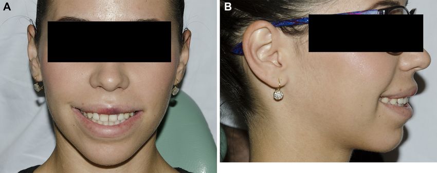
 PANDURIC
GABRIC         ET AL                                                                                                         e5

                                       FIGURE 3. Clinical view with reversible trial before surgery.
Gabric Panduric et al. Treatment of Excessive Gingival Display. J Oral Maxillofac Surg 2013.

scheduled on postoperative days 3 (Fig 9A, B), 5, 10                     cosa (Fig 11). Upper lip length (from the nasal base to
(Fig 10A, B), and 14, 3 months postoperatively, and                      the superior border of the upper lip vermillion)
6 months postoperatively, when clinical photographs                      increased from 10 mm at baseline to 16 mm at postop-
were taken. Postoperative healing occurred with min-                     erative day 14 and 15 mm at 3 and 6 months after sur-
imum discomfort, and she reported ‘‘tension’’ on the                     gery. Upper lip vermillion length (from the inferior
upper lip and ‘‘slight pain’’ when smiling and talking                   border of the upper lip) increased from 6 mm at base-
during the first week after surgery and feeling numb-                    line to 10 mm at postoperative day 14 and 3 and
ness on the left side of the upper lip. Sutures were                     6 months after surgery. The gingival display at baseline
removed 10 days later. The suture line healed in the                     was 5.5 to 10 mm and decreased significantly to 2 mm
form of scar that was not apparent when the patient                      in the medial line and 0 mm (when the lip covered part
smiled, because it was concealed in the upper lip mu-                    of clinical crowns) in the canine and molar regions

                                       FIGURE 4. Diode laser superficial incision and frenectomy.
Gabric Panduric et al. Treatment of Excessive Gingival Display. J Oral Maxillofac Surg 2013.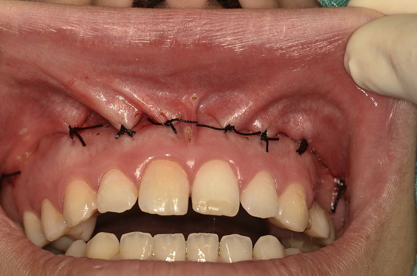
e6                                                                                   TREATMENT OF EXCESSIVE GINGIVAL DISPLAY

                          FIGURE 5. Intraoral comparison of left side after surgery and right side before surgery.
Gabric Panduric et al. Treatment of Excessive Gingival Display. J Oral Maxillofac Surg 2013.

bilaterally at postoperative day 14 and 3 and 6 months                   contrast to the first postoperative week, when she
after surgery. Results from the evaluation of the pa-                    felt tension when talking or smiling and numbness,
tient’s postoperative discomfort using a visual analog                   at 2 weeks and 3 and 6 months postoperatively she
scale (VAS), ranging from 1 for ‘‘no pain’’ to 10 for                    felt no tension or numbness. She reported that the
‘‘pain as bad as could possibly be,’’ were 8 the day after               worst part of this surgical procedure was the discom-
surgery, 3 at postoperative days 3 and 5, 2 at postoper-                 fort or inability to move the lip during the first week,
ative day 10, and 0 at postoperative day 14. The patient                 and the best part was the improvement of her smile
filled out the previously prepared questionnaire for pa-                 and facial esthetics (Fig 12A, B). Considering the over-
tient satisfaction with the surgical procedure. Preoper-                 all experience, she would likely choose to undergo the
atively, she was not satisfied with her smile and with                   surgery again.
the amount of gingival display, with the opposite effect
postoperatively. Postoperatively, she considered the
                                                                         Discussion
amount of displayed gingival to be ‘‘about right’’
compared with ‘‘way too much’’ preoperatively. In                           The lip repositioning technique is an excellent alter-
                                                                         native to more costly procedures with higher morbidity
                                                                         rates.9-12 The lip reposition surgery was originally
                                                                         described in the medical literature by Rubenstein and
                                                                         Kostianovsky in 1973.16 The LRS originally did not
                                                                         include severing the muscle attachments. Later on,
                                                                         different investigators modified the technique by pro-
                                                                         posing the detachment of the elevator muscle in cases
                                                                         of a short upper lip,10 myectomies or partial resection
                                                                         of 1 or 2 levator labii superior muscles,11 and partial tran-
                                                                         section of the lip elevator muscles and implantation of
                                                                         an alloplastic or autogenous spacer.18 All these modifica-
                                                                         tions were made to prevent relapses.
                                                                            In the past 7 years there have been several case re-
                                                                         ports and case series in the dental literature describing
                                                                         the use of LRS for the treatment of EGD,14-17 with the
FIGURE 6. Two strips of outlined mucosa removed intraopera-
                                                                         first by Rosenblatt and Simon.13 There are some differ-
tively, with a tissue thickness of approximately 1 mm.                   ences in the technique among investigators, with
Gabric Panduric et al. Treatment of Excessive Gingival Display. J      some leaving the frenulum intact14,15 and others
Oral Maxillofac Surg 2013.                                               including the frenulum in the partial-thickness flap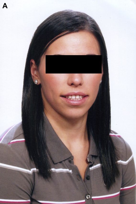
 PANDURIC
GABRIC         ET AL                                                                                                           e7

                  FIGURE 7. Continuous interlocking sutures for stabilization of the new mucosal margin to the gingiva.
Gabric Panduric et al. Treatment of Excessive Gingival Display. J Oral Maxillofac Surg 2013.

for removal.13,16,17 Leaving the frenulum intact helps                      Alternatives to LRS in the treatment of EGD caused by
maintain the position of the labial midline, prevent                     a hyperactive or short upper lip have been proposed by
changes in lip symmetry, and decrease the morbidity                      Polo8,20 and Ishida et al.12 Polo20 used botulinum toxin
associated with the procedure,14,15 but in the authors’                  type A to treat 30 patients with EGD. At the
opinion limits the possibility of correcting EGD in the                  second week after injection, the preinjection gingival
region of the maxillary central incisors. Because the                    display of 5.2  1.4 mm decreased to 0.09  1.06 mm.
present patient had an EGD larger than 10 mm, a                          The effect of the botulinum toxin was temporary, and
large correction had to be performed. In this case, the                  the gingival display gradually increased from the
amount of epithelium for excision was 1.5 times the                      second week to baseline values after the 32nd week.
amount of the EGD. The original plan was to decrease                     In their technique, Ishida et al12 combined and modified
the amount of EGD by 2 times, but with an EGD                            different procedures: myotomy of the levator labii supe-
larger than 10 mm, the superior incision line would                      rioris muscles11 and subperiosteal dissection21 associ-
be too close to the vermilion border. The scar form                      ated with a subcutaneous dissection and lip
after the surgery could violate the smile esthetics. To                  frenectomy.22 The surgery was performed in 14 patients
the best of the authors’ knowledge, the amount of the                    who showed a decrease of gingival display from 5.22
EGD corrected with the LRS technique and crown                           1.48 at baseline to 1.91  1.50 mm 6 months af-
lengthening reported in this case is the largest                         ter surgery.
described in the literature.13-17,19                                        All 3 techniques produce the same results in
   A novel addition to the technique has been pro-                       decreasing EGD. However, although the botulinum
posed, a reversible trial accomplished just by applying                  toxin injection20 is the least invasive treatment, the re-
sutures on the borders of the future split-thickness flap                sults are temporary and necessitate frequent retreat-
before starting the flap incision.19 In the present case,                ments. The approach used by Ishida et al12 is more
laser markings were used to depict the position of the                   aggressive, with higher morbidity compared with LRS.
incision line. Sutures were placed temporarily con-                         Some factors restrict the use of LRS. It is contraindi-
necting the upper and the lower markings, simulating                     cated in the presence of an inadequate amount of
the final result of the treatment. Using this technique,                 attached gingiva in the maxillary anterior sextant. It
the patient and the surgeon have the opportunity to                      will cause difficulty in flap design, suturing, and stabi-
preview the final result in advance. Because LRS is                      lization, which could lead to relapse.11 In addition, the
an elective surgery, it is important that the patient                    patient could be left with a shallower vestibule that
have realistic expectations related to the final result                  could compromise the ability to perform adequate
of the surgery. Therefore, the trial modification is a                   oral hygiene.14 Although LRS is not indicated for se-
good tool for communication between the surgeon                          vere maxillary excess,7,14 Humayun et al19 reported a
and the patient.                                                         case of mild maxillary excess treated with LRS.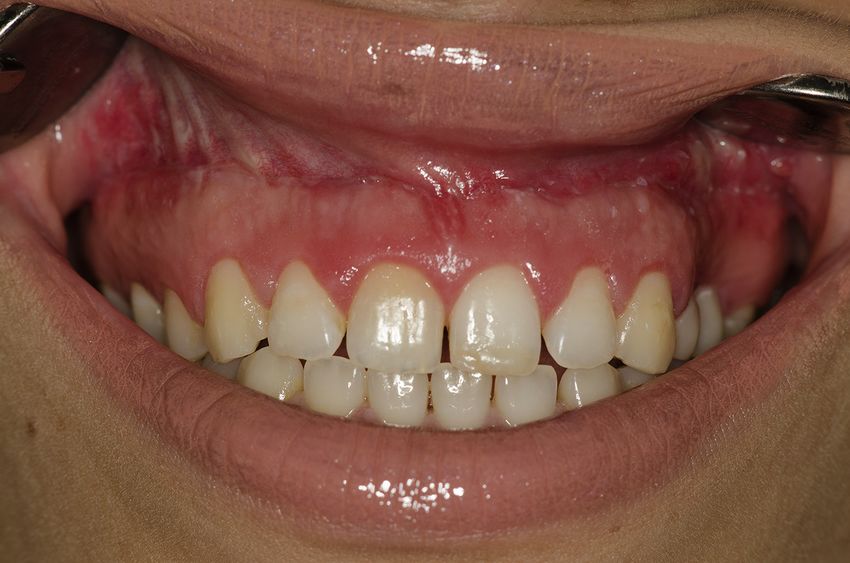
e8                                                                                   TREATMENT OF EXCESSIVE GINGIVAL DISPLAY

                              FIGURE 8. Immediately postoperatively. A, Enface view. B, Face profile view.
Gabric Panduric et al. Treatment of Excessive Gingival Display. J Oral Maxillofac Surg 2013.

   Rare complications have been described after LRS,                     In the first week after surgery, the present patient com-
such as discomfort, bruising, and swelling of the upper                  plained of mild discomfort (according to the VAS, the
lip.13,14,17 Rosenblatt and Simon13 reported on 1 pa-                    pain level at the third day after surgery was 3 and
tient with a mucocele that resolved without treatment.                   completely disappeared within 14 days) and tension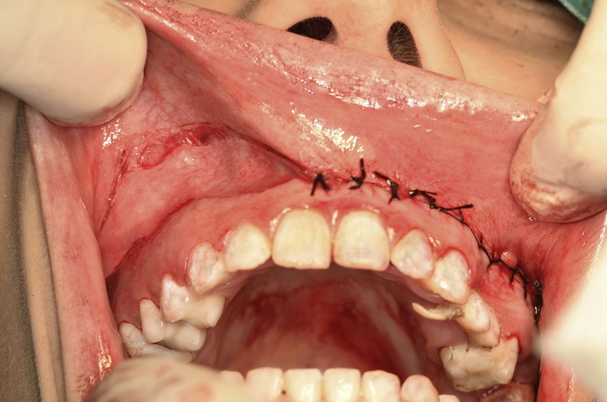
 PANDURIC
GABRIC         ET AL                                                                                                           e9

                          FIGURE 9. Follow-up on third postoperative day. A, Enface view. B, Face profile view.
Gabric Panduric et al. Treatment of Excessive Gingival Display. J Oral Maxillofac Surg 2013.

while talking and smiling and numbness of the left side                     The patient refused orthognathic surgery because
of the upper lip. On the left half of the upper lip, a he-               the morbidity and potential complication rate associ-
matoma had formed, which disappeared within                              ated with orthognathic surgery were not acceptable
2 weeks after surgery.                                                   to her for an elective cosmetic treatment. Therefore,
   This case presentation describes the treatment of a                   an alternative treatment was proposed: LRS and laser
young female patient with a combined etiology of                         gingivectomy, procedures with low morbidity and
EGD: altered passive eruption, vertical maxillary                        good acceptance by patients. With this treatment
excess, and a hyperactive upper lip. During maximum                      plan, 2 of 3 etiologic factors of EGD were corrected.7
smiling, the patient had a 10-mm EGD and Class 1A                           The outcome was successful, with a decrease of EGD
altered passive eruption according to Coslet et al.23                    from 10 to 1.5 mm in the region of the left and right cen-
The first treatment plan proposed to the patient was                     tral incisors, from 8.5 to 0 mm in the right canine re-
orthognathic surgery and gingivectomy.                                   gion, from 7 to 0 mm in the left canine region, from 8

                           FIGURE 10. Follow-up 10 days after surgery. A, Enface view. B, Face profile view.
Gabric Panduric et al. Treatment of Excessive Gingival Display. J Oral Maxillofac Surg 2013.
e10                                                                                  TREATMENT OF EXCESSIVE GINGIVAL DISPLAY

      FIGURE 11. Intraoral view 10 days after surgery, immediately after suture removal. The suture line healed in the form of scar.
Gabric Panduric et al. Treatment of Excessive Gingival Display. J Oral Maxillofac Surg 2013.

to 2.0 mm in the right first molar region, and from 5.5 to               after surgery, the patient expressed satisfaction with
1.0 mm in the left molar region at 6-month follow-up.                    her smile and with the decreased quantity of EGD, stat-
   Silva et al14 reported high patient satisfaction 2.5 years            ing that she would undergo the procedure again.
after surgery, with 70% of patients considering the post-                   LRS might be a valid alternative for the decrease of
operative amount of gingival display to be ‘ about right’’               EGD caused by a hyperactive or short upper lip.
and 90% willing to undergo the procedure again. Hence,                   Compared with alternative solutions, such as botulinum
LRS is a safe procedure with low morbidity and good                      toxin injections or a combined myotomy procedure, it
acceptance.14 This was in accord with present case.                      has a stable result and low morbidity. Furthermore, it
Based on the questionnaire filled out 3 and 6 months                     is well accepted by patients. This case presentation

                           FIGURE 12. Follow-up 6 months after surgery. A, Enface view. B, Face profile view.
Gabric Panduric et al. Treatment of Excessive Gingival Display. J Oral Maxillofac Surg 2013.
PANDURIC
GABRIC ET AL e11
suggests that LRS combined with laser gingivectomy for the correction of gummy smile. Plast Reconstr Surg 126:
1014, 2010
could be used as a minimally invasive alternative to or-
13. Rosenblatt A, Simon Z: Lip repositioning for reduction of exces-
thognathic surgery for cases of EGD with a com- sive gingival display: A clinical report. Int J Periodontics Restor-
plex etiology. ative Dent 26:433, 2006
14. Silva CO, Ribeiro-J
unior NV, Campos TV, et al: Excessive gingival
display: Treatment by a modified lip repositioning technique.
References J Clin Periodontol 40:260, 2013
15. Ribeiro-Junior NV, Campos TV, Rodrigues JG, et al: Treatment
1. Allen EP: Use of mucogingival surgical procedures to enhance of excessive gingival display using a modified lip reposition-
esthetics. Dent Clin North Am 32:307, 1988 ing technique. Int J Periodontics Restorative Dent 33:309,
2. Kokich VO, Kokich VG, Kiyak HA: Perceptions of dental profes- 2013
sionals and laypersons to altered dental esthetics: Asymmetric 16. Rubenstein A, Kostianovsky A: Cosmetic surgery for the malfor-
and symmetric situations. Am J Orthod Dentofacial Orthop mation of the laugh: Original technique. Prensa Med Argent 60:
130:141, 2006 952, 1973
3. McGuire MK: Periodontal plastic surgery. Dent Clin North Am 17. Jacobs PJ, Jacobs BP: Lip repositioning with reversible trial for
42:411, 1998 the management of excessive gingival display: A case series.
4. Chiche GJ, Pinault A: Esthetics of Anterior Fixed Prosthodontics. Int J Periodontics Restorative Dent 33:169, 2013
Chicago, IL, Quintessence, 1994 18. Ellenbogen R, Swara N: The improvement of the gummy smile
5. Tjan AH, Miller GD, The JG: Some esthetic factors in a smile. using the implant spacer technique. Ann Plast Surg 12:16,
J Prosthet Dent 51:24, 1984 1984
6. Peck S, Peck L, Kataja M: The gingival smile line. Angle Orthod 19. Humayun N, Kolhatkar S, Souiyas J, et al: Mucosal coronally
62:91, 1992 positioned flap for the management of excessive gingival
7. Silberberg N, Goldstein M, Smidt A: Excessive gingival display— display in the presence of hypermobility of the upper lip
Etiology, diagnosis, and treatment modalities. Quintessence Int and vertical maxillary excess: A case report. J Periodontol
40:809, 2009 81:1858, 2010
8. Polo M: Botulinum toxin type A in the treatment of excessive 20. Polo M: Botulinum toxin type A (Botox) for the neuromuscular
gingival display. Am J Orthod Dentofacial Orthop 127:214, 2005 correction of excessive gingival display on smiling (gummy
9. Ezquerra F, Berrazueta MJ, Ruiz-Capillas A, et al: New approach smile). Am J Orthod Dentofacial Orthop 133:195, 2008
to the gummy smile. Plast Reconstr Surg 104:1143, 1999 21. Rees TD, La Trenta GS: Aesthetic Plastic Surgery. Philadelphia,
10. Litton C, Fournier P: Simple surgical correction of the gummy PA, Saunders, 1994
smile. Plast Reconstr Surg 63:372, 1979 22. Edwards JG: A clinical study: The diastema, the frenum, the fre-
11. Miskinyar SA: A new method for correcting a gummy smile. Plast nectomy. Oral Health 67:51, 1977
Reconstr Surg 72:397, 1983 23. Coslet JG, Vanarsdall R, Weisgold A: Diagnosis and classification
12. Ishida LH, Ishida LC, Ishida J, et al: Myotomy of the levator labii of delayed passive eruption of the dentogingival junction in the
superioris muscle and lip repositioning: A combined approach adult. Alpha Omegan 70:24, 1977You can also read