Unusual Presentation of Neuroendocrine Tumor with Multiple Endocrine Neoplasia 1 Syndrome: A Rare Case Report
←
→
Page content transcription
If your browser does not render page correctly, please read the page content below
International Journal of Research and Review
                                                                                     Vol.7; Issue: 3; March 2020
                                                                                   Website: www.ijrrjournal.com
Case Report                                                              E-ISSN: 2349-9788; P-ISSN: 2454-2237

    Unusual Presentation of Neuroendocrine Tumor
    with Multiple Endocrine Neoplasia 1 Syndrome:
                  A Rare Case Report
                           Ankita Raj1, Reeta Dhar2, Piyush Sahu3
                                 1
                                  PG Student, 2Professor, 3Asst. Professor,
              Department of Pathology, M.G.M. Medical College, Kamothe, Navi Mumbai, India
                                        Corresponding Author: Reeta Dhar

ABSTRACT                                                           MEN1         (multiple     endocrine
                                                           neoplasia) is an autosomal dominant
Neuroendocrine tumor with multiple endocrine               inherited tumor of endocrine gland also
neoplasia type 1 (MEN1) syndrome is detected               known as Wermer Syndrome. It is due to
in a middle-aged lady presented with history of
                                                           loss of menin gene on chromosome 11q13.
fall leading to multiple fractures. On per rectal
                                                           Menin is a tumor suppressor gene associated
examination a polypoidal structure was found.
Further investigations confirmed the diagnosis             with cellular growth and function. [3]
of neuroendocrine tumor.                                   MEN1 causes neoplastic proliferation in
                                                           pituitary, parathyroid and pancreas.
Key words – Rectal polyp, Rectal neoplasm,                         Presenting here is an accidently
Neuroendocrine Tumor, MEN1                                 diagnosed case of rectal polyp (NET) in
                                                           MEN1 syndrome which presented to our
INTRODUCTION                                               hospital with history of fall leading to
        Neuroendocrine tumor shows neuro                   multiple non healing fractures.
and endocrine properties. [1] Neuro
properties is based on identification of                   Clinical History-
Dense Core Granules (DCG) and endocrine                            A 38 years old female was admitted
properties means secretion and synthesis of                to our hospital with history of sudden fall
monoamines. [1]                                            leading to non-healing multiple fractures.
        These       tumors      begin     in               Per rectal examination revealed a small
neuroendocrine cells. They occur in the                    rectal polypoidal growth.
digestive tract, lungs and brain. In the                   Hematological and the biochemical tests
digestive tract, they may involve the                      were within normal range with mild anemia
stomach, pancreas, small intestine, colon                  (9 gm/dl) and slightly raised serum calcium
and rectum. [1]                                            levels (12.8 mmol/L).
        The most common site for NET is                            Colonoscopy was performed which
gastrointestinal region accounting for 60 -                revealed a 3.5 mm rectal nodule 3-4 cm
67% of tumor. [2] Among all the colon and                  away from anal verge.
rectum tumors, rectal NET accounts for                             CT scan was done in view of non-
approximately 1-2%. [4] About 50 % of                      healing fracture that revealed Parathyroid
rectal NET are asymptomatic and are                        Adenoma,       Hyperparathyroidism      and
diagnosed incidentally or presents as polyp                Pancreatitis.
in colonoscopies. [4,6] Current incidence is                       Patient was admitted in view of
approximately 2 per lakh population with a                 multiple fractures, but these accidental
female preponderance. [2]                                  findings led to the diagnosis of

                      International Journal of Research and Review (ijrrjournal.com)                        375
                                         Vol.7; Issue: 3; March 2020
Ankita Raj et.al. Unusual presentation of neuroendocrine tumor with multiple endocrine neoplasia 1 syndrome:
a rare case report
neuroendocrine tumor with MEN1 Gross Examination - Received a single, soft
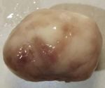
syndrome. Hence, patient was subjected to to firm, grey-white, globular tumor
surgical excision of the polyp and the tissue measuring 3.5 cm in diameter.
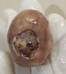
was sent for histopathological examination. External surface was smooth with one
Pathological Findings- ulcerated area measuring 2cmX1.5 cm.
Cut surface: homogenous, grey-white area
with yellowish tinge.
Figure 1: Gross appearance of rectal polypoidal mass.
Figure 2: Gross appearance of ulcerated surface of rectal mass
Figure 3: Cut surface of rectal mass- Tan Homogenous areas
Microscopic examination-
Entirely processed lesion showed lining mucosa and crypts lined by mucus secreting
columnar epithelium. The lamina propria and muscularis mucosa were unremarkable.
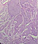
There was diffusely infiltrating neoplasm in the submucosa and muscularis propria composed
of uniform cells arranged in solid pattern (organoid), islands (insular), pseudo glandular,
festooning, trabeculae, syncytial and ribbons.
MICROSCOPY-
Figure 4- Shows epithelial lining (arrow) & Tumor Islands (arrow head).
Figure 5- Shows submucosal tumor.
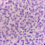
The individual tumor cells were uniform, having moderate cytoplasm and indistinct
cell borders. The nuclei were round to oval with powdery salt and pepper chromatin. Mitotic
activity of 3/10 HPF was noted. However, no necrosis or dysplasia of the overlying
epithelium was identified. The base of the polyp was involved by the tumor. No
lymphovascular or perineural invasion was seen. Hence, it was diagnosed as Well
Differentiated Neuroendocrine Tumor - Grade 1, Rectum- Base of polyp involved by tumor.
No lymphatic, vascular or perineural invasion seen.
International Journal of Research and Review (ijrrjournal.com) 376
Vol.7; Issue: 3; March 2020Ankita Raj et.al. Unusual presentation of neuroendocrine tumor with multiple endocrine neoplasia 1 syndrome:
a rare case report
Figure 6 - Tumor cells arranged in solid, organoid pattern(4x)
Figure 7- Tumor cells arranged in pseudo glandular pattern(10x)
Figure 8 & 9 - (40x or 100x) Uniform Tumor cells and nuclei show salt and pepper chromatin.
Immunohistochemistry-
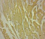
Figure 10: Synaptophysin- Positive Figure 11: Chromogranin A- Positive
[6]
DISCUSSION normal appearing mucosa.
NET can occur anywhere in body Neuroendocrine tumors are derived from
but most commonly occurs in digestive enterochromaffin cells which are located
system. They can be benign or malignant. throughout the intestine within the crypts of
NET is neuronal and endocrine hormonal Lieberkühn. The NET typically occurs deep
tumors. The five years survival rate for within these crypts. As these tumors
localized disease is 87% with an excellent progress, they invade the muscularis mucosa
prognosis. [7] Most of the benign cases are and sub mucosa resulting in there
asymptomatic, small, slow growing tumor. subepithelial appearance.
Generally, rectal NET appears as a single, MEN1 is clinically diagnosed by the
smooth submucosal nodule or polyp with presence of two or more associated tumors.
International Journal of Research and Review (ijrrjournal.com) 377
Vol.7; Issue: 3; March 2020Ankita Raj et.al. Unusual presentation of neuroendocrine tumor with multiple endocrine neoplasia 1 syndrome:
a rare case report
It is called familial if a patient is associated
with one tumor and a first degree relative Conflict of Interest: None
with MEN1. [5] It is diagnosed as genetic if a
patient has MEN1 gene mutation but no Abbreviation- MEN1 (Multiple Endocrine
clinical or biochemical MEN1 mutation (a Neoplasia 1), NET (Neuroendocrine Tumor),
mutant gene carrier). [1] DCG (Dense Core Granules)
30% of the pediatric NET is
REFERENCES
genetically inherited and are associated with 1. Nothing but NET : A review of NETs and
MEN1 syndrome, von Hippel Lindau carcinomas Volume 19 Number 12
syndrome, Neurofibromatosis type 1 and December 2017 pp. 991–1002
others. [8] The NET of rectum is rare but are 2. Epidemiology of Neuroendocrine Tumors.
being identified due to widespread use of https://pubmed.ncbi.nlm.nih.gov/15477707/
screening with colonoscopy. They show 3. Calcitonin-Secreting Pancreatic
indolent behavior, but they can be malignant Neuroendocrine Tumor In A Patient With
and can metastasize. Lesions showing Multiple Endocrine Neoplasia Type 1
similar microscopic features include - Small (AACE Clinical Case Rep. 2017;3:e317-
cell carcinoma lung, Lymphoma, e321)
Melanoma, Carcinoid tumor, Insulinoma, 4. A case of rectal neuroendocrine tumor
presenting as polyp ,
Gastrinoma, Renal cell carcinoma, http://dx.doi.org/10.1016/j.ijscr.2015.01.031
Medullary thyroid carcinoma, 5. Multiple Endocrine Neoplasia Type 1: A
Pheochromocytoma, PECOMA. Most of the Case Report With Review of Imaging
NETs show positivity for Chromogranin A Findings: Ochsner Journal 18:170–175,
and Synaptophysin. 2018
The current treatment for rectal NET 6. A small, well-differentiated rectal
of low grade that is confined within the neuroendocrine tumor with multiple lymph
submucosa is complete resection. Other node metastases: A case report, Oncology
factors to take into account are poor Letters 15: 7139-7143, 2018,
differentiation of tumor, invasion to 10.3892/ol.2018.8257
underlying muscles, high mitotic figures, 7. Neuroendocrine tumors of the
gastrointestinal tract: Case reports and
high Ki-67 index along with lymphatic/ literature review, World J Gastrointest
vascular or neural infiltration. Distance of Oncol 2014 August 15; 6(8): 301-310 ISSN
polyp from anal verge should also be 1948-5204
considered while resection. 8. WHO Grade 2 Neuroendocrine Tumor in a
15-Year-Old Male: A Case Report and
CONCLUSION Literature Review, Hindawi Publishing
We report a case of unusual Corporation Case Reports in Pathology
presentation of NET as rectal polyp. Volume 2014, Article ID 426161, 4 pages
Although rectal neuroendocrine tumors are http://dx.doi.org/10.1155/2014/426161
rare, still it should be kept in mind in the
differentials for rectal polyp. Despite their How to cite this article: Raj A, Dhar R, Sahu P.
indolent behavior, these tumors can be Unusual presentation of neuroendocrine tumor
with multiple endocrine neoplasia 1 syndrome: a
malignant and can metastasize. These
rare case report. International Journal of
tumors can occur many years after Research and Review. 2020; 7(3): 375-378.
resection, hence follow up is recommended.
******
International Journal of Research and Review (ijrrjournal.com) 378
Vol.7; Issue: 3; March 2020You can also read


