Unraveling Brain Microcircuits, Dendritic Spines, and Synaptic Processing Using Multiple Complementary Approaches
←
→
Page content transcription
If your browser does not render page correctly, please read the page content below
OPINION
published: 28 February 2022
doi: 10.3389/fphys.2022.831568
Unraveling Brain Microcircuits,
Dendritic Spines, and Synaptic
Processing Using Multiple
Complementary Approaches
Alberto A. Rasia-Filho 1,2*
1
Department of Basic Sciences/Physiology, Graduate Program in Biosciences, Universidade Federal de Ciências da Saúde
de Porto Alegre, Porto Alegre, Brazil, 2 Graduate Program in Neuroscience, Universidade Federal do Rio Grande do Sul,
Porto Alegre, Brazil
Keywords: brain cytology, neural networks, neural plasticity, neuronal morphology, higher-order processing,
synaptic plasticity
INTRODUCTION
Edited by:
James Todd Pearson, Innovative experimental approaches and technological advancements have provided an
National Cerebral and Cardiovascular unprecedented level of detail for the nervous system. New findings advanced our knowledge about
Center, Japan
the complexity of genetic profiles, neuroanatomical and connectional parcellation of cortical areas,
Reviewed by: and cytoarchitectonic and synaptic organization in humans compared to other species (DeFelipe,
Pirta Elina Hotulainen,
2011; Vogt, 2015; Bruner et al., 2017; Hodge et al., 2019; Assem et al., 2020; Benavides-Piccione
Minerva Foundation Institute for
et al., 2020; Eze et al., 2021; Girskis et al., 2021; Viscardi et al., 2021). We can now observe neuronal
Medical Research, Finland
Mathias De Roo, features on a nanoscale level and envisage possible links for cells and circuits when identifying
Université de Genève, Switzerland genes, constitutive proteins, subpopulations of neurons, networks with high-speed actions, higher-
*Correspondence:
ordered mental states, and a multitude of disparate behaviors (Fuzik et al., 2016; Turcotte et al.,
Alberto A. Rasia-Filho 2019; Hodge et al., 2020; Close et al., 2021; Demas et al., 2021; Helm et al., 2021). Therefore, we need
aarf@ufcspa.edu.br; to integrate different fields of knowledge about dendritic spines into a coherent vision of where this
rasiafilho@yahoo.com field of research is going using new techniques (Figure 1).
Dendritic spines are specialized postsynaptic compartments (Yuste, 2010; Stewart et al., 2014;
Specialty section: Helm et al., 2021) for monosynaptic or, in some cases, multisynaptic inputs (Brusco et al.,
This article was submitted to 2014; Dall’Oglio et al., 2015). Spines are significant in that their morphology appears to change
Integrative Physiology, with various inputs and brain disorders (Chidambaram et al., 2019; Baczyńska et al., 2021).
a section of the journal
Understanding their role in synaptic connectivity is a fruitful approach to elucidating relationships
Frontiers in Physiology
among connectivity of different cellular structures in the brain. However, the study of spines
Received: 08 December 2021 represents a huge challenge when considering neural networks that show region- and cell type-
Accepted: 26 January 2022
specific characteristics and differences within and among species [Lavenex et al., 2009; Cembrowski
Published: 28 February 2022
and Spruston, 2019; Hodge et al., 2019; Winnubst et al., 2019; Yang et al., 2019; BRAIN Initiative
Citation:
Cell Census Network (BICCN), 2021]. Complementary approaches to the structure-function
Rasia-Filho AA (2022) Unraveling
Brain Microcircuits, Dendritic Spines,
relationship of the components of an inherently intricate system as the brain are needed. For
and Synaptic Processing Using example, in vitro and in vivo studies demonstrate the high level of structural and functional
Multiple Complementary Approaches. complexity of the cerebral cortex (Scheperjans et al., 2008; Rigotti et al., 2013; Vogt, 2015; Glasser
Front. Physiol. 13:831568. et al., 2016; Real et al., 2018; Leopold et al., 2019; Assem et al., 2020; Finkelstein et al., 2021;
doi: 10.3389/fphys.2022.831568 Foster et al., 2021). In both allocortex and neocortex, morphological heterogeneity within classical
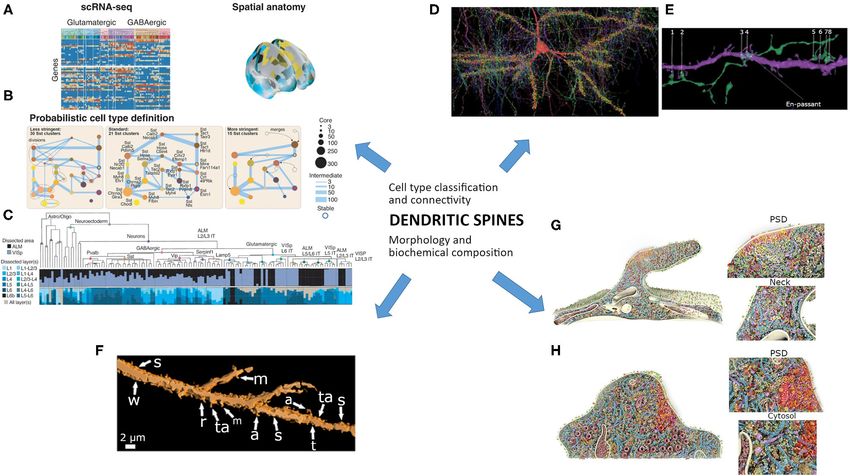
Frontiers in Physiology | www.frontiersin.org 1 February 2022 | Volume 13 | Article 831568Rasia-Filho Dendritic Spines and Synaptic Processing FIGURE 1 | The study of dendritic spines needs to integrate different current fields of knowledge into a coherent vision. Cell type classification and connectivity would be associated with morphology and nanoscale biochemical composition. (A–C) The probabilistic definition of a neuronal type needs that: (A) “A transcriptome-based cell-type taxonomy is constructed from scRNA-seq data, related epigenomic datasets and neuroanatomy. (B) Cell types are initially defined based on transcriptomic signatures in a probabilistic manner with multiresolution clustering and statistical analysis to identify robustness and variability. (C) Reproducible gene expression patterns identify hierarchies of putative cell types that are subject to further analyses and validation.” (A–C) are reproduced and adapted from Yuste et al. (2020) “A community-based transcriptomics classification and nomenclature of neocortical cell types” (a–c images from Figure 5 were reused without changes), Nat. Neurosci. 23, 1456–1468, doi: 10.1038/s41593-020-0685-8; used under CC BY 4.0 license and copyright. (D,E) Image of a petascale reconstruction of human parietal cortex for the study of cells and connectivity. (D) “The axonal innervation to a “pyramidal cell (red) is rendered along with all incoming synapses (yellow discs) and presynaptic axons.” (E) “Excitatory axon forming 8 synapses onto a spiny dendrite of an excitatory cell. One of these connections is en passant, the rest required directed growth of the axon to contact this same dendrite.” Note the axo-spinous contacts (numbered 1, 2, 5–8), which would be further studied by focused ion beam/scanning electron microscopy. (D,E) are reproduced and slightly adapted from Shapson-Coe et al. (2021) “A connectomic study of a petascale fragment of human cerebral cortex” (removed letters and changed number size from the original Figure 6), bioRxiv 2021.05.29.446289; doi: 10.1101/2021.05.29.446289; used under aCC-BY-NC-ND 4.0 International license and copyright. (F) Morphological features of 3D-reconstructed dendritic spines of a Golgi-impregnated neuron from the human cortical amygdala. Spine shapes include: stubby (s), wide (w), thin (t), mushroom-like (m), ramified (r), transitional (ta), atypical (a) or multiform. Reused and slightly adapted from Vásquez et al. (2018) “Neuronal types of the human cortical amygdaloid nucleus" (Figure 6e), J. Comp. Neurol. 526, 2776–2801. doi: 10.1002/cne.24527; under license # 5223180562839 from Copyright Clearance Center’s RightsLink® originally published by John Wiley & Sons, Inc. (G,H) 3D model of dendritic spines using large-scale nanoscopy and biochemistry analysis. Multiple constitutive proteins are colored and “shown to scale, with the copy numbers and locations measured in this study and configurations according to literature. For clarity, the highly abundant monomeric actin is not shown. (G) View into a mushroom spine. Magnifications into the postsynaptic density (PSD, highlighted with red glow) and neck are depicted. (H) View into a stubby spine. Again, a magnification of the PSD is shown and a zoom into the cytosolic region of the spine.” Reused and slight adaptation from Helm et al. (2021) manuscript) after Helm et al. (2021) “A large-scale nanoscopy and biochemistry analysis of postsynaptic dendritic spines” (removed letters in Figure 7), Nat. Neurosci. 24, 1151–1162. doi: 10.1038/s41593-021-00874-w under license # 5223201496130 from Copyright Clearance Center’s RightsLink® , originally published by Springer Nature. cell types can be the rule as occur for the spiny pyramidal intercellular interactions within the network in which the neurons (Morishima and Kawaguchi, 2006; Ramaswamy and cells are embedded” [BRAIN Initiative Cell Census Network Markram, 2015; Cembrowski and Spruston, 2019; Benavides- (BICCN), 2021]. Henceforth, the probabilistic definition of Piccione et al., 2020; Rasia-Filho et al., 2021). Indeed, discrete each neuronal type will require single-cell transcriptomic data and continuous variations may coexist and underlie cell-type associated to morphology (Figure 1A; Hodge et al., 2019; Yuste diversity, forming a “combination of specification through et al., 2020). Dendritic spines may be further studied in evolutionarily driven and developmentally regulated genetic specific subpopulations of neurons and circuits to address their mechanisms, and refinement of cellular identities through functional roles in information processing (Figure 1B). Frontiers in Physiology | www.frontiersin.org 2 February 2022 | Volume 13 | Article 831568
Rasia-Filho                                                                                                 Dendritic Spines and Synaptic Processing

BRAIN NETWORKS, CELLULAR                                                     or multiform spines, including “intermediate” shapes, “double”
CONNECTIVITY, AND THE RELEVANCE OF                                           spines, and thorny excrescences, among others (Fiala and
                                                                             Harris, 1999; Arellano et al., 2007; Bourne and Harris, 2007,
DENDRITIC SPINES
                                                                             2009; Stewart et al., 2014; Fuentealba-Villarroel et al., 2021;
Recent approaches advanced the study of brain cells,                         see also Ruszczycki et al., 2012; Pchitskaya and Bezprozvanny,
microcircuits, and connections. The connectomic study of                     2020). Spine shape involves local actin organization, second
a fragment of the human temporal cortex (1 mm3 , >5,000                      messengers, and organelles (e.g., endoplasmic reticulum and
slices cut at ∼30 nm), imaged using a high-speed multi-beam                  ribosomes, Yuste, 2010; Sala and Segal, 2014; Miermans
scanning electron microscopy (EM) and three-dimensional                      et al., 2017; Okabe, 2020; for mitochondria see Li et al.,
(3D) reconstruction, exhibited 57,216 cells and ∼133 million                 2004). This can lead to biochemical compartmentalization
synapses in a 1.4-petabyte volume (Shapson-Coe et al., 2021).                and affect the electrical signaling of synapses (Chen and
Dense digital reconstruction of a 0.3 mm3 cortical circuit                   Sabatini, 2012; Tønnesen and Nägerl, 2016; Obashi et al., 2021).
containing ∼31,000 neurons, ∼37 million excitatory and                       The balance between spine number, structure, and function
inhibitory synapses, and 55 morphological cell types served to               may represent synaptic processing for learning and memory
identify hub neurons that could modulate cortical dynamics                   (Bourne and Harris, 2007, 2009) with stimulus-specific features
(Gal et al., 2021). Additional highly multiplexed, high-resolution           (Knafo et al., 2005) in selective synaptic ensembles (Hayashi-
brain-wide cell-type mapping, and high-throughput spatially                  Takagi et al., 2015). Optogenetic manipulation allowed the
resolved transcriptomics approaches can link cell types with                 identification and erasure of specific synaptic memory traces
connectivity mapping and functional data (Close et al., 2021)                in potentiated spines of the mouse motor cortex (Hayashi-
for advanced molecular neuroanatomical maps (Ortiz et al.,                   Takagi et al., 2015). This was a remarkable achievement since
2021). Some techniques may link functional data with different               the functional mapping of single-spine synaptic inputs to the
spatial scales. For example, patch-clamp electrophysiology                   same dendrite can be highly heterogeneous, as revealed by high-
and single-cell semi-quantitative PCR would identify neuronal                resolution two-photon imaging of auditory-evoked NMDA-
subtypes (Fuzik et al., 2016). On the other hand, high-resolution            dependent calcium transients in mouse cortical neurons in vivo
magnetic resonance imaging (MRI) would locate different nuclei               (Chen et al., 2011).
in the brain (Saygin et al., 2017 for human amygdala) and help to               Dendritic spine dynamics in different neural circuits
identify likely borders for each area of interest (e.g., to separate         result from various phenomena. These include phylogenetic,
the medial and cortical amygdaloid nuclei, Dall’Oglio et al., 2013;          ontogenetic, and epigenetic events (García-López et al., 2010;
Vásquez et al., 2018). These data are relevant for understanding             DeFelipe, 2011; Reza-Zaldivar et al., 2020). Activity-dependent
the complex expression of emotion in different species (Quirk                and activity-independent actions promote stabilization,
                                                                             differentiation, and remodeling with enlargement or shrinkage
et al., 1995; Zebarjadi et al., 2021; including mice affiliative touch
                                                                             and pruning of spines (Oray et al., 2006; Zancan et al.,
in prosocial interaction, Wu et al., 2021) and what feelings are to
                                                                             2018; Runge et al., 2020; Kasai et al., 2021). Spines can be
humans (Zeki, 2007; Gendron and Barrett, 2009; de Boer et al.,
                                                                             found relatively isolated or in clusters in the same dendritic
2012; Diano et al., 2017; LeDoux and Brown, 2017; Fogazzi et al.,
                                                                             segments, as evidenced after 3D image reconstruction of
2020; Šimić et al., 2021).
                                                                             Golgi-impregnated neurons in humans from our laboratory
    To process manifold stimuli from external and internal
                                                                             (Reberger et al., 2018; Rasia-Filho et al., 2021) and other
milieux engenders specialization and functional integration of
                                                                             approaches using transmission EM (Arellano et al., 2007;
neural areas, cells, and networks (e.g., Rasia-Filho, 2006; Rasia-
                                                                             Bourne and Harris, 2009; Brusco et al., 2014; Stewart et al.,
Filho et al., 2018; Freiwald, 2020; Barnett et al., 2021). Dendritic
                                                                             2014), rapid structured illumination microscopy and enhanced
spine function comprises an important part of this complex
                                                                             resolution confocal microscopy (for spinules, Zaccard et al.,
scenario (Ramón y Cajal, 1909–1911; Bourne and Harris, 2009;
                                                                             2020), high-resolution transmission, focused ion beam (FIB)
von Bohlen und Halbach, 2009; Yuste, 2010; Spruston et al.,
                                                                             scanning and EM tomography (Rollenhagen et al., 2020), and/or
2013; Dall’Oglio et al., 2015; Helm et al., 2021). That is, spines
                                                                             FIB/scanning EM in humans and other animals (Cano-Astorga
increase the connectivity between neurons and the packing
                                                                             et al., 2021). Clustered spines can show spike-timing-dependent
density of synapses without increasing the brain’s overall volume            cooperativity and plasticity (Tazerart et al., 2020). Therefore,
(Bourne and Harris, 2009). This feature adds and maximizes the               synaptic integration made by each spine type can impact
connectivity repertoire governing the shape of dendritic arbors              cellular activity differently depending upon its location and
(Wen et al., 2009). Dendritic spines modulate the excitatory                 spatiotemporal processing along proximal to distal dendritic
synaptic transmission in the brain. The majority of input contacts           domains (Spruston et al., 2013). In addition, its passive
on dendritic spines are from glutamatergic axon terminals                    and/or active biophysical properties associated with those of
(Yuste, 2013; but see also GABAergic and dopaminergic                        parent dendrites may play a role (Sala and Segal, 2014; Gidon
innervation in Brusco et al., 2014; Kubota et al., 2016; Iino                et al., 2020; Obashi et al., 2021). Dendritic spines modulate
et al., 2020; Kasai et al., 2021). Spines are morphologically                both stable and/or transitory connections (Oray et al., 2006)
diverse, ranging in a continuum of number, shape, and size                   and synaptic plasticity using various molecules in variable
classified according to their head and neck features (Figure 1C).            biochemical pathways for short-term to long-term cellular effects
These include: stubby/wide, thin, mushroom, ramified, “atypical”             (Sala and Segal, 2014; Chidambaram et al., 2019).

Frontiers in Physiology | www.frontiersin.org                            3                                 February 2022 | Volume 13 | Article 831568
Rasia-Filho Dendritic Spines and Synaptic Processing There are many frontiers to explore the structure and dendritic spines are sexually dimorphic and/or affected by integrated function of dendritic spines for synaptic plasticity. gonadal steroids (Woolley and McEwen, 1993; Rasia-Filho The impact of heterogeneous glial cells and the role of the et al., 2012; Luine and Frankfurt, 2020), sexual experience and extracellular matrix in tetrapartite synapses need to be addressed motherhood (Rasia-Filho et al., 2004; Zancan et al., 2018). These (Chelini et al., 2018; Mederos et al., 2018; Tønnesen et al., 2018; phenomena are relevant to sexual differentiation in healthy brain Nguyen et al., 2020; Klimczak et al., 2021). The elucidation of connectivity and as a biological variable in neuropsychiatric the evolutionary reason for the divergence in gene expression research (Joel and McCarthy, 2017; Rubinow and Schmidt, 2019; patterns in the cerebral cortex, and the features that determine Arnold, 2020; Hidalgo-Lopez et al., 2021). neuronal diversity and specialization in humans are important Lastly, the human cerebral cortex shows a highly polygenic (Hodge et al., 2019, 2020; Kalmbach et al., 2021). Regarding architecture (Grasby et al., 2020) and ∼16 billion neurons the latter, some features of human cortical pyramidal neurons (Herculano-Houzel et al., 2014). One cortical pyramidal neuron include: (1) larger dendritic length and branch complexity than can form ∼30,000 synapses, 90% of them being excitatory macaque and mice (Mohan et al., 2015; Benavides-Piccione (DeFelipe, 2011). From ∼100 trillion spines in the human cortex et al., 2020); (2) a class of calcium-mediated graded dendritic (Kasai et al., 2021), ∼99.5% of all spines lie in pyramidal action potentials that would classify linearly non-separable neurons (Kubota et al., 2016; see also Foggetti et al., 2019) inputs (Gidon et al., 2020); and (3) membrane properties that for the organization of the ongoing synaptic transmission significantly enhance synaptic charge-transfer from dendrites from multiple neurochemical circuits (Palomero-Gallagher and to soma and spike propagation along the axon (Eyal et al., Zilles, 2019). This complexity is exemplified by the huge spine 2016). This indicates that extrapolations on some neuronal density and shape variation in a human CA1 pyramidal neuron features from other species to the human brain have to be related to circuits for memory modulation and self-identity done carefully. Human dendritic spines are systematically larger (see Figure 9 in Rasia-Filho et al., 2021). On a spine-by- and longer and exist at higher densities than in the mouse spine basis (Oray et al., 2006), there can be a high degree of cortex (Benavides-Piccione et al., 2020), likely increasing our synaptic processing arising from spatiotemporal and functional capacity of synaptic processing and plasticity (DeFelipe, 2011). heterogeneity among individual synapses on the same dendrite, Human spines, also, show a high diversity of size and shapes between different neurons, and across and between brain regions (Dall’Oglio et al., 2015; Vásquez et al., 2018; Rasia-Filho et al., (Grant and Fransén, 2020). Synaptic diversity and strength 2021). The functional implication of long “silent” spines (Yuste, are finely adjusted to code information (Grant and Fransén, 2013) and those of convoluted shapes, observed from subcortical 2020), enabling coincidence detection (Chabrol et al., 2015) to cortical human neurons (Dall’Oglio et al., 2015; Fuentealba- and merging multimodal inputs from parallel pathways (Soltesz Villarroel et al., 2021), need additional studies. Multiform spines and Losonczy, 2018). The integrated synaptic processing and likely indicate the existence of multisynaptic sites for signaling complex plasticity linked to the role of an increasing number of compartmentalization and further computational possibilities specialized neurons and glia cells within circuits may ultimately within functional microdomains (Chen and Sabatini, 2012; lead to the emergence of multiple sensorimotor, cognitive, Dall’Oglio et al., 2015; Reberger et al., 2018). emotional, abstract, creative and conscious elaborations, visceral The postsynaptic density (PSD) is a dense area behind reactions, and behavioral displays (for a parallel discussion see the postsynaptic membrane, as seen by EM. It consists Timo-Iaria and Valle, 1995; DeFelipe, 2011; Jezek et al., 2011; of many proteins, including receptors, ion channels, and Hodge et al., 2019; Freiwald, 2020; Grant and Fransén, 2020; adhesion proteins, shared with the membrane, cytoskeletal Rasia-Filho et al., 2021). proteins, and scaffolding proteins, all arranged in a hierarchical fashion (Cohen, 2013). Like dendritic spines, PSDs can display morphological alterations with various physiological and CONCLUSION behavioral inputs. PSD area can relate to spine head diameter (Arellano et al., 2007) depending on an NMDA receptor- Dendritic spines are key elements for innovative research in mediated long-term potentiation plasticity (Borczyk et al., 2019). integrative physiology. Various approaches can expand our Stubby and mushroom spines show similar average protein knowledge on spines studying them at both network-scale and copy number and topology for PSD composition identified after synapse-scale in the brain. For example, we still do not know summing EM, stimulated emission depletion microscopy, mass the implications of dendritic spines in different subpopulations spectrometry, fluorescence microscopy, and 3D reconstruction of neurons for the cytoarchitectonics, rich networks connections, procedures in cultured hippocampal neurons of rats (Helm et al., and complex information processing in the insular cortex. This is 2021; Figure 1D). However, proteins related to synaptic strength, an interesting cortical area that is strongly activated when “you spine dynamics, ion channels, endocytosis cofactors, cytoskeletal see the person you are in love with, try to listen to your own structure, signaling and trafficking, secretory proteins, and heartbeat, suffer from a headache, or crave for a chocolate cookie” ribosomes are more evident in mushroom spines (Helm et al., (Gogolla, 2017; see also Benarroch, 2019). As mentioned by 2021). These findings open the possibility to test different Mancuso et al. (2014): “Anatomical changes occur on a full range spines also in neuropathological conditions (Forrest et al., 2018; of scales from the trafficking of individual proteins, to alterations Chidambaram et al., 2019; Runge et al., 2020; Baczyńska et al., in synaptic morphology both individually and on a systems level, 2021; Montero-Crespo et al., 2021). From this perspective, to reductions in long-distance connectivity and brain volume.” Frontiers in Physiology | www.frontiersin.org 4 February 2022 | Volume 13 | Article 831568
Rasia-Filho                                                                                                                              Dendritic Spines and Synaptic Processing

Dendritic spines relate to all these processes in the brain and                                FUNDING
with a notable integrative complexity in humans. Unraveling
the dynamic role of dendritic spines for synaptic processing is a                              Grants from the Brazilian Agencies CAPES and CNPq (Brazilian
task that needs multiple complementary approaches. The level of                                Ministry of Science Technology and Innovation RRID), Grant/
complexity for this endeavor resides in the fact we are looking for                            Award Numbers: 314352/2020-1, SCR_002876.
representative data that is likely on the astonishing scale of 1015
ongoing connections in the human brain.
                                                                                               ACKNOWLEDGMENTS
AUTHOR CONTRIBUTIONS
                                                                                               I thank Prof. Rochelle S. Cohen for her valuable suggestions
The author confirms being the sole contributor of this work and                                on this manuscript and Mr. Josué Renner for his help with
has approved it for publication.                                                               figure preparation.

REFERENCES                                                                                     Chabrol, F. P., Arenz, A., Wiechert, M. T., Margrie, T. W., and DiGregorio, D. A.
                                                                                                  (2015). Synaptic diversity enables temporal coding of coincident multisensory
Arellano, J. I., Benavides-Piccione, R., DeFelipe, J., and Yuste, R. (2007).                      inputs in single neurons. Nat. Neurosci. 18, 718–727. doi: 10.1038/nn.3974
   Ultrastructure of dendritic spines: correlation between synaptic and spine                  Chelini, G., Pantazopoulos, H., Durning, P., and Berretta, S. (2018). The
   morphologies. Front. Neurosci. 1:131–143. doi: 10.3389/neuro.01.1.1.010.2007                   tetrapartite synapse: a key concept in the pathophysiology of schizophrenia.
Arnold, A. P. (2020). Sexual differentiation of brain and other tissues:                          Eur. Psychiatry 50, 60–69. doi: 10.1016/j.eurpsy.2018.02.003
   five questions for the next 50 years. Horm. Behav. 120, 104691.                             Chen, X., Leischner, U., Rochefort, N. L., Nelken, I., and Konnerth, A. (2011).
   doi: 10.1016/j.yhbeh.2020.104691                                                               Functional mapping of single spines in cortical neurons in vivo. Nature 475,
Assem, M., Glasser, M. F., Van Essen, D. C., and Duncan, J. (2020). A domain-                     501–505. doi: 10.1038/nature10193
   general cognitive core defined in multimodally parcellated human cortex.                    Chen, Y., and Sabatini, B. L. (2012). Signaling in dendritic spines
   Cereb. Cortex 30, 4361–4380. doi: 10.1093/cercor/bhaa023                                       and spine microdomains. Curr. Opin. Neurobiol. 22, 389–396.
Baczyńska, E., Pels, K. K., Basu, S., Włodarczyk, J., and Ruszczycki, B. (2021).                 doi: 10.1016/j.conb.2012.03.003
   Quantification of dendritic spines remodeling under physiological stimuli and               Chidambaram, S. B., Rathipriya, A. G., Bolla, S. R., Bhat, A., Ray, B.,
   in pathological conditions. Int. J. Mol. Sci. 22, 4053. doi: 10.3390/ijms22084053              Mahalakshmi, A. M., et al. (2019). Dendritic spines: revisiting the
Barnett, S. C., Parr-Brownliebde, L. C., Perry, B. A. L., Young, C. K., Wicky, H. E.,             physiological role. Prog. Neuropsychopharmacol. Biol. Psychiatry 92, 161–193.
   Hughes, S. M., et al. (2021). Anterior thalamic nuclei neurons sustain memory.                 doi: 10.1016/j.pnpbp.2019.01.005
   Curr. Res. Neurobiol. 2, 100022. doi: 10.1016/j.crneur.2021.100022                          Close, J. L., Long, B. R., and Zeng, H. (2021). Spatially resolved transcriptomics in
Benarroch, E. E. (2019). Insular cortex: functional complexity and clinical                       neuroscience. Nat. Methods 18, 23–25. doi: 10.1038/s41592-020-01040-z
   correlations. Neurology 93, 932–938. doi: 10.1212/WNL.0000000000008525                      Cohen, R. S. (2013). “The postsynaptic density,” in Neuroscience in the
Benavides-Piccione, R., Regalado-Reyes, M., Fernaud-Espinosa, I., Kastanauskaite,                 21st Century, ed D. W. Pfaff (New York, NY: Springer), 403–437.
   A., Tapia-González, S., León-Espinosa, G., et al. (2020). Differential structure of            doi: 10.1007/978-1-4614-1997-6_17
   hippocampal CA1 pyramidal neurons in the human and mouse. Cereb. Cortex                     Dall’Oglio, A., Dutra, A. C., Moreira, J. E., and Rasia-Filho, A. A.
   30, 730–752. doi: 10.1093/cercor/bhz122                                                        (2015). The human medial amygdala: structure, diversity, and
Borczyk, M., Sliwińska, M. A., Caly, A., and Bernas, T., Radwanska, K. (2019).                   complexity of dendritic spines. J. Anat. 227, 440–459. doi: 10.1111/joa.
   Neuronal plasticity affects correlation between the size of dendritic spine and                12358
   its postsynaptic density. Sci. Rep. 9, 1693. doi: 10.1038/s41598-018-38412-7                Dall’Oglio, A., Xavier, L. L., Hilbig, A., Ferme, D., Moreira, J. E., Achaval, M.,
Bourne, J., and Harris, K. M. (2007). Do thin spines learn to be                                  et al. (2013). Cellular components of the human medial amygdaloid nucleus.
   mushroom spines that remember? Curr. Opin. Neurobiol. 17, 381–386.                             J. Comp. Neurol. 521, 589–611. doi: 10.1002/cne.23192
   doi: 10.1016/j.conb.2007.04.009                                                             de Boer, A., Van Buel, E. M., and Ter Horst, G. J. (2012). Love is more than just
Bourne, J. N., and Harris, K. M. (2009). “Ultrastructural analysis of spine plasticity,”          a kiss: a neurobiological perspective on love and affection. Neuroscience 201,
   in Encyclopedia of Neuroscience, ed L. R. Squire (New York, NY: Elsevier),                     114–124. doi: 10.1016/j.neuroscience.2011.11.017
   11–17. doi: 10.1016/B978-008045046-9.01771-X                                                DeFelipe, J. (2011). The evolution of the brain, the human nature of
BRAIN Initiative Cell Census Network (BICCN). (2021). A multimodal cell                           cortical circuits, and intellectual creativity. Front. Neuroanat. 5, 29.
   census and atlas of the mammalian primary motor cortex. Nature 598, 86–102.                    doi: 10.3389/fnana.2011.00029
   doi: 10.1038/s41586-021-03950-0                                                             Demas, J., Manley, J., Tejera, F., Barber, K., Kim, H., Traub, F. M., et al.
Bruner, E., Preuss, T. M., Chen, X., and Rilling, J. K. (2017). Evidence for expansion            (2021). High-speed, cortex-wide volumetric recording of neuroactivity at
   of the precuneus in human evolution. Brain Struct. Funct. 222, 1053–1060.                      cellular resolution using light beads microscopy. Nat. Methods 18, 1103–1111.
   doi: 10.1007/s00429-015-1172-y                                                                 doi: 10.1038/s41592-021-01239-8
Brusco, J., Merlo, S., Ikeda, É. T., Petralia, R. S., Kachar, B., Rasia-Filho, A. A.,          Diano, M., Tamietto, M., Celeghin, A., Weiskrantz, L., Tatu, M. K., Bagnis, A., et al.
   et al. (2014). Inhibitory and multisynaptic spines, and hemispherical synaptic                 (2017). Dynamic changes in amygdala psychophysiological connectivity reveal
   specialization in the posterodorsal medial amygdala of male and female rats. J.                distinct neural networks for facial expressions of basic emotions. Sci. Rep. 7,
   Comp. Neurol. 522, 2075–2088. doi: 10.1002/cne.23518                                           1–13. doi: 10.1038/srep45260
Cano-Astorga, N., DeFelipe, J., and Alonso-Nanclares, L. (2021). Three-                        Eyal, G., Verhoog, M. B., Testa-Silva, G., Deitcher, Y., Lodder, J. C.,
   dimensional synaptic organization of layer III of the human temporal                           Benavides-Piccione, R., et al. (2016). Unique membrane properties and
   neocortex. Cereb. Cortex 31, 4742–4764. doi: 10.1093/cercor/bh                                 enhanced signal processing in human neocortical neurons. eLife 5:e16553.
   ab120                                                                                          doi: 10.7554/eLife.16553
Cembrowski, M. S., and Spruston, N. (2019). Heterogeneity within classical cell                Eze, U. C., Bhaduri, A., Haeussler, M., Nowakowski, T. J., and Kriegstein,
   types is the rule: lessons from hippocampal pyramidal neurons. Nat. Rev.                       A. R. (2021). Single-cell atlas of early human brain development
   Neurosci. 20, 193–204. doi: 10.1038/s41583-019-0125-5                                          highlights heterogeneity of human neuroepithelial cells and early

Frontiers in Physiology | www.frontiersin.org                                              5                                           February 2022 | Volume 13 | Article 831568
Rasia-Filho Dendritic Spines and Synaptic Processing radial glia. Nat. Neurosci. 24, 584–594. doi: 10.1038/s41593-020- numbers of neurons and average neuronal cell size. Front. Neuroanat. 8:77. 00794-1 doi: 10.3389/fnana.2014.00077 Fiala, J. C., and Harris, K. M. (1999). “Dendrite structure,” in Dendrites, eds Hidalgo-Lopez, E., Zeidman, P., Harris, T., Razi, A., and Pletzer, B. (2021). G. Stuart, N. Spruston, and M. Häusser (New York, NY: Oxford University Spectral dynamic causal modeling in healthy women reveals brain Press), 1–34. connectivity changes along the menstrual cycle. Commun Biol. 4:954. Finkelstein, A., Fontolan, L., Economo, M. N., Li, N., Romani, S., et al. doi: 10.1038/s42003-021-02447-w (2021). Attractor dynamics gate cortical information flow during Hodge, R. D., Bakken, T. E., Miller, J. A., Smith, K. A., Barkan, E. R., Graybuck, L. decision-making. Nat. Neurosci. 24, 843–850. doi: 10.1038/s41593-021-0 T., et al. (2019). Conserved cell types with divergent features in human versus 0840-6 mouse cortex. Nature 573, 61–68. doi: 10.1038/s41586-019-1506-7 Fogazzi, D. V., Neary, J. P., Sonza, A., Reppold, C. T., Kaiser, V., Scassola, C. Hodge, R. D., Miller, J. A., Novotny, M., Kalmbach, B. E., Ting, J. T., Bakken, M., et al. (2020). The prefrontal cortex conscious and unconscious response T. E., et al. (2020). Transcriptomic evidence that von Economo neurons are to social/emotional facial expressions involve sex, hemispheric laterality, and regionally specialized extratelencephalic-projecting excitatory neurons. Nat. selective activation of the central cardiac modulation. Behav. Brain Res. 393, Commun. 11, 1172. doi: 10.1038/s41467-020-14952-3 112773. doi: 10.1016/j.bbr.2020.112773 Iino, Y., Sawada, T., Yamaguchi, K., Tajiri, M., Ishii, S., Kasai, H., et al. (2020). Foggetti, A., Baccini, G., Arnold, P., Schiffelholz, T., and Wulff, P. (2019). Spiny Dopamine D2 receptors in discrimination learning and spine enlargement. and non-spiny parvalbumin-positive hippocampal interneurons show different Nature 579, 555–560. doi: 10.1038/s41586-020-2115-1 plastic properties. Cell Rep. 27, 3725–3732.e5. doi: 10.1016/j.celrep.2019.05.098 Jezek, K., Henriksen, E. J., Treves, A., Moser, E. I., and Moser, M.-B. (2011). Forrest, M. P., Parnell, E., and Penzes, P. (2018). Dendritic structural Theta-paced flickering between place-cell maps in the hippocampus. Nature plasticity and neuropsychiatric disease. Nat. Rev. Neurosci. 19, 215–234. 478, 246–249. doi: 10.1038/nature10439 doi: 10.1038/nrn.2018.16 Joel, D., and McCarthy, M. M. (2017). Incorporating sex as a biological variable Foster, N. N., Barry, J., Korobkova, L., Garcia, L., Gao, L., Becerra, M., et al. (2021). in neuropsychiatric research: where are we now and where should we be? The mouse cortico–basal ganglia–thalamic network. Nature 598, 188–194. Neuropsychopharmacology 42, 379–385. doi: 10.1038/npp.2016.79 doi: 10.1038/s41586-021-03993-3 Kalmbach, B. E., Hodge, R. D., Jorstad, N. L., Owen, S., de Frates, R., Yanny, Freiwald, W. A. (2020). Social interaction networks in the primate brain. Curr. A. M., et al. (2021). Signature morpho-electric, transcriptomic, and dendritic Opin. Neurobiol. 65, 49–58. doi: 10.1016/j.conb.2020.08.012 properties of human layer 5 neocortical pyramidal neurons. Neuron 109, Fuentealba-Villarroel, F. J., Renner, J., Hilbig, A., Bruton, O. J., and Rasia-Filho, 2914–2927.e5. doi: 10.1016/j.neuron.2021.08.030 A. A. (2021). Spindle-shaped neurons in the human posteromedial (precuneus) Kasai, H., Ziv, N. E., Okazaki, H., Yagishita, S., and Toyoizumi, T. (2021). Spine cortex. Front. Synaptic Neurosci. 13, 769228. doi: 10.3389/fnsyn.2021.769228 dynamics in the brain, mental disorders and artificial neural networks. Nat. Rev. Fuzik, J., Zeisel, A., Máté, Z., Calvigioni, D., Yanagawa, Y., Szabó, G., et al. (2016). Neurosci. 22, 407–422. doi: 10.1038/s41583-021-00467-3 Integration of electrophysiological recordings with single-cell RNA-seq data Klimczak, P., Rizzo, A., Castillo-Gómez, E., Perez-Rando, M., Gramuntell, Y., identifies neuronal subtypes. Nat. Biotech. 34, 175–183. doi: 10.1038/nbt.3443 Beltran, M., et al. (2021). Parvalbumin interneurons and perineuronal nets in Gal, E., Amsalem, O., Schindel, A., London, M., Schürmann, F., Markram, H., et al. the hippocampus and retrosplenial cortex of adult male mice after early social (2021). The role of hub neurons in modulating cortical dynamics. Front. Neural isolation stress and perinatal NMDA receptor antagonist treatment. Front. Circuits 15, 718270. doi: 10.3389/fncir.2021.718270 Synaptic Neurosci. 13:733989. doi: 10.3389/fnsyn.2021.733989 García-López, P., García-Marín, V., and Freire, M. (2010). Dendritic spines Knafo, S., Libersat, F., and Barkai, E. (2005). Olfactory learning-induced and development: towards a unifying model of spinogenesis - a present day morphological modifications in single dendritic spines of young rats. Eur. J. review of Cajal’s histological slides and drawings. Neural Plast. 2010, 769207. Neurosci. 21, 2217–2226. doi: 10.1111/j.1460-9568.2005.04041.x doi: 10.1155/2010/769207 Kubota, Y., Karube, F., Nomura, M., and Kawaguchi, Y. (2016). The Gendron, M., and Barrett, L. F. (2009). Reconstructing the past: A century diversity of cortical inhibitory synapses. Front. Neural Circuits 10:27. of ideas about emotion in psychology. Emotion Rev. 1, 316–339. doi: 10.3389/fncir.2016.00027 doi: 10.1177/1754073909338877 Lavenex, P., Lavenex, P. B., Bennett, J. L., and Amaral, D. G. (2009). Gidon, A., Zolnik, T. A., Fidzinski, P., Bolduan, F., Papoutsi, A., Poirazi, P., et al. Postmortem changes in the neuroanatomical characteristics of the primate (2020). Dendritic action potentials and computation in human layer 2/3 cortical brain: hippocampal formation. J. Comp. Neurol. 512, 27–51. doi: 10.1002/cne. neurons. Science 367, 83–87. doi: 10.1126/science.aax6239 21906 Girskis, K. M., Stergachis, A. B., DeGennaro, E. M., Doan, R. N., Qian, X., LeDoux, J. E., and Brown, R. (2017). Emotions as higher-order states Johnson, M. B., et al. (2021). Rewiring of human neurodevelopmental gene of consciousness. Proc. Natl. Acad. Sci. USA 114, E2016–E2025. regulatory programs by human accelerated regions. Neuron 109, 239–3251.e7. doi: 10.1073/pnas.1619316114 doi: 10.1016/j.neuron.2021.08.005 Leopold, D. A., Strick, P. L., Bassett, D. S., Bruno, R. M., Cuntz, H., Harris, K. M., Glasser, M. F., Coalson, T. S., Robinson, E. C., Hacker, C. D., Harwell, J., Yacoub, et al. (2019). “Functional architecture of the cerebral cortex,” in The Neocortex, E., et al. (2016). A multi-modal parcellation of human cerebral cortex. Nature ed W. Singer (Cambridge: MA: MIT Press), 141–164. 536, 171–178. doi: 10.1038/nature18933 Li, Z., Okamoto, K., Hayashi, Y., and Sheng, M. (2004). The importance of Gogolla, N. (2017). The insular cortex. Curr. Biol. 27, R580–586. dendritic mitochondria in the morphogenesis and plasticity of spines and doi: 10.1016/j.cub.2017.05.010 synapses. Cell 119, 873–887. doi: 10.1016/j.cell.2004.11.003 Grant, S. G., and Fransén, E. (2020). The synapse diversity dilemma: molecular Luine, V., and Frankfurt, M. (2020). Estrogenic regulation of memory: the first 50 heterogeneity confounds studies of synapse function. Front. Synaptic Neurosci. years. Horm. Behav. 121:104711. doi: 10.1016/j.yhbeh.2020.104711 12:590403. doi: 10.3389/fnsyn.2020.590403 Mancuso, J. J., Cheng, J.,Yin, Z.,Gilliam, J. C., Xia, X., Li, X., et al. Grasby, K. L., Jahanshad, N., Painter, J. N., Colodro-Conde, L., Bralten, J., Hibar, D. (2014). Integration of multi scale dendritic spine structure and P., et al. (2020). The genetic architecture of the human cerebral cortex. Science function data into systems biology models. Front. Neuroanat. 8:130. 367, eaay6690. doi: 10.1126/science.aay6690 doi: 10.3389/fnana.2014.00130 Hayashi-Takagi, A., Yagishita, S., Nakamura, M., Shirai, F., Wu, Y. I., Loshbaugh, Mederos, S., González-Arias, C., and Perea,. G. (2018). Astrocyte–neuron A. L., et al. (2015). Labeling and optical erasure of synaptic memory traces in networks: a multilane highway of signaling for homeostatic brain function. the motor cortex. Nature 525, 333–338. doi: 10.1038/nature15257 Front. Synaptic Neurosci. 10:45. doi: 10.3389/fnsyn.2018.00045 Helm, M. S., Dankovich, T. M., Mandad, S., Rammner, B., Jähne, S., Miermans, C. A., Kusters, R. P., Hoogenraad, C. C., and Storm, C. Salimi, V., et al. (2021). A large-scale nanoscopy and biochemistry (2017). Biophysical model of the role of actin remodeling on dendritic analysis of postsynaptic dendritic spines. Nat. Neurosci. 24, 1151–1162. spine morphology. PLoS ONE 12, e0170113. doi: 10.1371/journal.pone.01 doi: 10.1038/s41593-021-00874-w 70113 Herculano-Houzel, S., Manger, P. R., and Kaas, J. H. (2014). Brain scaling in Mohan, H., Verhoog, M. B., Doreswamy, K. K., Eyal, G., Aardse, R., Lodder, B. mammalian evolution as a consequence of concerted and mosaic changes in N., et al. (2015). Dendritic and axonal architecture of individual pyramidal Frontiers in Physiology | www.frontiersin.org 6 February 2022 | Volume 13 | Article 831568
Rasia-Filho                                                                                                                             Dendritic Spines and Synaptic Processing

   neurons across layers of adult human neocortex. Cereb. Cortex 25, 4839–4853.                  and synaptic plasticity in Alzheimer’s disease: a focus on microRNA. Front. Cell
   doi: 10.1093/cercor/bhv188                                                                    Dev. Biol. 8:255. doi: 10.3389/fcell.2020.00255
Montero-Crespo, M., Domínguez-Álvaro, M., Alonso-Nanclares, L.,                               Rigotti, M., Barak, O., Warden, M., Wang, X.-J., Daw, N. D., Miller, E. K., et al.
   DeFelipe, J., and Blazquez-Llorca, L. (2021). Three-dimensional                               (2013). The importance of mixed selectivity in complex cognitive tasks. Nature
   analysis of synaptic organization in the hippocampal CA1 field                                497, 585–590. doi: 10.1038/nature12160
   in Alzheimer’s disease. Brain 144, 553–573. doi: 10.1093/brain/                            Rollenhagen, A., Walkenfort, B., Yakoubi, R., Klauke, S. A., Schmuhl-Giesen, S. F.,
   awaa406                                                                                       Heinen-Weiler, J., et al. (2020). Synaptic organization of the human temporal
Morishima, M., and Kawaguchi, Y. (2006). Recurrent connection patterns of                        lobe neocortex as revealed by high-resolution transmission, focused ion beam
   corticostriatal pyramidal cells in frontal cortex. J. Neurosci. 26, 4394–4405.                scanning, and electron microscopic tomography. Int. J. Mol. Sci. 21, 5558.
   doi: 10.1523/JNEUROSCI.0252-06.2006                                                           doi: 10.3390/ijms21155558
Nguyen, P. T., Dorman, L. C., Pan, S., Vainchtein, I. D., Han, R. T., Nakao-Inoue,            Rubinow, D. R., and Schmidt, P. J. (2019). Sex differences and the
   H., et al. (2020). Microglial remodeling of the extracellular matrix promotes                 neurobiology of affective disorders. Neuropsychopharmacol 44, 111–128.
   synapse plasticity. Cell 182, 388–403.e15. doi: 10.1016/j.cell.2020.05.050                    doi: 10.1038/s41386-018-0148-z
Obashi, K., Taraska, J. W., and Okabe, S. (2021). The role of molecular diffusion             Runge, K., Cardoso, C., and de Chevigny, A. (2020). Dendritic spine
   within dendritic spines in synaptic function. J. Gen. Physiol. 153, e202012814.               plasticity: function and mechanisms. Front. Synaptic Neurosci. 12, 36.
   doi: 10.1085/jgp.202012814                                                                    doi: 10.3389/fnsyn.2020.00036
Okabe, S. (2020). Regulation of actin dynamics in dendritic spines: nanostructure,            Ruszczycki, B., Szepesi, Z., Wilczynski, G. M., Bijata, M., Kalita, K., Kaczmarek,
   molecular mobility, and signaling mechanisms. Mol. Cell. Neurosci. 109,                       L., et al. (2012). Sampling issues in quantitative analysis of dendritic spines
   103564. doi: 10.1016/j.mcn.2020.103564                                                        morphology. BMC Bioinformatics 13, 213. doi: 10.1186/1471-2105-13-213
Oray, S., Majewska, A., and Sur, M. (2006). Effects of synaptic activity on dendritic         Sala, C., and Segal, M. (2014). Dendritic spines: the locus of
   spine motility of developing cortical layer V pyramidal neurons. Cereb. Cortex                structural and functional plasticity. Physiol. Rev. 94, 141–188.
   16, 730–741. doi: 10.1093/cercor/bhj019                                                       doi: 10.1152/physrev.00012.2013
Ortiz, C., Carlén, M., and Meletis, K. (2021). Spatial transcriptomics:                       Saygin, Z. M., Kliemann, D., Iglesias, J. E., van der Kouwe, A. J. W., Boyd,
   molecular maps of the mammalian brain. Ann. Rev. Neurosci. 44, 547–562.                       E., Reuter, M., et al. (2017). High-resolution magnetic resonance imaging
   doi: 10.1146/annurev-neuro-100520-082639                                                      reveals nuclei of the human amygdala: manual segmentation to automatic atlas.
Palomero-Gallagher, N., and Zilles, K. (2019). Cortical layers: cyto-, myelo-,                   Neuroimage 155, 370–382. doi: 10.1016/j.neuroimage.2017.04.046
   receptor- and synaptic architecture in human cortical areas. NeuroImage 197,               Scheperjans, F., Eickhoff, S. B., Hömke, L., Mohlberg, H., Hermann, K.,
   716-741. doi: 10.1016/j.neuroimage.2017.08.035                                                Amunts, K., et al. (2008). Probabilistic maps, morphometry, and variability of
Pchitskaya, E., and Bezprozvanny, I. (2020). Dendritic spines shape analysis -                   cytoarchitectonic areas in the human superior parietal cortex. Cereb. Cortex 18,
   classification or clusterization? Perspective. Front. Synaptic Neurosci. 12, 31.              2141–2157. doi: 10.1093/cercor/bhm241
   doi: 10.3389/fnsyn.2020.00031                                                              Shapson-Coe, A., Januszewski, M., Berger, D. R., Pope, A., Wu, Y., Blakely, T., et al.
Quirk, G. J., Repa, J. C., and LeDoux, J. E. (1995). Fear conditioning                           (2021). A connectomic study of a petascale fragment of human cerebral cortex.
   enhances short-latency auditory responses of lateral amygdala neurons:                        bioRxiv. 2021.05.29.446289. doi: 10.1101/2021.05.29.446289
   parallel recordings in the freely behaving rat. Neuron 15, 1029–1039.                      Šimić, G., Tkalčić, M., Vukić, V., Mulc, D., Španić, E., Šagud, M. et al. (2021).
   doi: 10.1016/0896-6273(95)90092-6                                                             Understanding emotions: Origins and roles of the amygdala. Biomolecules 11,
Ramaswamy, S., and Markram, H. (2015). Anatomy and physiology of                                 823. doi: 10.3390/biom11060823
   the thick-tufted layer 5 pyramidal neuron. Front. Cell. Neurosci. 9:233.                   Soltesz, I., and Losonczy, A. (2018). CA1 pyramidal cell diversity enabling parallel
   doi: 10.3389/fncel.2015.00233                                                                 information processing in the hippocampus. Nat. Neurosci. 21, 484–493.
Ramón y Cajal (1909–1911). Histologie du système nerveux de l’homme et des                       doi: 10.1038/s41593-018-0118-0
   vertébrés, Paris: Maloine. doi: 10.5962/bhl.title.48637                                    Spruston, N., Häusser, M., and Stuart, G. (2013). “Information processing in
Rasia-Filho, A. A. (2006). Is there anything “autonomous” in the nervous system?                 dendrites and spines,” in Fundamental Neuroscience, eds L. R. Squire, D. Berg,
   Adv. Physiol. Educ. 30, 9–12. doi: 10.1152/advan.00022.2005                                   F. E. Bloom, S. du Lac, A. Ghosh, and N. C. Spitzer, (Waltham: Academic Press),
Rasia-Filho, A. A., Andrejew, R., and Belló-Klein, A. (2018). “Integrating concepts              231–260. doi: 10.1016/B978-0-12-385870-2.00011-1
   of resilience from cellular functioning to human behavior,” in Amygdala:                   Stewart, M. G., Popov, V. I., Kraev, I. V., Medvedev, N., and Davies, H. A.
   Mechanisms, Structure and Role in Disease, ed. A. Manu (Hauppauge: Nova                       (2014). “Structure and complexity of the synapse and dendritic spine,” in The
   Science Publishers), 1–30.                                                                    Synapse, eds V. Pickel and M. Segal (New York, NY: Academic Press), 1–20.
Rasia-Filho, A. A., Dalpian, F., Menezes, I. C., Brusco, J., and Moreira, J. E., Cohen,          doi: 10.1016/B978-0-12-418675-0.00001-8
   R. S. (2012). Dendritic spines of the medial amygdala: plasticity, density, shape,         Tazerart, S., Mitchell, D. E., Miranda-Rottmann, S., and Araya, R. (2020). A spike-
   and subcellular modulation by sex steroids. Histol. Histopathol. 8, 985–1011.                 timing-dependent plasticity rule for dendritic spines. Nat. Commun. 11:4276.
   doi: 10.14670/HH-27.985                                                                       doi: 10.1038/s41467-020-17861-7
Rasia-Filho, A. A., Fabian, C., Rigoti, K. M., and Achaval, M. (2004). Influence              Timo-Iaria, C., and Valle, A. C. (1995). The functional role of the conscious
   of sex, estrous cycle and motherhood on dendritic spine density in the rat                    process. Ciência e Cultura J. Braz. Assoc. Adv. Sci. 47, 221–234.
   medial amygdala revealed by the Golgi method. Neuroscience 126, 839–847.                   Tønnesen, J., Inavalli, V. V. G. K., and Nägerl, U. V. (2018). Super-
   doi: 10.1016/j.neuroscience.2004.04.009                                                       Resolution Imaging of the extracellular space in living brain tissue. Cell 172,
Rasia-Filho, A. A., Guerra, K. T. K., Vásquez, C. E., Dall’Oglio, A., Reberger,                  1108–1121.e15. doi: 10.1016/j.cell.2018.02.007
   R., Jung, C. R., et al. (2021). The subcortical-allocortical-neocortical                   Tønnesen, J., and Nägerl, V. (2016). Dendritic spines as tunable regulators of
   continuum for the emergence and morphological heterogeneity of                                synaptic signals. Front. Psychol. 7, 101. doi: 10.3389/fpsyt.2016.00101
   pyramidal neurons in the human brain. Front. Synaptic Neurosci. 13:616607.                 Turcotte, R., Liang, Y., Tanimoto, M., Zhang, Q., Li, Z., Koyama, M., et al.
   doi: 10.3389/fnsyn.2021.616607                                                                (2019). Dynamic super-resolution structured illumination imaging in the living
Real, R., Peter, M., Trabalza, A., Khan, S., Smith, M. A., Dopp, J., et al. (2018). In           brain. Proc. Natl. Acad. Sci. USA 116, 9586–9591. doi: 10.1073/pnas.181996
   vivo modeling of human neuron dynamics and Down syndrome. Science 362,                        5116
   eaau1810. doi: 10.1126/science.aau1810                                                     Vásquez, C. E., Reberger, R., Dall’Oglio, A., Calcagnotto, M. E., and Rasia-Filho, A.
Reberger, R., DallOglio, A., Jung, C. R., and Rasia-Filho, A. A. (2018). Structure               A. (2018). Neuronal types of the human cortical amygdaloid nucleus. J. Comp.
   and diversity of human dendritic spines evidenced by a new three-dimensional                  Neurol. 526, 2776–2801. doi: 10.1002/cne.24527
   reconstruction procedure for Golgi staining and light microscopy. J. Neurosci.             Viscardi, L. H., Imparato, D. O., Bortolini, M. C., and Dalmolin, R.
   Methods 293, 27–36. doi: 10.1016/j.jneumeth.2017.09.001                                       J. S. (2021). Ionotropic receptors as a driving force behind human
Reza-Zaldivar, E. E., Hernández-Sápiens, M. A., Minjarez, B., Gómez-Pinedo, U.,                  synapse establishment. Mol. Biol. Evol. 38, 735–744. doi: 10.1093/molbev/
   Sánchez-González, V. J., Márquez-Aguirre, A. L., et al. (2020). Dendritic spine               msaa252

Frontiers in Physiology | www.frontiersin.org                                             7                                           February 2022 | Volume 13 | Article 831568
Rasia-Filho                                                                                                                            Dendritic Spines and Synaptic Processing

Vogt, B. A. (2015). “Mapping cingulate subregions,” in Brain Mapping: An                     Zaccard, C. R., Shapiro, L., Martin-de-Saavedra, M. D., Pratt, C., Myczek, K., Song,
   Encyclopedic Reference, ed A. W. Toga, (Oxford: Academic Press), 325–339.                    A., et al. (2020). Rapid 3D enhanced resolution microscopy reveals diversity
   doi: 10.1016/B978-0-12-397025-1.00230-X                                                      in dendritic spinule dynamics, regulation, and function. Neuron 107, 522–537.
von Bohlen und Halbach, O. (2009). Structure and function of                                    doi: 10.1016/j.neuron.2020.04.025
   dendritic spines within the hippocampus. Ann. Anat. 191, 518–531.                         Zancan, M., da Cunha, R. S. R., Schroeder, F., Xavier, L. L., and Rasia-Filho,
   doi: 10.1016/j.aanat.2009.08.006                                                             A. A. (2018). Remodeling of the number and structure of dendritic spines
Wen, Q., Stepanyants, A., Elston, G. N., Grosberg, A. Y., and Chklovskii, D. B.                 in the medial amygdala: from prepubertal sexual dimorphism to puberty
   (2009). Maximization of the connectivity repertoire as a statistical principle               and effect of sexual experience in male rats. Eur. J Neurosci. 48, 1851–1865.
   governing the shapes of dendritic arbors. Proc. Natl. Acad. Sci. USA 106,                    doi: 10.1111/ejn.14052
   12536–12541. doi: 10.1073/pnas.0901530106                                                 Zebarjadi, N., Adler, E., Kluge, A., Jääskeläinen, I. P., Sams, M., and Levy, J.
Winnubst, J., Bas, E., Ferreira, T. A., Wu, Z., Economo, M. N., Edson, P., et al.               (2021). Rhythmic neural patterns during empathy to vicarious pain: beyond
   (2019)... Reconstruction of 1,000 projection neurons reveals new cell types                  the affective-cognitive empathy dichotomy. Front. Hum. Neurosci. 15:708107.
   and organization of long-range connectivity in the mouse brain. Cell 179,                    doi: 10.3389/fnhum.2021.708107
   268–281.e13. doi: 10.1016/j.cell.2019.07.042                                              Zeki, S. (2007). The neurobiology of love. FEBS Lett. 581, 2575–2579.
Woolley, C. S., and McEwen, B. S. (1993). Roles of estradiol and progesterone in                doi: 10.1016/j.febslet.2007.03.094
   regulation of hippocampal dendritic spine density during the estrous cycle in
   the rat. J. Comp. Neurol. 336, 293–306. doi: 10.1002/cne.903360210                        Conflict of Interest: The author declares that the research was conducted in the
Wu, Y. E., Dang, J., Kingsbury, L., Zhang, M., Sun, F., Hu, R. K., et al. (2021).            absence of any commercial or financial relationships that could be construed as a
   Neural control of affiliative touch in prosocial interaction. Nature 599, 262–267.        potential conflict of interest.
   doi: 10.1038/s41586-021-03962-w
Yang, L., Yang, Y., Yuan, J., Sun, Y., Dai, J., and Su, B. (2019). Transcriptomic            Publisher’s Note: All claims expressed in this article are solely those of the authors
   landscape of von Economo neurons in human anterior cingulate cortex                       and do not necessarily represent those of their affiliated organizations, or those of
   revealed by microdissected-cell RNA sequencing. Cereb. Cortex 29, 838–851.
                                                                                             the publisher, the editors and the reviewers. Any product that may be evaluated in
   doi: 10.1093/cercor/bhy286
                                                                                             this article, or claim that may be made by its manufacturer, is not guaranteed or
Yuste,     R.    (2010).     Dendritic     Spines.     Cambridge:      MIT      Press.
   doi: 10.7551/mitpress/9780262013505.001.0001                                              endorsed by the publisher.
Yuste, R. (2013). Electrical compartmentalization in dendritic spines.
   Annu. Rev. Neurosci. 36, 429–449. doi: 10.1146/annurev-neuro-062111-1                     Copyright © 2022 Rasia-Filho. This is an open-access article distributed under the
   50455                                                                                     terms of the Creative Commons Attribution License (CC BY). The use, distribution
Yuste, R., Hawrylycz, M., Aalling, N., Aguilar-Valles, A., Arendt, D., Armañanzas,           or reproduction in other forums is permitted, provided the original author(s) and
   R., et al. (2020). A community-based transcriptomics classification and                   the copyright owner(s) are credited and that the original publication in this journal
   nomenclature of neocortical cell types. Nat. Neurosci. 23, 1456–1468.                     is cited, in accordance with accepted academic practice. No use, distribution or
   doi: 10.1038/s41593-020-0685-8                                                            reproduction is permitted which does not comply with these terms.

Frontiers in Physiology | www.frontiersin.org                                            8                                           February 2022 | Volume 13 | Article 831568
You can also read