Ultrafast spectroscopy reveals singlet fission, ionization and excimer formation in perylene film
←
→
Page content transcription
If your browser does not render page correctly, please read the page content below
www.nature.com/scientificreports
OPEN Ultrafast spectroscopy reveals
singlet fission, ionization
and excimer formation in perylene
film
Wenjun Ni1, Licheng Sun1,2 & Gagik G. Gurzadyan1*
Singlet exciton fission (SF) is a spin-allowed process whereby two triplet excitons are created from
one singlet exciton. This phenomenon can offset UV photon energy losses and enhance the overall
efficiency in photovoltaic devices. For this purpose, it requires photostable commercially available
SF materials. Excited state dynamics in pure perylene film, ease of commercial production, is studied
by time-resolved fluorescence and femtosecond transient absorption techniques under different
photoexcitation energies. In film, polycrystalline regions contain perylene in H-type aggregate
form. SF takes place from higher excited states of these aggregates in ultrafast time scale < 30 fs,
reaching a triplet formation quantum yield of 108%. Moreover, at λex = 450 nm singlet fission was
detected as a result of two-quantum absorption. Other competing relaxation channels are excimer
(1 ps) and dimer radical cation formation (< 30 fs). Excimer radiatively relaxes within 19 ns and radical
cation recombines in 3.2 ns. Besides, exciton self-trapping by crystal lattice distortions occurs within
hundreds of picosecond. Our results highlight potential of simple-fabricated perylene films with
similar properties as high-cost single crystal in SF based photovoltaic applications.
Singlet fission is a phenomenon that happens in selected systems where a singlet excited molecule shares its
energy with a neighboring molecule in its ground state, both molecules forming a pair of triplet excitons. Triplet
formation via SF is a spin-allowed process, in contrast to spin-forbidden intersystem crossing, which usually
proceeds in much faster femtosecond/picosecond time scale. SF materials are supposed to boost efficiency of pho-
tovoltaic cells through novel m echanism1–3. On one hand, nonlinear optoelectronic process SF (the absorption
of one photon generates two triplet exciton-hole pairs) can theoretically break the Shockly-Queisser limit of 34%
for single-junction conventional photovoltaics (one photon produces at most one exciton). On the other hand,
important part of solar radiation is emitted in blue or UV spectral regions; after relaxation to the reactive state
most part of excitation is transferred to heat, i.e. is lost. However, designing and synthesizing new SF molecular
systems for real applications is attractive and quite challenging. SF studies are mostly focused on acenes, e.g.
tetracene, pentacene, rubrene and related systems: molecular crystals, films and covalently linked dimers.
Perylene and its derivatives aroused a considerable interest in SF field due to their attractive properties, i.e.
high absorption coefficients, photostability; they are widely used in various photovoltaic applications. In these
systems perylenediimides stand out as a result of easy chemical modification and better energetics for singlet
fission4–7.
In α- and β-perylene crystals and dimers, highly efficient ultrafast SF formation was d emonstrated8–10. Sin-
glet and triplet states of perylene are located at 2.86 and 1.53 eV, respectively. Therefore in order to realize SF,
i.e. to fulfill requirement E ( S1) ≥ 2 E (T1)11,12 one needs to excite higher states. Thus, for pure perylene exists a
threshold E = 3.06 eV for S F13. As it was demonstrated in α-perylene crystal and perylene dimeric system, SF
proceeds directly from higher vibrational states of S 1 and upper electronic singlet state S 2 state bypassing the
lowest S1 state in t < 100 f s8,9. To the best of our knowledge, the excited state dynamics was not studied yet in
perylene films. Compared with single crystal, fabrication of low-cost films is much simpler and more attractive
for further applications.
1
State Key Laboratory of Fine Chemicals, Institute of Artificial Photosynthesis, Dalian University of Technology,
Dalian 116024, People’s Republic of China. 2Department of Chemistry, School of Engineering Sciences in
Chemistry, Biotechnology and Health, KTH Royal Institute of Technology, 10044 Stockholm, Sweden. *email:
gurzadyan@dlut.edu.cn
Scientific Reports | (2021) 11:5220 | https://doi.org/10.1038/s41598-021-83791-z 1
Vol.:(0123456789)
www.nature.com/scientificreports/
Figure 1. (a) Steady-state absorption, fluorescence and excitation spectra (solid curves) of perylene film
(λex = 390 nm, λem = 510 nm). Absorption and emission spectra (dotted curves) of α-perylene crystal are offered
as reference (λex = 400 nm)8. (b) XRD spectrum of perylene film (red curve) and of α-perylene bulk crystal
(black curve). Insets show the AFM images of perylene film.
In this work, we have fabricated perylene films by thermal vacuum evaporation and applied the ultrafast time-
resolved techniques to excavate the possibility of singlet fission. Moreover, evolution of competing processes, e.g.
excimer, dimer radical cation and self-tapped excitons formation/recombination was tracked as well.
Results and discussion
Film characterization. Morphological characterization of perylene film has been performed by use of
scanning electron microscope (SEM) and atomic force microscopy (AFM) techniques. The cubic perylene
nanoaggregates are clearly seen from SEM (Fig. S1a), distributed homogenously on the surface. As shown in
Figure S1b, the particle size distribution was estimated in the range of 50–300 nm. AFM images (Fig. S2) also
reveal the height of particles as 100–200 nm, which indicates that nanoaggregates have almost cubic shape.
The thickness of film is estimated as 50–100 nm. In addition, to elucidate the real molecular packing in the
film, X-ray diffraction (XRD) spectra of both perylene film and α-perylene single crystal have been recorded
(Fig. 1b). All sharp lines position of our film spectrum in the 2θ range of 5°–50° match quite well with those of
standard α-perylene crystal and the relative intensities are only slightly different. Therefore, we infer that here
the molecular packing is mostly similar as in dimeric α-perylene single crystal. However, we cannot fully rule
out existence of small fraction of monomeric perylene as well.
Steady‑state spectra. The absorption, fluorescence and excitation emission spectra of perylene film evap-
orated onto the fused silica plate are shown in Fig. 1a (solid curves). The film absorption spectrum shows a spe-
cific broad band between 320 and 500 nm with four humps at 468, 435, 342 and 328 nm. To resolve the transition
assignments of each absorption band, Gaussian multipeak analysis was performed (Fig. S3) and the fit maxima
were obtained (Table S1). The resulting constant energy spacing of 0.19 eV, corresponding to the C=C stretching
energy, agrees well with the respective data for perylene solution (pink dotted curve in Fig. S3). Therefore, the
absorption band between 2.66 and 3.62 eV corresponds to S 0 → S1 transition14. As previously reported by Spano
et al., the Coulombic coupling due to molecular aggregation can impact optical absorption spectra of perylene
derivatives and lead to shift of the absorption maxima: red or blue shift for H- and J-aggregates, respectively15,16.
For our perylene film, we found small red shift 0.02 eV compared with perylene solution (Fig. S3). It is indicative
for existence of H-type aggregates in our film samples.
Another stronger absorption band below 320 nm is assigned to the S0 → S2 transition, supported by S2 absorp-
tion band at 250 nm in both perylene solution and crystal8,9. For comparison, absorption spectrum (red dotted
curve) of α-perylene crystal is displayed as well. The shapes of the absorption spectra for film and crystal between
340 and 480 nm are almost the same, which points out that most molecules in film exist in highly ordered
arrangement with dimeric H-type aggregated form. Existence of nanoaggregates and of α-form perylene con-
formation is further supported by SEM (Fig. S1) and XRD spectra (Fig. 1b), respectively. Additionally, there is a
Scientific Reports | (2021) 11:5220 | https://doi.org/10.1038/s41598-021-83791-z 2
Vol:.(1234567890)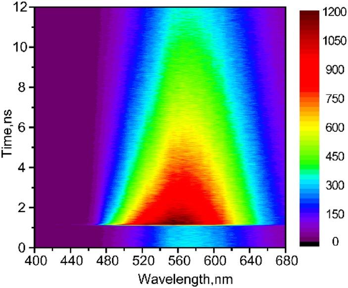
www.nature.com/scientificreports/
Figure 2. Time-resolved fluorescence map of perylene film, measured by TCSPC, λex = 250 nm.
broad unstructured band ranging from 500 to 800 nm, which can be assigned to the light scattering of nano or
microcrystals in the film17. Ishino et al. investigated the nanocrystals of α-perylene with size from 74 to 318 nm18.
The phenomenon of size-dependent redshift and tail towards longer wavelengths were described in terms of light
scattering and were supported by theoretical calculations18. The above shape of the absorption tail is indicative
for existence of large amount of nanocrystals of various sizes, in agreement with our SEM characterization of
50–300 nm nanoaggregates.
Fluorescence emission with maximum at 606 nm is due to excimer; it is blue shifted compared to dimeric
systems (613 nm) or crystal (610 nm)9,19. Compared with the reference fluorescence of crystal (blue dotted curve),
the shoulder at about 500 nm can be ascribed to perylene monomer emission, supported by TCSPC results below.
It should be noted that the discrepancy between absorption and fluorescence excitation spectra in the range of
250–310 nm is indicative of a photophysical process, which proceeds directly from the higher excitonic state
bypassing S1 state, viz. singlet fission, which will be discussed below in detail.
Time‑resolved fluorescence spectra. The fluorescence map of film in the wavelength range 400–680 nm
was first collected by use of TCSPC: λex = 250 nm (Fig. 2) and 380 nm (Fig. S4). Both maps exhibit the same
kinetics, which indicates that excimer emission in film is independent on excitation wavelength. Kinetics at
longer emission wavelengths (540–680 nm) exhibits single-exponential decay with time τ > 10 ns (Fig. S5). How-
ever, emission between 480 to 520 nm results in additional two shorter time components: τ1 ≤ 20 ps (limited by
instrument response function) and the longer time component τ2 = 3.8–4.5 ns with varying amplitudes. The data
at λex = 380 nm were globally fit to three exponentials: resulting decay-associated spectra (DAS) are shown in
Figure S4(b). The DAS contains three fluorescence spectra with maxima at 500 nm (0.06 ns), 520 nm (3.2 ns) and
590 nm (19 ns). The emission maximum at ~ 590 nm is in good agreement with excimer fluorescence in crystal
and dimers8, 20,21. The spectrum with maximum at 520 nm (τ = 3.2 ns) is due to existence of certain amount of
perylene monomers, which is consistent with τ = 4 ns for perylene in hexane22. The shortest time component
(< 0.06 ns) results from the locally excited singlet state and hot excimer state, which was further quantified by
up-conversion technique with higher time resolution.
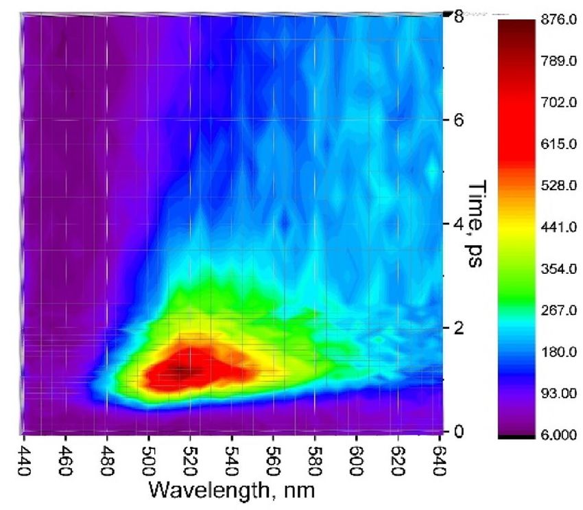
Figure 3 records fluorescence map at short time range 0–8 ps with 100 fs resolution. An ultrafast decay com-
ponent of τ = 850 fs was observed in the range of 460–500 nm (Table S2). This emission band can be attributed to
the quenched locally excited singlet state of perylene9,21. The other broad region, 500–560 nm, originates from hot
excimer, in agreement with Refs.23–25. This hot excimer relaxes within 0.45–1 ps; compare with 0.5–3 ps in c rystal8.
Femtosecond transient absorption (fsTA) spectra. In order to study higher excited states relaxa-
tion processes in perylene film, fsTA measurements employing 250 nm excitation were made. The fsTA spectra
(Fig. 4a) display a single positive band ranging from 440 to 550 nm and a weaker broad tail between 600 and
800 nm. The structured excited state absorption (ESA) band (440–550 nm) exhibits three maxima: 450, 480,
510 nm. Obviously, ESA at 510 nm (Fig. 4b) decays much faster than that at 480 nm, indicating that they belong
to different transient species.
In entire TA spectra, we did not observe ground state bleaching (GSB) and stimulated emission (SE) signal,
similar to the case of α-perylene c rystal8. This can occur because of several reasons. First, the ESA is much
stronger than GSB, e.g. the absorption cross section of the S 1 → SN transition in α-perylene crystal is 50 times
larger than that of S 0 → S18. Free carriers which are generated during the laser excitation can strongly (orders of
magnitude) enhance the absorption cross-section due to “infrared-activated vibrations” (IRAV)26. Second, bright
excitons reside at the band-top in H-type aggregates, i.e. these states have high transition dipole moments. We
excite molecules to band-top, and further excitons in ultrafast scale relax to the band-bottom, which has very
weak transition dipole moment. Therefore, observed ESA is much stronger than the GSB27.
Dimer radical cation formation. In order to identify the states/photoproducts produced from singlet
excitons, we compare our spectra with known TA spectral features in α-perylene crystal (Fig. 4a, red dashed
curve). The shape of TA curve of film follows the trace of crystal. In α-crystal this well-marked 510 nm ESA was
Scientific Reports | (2021) 11:5220 | https://doi.org/10.1038/s41598-021-83791-z 3
Vol.:(0123456789)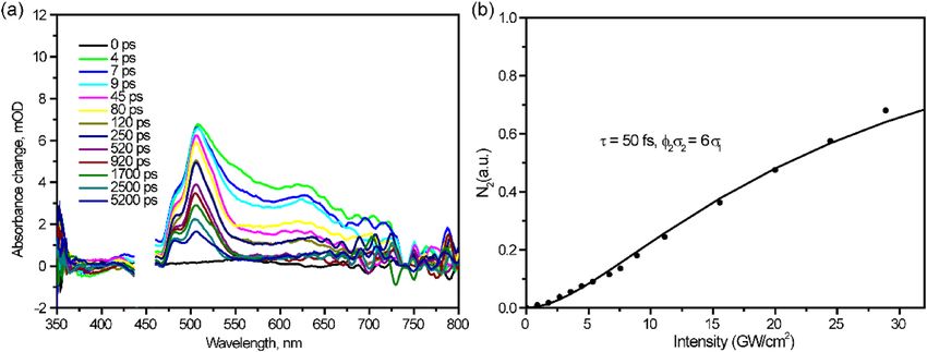
www.nature.com/scientificreports/
Figure 3. Up-converted fluorescence map of perylene film, λex = 400 nm.
Figure 4. Femtosecond transient absorption spectra (a) and kinetics (b) in perylene film, λex = 250 nm.
the result of photoionization with formation of perylene dimer cation radicals. They were observed and studied
in gas phase and liquid sulfuric acid28,29. This spectral feature was reported in different systems: perylene mono-
mer in boric acid with λmax = 508 nm29, perylene dimer in hexane and acetonitrile with λmax = 520–600 nm9,21,30,
in α-perylene crystal with λmax = 503 nm8. Therefore, our observed ESA at 510 nm we assign to the perylene
dimer radical cation (Per •+).
In our films, ESA at 510 nm, rises instantaneously (< 30 fs) and decays triexponentially with τ1 = 13 ps (26%),
τ2 = 410 ps (43%) and τ3 = 3.3 ns (31%). Here Per •+ formation proceeds in < 30 fs and final recombination occurs
within 3.3 ns. In the previous publications, Per •+ lifetimes were studied in concentrated sulfuric acid t = 15 ps31,
or 26 ps32, in boric acid glass t = 35 ps31, in freon glass at 77 K t = 100 ps32. Besides, they also mentioned that glass
defects can also quench the lifetime of radicals. Note that in similar aromatic hydrocarbon molecules, e.g. biphe-
nyl, naphthalene and anthracene, cation radicals decay even much faster, within 200 f s33. In contrary, changing
pH from 2 to 7 results in increase of Per •+ lifetime from picoseconds to m icroseconds34. The second lifetime in
ESA decay (410 ps) we assign to self-trapping of excitons (see below).
Triplet formation. The 480 nm shoulder of ESA band in film (Fig. 4a) is more pronounced than in
α-perylene crystal (blue dash curve)8. This ESA band rises within the instrument response (< 30 fs) and sub-
sequently decays with three time components: τ1 = 18 ps, τ2 = 650 ps and τ3 > > 10 ns. This 480 nm TA band we
assign to triplet–triplet absorption, in agreement with previous publications: 485 nm in perylene monomer35–37,
480–525 nm in perylene dimer9, 480 nm in α-perylene crystal8.
Well-known pathways of triplet formation are spin-forbidden intersystem crossing (ISC) S 1 → T1, and spin-
not efficient, ϕisc < 0.0138. However, in perylene film we have observed triplet TA at λex = 250 nm, well above the
allowed SF S1 → T1 + T1. Because of large S1-T1 energy gap and planarity of the molecule, ISC in perylene is
Scientific Reports | (2021) 11:5220 | https://doi.org/10.1038/s41598-021-83791-z 4
Vol:.(1234567890)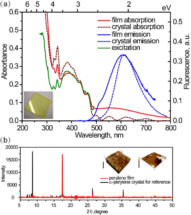
www.nature.com/scientificreports/
energy threshold. Thus, we claim that it forms due to SF. The yield of singlet fission was calculated by using the
methodology used in Refs.7,39. For this purpose we need to know the extinction coefficients of triplet–triplet
absorption of perylene in film. Therefore, we have performed three different measurements: a) perylene triplet
sensitization7,40 by using BODIPY or b) anthracene as a photosensitizer; c) comparison with a known molecule
(or transient39): 3-bromoperylene. First two methods give large uncertainty (see Supplementary Information, pp.
is ϕT = 108%, accordingly, ϕSF = 54%.
9–14). Our estimation of the quantum yield of triplet formation based on the third method (3-bromoperylene)
It was demonstrated that molecular packing has a strong effect on the yield of SF in organic semiconductors:
slip-stacking favors delocalization of excitation and enhances exciton fi ssion41. J-type tetracene aggregates lead
to fast triplet formation by S F42. Quantum interference theoretical calculations show that in J or H type aggre-
gates SF is more efficient43. Here perylene is organized as H-type aggregates and homogenous distributed in the
film surface; it is favorable condition for SF. Moreover, shortening of triplet lifetime in our case is also a strong
indication for existence of SF. Hence, we conclude that triplets form via SF between perylene aggregates within
t < 30 fs at λex = 250 nm. Note that competition between SF and excimer formation was studied in detail also in
films containing various perylene d iimides6. It was demonstrated that molecular packing due to introduction of
bromine atoms is crucial in excitonic interactions.
Spin-obit charge-transfer enhanced intersystem crossing mechanism (SOCT-ISC) may also contribute in
triplet formation. Intersystem crossing via a symmetry-breaking charge transfer in zinc dipyrrin complex was
observed within 1–5 ps. In this case, real donor/acceptor interface is expected inside the complex44. However,
in our film is less probable generating triplets via this mechanism. First, our film was constructed by use of pure
perylene molecules. Strong charge transfer is not expected between two identical perylene molecules. Second,
there are few molecular configuration and electronic coupling requirements for the charge transfer mechanism.
Parameters that influence the efficiency of charge transfer enhanced intersystem crossing are the following: degree
of charge transfer from donor to acceptor, the dihedral angle θ between their π systems, and the magnitude of
electronic coupling between donor and acceptor45. Moreover, the efficiency is maximal at θ = 90°. In our previ-
ous work on multicomponent molecular crystal (perylene-TCNQ), we observed the triplet formation via charge
transfer mechanism between donor perylene and acceptor TCNQ. Note that in that case perylene stays almost
perpendicular to T CNQ46. In our present case with perylene film, the steady-state absorption is almost identical
to α-perylene crystal. It indicates that our films are of H-type aggregated morphology with face-to-face molecular
packing (as in the crystal), but not orthogonal. Even though, we cannot fully rule out contribution of charge
transfer intermediate to enhancement of triplet states formation. It should also be mentioned that existence of
dimer radical cation (as a result of ionization) cannot be involved in ISC process. In several publications with
various systems it was clearly demonstrated that separated radical anions or cations do not lead to generation
of triplets47–49.
Moreover, we have observed an ultrafast (< 30 fs) rise of the triplet transient, which is an additional argu-
ment for the triplet formation directly from upper excited states via SF and not through SOCT or radical cation.
In our case, perylene triplet TA band overlaps considerably with the absorption of dimer radical cation. The
triplet TA did not decay at 6 ns, i.e. the longest delay time of our setup. However, in contrast with the α-perylene
crystal (Fig. 4a, blue dashed curve), where the triplet TA remains at t = 1 ms, in film we did not see any remain-
ing triplet transients at “negative times”. Note that negative times mean that the probe beam comes earlier than
the pump beam. Because the repetition rate of our laser is 1 kHz, we can consider that the observed signal is
generated by the previous pulse, i.e. the actual time delay between pump and probe pulses is 1 ms. In order to
resolve our lifetime of triplet state in film, nanosecond flash photolysis technique was applied at λex = 355 nm
and we probe in the triplet–triplet transition range at λprobe = 480 nm (Fig. S6). The lifetime of triplet state was
obtained: τ = 85 ns.
Our obtained triplet lifetimes for perylene films are significantly shorter than in deoxygenated solution
(4.8 ms)35. The shortening of the triplet state lifetime in films is a well-known phenomenon. It was demonstrated
that the triplet lifetime of phthalocyanine films decreased from 75 to 11 µs compared with isolated molecules in
solvent. This effect was explained in terms of electron and energy transfer between adjacent m olecules50. Similar
shortening of triplet lifetimes was demonstrated in hexathiophene films resulting from numerous defects and
aggregates traps51. Triplet–triplet annihilation (TTA) was also shown to shorten triplet lifetimes, e.g. in caro-
tene aggregates τT < 1 µs relative to 20 µs for monomeric molecule52. Therefore, shortening of triplet lifetime in
perylene film is considered to be due to charge/energy transfer between perylene species or from perylene to
the defect/trap states of quartz.
In our case, the shortening of triplet lifetime can also be due to TTA from neighboring triplet excited mole-
cules, which are formed via SF. In film, there exists energy level matching: 2E(T1) > E(S1). Note that E(S1) = 2.86 eV,
E(T1) = 1.53 eV. This condition is favorable that two triplet excitons can annihilate efficiently to form the S 1 state,
which could in turn produce delayed fluorescence. In order to proof this hypothesis, we have measured Time-
resolved photoluminescence (PL) in the larger time window with λex = 450 and 389 nm, i.e. below and above SF
threshold (Fig. 5). At λex = 450 nm, decay is monoexponential with τ = 17 ns, whereas at λex = 389 nm kinetics
exhibits two time components: τ1 = 17 ns, τ2 = 82 ns. The shorter lifetime (17 ns) is assigned to the excimer PL,
which corresponds to above τ = 19 ns (Fig. S4b). The longer time 82 ns at λex = 389 nm agrees well with the triplet
lifetime 85 ns obtained from ns flash photolysis. It means that the longest PL at λex = 389 nm can be assigned to
delayed fluorescence due to TTA, which strongly supports our arguments on triplet formation via singlet fission.
Two‑quantum induced singlet fission. The fsTA spectra at 450 nm excitation are shown in Fig. 6a and
accordingly kinetics is presented in Table S3. Note that the excitation photon energy is below the threshold
for SF. However, here triplet TA signal at 480 nm still dominates the whole spectrum. Moreover, the inten-
Scientific Reports | (2021) 11:5220 | https://doi.org/10.1038/s41598-021-83791-z 5
Vol.:(0123456789)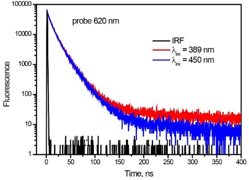
www.nature.com/scientificreports/
Figure 5. Time-resolved photoluminescence decay trace of perylene film ar 620 nm, λex = 389 and 450 nm.
Figure 6. (a) fsTA spectra of perylene film atλex = 450 nm. (b) Intensity dependence of the 480 nm transient
(TT absorption) versus the laser excitation intensity.
sity dependence of the TA signal is obviously nonlinear. Similar to the case for perylene c rystal8, we explain
(Scheme S1). The best fit is obtained for ϕ2σ2 = 6σ1, where σ1 and σ2 are absorption cross-sections from S 0 and S 1
this nonlinear dependence in terms of consecutive two-quantum absorption (TQA) via intermediate state S1
states, respectively, and ϕ2 is the quantum yield of two-quantum photoproduct (Fig. 6b). The analytical formula
for TQA and fit process is described in Supplementary Information. On the basis of the above considerations,
we claim that singlet fission at λex = 450 nm is due to TQA. Even though 450 nm two-quantum excitation leads to
population of higher electronic states (5.5 eV) than upon direct 250 nm excitation (5 eV), SF in the former case
proceeds slower: compare 170 fs with < 30 fs.
Other photophysical processes. A closer look to the femtosecond transient absorption spectra depicts
weak ESA around 700 nm, similar to singlet–singlet absorption of perylene m onomer19. This band decays with
τ = 1.0 ps (Fig. S7), which corresponds well to S 1 fluorescence lifetime of perylene film (τ = 850 fs). Another weak
broad band at λmax = 620 nm is due to excimer, in agreement with our previous publications for perylene dimers
and α-perylene crystal8,9. We cannot clearly resolve the formation time of excimer because of its strong overlap
with ESA of self-trapped exciton. However, by use of time-resolved fluorescence, hot excimer relaxation time
and excimer formation time were clearly resolved: 0.4–0.8 ps and 1 ps, respectively. In fact, with λex = 380 nm
(TCSPC) and λex = 400 nm (up-conversion), we were exciting the S 1 state. Accordingly, we resolved lifetimes
of different states: the locally excited S 1 state (850 fs); hot excimer (0.45–1 ps); relaxed excimer (19 ns), as well
as the remaining perylene monomer (3.2 ns). In addition, by using pump–probe technique, when excited S1
(450 nm) and also S2 state (250 nm), we monitored formation of the dimer radical cation (510 nm) and triplet
state (480 nm) with lifetimes 3.3 and 82 ns, respectively. Moreover, weak broad excimer ESA at about 620 nm
with τ > 6 ns and S1 → SN absorption at 700 nm with τ = 1 ps were also observed. Both TRPL and TA data give for
Scientific Reports | (2021) 11:5220 | https://doi.org/10.1038/s41598-021-83791-z 6
Vol:.(1234567890)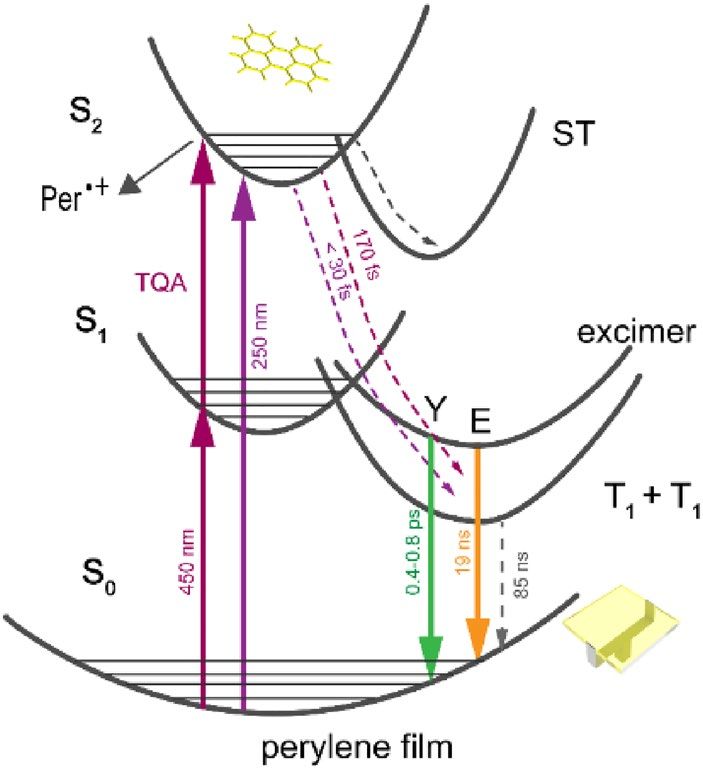
www.nature.com/scientificreports/
Figure 7. Photophysical pathways of perylene film at λex = 450 and 250 nm. Per •+ is perylene dimer radical
cation. Y is hot excimer and E is relaxed excimer. T1 + T1 is the triplet pair state generated through singlet fission.
ST is self-trapped excitonic state.
S1 state lifetime t = 1 ps and for excimer lifetime t = 19 ns. Therefore both methods lead to reliable and consistent
results.
In order to view the full relaxation picture, the fsTA data at λex = 250 nm were globally fit, and the resulting
Evolution-Associated Spectra (EAS) (Fig. S8) exhibit maxima at 480 nm (> 10 ns), 504 nm (3.2 ns), 515 and
550 nm (56 and 1.5 ps). The 504 nm band is assigned to dimer radical cation, in agreement with Ref.28,29. The
longest time component (> > 10 ns) is triplet–triplet absorption. In addition, the broad EAS with the shorter
time constant 56 ps is assigned to self-trapped exciton. Similar TA band in transient absorption microscopy with
λex = 680 nm was observed in β-perylene crystal and was assigned to self-trapping of free excitons (formation time
15 ps)53. Excitons were stabilized by self-induced crystal lattice distortion through phonon-exciton coupling. In
α-perylene crystal with dimeric molecules in unit cell, exciton self-trapping runs with two time constants: < 100 fs
and 2 ps19,53. Besides, self-trapped excitons were also observed in aggregated molecular n anostructures54. Inter-
estingly, lifetime of self-trapped excitons formed after 250 nm excitation are longer than that formed after
λex = 450 nm. In addition, the shortest time constant 1.5 ps is a result of intramolecular vibrational redistribution
(IVR) in S 1 state, which was observed after excitation of S 2 state.
The observed relaxation processes in perylene film after one- or two-quantum excitation at 450 and 250 nm
are summarized in Fig. 7. Triplet excitons are produced directly from upper excited electronic states with 108%
quantum yield via singlet fission within < 30 fs. This ultrashort formation time is due to existence of large poly-
crystalline regions of H-aggregated perylenes and results from the strong coupling between upper singlet and
triplet states and thus facilitating SF directly from S2 bypassing S1 state (violation of classical Kasha-Vavilov
rule). Lifetime of triplet state is drastically shortened compared to crystal, as a result of efficient triplet–triplet
annihilation. Competing processes are excimer formation and photoionization. Excimer is generated from singlet
exciton within ~ 1 ps and subsequently emits yellow fluorescence. Photoionization results instantaneously after
one- or two-quantum excitation (5–5.5 eV) with formation of dimeric radical cation. In addition, excitons are
self-trapped due to a strong-field-induced lattice distortion through phonon-exciton coupling inside molecular
packing. The lifetimes of self-trapped states are several hundreds of ps. Suppression of self-trapping and excimer
formation by optimizing the film packing arrangement can be considered as the next step towards development
of perylene based SF materials.
Methods
Film preparation and characterization. Perylene was purchased from Sigma-Aldrich and used as
received. Films were prepared by thermal evaporation in vacuum evaporation pump device (QHV-Z350C, Pana
Instruments), under low pressure of 1 0–4 Pa. The deposition rate was controlled at ~ 1.5 Å/s. Prior to deposition,
the quartz substrates were cleaned with ethanol in an ultrasonic bath for 30 min and soaked in ethanol (12 h),
water, acetone, ethanol (30 min each). The substrates were subsequently treated with plasma device (FEMTO SR
CE, Diener) (0.3 mbar, 70 W) under O3 twice for 10 min.
X-ray diffraction (XRD) data of film were collected using a Rigaku D/Max-2400 X-ray diffractometer in par-
allel beam geometry employing Cu Kα line focused radiation at 9000 W (45 kV, 200 mA) power and equipped
with a position sensitive detector with 10.0 nm radiation entrance slit. Samples were counted on zero background
sample holders during the data acquisition. The best counting statistics were achieved by collecting samples using
a 0.01° 2θ step scan from 5° to 80° with exposure time of 30 s per step.
Atomic Force Microscopy (AFM) was conducted on Tecnai F30 operated at 300 kV, HITACHI UHR FE-SEM
SU8220 and Park Systems XE-70 with non-contact mode, respectively to characterize the surface structure and
flatness of film.
Scientific Reports | (2021) 11:5220 | https://doi.org/10.1038/s41598-021-83791-z 7
Vol.:(0123456789)www.nature.com/scientificreports/
The structure and morphology of film were investigated by use of field emission scanning electron microscope
(FESEM, Nova Nanosem 450). SEM images were measured under 10 kV accelerating voltage and 10 µA current.
Steady‑state spectra. Absorption and fluorescence spectra were obtained by UV–visible spectrophotom-
eter (Cary 100, Agilent) and spectrofluorometer (Fluorolog-3, Horiba Jobin Yvon), respectively.
Time‑resolved fluorescence spectra. Fluorescence lifetime measurements in sub-nanosecond range
were carried out at room temperature by the time-correlated single photon counting (TCSPC) technique (Pico-
Harp 300, PicoQuant). By use of deconvolution/fit program (FluFit, PicoQuant) the time resolution was reached
down to 10 ps. The second harmonic of a Ti-sapphire laser at 380 nm was used as the excitation source. Fluores-
cence lifetimes down to 100 fs were recorded by using up-conversion technique (TRFLS, Newport) in combina-
tion with the same laser (Mai Tai DeepSee, Spectra-Physics) (150 fs, 80 MHz)9,55.
Time-resolved PL spectra for longer decay times (450 ns) were measured using a fluorescence lifetime spec-
trometer (TemPro-01). Samples were excited at 389 and 450 nm with 1 MHz repetition rate.
Femtosecond pump‑probe spectra. Femtosecond transient absorption spectra were obtained by use
of a home-made pump-probe s etup9,55. Briefly, it consists of a mode-locked Ti–Sapphire amplified laser system
(Spitfire Ace, Spectra-Physics). The output laser beam was at 800 nm with pulses width of 35 fs, repetition rate
of 1 kHz and average power of 4 W. Pump beam (240–2400 nm) was generated through optical parametric
amplifier (TOPAS, Light Conversion). The pump pulse duration was 30–40 fs (measured from the risetime of
TA kinetics for rhodamine 6G). Probe beam, white light continuum (WLC), was generated in CaF2 rotating
plate. The experimental data were fitted to a multiexponential decay function convoluted with the instrument
response function; resulting time resolution was 20–30 fs. The pump beam diameter on the sample was 200 µm
and 100 µm; power was 0.075 mW and 0.1 mW at 250 and 450 nm, respectively.
Nanosecond flash photolysis. Nanosecond time-resolved transient absorption spectra were recorded on
a LP980 laser flash photolysis spectrometer (Edinburgh Instruments Ltd.) in combination with a Nd:YAG laser
(Surelite I-10, Continuum Electro-Optics, Inc.). Sample was excited with 355 nm laser pulse (1 Hz, 100 mJ per
pulse, FWHM ≈ 7 ns).
Global analysis. Global lifetime analysis of fsTA measurements were performed by Glotaran s oftware56.
The Decay Associated Spectra (DAS) allow separating several overlapping spectra. Parallel model was used for
global analysis of time-resolved fluorescence and absorption spectra.
Received: 13 August 2020; Accepted: 2 February 2021
References
1. Smith, M. B. & Michl, J. Singlet fission. Chem. Rev. 110, 6891–6936 (2010).
2. Rao, A. & Friend, R. H. Harnessing singlet exciton fission to break the Shockley-Queisser limit. Nat. Rev. Mater. 2 (2017).
3. Miyata, K., Conrad-Burton, F. S., Geyer, F. L. & Zhu, X. Y. Triplet pair states in singlet fission. Chem. Rev. 119, 4261–4292 (2019).
4. Le, A. K. et al. Singlet fission involves an interplay between energetic driving force and electronic coupling in perylenediimide
films. J. Am. Chem. Soc. 140, 814–826 (2018).
5. Farag, M. H. & Krylov, A. I. Singlet fission in perylenediimide dimers. J. Phys. Chem. C 122, 25753–25763 (2018).
6. Felter, K. M., Dubey, R. K. & Grozema, F. C. Relation between molecular packing and singlet fission in thin films of brominated
perylenediimides. J. Chem. Phys. 151, 094301 (2019).
7. Korovina, N. V., Chang, C. H. & Johnson, J. C. Spatial separation of triplet excitons drives endothermic singlet fission. Nat. Chem.
12, 391 (2020).
8. Ma, L. et al. Excited-state dynamics in an alpha-perylene single crystal: two-photon- and consecutive two-quantum-induced singlet
fission. J. Phys. Chem. A 118, 838–843 (2014).
9. Ni, W. et al. Singlet fission from upper excited electronic states of cofacial perylene dimer. J. Phys. Chem. Lett. 10, 2428–24331
(2019).
10. Sato, K. & Katoh, R. Fluorescence properties of beta-perylene crystals prepared by a physical vapor transport method under
atmospheric pressure. Chem. Phys. Lett. 730, 312–315 (2019).
11. Clarke, R. H. & Hochstrasser, R. M. Location and assignment of the lowest triplet state of perylene. J. Mol. Spectrosc. 32, 309–319
(1969).
12. Giri, G., Prodhan, S., Pati, Y. A. & Ramasesha, S. A model exact study of the properties of low-lying electronic states of perylene
and substituted perylenes. J. Phys. Chem. A 122, 8650–8658 (2018).
13. Albrecht, W., Michel-Beyerle, M. & Yakhot, V. Exciton fission in excimer forming crystal. Dynamics of an excimer build-up in
α-perylene. Chem. Phys. 35, 193–200 (1978).
14. Matsunuma, S. et al. Sn ← S1 and S 1 → S0 resonance CARS spectra of perylene in the S 1 state. J. Chem. Phys. 88, 2956–2961 (1988).
15. Yamagata, H. et al. HJ-aggregate behavior of crystalline 7,8,15,16-tetraazaterrylene: Introducing a new design paradigm for organic
materials. J. Phys. Chem. C 118, 28842–28854 (2014).
16. Hestand, N. J. & Spano, F. C. Molecular aggregate photophysics beyond the Kasha model: Novel design principles for organic
materials. Acc. Chem. Res. 50, 341–350 (2017).
17. Ishino, H. et al. Nonlinear absorption microspectroscopy of single perylene nanocrystals with a multichannel double lock-in
amplifier. Opt. Rev. 17, 337–340 (2010).
18. Ishino, H., Nair, S. V., Nakagawa, K., Kobayashi, T. & Tokunaga, E. Effect of light scattering on the transmission spectra of organic
nanocrystals. Appl. Phys. Lett. 99 (2011).
Scientific Reports | (2021) 11:5220 | https://doi.org/10.1038/s41598-021-83791-z 8
Vol:.(1234567890)www.nature.com/scientificreports/
19. Furube, A., Murai, M., Tamaki, Y., Watanabe, S. & Katoh, R. Effect of aggregation on the excited-state electronic structure of
perylene studied by transient absorption spectroscopy. J. Phys. Chem. A 110, 6465–6471 (2006).
20. Katoh, R., Sinha, S., Murata, S. & Tachiya, M. Origin of the stabilization energy of perylene excimer as studied by fluorescence and
near-IR transient absorption spectroscopy. J. Photochem. Photobiol. A 145, 23–34 (2001).
21. Cook, R. E. et al. Excimer formation and symmetry-breaking charge transfer in cofacial perylene dimers. J. Phys. Chem. A 121,
1607–1615 (2017).
22. Johansson, L. B., Molotkovskii, Y. G. & Bergel’son, L. D. Fluorescence and absorption properties of perylenyl and perylenoyl probe
molecules in solvents and liquid crystals. J. Am. Chem. Soc. 109, 7374–7381 (1987).
23. Auweter, H., Ramer, D., Kunze, B. & Wolf, H. C. The dynamics of excimer formation in perylene crystals. Chem. Phys. Lett. 85,
325–329 (1982).
24. Walker, B., Port, H. & Wolf, H. C. The 2-step excimer formation in perylene crystals. Chem. Phys. 92, 177–185 (1985).
25. Mizoguchi, R., Kano, S. S. & Wada, A. Optical control of excited states of alpha-perylene crystal using optimized pulse shaping
method. Chem. Phys. Lett. 379, 319–324 (2003).
26. Stallhofer, K. et al. Dynamics of short-lived polaron pairs and polarons in polythiophene derivatives observed via infrared-activated
vibrations. J. Phys. Chem. C 123, 28100–28105 (2019).
27. Hestand, N. J. & Spano, F. C. Expanded theory of H- and J-molecular aggregates: the effects of vibronic coupling and intermolecular
charge transfer. Chem. Rev. 118, 7069–7163 (2018).
28. Shida, T. & Iwata, S. Electronic-spectra of ion radicals and their molecular-orbital interpretation. 3. Aromatic-hydrocarbons. J.
Am. Chem. Soc. 95, 3473–3483 (1973).
29. Turner, J. M., Karl, M. W. & Kauffman, J. F. Spectroscopic signatures of protonated perylene in concentrated sulfuric acid. J. Pho-
tochem. Photobiol. A 163, 433–438 (2004).
30. Sauer, M. C. & Jonah, C. D. Electron transfer from carbon dioxide anion radical to perylene in cyclohexane. J. Phys. Chem. 96,
5872–5875 (1992).
31. Gumy, J. C. & Vauthey, E. Investigation of the excited-state dynamics of radical ions in the condensed phase using the picosecond
transient grating technique. J. Phys. Chem. A 101, 8575–8580 (1997).
32. Brodard, P., Sarbach, A., Gumy, J. C., Bally, T. & Vauthey, E. Excited-state dynamics of organic radical ions in liquids and in low-
temperature matrices. J. Phys. Chem. A 105, 6594–6601 (2001).
33. Tokmachev, A.M., Boggio-Pasqua, M., Mendive-Tapia, D., Bearpark, M.J. & Robb, M.A. Fluorescence of the perylene radical
cation and an inaccessible D-0/D-1 conical intersection: An MMVB, RASSCF, and TD-DFT computational study. J. Chem. Phys.
132 (2010).
34. Shkrob, I. A., Sauer, M. C., Liu, A. D., Crowell, R. A. & Trifunac, A. D. Reactions of photoexcited aromatic radical cations with
polar solvents. J. Phys. Chem. A 102, 4976–4989 (1998).
35. Parker, C. & Joyce, T.A. Formation efficiency and energy of the perylene triplet. Chem. Commun. (London), 108b–109 (1966).
36. Tamai, N., Porter, C. F. & Masuhara, H. Femtosecond transient absorption wpectroscopy of a single perylene microcrystal under
a microscopy. Chem. Phys. Lett. 211, 364–370 (1993).
37. Cui, X., El-Zohry, A. M., Wang, Z., Zhao, J. & Mohammed, O. F. Homo- or hetero-triplet–triplet annihilation? A case study with
perylene-BODIPY dyads/triads. J. Phys. Chem. C 121, 16182–16192 (2017).
38. Stevens, B. & Algar, B. Intersystem crossing yields in anthanthrene and perylene from photosensitised peroxidation. Chem. Phys.
Lett. 1, 219–220 (1967).
39. Roberts, S. T. et al. Efficient singlet fission discovered in a disordered acene film. J. Am. Chem. Soc. 134, 6388–6400 (2012).
40. Wu, W., Guo, H., Wu, W., Ji, S. & Zhao, J. Organic triplet sensitizer library derived from a single chromophore (BODIPY) with
long-lived triplet excited state for triplet-triplet annihilation based upconversion. J. Org. Chem. 76, 7056–7064 (2011).
41. Kolata, K., Breuer, T., Witte, G. & Chatterjee, S. Molecular packing determines singlet exciton fission in organic semiconductors.
ACS Nano 8, 7377–7383 (2014).
42. Aggarwal, N. & Patnaik, A. Dimeric conformation sensitive electronic excited states of tetracene congeners and their unconven-
tional non-fluorescent behaviour. J. Chem. Sci. 131 (2019).
43. Zang, H., Zhao, Y. & Liang, W. Quantum interference in singlet fission: J- and H-aggregate behavior. J. Phys. Chem. Lett. 8,
5105–5112 (2017).
44. Trinh, C. et al. Symmetry-breaking charge transfer of visible light absorbing systems: Zinc dipyrrins. J. Phys. Chem. C 118,
21834–21845 (2014).
45. Dance, Z. E. X. et al. Intersystem crossing mediated by photoinduced intramolecular charge transfer: Julolidine-anthracene mol-
ecules with perpendicular systems. J. Phys. Chem. A 112, 4194–4201 (2018).
46. Ni, W. et al. Ultrafast tuning of various photochemical pathways in perylene—TCNQ charge-transfer crystals. J. Phys. Chem. C
124, 13894–13901 (2020).
47. Verhoeven, J. W., van Ramesdonk, H. J., Groeneveld, M. M., Benniston, A. C. & Harriman, A. Long-lived charge-transfer states
in compact donor-acceptor dyads. ChemPhysChem 6, 2251–2260 (2005).
48. Verhoeven, J. W. On the role of spin correlation in the formation, decay, and detection of long-lived, intramolecular charge-transfer
states. J. Photochem. Photobiol. C 7, 40–60 (2006).
49. Hou, Y. et al. Charge separation, charge recombination, long-lived charge transfer state formation and intersystem crossing in
organic electron donor/acceptor dyads. J. Mater. Chem. C 7, 12048–12074 (2019).
50. Zhang, X.-F., Xi, Q. & Zhao, J. Fluorescent and triplet state photoactive J-type phthalocyanine nano assemblies: Controlled forma-
tion and photosensitizing properties. J. Mater. Chem. 20, 6726–6733 (2010).
51. Frolov, S. V., Kloc, C., Berg, S., Thomas, G. A. & Batlogg, B. Transient spectroscopy of Frenkel excitons in alpha-hexathiophene
single crystals. Chem. Phys. Lett. 326, 558–566 (2000).
52. Chang, H.-T. et al. Singlet fission reaction of light-exposed beta-carotene bound to bovine serum albumin. A novel mechanism
in protection of light exposed tissue by dietary carotenoids. J. Agric. Food Chem. 65, 6058–6062 (2017).
53. Yago, T., Tamaki, Y., Furube, A. & Katoh, R. Self-trapping limited exciton diffusion in a monomeric perylene crystal as revealed
by femtosecond transient absorption microscopy. Phys. Chem. Chem. Phys. 10, 4435–4441 (2008).
54. Malyukin, Y. V., Sorokin, A. V. & Semynozhenko, V. P. Features of exciton dynamics in molecular nanoclusters (J-aggregates):
Exciton self-trapping. Low Temp. Phys. 42, 429–440 (2016).
55. Li, X. et al. Ultrafast relaxation dynamics in zinc tetraphenylporphyrin surface-mounted metal organic framework. J. Phys. Chem.
C 122, 50–61 (2018).
56. Snellenburg, J. J., Laptenok, S. P., Seger, R., Mullen, K. M. & van Stokkum, I. H. M. Glotaran: A Java-based graphical user interface
for the R package TIMP. J. Stat. Softw. 49, 1–22 (2012).
Acknowledgements
We thank Dr. Saran Long for help in performing ns flash photolysis measurements. We are indebted to Prof.
Jianzhang Zhao (Dalian University of Technology) Prof. Maxim Gelin (Dianzi University of Hangzhou) and Prof.
Hristo Iglev (Technical University of Munich) for useful discussions. This work was supported by DUT startup
grant, DUT basic research funding (DUT18GJ205).
Scientific Reports | (2021) 11:5220 | https://doi.org/10.1038/s41598-021-83791-z 9
Vol.:(0123456789)www.nature.com/scientificreports/
Author contributions
W.N. prepared and characterized the perylene film. W.N. and G.G.G. performed all spectroscopic measure-
ments and analysis. G.G.G. and L.S. supervised the research. All authors contributed in discussion and writing
the manuscript.
Competing interests
The authors declare no competing interests.
Additional information
Supplementary Information The online version contains supplementary material available at https://doi.org/
10.1038/s41598-021-83791-z.
Correspondence and requests for materials should be addressed to G.G.G.
Reprints and permissions information is available at www.nature.com/reprints.
Publisher’s note Springer Nature remains neutral with regard to jurisdictional claims in published maps and
institutional affiliations.
Open Access This article is licensed under a Creative Commons Attribution 4.0 International
License, which permits use, sharing, adaptation, distribution and reproduction in any medium or
format, as long as you give appropriate credit to the original author(s) and the source, provide a link to the
Creative Commons licence, and indicate if changes were made. The images or other third party material in this
article are included in the article’s Creative Commons licence, unless indicated otherwise in a credit line to the
material. If material is not included in the article’s Creative Commons licence and your intended use is not
permitted by statutory regulation or exceeds the permitted use, you will need to obtain permission directly from
the copyright holder. To view a copy of this licence, visit http://creativecommons.org/licenses/by/4.0/.
© The Author(s) 2021, corrected publication 2021
Scientific Reports | (2021) 11:5220 | https://doi.org/10.1038/s41598-021-83791-z 10
Vol:.(1234567890)You can also read