Transformer based multiple instance learning for weakly supervised histopathology image segmentation
←
→
Page content transcription
If your browser does not render page correctly, please read the page content below
Transformer based multiple instance learning for
                                             weakly supervised histopathology image
                                                         segmentation

                                         Ziniu Qian1 , Kailu Li1 , Maode Lai2,3 , Eric I-Chao Chang4 , Bingzheng Wei5 ,
                                                                    Yubo Fan1 , Yan Xu1( )
arXiv:2205.08878v1 [cs.CV] 18 May 2022

                                          1
                                           School of Biological Science and Medical Engineering, State Key Laboratory of
                                              Software Development Environment, Key Laboratory of Biomechanics,
                                         Mechanobiology of Ministry of Education and Beijing Advanced Innovation Centre
                                              for Biomedical Engineering, Beihang University, Beijing 100191, China
                                                                         xuyan04@gmail.com
                                                    2
                                                      China Pharmaceutical University, Nanjing 210009, China
                                                          3
                                                              Zhejiang University, Hangzhou 310058, China
                                                             4
                                                               Microsoft Research, Beijing 100080, China
                                                            5
                                                               Xiaomi Corporation, Beijing 100085, China

                                                 Abstract. Hispathological image segmentation algorithms play a crit-
                                                 ical role in computer aided diagnosis technology. The development of
                                                 weakly supervised segmentation algorithm alleviates the problem of med-
                                                 ical image annotation that it is time-consuming and labor-intensive. As a
                                                 subset of weakly supervised learning, Multiple Instance Learning (MIL)
                                                 has been proven to be effective in segmentation. However, there is a
                                                 lack of related information between instances in MIL, which limits the
                                                 further improvement of segmentation performance. In this paper, we
                                                 propose a novel weakly supervised method for pixel-level segmentation
                                                 in histopathology images, which introduces Transformer into the MIL
                                                 framework to capture global or long-range dependencies. The multi-head
                                                 self-attention in the Transformer establishes the relationship between in-
                                                 stances, which solves the shortcoming that instances are independent of
                                                 each other in MIL. In addition, deep supervision is introduced to over-
                                                 come the limitation of annotations in weakly supervised methods and
                                                 make the better utilization of hierarchical information. The state-of-the-
                                                 art results on the colon cancer dataset demonstrate the superiority of
                                                 the proposed method compared with other weakly supervised methods.
                                                 It is worth believing that there is a potential of our approach for various
                                                 applications in medical images.

                                                 Keywords: Weakly supervised learning · Transformer · Multiple in-
                                                 stance learning · Segmentation.

                                          ∗
                                              Ziniu Qian and Kailu Li are contributed equally to this work.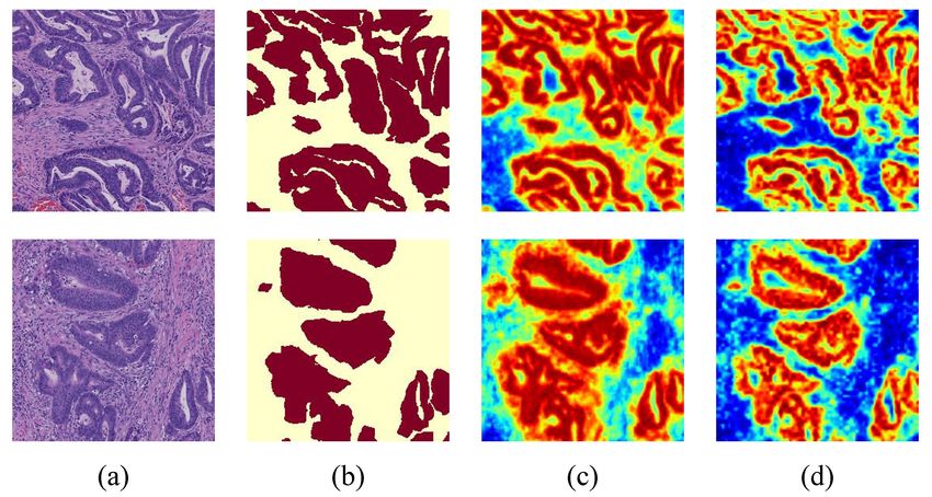
2       Qian et al.

1   Introduction
Histopathology image segmentation is of great significance in computer-assisted
diagnostics (CAD), which assists doctors quickly trace abnormal tissue areas.
Recently, effective supervised methods always rely on a large amount of high-
quality annotations [1,2,3]. However, it is time-consuming and labor-intensive for
doctors to manually label regions of interest (ROIs) in histopathology images [4].
Therefore, there is a strong requirement of automated methods to segment and
trace ROIs in histopathology images with weakly supervised methods. In this
paper, we consider the design of weakly supervised segmentation methods with
image-level annotations for their low cost and wide applicability [5].
    Multiple instance learning (MIL) [6] is a subset of weakly supervised meth-
ods, which has demonstrated its effectiveness on segmentation tasks in previous
studies [7,8,9]. Training datasets of MIL are set as several bags that contain mul-
tiple instances. The available labels are only assigned at the bag-level. MIL is
able to predict instance-level labels except performing bag-level classification. In
this work, cancerous images or not are regarded as bags, while pixels in images
are regarded as instances. Labels of pixels can be predicted by MIL with the
annotations of images so that semantic segmentation can be performed. How-
ever, MIL methods are all limited by the fact that the instances within the bags
are independent of each other. The fact leads to a lack of relationships between
instances with similar contextual information.
    Transformer [10] has demonstrated its excellent performance both on Natural
Language Process (NLP) and Computer Vision (CV). Different from convolu-
tional neural networks (CNNs) that focus on the local receptive field at each
convolution layer, Transformer can capture global or long-range dependencies
with a self-attention mechanism [11]. Self-attention mechanism computes the
response at a position in a sequence by attending to all positions and taking
their weighted average in an embedding space. That is, self-attention mecha-
nism aggregates contextual information from other instances in a bag in MIL.
By weighting value with attention matrix, self-attention mechanism increases the
difference between classes, which is the distance between foreground and back-
ground in semantic segmentation. Therefore, the feature maps from Transformer
implicitly include relationships between instances in MIL. In previous studies,
several researchers have proposed MIL methods integrated with Transformer
that achieve superior performance over CNNs models [12,13,14]. However, ex-
isting studies in this field mostly focus on image classification, rarely addressing
the challenge of segmentation. To the best of our knowledge, we are the first
to attack semantic segmentation on histopathology images using Transformer
combined with MIL.
    Among the developed Transformer methods, Swin Transformer [15] con-
structs multi-scale feature maps and achieves state-of-the-art performance on
several vision tasks. It is obvious that algorithms utilizing hierarchical informa-
tion from multi-scale feature maps show better performance [16]. On the other
hand, Swin Transformer provides higher resolution feature maps than other
Transformer methods, which is benefit to facilitate prediction maps for lower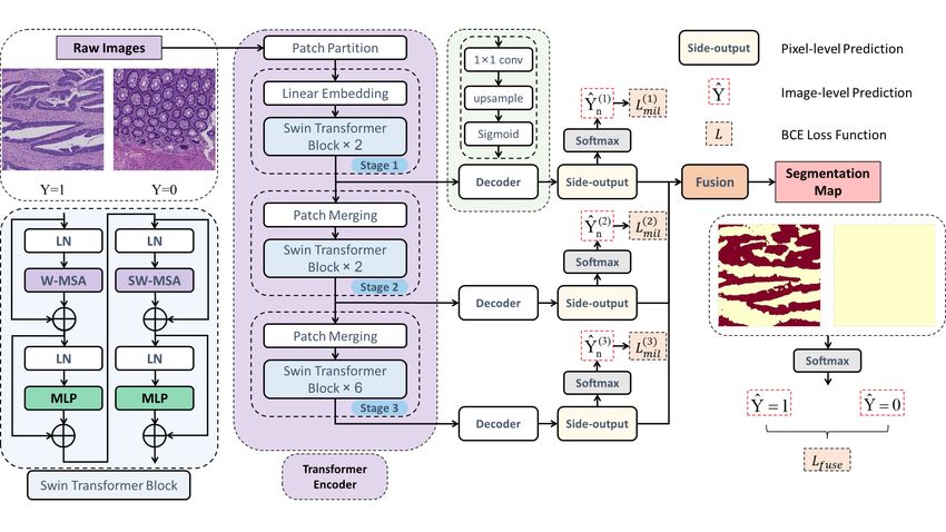
Swin Transformer Based MIL          3

upsampling ratio. Therefore, we explore adapting Swin Transformer in the MIL
framework. In addition, it is difficult to constrain the learning process in the MIL
method due to a lack of supervision. On the other hand, Transformer relies more
on large datasets than CNNs methods, which are difficult to obtain in medical
image analysis. To address these problems, we introduce deep supervision [9,17]
to make better use of image-level annotations and strengthen constraints.
    In this work, we introduce the Transformer into the MIL framework to per-
form histopathology image segmentation for the first time. We propose a novel
MIL method leveraging Swin Transformer encoder, decoder and deep super-
vision to build a trainable bag embedding module for generating prediction
masks. Swin Transformer encoder learns deep feature representations, where
attention weights are assigned to features. To extend constraints, we introduce
deep supervision by producing multi-scale side-outputs from the encoder which
can be leveraged adequately by a fusion layer. Our code will be released on
https://github.com/Nexuslkl/Swin MIL. The contributions can be summarized
as follows:

 – We propose a novel Transformer based method for weakly supervised seman-
   tic segmentation. Deep supervision is leveraged in the framework to utilize
   multi-scale features and provide more supervised information.
 – We explore the combination of Transformer and MIL for weakly supervised
   segmentation on histopathology images, which, to our best knowledge, is the
   first attempt at this task.
 – Multi-head self-attention of Transformer builds long-range dependencies to
   solve the problem that instances in MIL are independent of each other.

2   Method

In this paper, the motivation is to introduce Transformer to solve the short-
coming of independent instances in MIL for histopathology image segmentation.
The proposed Swin-MIL consists of Swin Transformer encoder, decoder and
deep supervision. Swin Transformer encoder plays a role in assigning attention
weights to features. By capturing global information, the self-attention module
effectively highlights features that have better interpretability at each stage. De-
coder of each stage generates pixel-level predictions as side-outputs, where the
final segmentation maps are fused with side-outputs across all stages. Deep su-
pervision is to constrain the training process with only image-level annotations.
An overall framework of the proposed Swin-MIL is illustrated in Figure 1.

Definition: Let S = {(Xn , Yn ), n = 1, 2, 3, ..., m} donates our training set,
where Xn ∈ RH×W denotes the nth input image and Yn ∈ {0, 1} refers to
the image-level label assigned to the nth input image. Here Yn = 1 refers to a
positive image and Yn = 0 refers to a negative image.
4 Qian et al.
Swin Transformer encoder: MIL does not perform as well as fully super-
vised semantic segmentation methods. There is a problem that instances in the
standard MIL maintain their independencies, which is in conflict with a char-
acteristic named category consistency in semantic segmentation tasks. It means
that pixels in the same class should have similar features, while those in a sep-
arate class should have dissimilar features [18]. To address the shortcoming of
MIL, Swin Transformer encoder is introduced that incorporates the self-attention
mechanism into the MIL setting. By introducing a shifted window partitioning
approach which alternates between two partitioning configurations in consec-
utive Swin Transformer blocks, Swin Transformer encoder builds relationships
between long-range tokens with an efficient computation. Therefore, similar fea-
tures get high attention weights while dissimilar ones get low attention weights,
which leads to an improvement in distinguishing foreground and background.
The image Xn is cropped into non-overlapping patches with patch size of
4 × 4 firstly. Each patch is treated as a “Token” and fed into several Swin Trans-
former blocks, which enhance the significant regions in feature maps and inhibit
the influence of irrelevant regions through multi-head self-attention. The Swin
Transformer blocks refer to as “Stage 1” together with a linear embedding layer,
which projects the raw-valued feature to an arbitrary dimension. To produce a
multi-scale feature representation, patch merging layers are introduced to reduce
the number of tokens as the network gets deeper. The patch merging layers and
several Swin Transformer blocks are denoted as “Stage 2” and “Stage 3”, which
output feature maps with resolution of H W H W
8 × 8 and 16 × 16 , respectively. It is
evident in our experiments that the backbone with three stages is sufficiently
powerful to extract features. Then the output feature maps are fed into decoder.
Decoder: The channel size of the output feature map is squeezed to 1 by a 1×1
convolutional layer. After a bilinear upsampling operation, it is restored to the
original size (H ×W ) and finally activated by the sigmoid to generate a predicted
probabilistic map as side-outputs. A fusion layer is proposed to adequately lever-
age the multi-scale side-outputs across all stages to predict final segmentation
maps. We can predict the classification of the image by each instance in the bag.
Ŷn (i, j) is denoted as the probability of the pixel pij in the nth image, where
(i, j) denotes the position of the pixel pij in Xn . Therefore, a softmax function
is often used to replace the hard maximum approach. Generalized Mean (GM)
[19] is utilized as our softmax function, which is defined as:
r1
1 X h ir
Ŷn = Ŷn (i, j) , (1)
|Xn | i,j
where the parameter r controls the sharpness and proximity to the hard function:
Ŷn → max Ŷn (i, j) as r → ∞.
i,j
Deep supervision: To address the lack of supervision information in MIL and
utilize hierarchical information effectively, we introduce deep supervision. OurSwin Transformer Based MIL 5
Fig. 1. An overview of our method. Under the MIL setting, we adopt the first three
stages of the Swin Transformer encoder. At each stage, the deep supervision layer
produces side-outputs which can be seen as predictions. In addition, a fusion layer is
proposed to adequately leverage the multi-scale predictions across all side-outputs.
goal is to train the model by minimizing the loss between output predictions and
ground truths, which is designed in the form of the cross-entropy loss function
as follows:
(t)
X
Lmil = − I (Yn = 1) log Ŷn(t) + I (Yn = 0) log(1 − Ŷn(t) ) , (2)
n
X
Lf use = − (I (Yn = 1) log Ŷn,f use + I (Yn = 0) log(1 − Ŷn,f use )), (3)
n
(t)
where t is denoted to the number of stage, Ŷn and Ŷn,f use are calculate by
Equation (1), and I(·) is an indicator function.
Loss function: The final objective loss function is defined as below:
3
(t)
X
L= Lmil + Lf use . (4)
t=1
In each side-output and fusion layer, the loss function is computed in the
form of deep supervision without any additional pixel-level label supervision.
After that, the parameters are learned by minimizing the objective function via
backpropagation using the stochastic gradient descent algorithm.6 Qian et al.
3 Experiment
3.1 Dataset
A hispathological tissue dataset was selected to evaluate the effectiveness of our
approach. The dataset is a Haematoxylin and Eosin (H&E) stained histopathol-
ogy image dataset of colon cancer reported by Jia et al. [9], which consists of 330
cancer (positive) and 580 non-cancer (negative) images. In this dataset, 250 posi-
tive and 500 negative images were used for training; 80 cancer and 80 non-cancer
images were used for testing. These images were obtained from the NanoZoomer
2.0HT digital slide scanner produced by Hamamatsu Photonics with a magnifi-
cation factor of 40, i.e. 226 nm/pixel. Each image has a resolution of 3, 000 × 3,
000. Due to memory limits, the original 3, 000 × 3, 000 pixels can not be loaded
directly. Thus, in all experiments, images were resized to 256 × 256 pixels. For
simplicity, we used Pos to refer to cancer images and Neg to refer to non-cancer
images. The ground truths are pixel-level annotations which were provided by
two pathologists. (1) If the overlap between the two cancerous regions labeled
by the two pathologists is larger than 80%, we use the intersection of the two
regions as the ground truth; (2) if the overlap is less than 80%, or if a cancerous
region is annotated by one pathologist but ignored by another, a third senior
pathologist will step in to help determine whether the region is cancerous or not.
3.2 Implementation
All experiments were implemented on the PyTorch framework and conducted on
NVIDIA GeForce RTX 3090 GPUs with 24G memory. For training, the Adam
optimizer is employed to train the model with a weight decay of 5e-4 and a fixed
learning rate of 1e-6. The batch size is set to 4 per GPU, with an epoch of 60. The
parameter r of the generalized mean function is set to 4. Learnable weights will
cause over-segmentation problems and degrade performance. With the limited
supervision of image-level annotation, MIL methods with learnable weights has a
tendency to perform better classification but not segmentation. Thus, we adopt
a strategy, using fixed weights, to preserve multi-scale information. The fixed
weight values we set are optimal settings determined by experiments. Weights
of 0.3, 0.4, 0.3 are selected for the three side-output layers. The learning rates
of the side-output layers are set to 1/100 of the global learning rate. We used
the pretrained model of Swin-T from ImageNet on our backbone and Xavier
initialization [20] to initialize the side-output layers.
F1-score (F1) and Hausdorff Distance (HD) are employed to evaluate the
quality of semantic segmentation and the boundary of prediction mask, respec-
tively. Here, the F1-score in positive images is equivalent to the dice coefficient.
3.3 Comparisons
Attention mechanism is the core of Transformer to capture global or long-range
dependencies. Hence, some attention-based MIL methods [23,21,22] which canSwin Transformer Based MIL 7
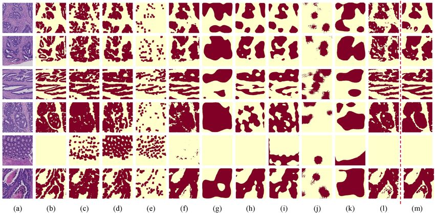
Fig. 2. Visualization results of all methods. (a) Raw Image. (b) Ground Truth. (c)
DA-MIL. (d) DeepAttnMISL. (e) GA-MIL. (f) DWS-MIL. (g) OAA. (h) MDC-UNet.
(i) MDC-CAM. (j) PRM. (k) VGG-CAM. (l) Ours. (m) U-Net. The rows 1-4 are the
results on positive images. The row 5 are the results on negative images. The row 6
are the failure cases.
Table 1. Comparisons with weakly supervised methods and fully supervised methods.
Pos and Neg means the results are conducted on positive images and negative im-
ages, respectively. WSL and FSL are denoted to weakly supervised learning and fully
supervised learning, respectively.
Running
Type Method F1Pos HDPos F1Neg
Time(s)
Swin-MIL (Ours) 0.850 10.463 0.999 0.0226
DA-MIL (2020) [21] 0.791 12.962 0.755 0.1635
MIL based WSL: DeepAttnMISL (2020) [22] 0.772 11.111 0.634 0.1389
GA-MIL (2018) [23] 0.355 16.289 0.939 0.1695
DWS-MIL (2017) [9] 0.833 15.179 0.986 0.0142
OAA (2021) [24] 0.744 28.972 0.999 0.0190
MDC-UNet (2018) [25] 0.744 17.071 0.998 0.0157
CAM based WSL: MDC-CAM (2018) [25] 0.726 13.754 0.760 0.0167
PRM (2018) [26] 0.561 24.468 0.995 0.9277
VGG-CAM (2016) [27] 0.675 23.665 0.645 0.0160
FSL: U-Net (2015) [1] 0.885 7.428 0.997 0.0153
achieve patch-level segmentation by visualizing the attention map with image-
level annotations, were conducted for comparison. Weakly supervised segmenta-
tion methods for natural images are also reimplemented on our dataset. Besides,
we reimplemented DWS-MIL[9] framework using PyTorch instead of Caffe tool-
box and introduced a batch normalization layer between convolutional layer and
RELU for fair comparison. As shown in Table 1 and Figure 2, compared with re-8 Qian et al. cent methods [24,22] relied on image-level annotations, our method significantly outperforms those methods (state-of-the-art) and is close to the fully-supervised U-Net [1]. Attention-based MIL methods mainly focus on classification tasks, where the limitation of patch-level segmentation leads to a low-quality bound- ary. Accurate class activation maps (CAMs) are generated CAM-based weakly supervised methods [27,25,26,24], which are sensitive to the location of can- cerous regions while ineffective in accurately segmentation. The MDC methods use dilated convolutional layers to obtain larger receptive fields, which achieve better performance than VGG-CAM. PRM and OAA can perform weakly su- pervised segmentation based on class activation maps to generate a complete mask prediction. However, it is obvious that these methods do not perform well in histopathology images. Because the natural image instance segmentation objects (people, vehicles, and animals) often have regular shapes and charac- teristics, the object regions are relatively complete. Nevertheless, the objects in the histopathology images are often of irregular shapes. It is difficult to generate accurate predictions by expanding which is sensitive to boundaries. Besides, we show the failure case in the row 6 of Figure 1. Given the complexity and the diversity of the tissue appearance, our method misses the accurate segmentation boundary under the limited supervision with image-level annotation. 3.4 Ablation Study Effect of different backbone: Table 2 shows the comparison results adopting CNN model (VGG-16 [28] & ResNet18 [29]) as the backbone. The VGG back- bone can generate segmentation maps with higher boundary quality, while the ResNet backbone is able to extract more semantic information. However, be- cause of the locality of convolution operations, the CNN backbones are unable to extract global or long-range dependencies, which can not solve the lack of re- lations between instances in MIL. By comparison, the Transformer backbone can learn global or long-range dependencies through multi-head self-attention, which leads to good performance both on F1-score (F1) and Hausdorff Distance (HD). Some samples of feature maps of Transformer-based method and CNNs-based method are shown in Figure 3. Effect of the number of stages in backbone: Table 3 summarizes the performances of the different number of stages in the Transformer backbone. It can be observed that the first two stages of Transformer backbone are unable to extract enough semantic information to perform segmentation accurately. On the other hand, the fourth stage incorporates semantic information more than necessary, which affects the performance of the boundary accuracy and fails to achieve effective improvements. Thus, taking the first three stages as our backbone is the optimal option.
Swin Transformer Based MIL            9

Fig. 3. Samples of feature maps of Transformer-based method and CNNs-based
method. (a) Raw Image. (b) Ground Truth. (c) DWS-MIL. (d) Swin-MIL.

 Table 2. Effect of different backbone    Table 3. Effect of the number of stages
 in MIL framework                         in backbone
  Backbone    F1Pos    HDPos     F1Neg      Methods     F1Pos    HDPos    F1Neg
   Swin-T     0.850    10.463    0.999      2 stages    0.814    12.980   0.998
   VGG-16     0.833    15.179    0.986      3 stages    0.850    10.463   0.999
  ResNet-18   0.838    17.852    0.948      4 stages    0.759    37.738   1.000

Effect of side-outputs and fusion: To illustrate the effectiveness of deep
supervision, Table 4 summarizes the performance of the different side-outputs.
Due to the insufficient ability of feature extraction, the side-output of Stage 1
is limited in F1-score but gets the best boundary accuracy. The side-output of
Stage 2 has the highest performance on F1-score and good boundary accuracy
close to Stage 1. The side-output of Stage 3 contains most semantic information
but lacks almost all boundary features. The fusion of three side-output feature
maps can achieve optimal performance.

                    Table 4. Effect of side-outputs and fusion

                      Methods   F1Pos     HDPos        F1Neg
                       Side-1   0.605     10.383       0.964
                       Side-2   0.810      10.413      0.995
                       Side-3   0.743     100.965      1.000
                       Fusion   0.850      10.463      0.999
10 Qian et al.
4 Conclusion
In this work, we propose a novel weakly supervised method for histopathology
image segmentation, which combines Transformer with MIL to capture global
or long-range dependencies. Multi-head self-attention in Transformer solves the
independent instances of MIL by learning relations between instances, which
significantly facilitates segmentation maps and makes the weakly supervised
method more interpretable. It effectively utilizes hierarchical information from
multi-scale feature maps through the introduction of deep supervision. The ex-
periments demonstrate that our method achieves state-of-the-art performance
on the colon cancer dataset. With a broad scope, the proposed method has the
potential to be applied to a wide range of medical images in the future.
References
1. Ronneberger, O., Fischer, P., Brox, T.: U-net: Convolutional networks for biomedi-
cal image segmentation. In: International Conference on Medical image computing
and computer-assisted intervention. pp. 234–241. Springer (2015)
2. Chen, H., Qi, X., Yu, L., Heng, P.A.: Dcan: deep contour-aware networks for ac-
curate gland segmentation. In: Proceedings of the IEEE conference on Computer
Vision and Pattern Recognition. pp. 2487–2496 (2016)
3. Xing, F., Shi, X., Zhang, Z., Cai, J., Xie, Y., Yang, L.: Transfer shape modeling
towards high-throughput microscopy image segmentation. In: International Con-
ference on Medical Image Computing and Computer-Assisted Intervention. pp.
183–190. Springer (2016)
4. Yu, G., Zare, A., Xu, W., Matamala, R., Reyes-Cabrera, J., Fritschi, F.B., Juenger,
T.E.: Weakly supervised minirhizotron image segmentation with mil-cam. In: Eu-
ropean Conference on Computer Vision. pp. 433–449. Springer (2020)
5. Zhou, Z.H.: A brief introduction to weakly supervised learning. National science
review 5(1), 44–53 (2018)
6. Dietterich, T.G., Lathrop, R.H., Lozano-Pérez, T.: Solving the multiple instance
problem with axis-parallel rectangles. Artificial intelligence 89(1-2), 31–71 (1997)
7. Xu, Y., Zhu, J.Y., Chang, E., Tu, Z.: Multiple clustered instance learning for
histopathology cancer image classification, segmentation and clustering. In: 2012
IEEE Conference on Computer Vision and Pattern Recognition. pp. 964–971. IEEE
(2012)
8. Xu, Y., Zhu, J.Y., Eric, I., Chang, C., Lai, M., Tu, Z.: Weakly supervised
histopathology cancer image segmentation and classification. Medical image anal-
ysis 18(3), 591–604 (2014)
9. Jia, Z., Huang, X., Eric, I., Chang, C., Xu, Y.: Constrained deep weak supervision
for histopathology image segmentation. IEEE transactions on medical imaging
36(11), 2376–2388 (2017)
10. Vaswani, A., Shazeer, N., Parmar, N., Uszkoreit, J., Jones, L., Gomez, A.N., Kaiser,
L., Polosukhin, I.: Attention is all you need. Advances in neural information pro-
cessing systems 30 (2017)
11. Chen, J., Lu, Y., Yu, Q., Luo, X., Adeli, E., Wang, Y., Lu, L., Yuille, A.L., Zhou,
Y.: Transunet: Transformers make strong encoders for medical image segmentation.
arXiv preprint arXiv:2102.04306 (2021)Swin Transformer Based MIL 11
12. Li, H., Yang, F., Zhao, Y., Xing, X., Zhang, J., Gao, M., Huang, J., Wang, L., Yao,
J.: Dt-mil: Deformable transformer for multi-instance learning on histopathological
image. In: International Conference on Medical Image Computing and Computer-
Assisted Intervention. pp. 206–216. Springer (2021)
13. Yu, S., Ma, K., Bi, Q., Bian, C., Ning, M., He, N., Li, Y., Liu, H., Zheng, Y.: Mil-vt:
Multiple instance learning enhanced vision transformer for fundus image classifi-
cation. In: International Conference on Medical Image Computing and Computer-
Assisted Intervention. pp. 45–54. Springer (2021)
14. Shao, Z., Bian, H., Chen, Y., Wang, Y., Zhang, J., Ji, X., et al.: Transmil: Trans-
former based correlated multiple instance learning for whole slide image classifica-
tion. Advances in Neural Information Processing Systems 34 (2021)
15. Liu, Z., Lin, Y., Cao, Y., Hu, H., Wei, Y., Zhang, Z., Lin, S., Guo, B.: Swin
transformer: Hierarchical vision transformer using shifted windows. In: Proceedings
of the IEEE/CVF International Conference on Computer Vision. pp. 10012–10022
(2021)
16. Yi, J., Wu, P., Huang, Q., Qu, H., Liu, B., Hoeppner, D.J., Metaxas, D.N.: Multi-
scale cell instance segmentation with keypoint graph based bounding boxes. In:
International conference on medical image computing and computer-assisted in-
tervention. pp. 369–377. Springer (2019)
17. Lee, C.Y., Xie, S., Gallagher, P., Zhang, Z., Tu, Z.: Deeply-supervised nets. In:
Artificial intelligence and statistics. pp. 562–570. PMLR (2015)
18. Huang, Z., Wang, X., Huang, L., Huang, C., Wei, Y., Liu, W.: Ccnet: Criss-cross
attention for semantic segmentation. In: Proceedings of the IEEE/CVF Interna-
tional Conference on Computer Vision. pp. 603–612 (2019)
19. Zhang, C., Platt, J., Viola, P.: Multiple instance boosting for object detection.
Advances in neural information processing systems 18, 1417–1424 (2005)
20. Glorot, X., Bengio, Y.: Understanding the difficulty of training deep feedforward
neural networks. In: Proceedings of the thirteenth international conference on ar-
tificial intelligence and statistics. pp. 249–256. JMLR Workshop and Conference
Proceedings (2010)
21. Hashimoto, N., Fukushima, D., Koga, R., Takagi, Y., Ko, K., Kohno, K.,
Nakaguro, M., Nakamura, S., Hontani, H., Takeuchi, I.: Multi-scale domain-
adversarial multiple-instance cnn for cancer subtype classification with unanno-
tated histopathological images. In: Proceedings of the IEEE/CVF conference on
computer vision and pattern recognition. pp. 3852–3861 (2020)
22. Yao, J., Zhu, X., Jonnagaddala, J., Hawkins, N., Huang, J.: Whole slide images
based cancer survival prediction using attention guided deep multiple instance
learning networks. Medical Image Analysis 65, 101789 (2020)
23. Ilse, M., Tomczak, J., Welling, M.: Attention-based deep multiple instance learning.
In: International conference on machine learning. pp. 2127–2136. PMLR (2018)
24. Jiang, P.T., Han, L.H., Hou, Q., Cheng, M.M., Wei, Y.: Online attention accumu-
lation for weakly supervised semantic segmentation. IEEE Transactions on Pattern
Analysis and Machine Intelligence (2021)
25. Wei, Y., Xiao, H., Shi, H., Jie, Z., Feng, J., Huang, T.S.: Revisiting dilated convo-
lution: A simple approach for weakly-and semi-supervised semantic segmentation.
In: Proceedings of the IEEE Conference on Computer Vision and Pattern Recog-
nition. pp. 7268–7277 (2018)
26. Zhou, Y., Zhu, Y., Ye, Q., Qiu, Q., Jiao, J.: Weakly supervised instance segmenta-
tion using class peak response. In: Proceedings of the IEEE Conference on Com-
puter Vision and Pattern Recognition. pp. 3791–3800 (2018)12 Qian et al.
27. Zhou, B., Khosla, A., Lapedriza, A., Oliva, A., Torralba, A.: Learning deep features
for discriminative localization. In: Proceedings of the IEEE conference on computer
vision and pattern recognition. pp. 2921–2929 (2016)
28. Simonyan, K., Zisserman, A.: Very deep convolutional networks for large-scale
image recognition. arXiv preprint arXiv:1409.1556 (2014)
29. Targ, S., Almeida, D., Lyman, K.: Resnet in resnet: Generalizing residual archi-
tectures. arXiv preprint arXiv:1603.08029 (2016)You can also read