Towards novel herbicide modes of action by inhibiting lysine biosynthesis in plants - eLife
←
→
Page content transcription
If your browser does not render page correctly, please read the page content below
RESEARCH ARTICLE

                                        Towards novel herbicide modes of action
                                        by inhibiting lysine biosynthesis in plants
                                        Tatiana P Soares da Costa1†*, Cody J Hall1†, Santosh Panjikar2,3, Jessica A Wyllie1,
                                        Rebecca M Christoff4, Saadi Bayat4, Mark D Hulett1, Belinda M Abbott4,
                                        Anthony R Gendall5,6, Matthew A Perugini1*
                                        1
                                          Department of Biochemistry and Genetics, La Trobe Institute for Molecular
                                        Science, La Trobe University, Bundoora, Australia; 2Australian Synchrotron, ANSTO,
                                        Clayton, Australia; 3Department of Molecular Biology and Biochemistry, Monash
                                        University, Melbourne, Australia; 4Department of Chemistry and Physics, La Trobe
                                        Institute for Molecular Science, La Trobe University, Bundoora, Australia;
                                        5
                                          Department of Animal, Plant and Soil Sciences, AgriBio, La Trobe University,
                                        Bundoora, Australia; 6Australian Research Council Research Hub for Medicinal
                                        Agriculture, Bundoora, Australia

                                        Abstract Weeds are becoming increasingly resistant to our current herbicides, posing a
                                        significant threat to agricultural production. Therefore, new herbicides with novel modes of action
                                        are urgently needed. In this study, we exploited a novel herbicide target, dihydrodipicolinate
                                        synthase (DHDPS), which catalyses the first and rate-limiting step in lysine biosynthesis. The first
                                        class of plant DHDPS inhibitors with micromolar potency against Arabidopsis thaliana DHDPS was
                                        identified using a high-throughput chemical screen. We determined that this class of inhibitors
                                        binds to a novel and unexplored pocket within DHDPS, which is highly conserved across plant
*For correspondence:                    species. The inhibitors also attenuated the germination and growth of A. thaliana seedlings and
t.soaresdacosta@latrobe.edu.au          confirmed their pre-emergence herbicidal activity in soil-grown plants. These results provide proof-
(TPSC);
                                        of-concept that lysine biosynthesis represents a promising target for the development of
Matt.Perugini@gmail.com (MAP)
                                        herbicides with a novel mode of action to tackle the global rise of herbicide-resistant weeds.
†
 These authors contributed
equally to this work

Competing interest: See
page 14
                                        Introduction
Funding: See page 14                    Our ability to provide food security for a growing world population is increasingly challenged by the
Received: 15 April 2021                 emergence and spread of herbicide-resistant weeds. Resistance has now been observed to the most
Preprinted: 06 May 2021                 widely used classes of herbicides. This includes amino acid biosynthesis inhibitors such as chlorsul-
Accepted: 27 July 2021                  furon, glufosinate, and glyphosate, which target enzymes in the biosynthetic pathways leading to
Published: 27 July 2021                 the production of branched-chain amino acids, glutamine, and aromatic amino acids, respectively
                                        (Gaines et al., 2020; Hall et al., 2020). The impact of herbicide resistance is exacerbated by the
Reviewing editor: Todd Gaines,
Colorado State University,
                                        lack of new herbicides entering the market in the past 30 years, especially those with new mecha-
United States                           nisms of action (Duke, 2012). Nevertheless, the successful commercialisation of such herbicides pro-
                                        vides proof-of-concept that targeting the biosynthesis of amino acids offers an excellent strategy for
    Copyright Soares da Costa et
                                        herbicide development (Hall et al., 2020). Amino acids are not only essential building blocks for
al. This article is distributed under
                                        protein biosynthesis, but they also play important roles in physiological processes that are critical for
the terms of the Creative
Commons Attribution License,            plant growth and development, such as carbon and nitrogen metabolism, in addition to serving as
which permits unrestricted use          precursors to a wide range of secondary metabolites (Hildebrandt et al., 2015).
and redistribution provided that            One amino acid biosynthesis pathway that remains largely unexplored for herbicide development
the original author and source are      is the diaminopimelate (DAP) pathway (Figure 1), which is responsible for the production of L-lysine
credited.                               (here after referred to as lysine) in plants and bacteria (Figure 1; Hall and Soares da Costa, 2018).

Soares da Costa, Hall, et al. eLife 2021;10:e69444. DOI: https://doi.org/10.7554/eLife.69444                                              1 of 17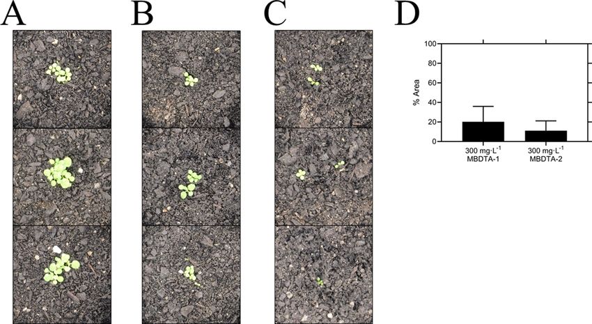
Research article                                                                         Biochemistry and Chemical Biology Plant Biology

                                    Figure 1. Lysine biosynthesis in plants. Plants utilise the diaminopimelate (DAP) pathway, a branch of the
                                    aspartate-derived super-pathway, to synthesise L-lysine. Firstly, L-aspartate semialdehyde (ASA) and pyruvate are
                                    converted to (4S) 4-hydroxy-2,3,4,5-tetrahydro-(2S)-dipicolinic acid (HTPA) in a condensation reaction catalysed by
                                    dihydrodipicolinate synthase (DHDPS). Dihydrodipicolinate reductase (DHDPR) then catalyses an NAD(P)H-
                                    dependent reduction of HTPA to produce 2,3,4,5-tetrahydrodipicolinate (THDP). THDP subsequently undergoes
                                    an amino-transfer reaction with L-glutamate, catalysed by diaminopimelate aminotransferase (DAPAT), to yield L,L-
                                    DAP. L,L-DAP is converted to meso-DAP by diaminopimelate epimerase (DAPEpi) and lastly, meso-DAP is
                                    decarboxylated by diaminopimelate decarboxylase (DAPDC) to yield L-lysine, which imparts a negative feedback
                                    loop on DHDPS.

                                    Animals, including humans, do not synthesise lysine, and therefore, must acquire it from dietary sour-
                                    ces (Galili and Amir, 2013; Tomé and Bos, 2007). Consequently, specific chemical inhibition of the
                                    DAP pathway in plants is unlikely to be harmful to animals and humans (Hutton et al., 2007). The
                                    DAP pathway commences with a condensation reaction between L-aspartate semialdehyde (ASA)
                                    and pyruvate to form (4S) 4-hydroxy-2,3,4,5-tetrahydro-(2S)-dipicolinic acid (HTPA), catalysed by
                                    HTPA synthase (EC 4.2.1.52), also known as dihydrodipicolinate synthase (DHDPS) (Griffin et al.,
                                    2012; Soares da Costa et al., 2018; Soares da Costa et al., 2015). HTPA is then reduced by dihy-
                                    drodipicolinate reductase (DHDPR, EC 1.3.1.26) in the presence of NAD(P)H to produce 2,3,4,5-tet-
                                    rahydrodipicolinate (THDP) (Christensen et al., 2016). In plants, THDP undergoes an amino-transfer
                                    by diaminopimelate aminotransferase (DAPAT, EC 2.6.1.83) to form L,L-DAP, which is converted to
                                    meso-DAP by diaminopimelate epimerase (DAPEpi, EC 5.1.1.7) (Hudson et al., 2005; McCoy et al.,
                                    2006). Lastly, meso-DAP is irreversibly decarboxylated by diaminopimelate decarboxylase (DAPDC,
                                    EC 4.1.1.20) to produce lysine (Peverelli and Perugini, 2015). Lysine regulates flux through the

Soares da Costa, Hall, et al. eLife 2021;10:e69444. DOI: https://doi.org/10.7554/eLife.69444                                                    2 of 17
Research article                                                                    Biochemistry and Chemical Biology Plant Biology

                                    pathway by binding allosterically to DHDPS and inhibiting the enzyme. Thus, DHDPS catalyses the
                                    rate-limiting step of the DAP pathway (Geng et al., 2013; Soares da Costa et al., 2016).
                                        Due to the central role of DHDPS in lysine production in plants, this enzyme has been proposed
                                    as a potential target for the development of herbicides (Griffin et al., 2012; Soares da Costa et al.,
                                    2018). Indeed, the lysine analogue, S-(2-aminoethyl)-L-cysteine, halts rooting of potato tuber discs at
                                    mid-micromolar concentrations (Perl et al., 1993; Ghislain et al., 1995). However, given its poor in
                                    vitro potency against plant DHDPS, it is believed that this analogue inhibits plant growth by compet-
                                    ing with lysine for incorporation into proteins rather than inhibition of the DHDPS enzyme
                                    (Ghislain et al., 1995; Perl et al., 1993). Plants typically have two annotated DHDPS-encoding
                                    genes (DHDPS) (Figure 2—figure supplement 1; Craciun et al., 2000; Sarrobert et al., 2000;
                                    Vauterin and Jacobs, 1994). In Arabidopsis thaliana, these genes are At3G60880 (DHDPS1) and
                                    At2G45440 (DHDPS2), which encode chloroplast-targeted AtDHDPS1 and AtDHDPS2, respectively
                                    (Jones-Held et al., 2012). RNA sequencing data have elucidated that both DHDPS-encoding genes
                                    are expressed at all stages of A. thaliana development, with the most prominent expression at the
                                    seed development stages, in the dry seeds and during germination (Klepikova et al., 2016). Inter-
                                    estingly, maximal DHDPS2 expression is approximately 3-fold greater than that of DHDPS1
                                    (Klepikova et al., 2016). Moreover, upon comparison to the known glyphosate herbicide target, 5-
                                    enolpyruvyl-shikimate 3-phosphate synthase (At1G48860), expression of DHDPS1 is considerably
                                    lower at almost all developmental stages, while expression of DHDPS2 is comparable at all stages,
                                    except in the dry seeds (Klepikova et al., 2016). This is a key consideration in target validation as
                                    low expressing targets will require less inhibitor to achieve phytotoxicity. Double knockouts of
                                    DHDPS1 and DHDPS2 result in non-viable embryos even after exogenous supplementation with
                                    lysine, indicating that DHDPS activity is essential (Jones-Held et al., 2012). AtDHDPS enzymes exist
                                    as homotetramers (Figure 2A), with the active site located within the (b/a)8-barrel (Figure 2B) and
                                    the allosteric cleft in the tight-dimer interface located in the interior of the structure
                                    (Figure 2C; Griffin et al., 2012; Hall et al., 2021).
                                        In this study, we describe a new class of plant DHDPS inhibitors. We show that these compounds
                                    display micromolar potency in vitro and in planta against A. thaliana using a combination of enzyme
                                    kinetics, seedling, and soil assays, whilst exhibiting no cytotoxic effects in bacterial or human cell
                                    lines at equivalent concentrations. Furthermore, we employ X-ray crystallography to show that these
                                    compounds target a previously unexplored binding site within DHDPS, which is highly conserved
                                    amongst plant species. Thus, this study provides proof-of-concept that lysine biosynthesis represents
                                    a promising pathway to target for the development of herbicides with a new mode of action and
                                    highlights a novel DHDPS binding pocket to assist in the discovery of herbicide candidates.

                                    Results
                                    High-throughput chemical screen for inhibitor discovery
                                    A high-throughput screen of a library of 87,648 compounds was conducted against recombinant
                                    DHDPS enzyme by the Walter and Eliza Hall Institute High Throughput Chemical Screening Facility
                                    (Melbourne, Australia). The o-aminobenzaldehyde (o-ABA) colourimetric assay was used to estimate
                                    DHDPS activity via the formation of a purple chromophore that can be measured at 520–540 nm
                                    (Yugari and Gilvarg, 1965). Using a cut-off equal to the mean ± 3 standard deviation for classifica-
                                    tion as a hit compound, 435 compounds out of 87,648 were identified as hits at 20 mM (hit rate =
                                    0.50%). The activity of these 435 compounds was confirmed at the same concentration as the pri-
                                    mary screen, resulting in 38 compounds demonstrating >40% inhibition of the DHDPS enzymatic
                                    reaction (confirmation rate = 8.7%). A counter screen was employed to exclude false-positive com-
                                    pounds i.e., compounds that interacted with o-ABA detection or absorbance quantification. The
                                    compounds that displayed confirmed DHDPS inhibition were subsequently progressed to full dose
                                    response titration assays using recombinant DHDPS. One promising compound from the screen was
                                    (Z) 2-(5-(4-fluorobenzylidene) 2,4-dioxothiazolidin-3-yl)acetic acid (FBDTA). The characterisation of
                                    two thiazolidinedione analogues containing methoxy substituents, MBDTA-1 and MBDTA-2, will be
                                    discussed here (Figure 3, Supplementary file 1).

Soares da Costa, Hall, et al. eLife 2021;10:e69444. DOI: https://doi.org/10.7554/eLife.69444                                           3 of 17
Research article Biochemistry and Chemical Biology Plant Biology
Figure 2. Structure and sequence conservation of plant DHDPS enzymes. (A) Cartoon structure of Arabidopsis thaliana (At) DHDPS1 (PDB: 6VVI) in the
unliganded form illustrating the ‘back-to-back’ homotetramer conformation. (B) Cartoon structure of AtDHDPS1, with residues critical for catalysis
shown as sticks. (C) Multiple sequence alignment of residues important for catalysis. (D) Cartoon structure of AtDHDPS1, with residues important for
lysine binding and allosteric regulation shown as sticks. Residues are coloured by nitrogen (blue), oxygen (red), and sulfur (yellow). Images were
generated using PyMOL v 2.2 (Schrödinger). (E) Multiple sequence alignment of residues involved in allosteric lysine binding. Sequences are AtDHDPS1
(At1; UNIPROT ID: Q9LZX6), AtDHDPS2 (At2; UNIPROT ID: Q9FVC8), Arabidopsis lyrata DHDPS1 (Al1; UNIPROT ID: D7LRV3), Nicotiana tabacum
DHDPS (Nt; UNIPROT ID: Q42948), Oryza sativa DHDPS1 (Os1; UNIPROT ID: A0A0K0K9A6), Solanum lycopersicum DHDPS1 (Sl1; UNIPROT ID:
A0A3Q7IMG0), Triticum aestivum DHDPS1 (Ta1; UNIPROT ID: P24846), Vitis vinifera DHDPS1 (Vv1; UNIPROT ID: A0A438E022), Zea mays DHDPS1 (Zm1;
UNIPROT ID: P26259), and Escherichia coli (Ec) DHDPS (UNIPROT ID: P0A6L2). Residues are numbered according to AtDHDPS1 with dots (.)
representing interspacing residues. Sequences were aligned in BioEdit (v 7.2.5) (Hall, 1999) using the ClustalW algorithm (Thompson et al., 1994).
The online version of this article includes the following figure supplement(s) for figure 2:
Figure supplement 1. Sequence alignment of plant DHDPS enzymes.
Efficacy of inhibitors on recombinant DHDPS
The inhibitory activity of MBDTA-1 and MBDTA-2 against both recombinant A. thaliana DHDPS pro-
teins was quantitated using a DHDPS–DHDPR coupled assay (Atkinson et al., 2013). This was
achieved by titrating different concentrations of each compound with substrates fixed at previously
determined Michaelis–Menten constant values (Griffin et al., 2012; Hall et al., 2021). The IC50 val-
ues of MBDTA-1 and MBDTA-2 against AtDHDPS1 were 126 ± 6.50 mM and 63.3 ± 1.80 mM, respec-
tively (Figure 4A). Similarly, the dose–response curves for AtDHDPS2 yielded IC50 values of 116 ±
5.20 mM and 64.0 ± 1.00 mM for MBDTA-1 and MBDTA-2, respectively (Figure 4B). As these com-
pounds represent a novel class of inhibitors of plant DHDPS, we set out to assess the mechanism of
inhibition by examining the binding of MBDTA-2 to DHDPS using X-ray crystallography.
Molecular basis for inhibitor binding
To probe the molecular determinants for inhibition, recombinant AtDHDPS1 was co-crystallised with
MBDTA-2 using the same crystallisation conditions as for the apo enzyme with the addition of inhibi-
tor (Hall et al., 2021). Diffraction data were obtained at a maximal resolution of 2.29 Å, phases
solved by molecular replacement and repeating rounds of model building and refinement were per-
formed that allowed us to generate an atomic inhibitor-bound model (Figure 5A, Table 1). We ini-
tially found several MBDTA-2 molecules at the crystal contact formed between protein molecules at
chains B and D. Specifically, two parallel MBDTA-2 molecules were bound to H187 (of the symmetry
mate) and F210 with complete occupancy (Figure 5—figure supplement 1). However, given that
these molecules were found solely at the crystal interface and were absent in chains A and C, they
were assumed to be a result of non-specific binding.
Soares da Costa, Hall, et al. eLife 2021;10:e69444. DOI: https://doi.org/10.7554/eLife.69444 4 of 17Research article Biochemistry and Chemical Biology Plant Biology
Figure 3. Structure of DHDPS inhibitors. Chemical structures of the hit compound, (Z) 2-(5-(4-fluorobenzylidene)
2,4-dioxothiazolidin-3-yl)acetic acid (FBDTA), and two analogues, (Z) 2-(5-(2-methoxybenzylidene) 2,4-
dioxothiazolidin-3-yl)acetic acid (MBDTA-1) and (Z) 2-(5-(4-methoxybenzylidene) 2,4-dioxothiazolidin-3-yl)acetic
acid (MBDTA-2).
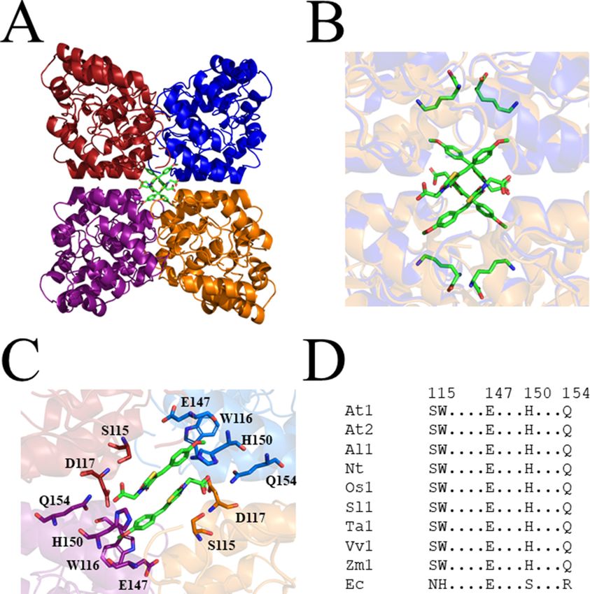
Closer inspection of the crystal structure revealed the presence of four MBDTA-2 molecules
bound at the centre of the homotetrameric protein (Figure 5A), in antiparallel pairs with each mole-
cule, which were stabilised by interactions across three of the monomers (Figure 5B). Interestingly,
this pocket, albeit distinct to the lysine binding site, shares two residues with it, namely W116 and
Figure 4. In vitro potency of DHDPS inhibitors. Dose responses of MBDTA-1 (. or &) and MBDTA-2 (○ or □) against recombinant (A) AtDHDPS1 and (B)
AtDHDPS2. Initial enzyme rates were normalised against a vehicle control (1% [v/v] DMSO). Normalised data (% activity remaining) is plotted as a
function of log10[inhibitor] and fitted to a nonlinear regression model (solid line) (R2 = 0.99). Data represents mean ± S.E.M. (N = 3).
The online version of this article includes the following source data for figure 4:
Source data 1. In vitropotency of DHDPS inhibitors.
Soares da Costa, Hall, et al. eLife 2021;10:e69444. DOI: https://doi.org/10.7554/eLife.69444 5 of 17Research article Biochemistry and Chemical Biology Plant Biology
Figure 5. Crystal structure of AtDHDPS1 bound to MBDTA-2. (A) Cartoon view of overall AtDHDPS1 quaternary (tetrameric) structure, illustrating the
binding sites for MBDTA-2 at the centre of the tetramer. (B) Overlay of the lysine-bound (PDB: 6VVH) and MBDTA-2-bound structures. (C) Close-up of
inhibitor binding pocket, with interacting residues shown as sticks. Lysine and MBDTA-2 are shown as green sticks and coloured by nitrogen (blue),
oxygen (red), and sulfur (yellow). Images were generated using PyMOL v 2.2 (Schrödinger). (D) Sequence alignment of residues involved in MBDTA-2
binding from A. thaliana DHDPS1 (At1; UNIPROT ID: Q9LZX6), A. thaliana DHDPS2 (At2; UNIPROT ID: Q9FVC8), A. lyrata DHDPS1 (Al1; UNIPROT ID:
D7LRV3), N. tabacum DHDPS (Nt; UNIPROT ID: Q42948), O. sativa DHDPS1 (Os1; UNIPROT ID: A0A0K0K9A6), S. lycopersicum DHDPS1 (Sl1; UNIPROT
ID: A0A3Q7IMG0), T. aestivum DHDPS1 (Ta1; UNIPROT ID: P24846), V. vinifera DHDPS1 (Vv1; UNIPROT ID: A0A438E022), Z. mays DHDPS1 (Zm1;
UNIPROT ID: P26259), and E. coli (Ec) DHDPS (UNIPROT ID: P0A6L2). Residues are numbered according to A. thaliana DHDPS1 with dots (.)
representing interspacing residues. Sequences were aligned in BioEdit (v 7.2.5) using the ClustalW algorithm.
The online version of this article includes the following figure supplement(s) for figure 5:
Figure supplement 1. Non-specific binding of MBDTA-2 between symmetry mates.
E147 (Figure 2C). Specifically, the methoxy group of MBDTA-2 interacts with W116, E147, and H150
from chain B, while the pendant carboxylic acid interacts with S115 from chain A as well as H150 and
Q154 from chain C (Figure 5C). Additionally, we observed that upon binding, MBDTA-2 forces
D117 to adopt a different rotamer conformation, which in turn, results in W116 assuming a different
conformation. It must be noted that the four MBDTA-2 molecules were present with 50% occupancy.
Consequently, each of the two moving residues, D117 and W116, adopt two distinct rotamer confor-
mations, one of the apo- and ligand-bound states of AtDHDPS1. Given that no major rotamer
changes or movement of catalytically important residues were noted, the exact mechanism of
Soares da Costa, Hall, et al. eLife 2021;10:e69444. DOI: https://doi.org/10.7554/eLife.69444 6 of 17Research article Biochemistry and Chemical Biology Plant Biology
Table 1. Summary of MBDTA-2-bound AtDHDPS1 crystallographic data collection, processing, and
refinement statistics.
Data collection AtDHDPS1 + MBDTA-2
Space group P41212
Unit-cell parameters (Å) 94.47, 94.47, 181.41
Resolution (Å) 20–2.29 (2.43–2.29)
No. of observations 491,320 (74,297)
No. of unique reflections 37,390 (5768)
Completeness (%) 99.4 (96.6)
Redundancy 13.1 (12.8)
Rmerge (%) 9.9 (39.1)
Rmeas (%) 10.0 (40.7)
CC1/2 99.9 (97.8)
Average I/s(I) 27.9 (7.9)
Refinement
R (%) 18.3
Rfree (%) 22.6
No. (%) of reflections in test set 1071
No. of protein molecules per asu 2
r.m.s.d bond length (Å) 0.007
r.m.s.d bond angle (˚) 1.415
2 *
Average B-factors (Å )
Protein molecules 44.52
Ligand molecules 60.01
Water molecules 40.33
Ramachandran plot†
Residues other than Gly and Pro in:
Most favored regions (%) 98.0
Additionally allowed regions (%) 2.0
Disallowed regions (%) 0.0
PDB code 7MDS
Values in parentheses are for the highest-resolution shell.
*
Calculated by BAVERAGE in CCP4 Suite (Winn et al., 2011).
†
Calculated using MolProbity (Chen et al., 2010).
inhibition remains elusive. Nevertheless, this indicates the presence of a novel DHDPS allosteric
pocket that has not been previously exploited for inhibitor discovery. Moreover, an alignment of the
primary structure of several DHDPS enzymes from plant species indicates that the residues involved
in MBDTA-2 binding are highly conserved across both monocotyledons and dicotyledons
(Figure 5D) and therefore should allow for broad-spectrum inhibition.
Specificity of DHDPS inhibitors
Following determination of the binding site, we examined the specificity of MBDTA-1 and MBDTA-2
to determine whether any future applications would have off-target effects. First, the cytotoxicity of
the inhibitors was examined against the human cell lines, HepG2 and HEK293, using the 3-(4,5-dime-
thylthiazol-2-yl) 2,5-diphenyltetrazolium bromide (MTT) assay (Figure 6A,B). At the highest concen-
tration assessed (400 mM), treatment with the inhibitors did not affect the viability of either cell line
relative to the vehicle control. Second, the effect of the inhibitors on several bacterial species com-
monly found in the human flora and soil microbiome was assessed by measuring their minimum
Soares da Costa, Hall, et al. eLife 2021;10:e69444. DOI: https://doi.org/10.7554/eLife.69444 7 of 17Research article Biochemistry and Chemical Biology Plant Biology
Figure 6. Effect of compounds on the viability of human cell lines. Toxicity of MBDTA-1 (black) and MBDTA-2 (grey), compared to the positive control
defensin (white), assessed against (A) HepG2 and (B) HEK293 human cell lines using the MTT assay. Data were normalised against a vehicle control
(1% [v/v] DMSO) and plotted against inhibitor concentration. Data represents mean ± S.E.M. (N = 3).
The online version of this article includes the following source data for figure 6:
Source data 1. Minimum inhibitory concentration(MIC) values for MBDTA-1 and MBDTA-2 against several bacterial strains.
inhibitory concentrations. No inhibition of bacterial growth was observed up to 128 mgmL 1 (equiva-
lent to ~400 mM) (Table 2), indicating that these DHDPS inhibitors have specificity directed towards
plants.
Herbicidal efficacy
Given the promising in vitro properties of the inhibitors, we determined their herbicidal efficacy
against A. thaliana, initially using seedling agar assays. At high micromolar concentrations of both
MBDTA-1 and MBDTA-2, growth was completely attenuated, and most seeds were unable to germi-
nate. Upon quantitation of root lengths, we determined an IC50 of 98.1 ± 4.34 mM and 47.4 ± 0.450
mM for MBDTA-1 (Figure 7A) and MBDTA-2 (Figure 7B), respectively. Based on these results, we
Table 2. Minimum inhibitory concentration (MIC) values for MBDTA-1 and MBDTA-2 against several
bacterial strains.
1 1
MBDTA-1 MIC (mgmL ) MBDTA-2 MIC (mgmL )
Human flora
Enterococcus spp. >128 >128
Staphylococcus aureus >128 >128
Escherichia coli >128 >128
Soil bacteria
Enterobacter ludwigii >128 >128
Arthrobacter sp. >128 >128
Enterobacter cancerogenus >128 >128
Cedecea davisae >128 >128
Rhodococcus erthropolis >128 >128
The online version of this article includes the following source data for Table 2:
Source data 1. Minimum inhibitory concentration(MIC) values for MBDTA-1 and MBDTA-2 against several bacterial strains.
Soares da Costa, Hall, et al. eLife 2021;10:e69444. DOI: https://doi.org/10.7554/eLife.69444 8 of 17Research article Biochemistry and Chemical Biology Plant Biology
Figure 7. Effect of MBDTA compounds on agar-grown A. thaliana. A thaliana root lengths after treatment with increasing concentrations of either (A)
MBDTA-1 or (B) MBDTA-2. Root lengths were determined using ImageJ v 1.53b and normalised against a vehicle control (1% [v/v] DMSO). Normalised
data (.) (% root length) is plotted as a function of log10[inhibitor] and fitted to a nonlinear regression model (solid line) (R2 = 0.99). Data represents mean
± S.E.M. (N = 3).
The online version of this article includes the following source data for figure 7:
Source data 1. Effect of MBDTA compounds on agar-grown A. thaliana.
examined their pre-emergence effect on soil-grown A. thaliana. Specifically, compounds were dis-
solved in a solution containing a non-ionic organic surfactant (Agral) and seeds were treated immedi-
ately after sowing on soil. The vehicle control-treated plants (Figure 8A) were used as a benchmark
to visually assess the effects of inhibitors. The growth of A. thaliana in the presence of MBDTA-1
(Figure 8B) or MBDTA-2 (Figure 8C) at 300 mgL 1 was severely impeded as evidenced by the
Figure 8. Pre-emergence efficacy of inhibitors on A. thaliana grown in soil. Treatments of (A) vehicle control (1% [v/v] DMSO), (B) 300 mgL 1 MBDTA-1,
and (C) 300 mgL 1 MBDTA-2 given at day 0 (first day under controlled environment room conditions). A representative in triplicate of the biological
replicates is shown vertically. (D) Leaf area of MBDTA-1/2-treated A. thaliana. Area was determined using ImageJ v 1.53b and normalised against a
vehicle control (1% [v/v] DMSO). Data represents mean ± S.D. (n = 3).
The online version of this article includes the following source data for figure 8:
Source data 1. Pre-emergence efficacy of inhibitors on A. thaliana grown in soil.
Soares da Costa, Hall, et al. eLife 2021;10:e69444. DOI: https://doi.org/10.7554/eLife.69444 9 of 17Research article Biochemistry and Chemical Biology Plant Biology
growth area relative to the DMSO control (Figure 8D), wherein few seeds were able to germinate.
This is consistent with the results observed at the highest concentrations of inhibitor on agar. Fur-
thermore, the A. thaliana seeds capable of germinating in the presence of 300 mgL 1 MBDTA-2
were halted at the cotyledon stage before the generation of true leaves. As such, our newly discov-
ered MBDTA compounds represent the first DAP pathway inhibitors with soil efficacy against plants.
Discussion
The lack of herbicides with novel modes of action entering the market in the past three decades has
led to an over-reliance on our current agrichemicals, which has contributed to the rapid generation
of resistance. Although the DAP pathway has gained attention as a way to increase the nutritional
content of lysine in crops (Wang et al., 2017), it has remained an unexplored target for the develop-
ment of herbicides until now. DHDPS catalyses the first step of the DAP pathway and is commonly
duplicated in plant species, including A. thaliana. Both DHDPS proteins are localised to the chloro-
plast and share >85% of primary structure identity, with the majority of differences at the N-termi-
nus. Although DHDPS is essential in A. thaliana (Jones-Held et al., 2012), there have been no
published inhibitors of the plant enzymes, with much of the focus on inhibitors of bacterial DHDPS
as possible new antibiotics. Several iterations of active site inhibitors against bacterial DHDPS
enzymes have been developed, but most of these have, at best, high micromolar potency
(Christoff et al., 2019). As the active site of DHDPS is seemingly difficult to target, the allosteric site
of the enzyme could represent a more fruitful route for the development of inhibitors. Indeed, lysine
is the most potent inhibitor of plant DHDPS orthologues. Furthermore, the most potent bacterial
DHDPS inhibitor to date is bislysine, whereby two lysine molecules are linked together via an ethyl-
ene bridge (Skovpen et al., 2016). Although herbicides commonly directly target, or bind closely
to, the active site of their respective enzyme target, this study shows that allosteric site inhibitors
can be employed and afford lethal phytotoxicity.
Our study describes the discovery of the first plant DHDPS inhibitors, with two MBDTA analogues
identified and characterised here. The mode of inhibition is via a novel binding pocket adjacent to
the lysine binding site, which results in the allosteric inhibition of the enzyme. Lysine has recently
been shown to differentially inhibit the AtDHDPS isoforms, with AtDHDPS1 being 10-fold more sen-
sitive to the allosteric inhibitor (Hall et al., 2021). In this study, we demonstrate that the MBDTA
compounds have similar inhibitory effects against both enzymes. This further supports crystallogra-
phy data demonstrating that MBDTA-2 binds in a pocket adjacent to the lysine allosteric site and is
likely acting in a different way. However, the exact mechanism of inhibition, much like lysine-medi-
ated allostery, remains elusive. Moreover, this binding pocket is conserved across multiple plant spe-
cies, including both monocots and dicots. Importantly, our compounds lacked off-target toxicity,
whilst resulting in the inhibition of germination and growth of A. thaliana seedlings on solidified
media and in soil. However, as expected, plant inhibition was more pronounced on media likely due
to the stability, distribution, and persistence of the compounds. Nevertheless, the assays performed
on soil demonstrate their potential applicability as pre-emergence treatments. It would also be of
interest to investigate the metabolic shifts in plants treated with inhibitors and determine if there is
a toxic build-up of other amino acids such as threonine, which has been observed in DHDPS knock-
out experiments (Sarrobert et al., 2000). Indeed, a common trait of systemic herbicides is that their
efficacy is often related to the cascading consequences of inhibiting a key reaction, rather than inhi-
bition of the reaction itself (Hall et al., 2020). Additionally, it would be of interest to determine the
metabolic state of the compounds after uptake into the cells. This would elucidate whether the
MBDTA compounds act as proherbicides, potentially through the demethylation of their aryl methyl
ethers. As such, it would also be of interest to test demethylated analogues in vitro and in planta to
assess any changes in potency relative to the methoxy compounds.
Developing enzyme inhibitors into a commercial product is an arduous and costly process. Opti-
misation of phytotoxicity, water solubility, cell wall penetration, translocation, soil/water persistence,
and formulation must all be considered. The MBDTA compounds described here represent an
attractive avenue to pursue, and with the elucidation of a novel binding pocket within DHDPS, it
may be possible to rationally improve their potency guided by the crystallography data. Specifically,
it would be of interest to build compounds that extend into the lysine binding pocket, which could
improve potency. Alternatively, novel chemical scaffolds could be explored to target the DHDPS
Soares da Costa, Hall, et al. eLife 2021;10:e69444. DOI: https://doi.org/10.7554/eLife.69444 10 of 17Research article Biochemistry and Chemical Biology Plant Biology
pocket identified. The inhibitors must be able to traverse the chloroplast membrane in order to
reach the DHDPS target and be amenable to post-emergence application. It would also be of inter-
est to study inhibitors with increased hydrophobicity and thus, potentially enhanced transport
through the epidermis, cell wall, and plastid membrane, to reach the DHDPS target. However, it is
important to recognise that there is a fine balance between membrane permeability and aqueous
solubility that must be achieved for any new herbicides. Following the synthesis of more potent ana-
logues, it would be of interest to test inhibitors against weed species. To further pursue the com-
pounds described herein and eventually deploy more potent analogues as herbicides, tolerant crops
will eventually need to be engineered. As the MBDTA binding pocket is highly conserved among
plant species, these inhibitors will likely have broad-spectrum phytotoxicity. Given that the inhibitors
do not bind at the active site, it should be possible to engineer tolerant crops with mutant DHDPS
enzymes that are not susceptible to inhibition with minimal effects on enzyme function. Importantly,
DHDPS inhibitors could also be used in conjunction with other herbicides as part of a combinatorial
treatment to yield synergistic responses and circumvent resistance mechanisms to tackle the global
rise in herbicide-resistant weeds.
Materials and methods
Key resources table
Reagent type
(species) or resource Designation Source or reference Identifiers Additional information
Gene DHDPS1 TAIR At3G60880
(Arabidopsis thaliana)
Gene DHDPS2 TAIR At2G45440
(Arabidopsis thaliana)
Cell line HepG2 ATCC ATCC: HB-8065
(Homo sapiens) RRID:CVCL_0027
Cell line HEK293 ATCC ATCC:ACS-4500
(Homo sapiens) RRID:CVCL_0063
High-throughput chemical screen and analogue synthesis
A high-throughput screen of a library of 87,648 compounds was conducted against recombinant
DHDPS enzyme by the Walter and Eliza Hall Institute High Throughput Chemical Screening Facility
(Melbourne, Australia). The o-ABA colourimetric assay employed assesses DHDPS activity via the for-
mation of a purple chromophore that can be measured at 520–540 nm (Yugari and Gilvarg, 1965).
The assay was miniaturised, so it could be performed in 384-well plates. For the primary screen,
reactions comprised 0.5 mgmL 1 DHDPS, 0.5 mM sodium pyruvate, and 0.5 mM ASA. Library com-
pounds were added at final concentrations of 20 mM, with DMSO concentrations kept at 0.4% (v/v).
After ASA addition, reactions were incubated at 25˚C for 15 min, before a final concentration of 350
mM HCl was added to stop the reaction. o-ABA was subsequently added to a final concentration of
0.44 mgmL 1, plates incubated at room temperature for 1 hr, and absorbance quantified at 540
nm. Vehicle (DMSO) was used as positive controls, and negative controls lacked ASA. For the sec-
ondary screen, 11-point dose–response curves were generated using the same reactions as
described above. A counter screen was conducted using the same set-up albeit without the inclusion
library compounds before the addition of 350 mM HCl. Library compounds were then added after
the reaction was stopped, followed by o-ABA to a final concentration of 0.44 mgmL 1. The plates
were subsequently incubated at room temperature for 1 hr, and absorbance was quantified at 540
nm. Analogues were designed and synthesised using the methods described in previous and con-
temporary work (Perugini et al., 2018).
Expression and purification of A. thaliana DHDPS enzymes
Both DHDPS isoforms from A. thaliana were expressed and purified as previously described
(Hall et al., 2021). Briefly, AtDHDPS isoforms were expressed in Escherichia coli BL21 (DE3) cells,
with AtDHDPS2 requiring the GroEL/ES chaperone complex to facilitate correct folding. Purification
was performed using immobilised metal affinity chromatography. Lastly, fusion tags were cleaved by
Soares da Costa, Hall, et al. eLife 2021;10:e69444. DOI: https://doi.org/10.7554/eLife.69444 11 of 17Research article Biochemistry and Chemical Biology Plant Biology
human rhinovirus 3C or tobacco etch virus protease for AtDHDPS1 and AtDHDPS2, respectively,
whilst simultaneously dialysing into storage buffer (20 mM Tris, 150 mM NaCl, 0.5 mM TCEP, pH
8.0).
Enzyme kinetics
DHDPS enzyme activity was determined using the DHDPS–DHDPR coupled assay as previously
described by measuring the oxidation of NADPH (Atkinson et al., 2013; Hall et al., 2021). Assays
were carried out in a Cary 4000 UV/Vis spectrophotometer at 30˚C with substrates fixed at the previ-
ously determined Michaelis–Menten constant values (Griffin et al., 2012; Hall et al., 2021). Inhibitor
was titrated against AtDHDPS enzymes, and reactions were incubated at 30˚C for 12 min before initi-
ation with ASA. Initial velocity data were normalised against a vehicle (DMSO) control and analysed
using Equation 1 (log(inhibitor) vs. normalised response – variable slope, GraphPad Prism v 8.3).
Dose responses were performed with three technical replicates for each concentration of compound.
Dose responses were repeated with three biological replicates, each using a new stock of reagents.
Y ¼ 100=ð1 þ 10ððLogIC50 XÞHillSlopeÞ
Þ (1)
where Y is the normalised rate, logIC50 is the logarithmic concentration of ligand resulting in 50%
activity, X is the concentration of ligand, and Hill Slope is the steepness of the curve.
X-ray crystallography
AtDHDPS1 was co-crystallised as previously described in the presence of MBDTA-2 (Hall et al.,
2021). Briefly, protein (8.5 mgmL 1) was incubated at 20˚C with MBDTA-2 at a final concentration
of 1 mM (in 2% [v/v] DMSO) before being added in a 1:1 ratio to a reservoir solution containing 1.4
M (NH4)2SO4, 0.1 M NaCl, 0.1 M HEPES (pH 7.5), and 1 mM MBDTA-2 (in 2% [v/v] DMSO). Plates
were incubated at 20˚C. Crystals were briefly dipped in cryo-protectant (1.4 M (NH4)2SO4, 0.1 M
NaCl, 0.1 M HEPES [pH 7.5], 1 mM MBDTA-2 [in 2% (v/v) DMSO], and 20% [v/v] glycerol) and flash
frozen in liquid nitrogen. Data were collected at the Australian Synchrotron using the MX2 beamline
(Aragão et al., 2018). A total of 1800 diffraction images were collected with 0.1˚ oscillation using an
EIGER 16M detector at a distance of 350 mm, with 20% beam attenuation for a total exposure time
of 18 s. X-ray data were integrated using XDS (Kabsch, 2010) and scaled with AIMLESS (Evans and
Murshudov, 2013) before phases were determined by molecular replacement through Auto-Rick-
shaw (Panjikar et al., 2005) with AtDHDPS1 (PDB ID: 6VVI) used as a search model (Hall et al.,
2021). Manual building was performed in COOT Emsley et al., 2010 followed by refinement
employing REFMAC5 in the CCP4i2 (v7.0) software suite (Emsley et al., 2010; Murshudov et al.,
2011; Winn et al., 2011). SMILES string of the inhibitor (MBDTA-2) was processed through AceDRG
to generate the coordinate and cif file (Long et al., 2017). Validation was completed using MolPro-
bity (Chen et al., 2010). The structure of MBDTA-2 bound to AtDHDPS1 is deposited in the Protein
Data Bank as 7MDS.
Cell lines
Cell lines used were sourced from the American Type Culture Collection and were authenticated
using STR DNA profiling, and no mycoplasma contamination was detected.
Toxicity assays
The toxicity of inhibitors against human HepG2 and HEK293 cell lines was assessed using the MTT
viability assay as previously described (Soares da Costa et al., 2012). In brief, the cells were sus-
pended in Dulbecco-modified Eagle’s medium containing 10% (v/v) fetal bovine serum and then
seeded in 96-well tissue culture plates at 5000 cells per well. After 24 hr, cells were treated with 50–
400 mM of MBDTA-1 or MBDTA-2, such that the DMSO concentration was consistent at 1% (v/v) in
all wells. Alternatively, cells were treated with the cytotoxic defensin protein at 100 mM
(Baxter et al., 2017). After treatment for 48 hr, MTT cell proliferation reagent was added to each
well and incubated for 3 hr at 37˚C. The percentage viability remaining reported is relative to the
vehicle control of 1% (v/v) DMSO. Assays were performed in three biological replicates, using a dif-
ferent batch of reagents and cells.
Soares da Costa, Hall, et al. eLife 2021;10:e69444. DOI: https://doi.org/10.7554/eLife.69444 12 of 17Research article Biochemistry and Chemical Biology Plant Biology
Antibacterial assays
The minimum inhibitory concentration (MIC) for MBDTA-1 and MBDTA-2 was determined against a
panel of Gram-positive and Gram-negative bacteria using a broth microdilution method according
to guidelines defined by the Clinical Laboratory Standards Institute (National Committee for Clini-
cal Laboratory Standards, 2004; National Committee for Clinical Laboratory Standards, 2003).
An inoculum of 1 105 colony forming units/mL was used, and the testing conducted using tryptic
soy broth in 96-well plates. Growth was assessed after incubation at 37˚C for 20 hr by measuring the
absorbance at 600 nm. The MIC value is defined as the lowest concentration of inhibitor where no
bacterial growth is observed. Experiments were performed in three biological replicates, using a dif-
ferent stock of reagents and bacterial culture.
Seedling assays
Inhibitors were dissolved in 1 Gamborg modified/Murashige Skoog (GM/MS) media to final con-
centrations of 8–1000 mM. Specifically, media were prepared with 0.8% (w/v) plant grade agar and
1% (w/v) sucrose before sterilisation (Lindsey et al., 2017). A. thaliana seeds were surface sterilised
by soaking in 80% (v/v) ethanol for 5 min, followed by a 15 min incubation in bleach solution contain-
ing 1% (v/v) active NaClO and rinsed in excess sterile water before placing onto agar-containing
inhibitors (Boyes et al., 2001). A. thaliana seeds were stratified at 4˚C for 72 hr in the dark prior to
relocation to a controlled environment room (CER), where seeds were grown at 22 ± 0.5˚C at 60 ±
10% humidity with light produced by cool white fluorescent lights at a rate of ~110 mmolm 2s 1
over long-day conditions (16 hr light:8 hr dark) (Boyes et al., 2001). Plates were positioned upright
to allow roots to grow downwards, and after 7 days, images were taken, and root length
was determined using ImageJ (v 1.53b) (Rasband, 2011). Outliers were identified using the 1.5
interquartile range method (Tukey, 1977). Resulting data were analysed using Equation 1 (log(inhib-
itor) vs. normalised response – variable slope, GraphPad Prism v 8.3). No DMSO and vehicle (1% [v/
v] DMSO) controls were also employed. Assays were carried out with 20 technical replicates (i.e.
seeds) per experiment and were repeated in three biological replicates, with each biological repli-
cate using a different stock of reagents and batch of seeds.
Soil assays
Inhibitors were prepared in DMSO and diluted to 300 mgL 1 (1% [v/v] DMSO) in H2O containing
0.01% (v/v) Agral (Syngenta, North Ryde, NSW, Australia). A. thaliana seeds were surface sterilised
as above and resuspended in 0.1% (w/v) agar before stratification. Subsequently, ~30 seeds were
sown into a small indentation (depth ~1 cm) in moist seed raising soil (pH 5.5) (Biogro, Dandenong
South, VIC, Australia), supplemented with 0.22% (w/w) Nutricote N12 Micro 140 day-controlled
release fertiliser (Yates, Sydney, NSW, Australia). Seeds were treated with 1 mL (equivalent to 1200
gha 1) of compound or vehicle control by pipetting and covered with soil just prior to transfer to a
CER and images taken after 7 days. Area analysis was performed using colour thresholding in
ImageJ (v 1.53b) and normalised against the DMSO control (Corral et al., 2017; Rasband, 2011).
Assays were carried out across three technical replicates (i.e. pots) using the same batch of reagents
and seed stock.
Acknowledgements
TPSC would like to thank the National Health and Medical Research Council of Australia
(APP1091976) and Australian Research Council (DE190100806) for fellowship and funding support,
and MAP and SP thank the Australian Research Council for funding support (DP150103313). ARG
would like to thank the Australian Research Council Research Hub for Medicinal Agriculture
(IH180100006) for support. CJH is supported by La Trobe University Postgraduate Research scholar-
ships. RMC is a recipient of an Australian Government Research Training Program Scholarship and a
LIMS Write-Up Award. This research was supported by the Defence Science Institute, an initiative of
the State Government of Victoria, with a scholarship awarded to JAW, who is also the recipient of a
Research Training Program scholarship. We thank Dr Grant Pearce (University of Canterbury, New
Zealand) for supplying pET151/D-Topo harbouring the DHDPS2/dapA2 gene and Professor Ashley
Franks (La Trobe University, Australia) for supplying soil bacterial isolates. We acknowledge the use
Soares da Costa, Hall, et al. eLife 2021;10:e69444. DOI: https://doi.org/10.7554/eLife.69444 13 of 17Research article Biochemistry and Chemical Biology Plant Biology
of the MX2 beamline at the Australian Synchrotron, part of ANSTO and employed the Australian
Cancer Research Foundation (ACRF) detector. We acknowledge the CSIRO Collaborative Crystallisa-
tion Centre (http://www.csiro/C3; Melbourne, Australia). We also thank the La Trobe University
Comprehensive Proteomics Platform for providing infrastructure support.
Additional information
Competing interests
Tatiana P Soares da Costa, Belinda M Abbott, Matthew A Perugini: is listed as an inventor on a pat-
ent pertaining to inhibitors described in the manuscript. Patent Title: Heterocyclic inhibitors of lysine
biosynthesis via the diaminopimelate pathway; International patent (PCT) No.: WO2018187845A1;
Granted: 18/10/2018. The other authors declare that no competing interests exist.
Funding
Funder Grant reference number Author
National Health and Medical APP1091976 Tatiana P Soares da Costa
Research Council of Australia
Australian Research Council DE190100806 Tatiana P Soares da Costa
Australian Research Council DP150103313 Santosh Panjikar
Matthew A Perugini
Australian Research Council IH180100006 Anthony R Gendall
The funders had no role in study design, data collection and interpretation, or the
decision to submit the work for publication.
Author contributions
Tatiana P Soares da Costa, Conceptualization, Resources, Data curation, Formal analysis, Supervi-
sion, Funding acquisition, Validation, Investigation, Visualization, Methodology, Writing - original
draft, Project administration, Writing - review and editing; Cody J Hall, Data curation, Formal analy-
sis, Validation, Investigation, Visualization, Methodology, Writing - original draft, Writing - review
and editing; Santosh Panjikar, Resources, Formal analysis, Validation, Investigation, Methodology,
Writing - review and editing; Jessica A Wyllie, Formal analysis, Validation, Investigation, Methodol-
ogy, Writing - review and editing; Rebecca M Christoff, Saadi Bayat, Resources, Methodology, Writ-
ing - review and editing; Mark D Hulett, Resources, Writing - review and editing; Belinda M Abbott,
Resources, Supervision, Visualization, Methodology, Writing - review and editing; Anthony R Gen-
dall, Resources, Supervision, Funding acquisition, Methodology, Writing - review and editing; Mat-
thew A Perugini, Conceptualization, Resources, Supervision, Funding acquisition, Methodology,
Project administration, Writing - review and editing
Author ORCIDs
Tatiana P Soares da Costa https://orcid.org/0000-0002-6275-7485
Santosh Panjikar http://orcid.org/0000-0001-7429-3879
Mark D Hulett http://orcid.org/0000-0003-2072-5968
Anthony R Gendall http://orcid.org/0000-0002-2255-3939
Decision letter and Author response
Decision letter https://doi.org/10.7554/eLife.69444.sa1
Author response https://doi.org/10.7554/eLife.69444.sa2
Additional files
Supplementary files
. Supplementary file 1. Physicochemical properties of MBDTA-1/2.
Soares da Costa, Hall, et al. eLife 2021;10:e69444. DOI: https://doi.org/10.7554/eLife.69444 14 of 17Research article Biochemistry and Chemical Biology Plant Biology
. Transparent reporting form
Data availability
Diffraction data have been deposited in PDB under the accession code 7MDS. The validation report
has been uploaded as a ’Related Manuscript File’. Other data sets have been uploaded as ’Source
Data’ files.
References
Aragão D, Aishima J, Cherukuvada H, Clarken R, Clift M, Cowieson NP, Ericsson DJ, Gee CL, Macedo S, Mudie
N, Panjikar S, Price JR, Riboldi-Tunnicliffe A, Rostan R, Williamson R, Caradoc-Davies TT. 2018. MX2: a high-flux
undulator microfocus beamline serving both the chemical and macromolecular crystallography communities at
the Australian Synchrotron. Journal of Synchrotron Radiation 25:885–891. DOI: https://doi.org/10.1107/
S1600577518003120
Atkinson SC, Dogovski C, Downton MT, Czabotar PE, Dobson RCJ, Gerrard JA, Wagner J, Perugini MA. 2013.
Structural, kinetic and computational investigation of Vitis vinifera DHDPS reveals new insight into the
mechanism of lysine-mediated allosteric inhibition. Plant Molecular Biology 81:431–446. DOI: https://doi.org/
10.1007/s11103-013-0014-7
Baxter AA, Poon IK, Hulett MD. 2017. The plant defensin NaD1 induces tumor cell death via a non-apoptotic,
membranolytic process. Cell Death Discovery 3:16102. DOI: https://doi.org/10.1038/cddiscovery.2016.102,
PMID: 28179997
Boyes DC, Zayed AM, Ascenzi R, McCaskill AJ, Hoffman NE, Davis KR, Görlach J. 2001. Growth stage–based
phenotypic analysis of Arabidopsis: a model for high throughput functional genomics in plants. The Plant Cell
13:1499–1510. DOI: https://doi.org/10.1105/tpc.010011, PMID: 11449047
Chen VB, Arendall WB, Headd JJ, Keedy DA, Immormino RM, Kapral GJ, Murray LW, Richardson JS, Richardson
DC. 2010. MolProbity: all-atom structure validation for macromolecular crystallography. Acta Crystallographica
Section D Biological Crystallography 66:12–21. DOI: https://doi.org/10.1107/S0907444909042073,
PMID: 20057044
Christensen JB, Soares da Costa TP, Faou P, Pearce FG, Panjikar S, Perugini MA. 2016. Structure and function of
cyanobacterial DHDPS and DHDPR. Scientific Reports 6:37111. DOI: https://doi.org/10.1038/srep37111,
PMID: 27845445
Christoff RM, Gardhi CK, Soares da Costa TP, Perugini MA, Abbott BM. 2019. Pursuing DHDPS: an enzyme of
unrealised potential as a novel antibacterial target. MedChemComm 10:1581–1588. DOI: https://doi.org/10.
1039/C9MD00107G
Corral MG, Leroux J, Stubbs KA, Mylne JS. 2017. Herbicidal properties of antimalarial drugs. Scientific Reports
7:45871. DOI: https://doi.org/10.1038/srep45871
Craciun A, Jacobs M, Vauterin M. 2000. Arabidopsis loss-of-function mutant in the lysine pathway points out
complex regulation mechanisms. FEBS Letters 487:234–238. DOI: https://doi.org/10.1016/S0014-5793(00)
02303-6, PMID: 11150516
Duke SO. 2012. Why have no new herbicide modes of action appeared in recent years? Pest Management
Science 68:505–512. DOI: https://doi.org/10.1002/ps.2333
Emsley P, Lohkamp B, Scott WG, Cowtan K. 2010. Features and development of Coot. Acta Crystallographica
Section D Biological Crystallography 66:486–501. DOI: https://doi.org/10.1107/S0907444910007493
Evans PR, Murshudov GN. 2013. How good are my data and what is the resolution? Acta Crystallographica
Section D Biological Crystallography 69:1204–1214. DOI: https://doi.org/10.1107/S0907444913000061
Gaines TA, Duke SO, Morran S, Rigon CAG, Tranel PJ, Küpper A, Dayan FE. 2020. Mechanisms of evolved
herbicide resistance. Journal of Biological Chemistry 295:10307–10330. DOI: https://doi.org/10.1074/jbc.
REV120.013572
Galili G, Amir R. 2013. Fortifying plants with the essential amino acids lysine and methionine to improve
nutritional quality. Plant Biotechnology Journal 11:211–222. DOI: https://doi.org/10.1111/pbi.12025
Geng F, Chen Z, Zheng P, Sun J, Zeng A-P. 2013. Exploring the allosteric mechanism of dihydrodipicolinate
synthase by reverse engineering of the allosteric inhibitor binding sites and its application for lysine production.
Applied Microbiology and Biotechnology 97:1963–1971. DOI: https://doi.org/10.1007/s00253-012-4062-8
Ghislain M, Frankard V, Jacobs M. 1995. A dinucleotide mutation in dihydrodipicolinate synthase of Nicotiana
sylvestris leads to lysine overproduction. The Plant Journal 8:733–743. DOI: https://doi.org/10.1046/j.1365-
313X.1995.08050733.x
Griffin MD, Billakanti JM, Wason A, Keller S, Mertens HD, Atkinson SC, Dobson RC, Perugini MA, Gerrard JA,
Pearce FG. 2012. Characterisation of the first enzymes committed to lysine biosynthesis in Arabidopsis thaliana.
PLOS ONE 7:e40318. DOI: https://doi.org/10.1371/journal.pone.0040318, PMID: 22792278
Hall T. 1999. BioEdit: a user-friendly biological sequence alignment editor and analysis program for windows 95/
98/NT. Nucelic Acids Symp Ser 41:95–98. DOI: https://doi.org/10.14601/Phytopathol_Mediterr-14998u1.29
Hall CJ, Mackie ER, Gendall AR, Perugini MA, Soares da Costa TP. 2020. Review: amino acid biosynthesis as a
target for herbicide development. Pest Management Science 76:3896–3904. DOI: https://doi.org/10.1002/ps.
5943, PMID: 32506606
Soares da Costa, Hall, et al. eLife 2021;10:e69444. DOI: https://doi.org/10.7554/eLife.69444 15 of 17Research article Biochemistry and Chemical Biology Plant Biology
Hall CJ, Lee M, Boarder MP, Mangion AM, Gendall AR, Panjikar S, Perugini MA, Soares da Costa TP. 2021.
Differential lysine-mediated allosteric regulation of plant dihydrodipicolinate synthase isoforms. FEBS J Febs
10:15766. DOI: https://doi.org/10.1111/febs.15766
Hall CJ, Soares da Costa TP. 2018. Lysine: biosynthesis, catabolism and roles. WikiJournal of Science 1:4.
DOI: https://doi.org/10.15347/wjs/2018.004
Hildebrandt TM, Nunes Nesi A, Araújo WL, Braun HP. 2015. Amino acid catabolism in plants. Molecular Plant 8:
1563–1579. DOI: https://doi.org/10.1016/j.molp.2015.09.005, PMID: 26384576
Hudson A, Bless C, Macedo P, Chatterjee SP, Singh BK, Gilvarg C, Leustek T. 2005. Biosynthesis of lysine in
plants: evidence for a variant of the known bacterial pathways. Biochimica Et Biophysica Acta (BBA) - General
Subjects 1721:27–36. DOI: https://doi.org/10.1016/j.bbagen.2004.09.008
Hutton CA, Perugini MA, Gerrard JA. 2007. Inhibition of lysine biosynthesis: an evolving antibiotic strategy.
Molecular BioSystems 3:458. DOI: https://doi.org/10.1039/b705624a
Jones-Held S, Ambrozevicius LP, Campbell M, Drumheller B, Harrington E, Leustek T. 2012. Two Arabidopsis
thaliana dihydrodipicolinate synthases, DHDPS1 and DHDPS2, are unequally redundant. Functional Plant
Biology 39:1058–1067. DOI: https://doi.org/10.1071/FP12169
Kabsch W. 2010. XDS. Acta Crystallogr, Sect D: Struct Biol 66:125–132. DOI: https://doi.org/10.1107/
S0907444909047337
Klepikova AV, Kasianov AS, Gerasimov ES, Logacheva MD, Penin AA. 2016. A high resolution map of the
Arabidopsis thaliana developmental transcriptome based on RNA-seq profiling. The Plant Journal 88:1058–
1070. DOI: https://doi.org/10.1111/tpj.13312
Lindsey BE, Rivero L, Calhoun CS, Grotewold E, Brkljacic J. 2017. Standardized method for High-throughput
sterilization of Arabidopsis Seeds. Journal of Visualized Experiments 17:56587. DOI: https://doi.org/10.3791/
56587
Long F, Nicholls RA, Emsley P, Gražulis S, Merkys A, Vaitkus A, Murshudov GN. 2017. AceDRG : a
stereochemical description generator for ligands. Acta Crystallographica Section D Structural Biology 73:112–
122. DOI: https://doi.org/10.1107/S2059798317000067
McCoy AJ, Adams NE, Hudson AO, Gilvarg C, Leustek T, Maurelli AT. 2006. L,L-diaminopimelate
aminotransferase, a trans-kingdom enzyme shared by Chlamydia and plants for synthesis of diaminopimelate/
lysine. PNAS 103:17909–17914. DOI: https://doi.org/10.1073/pnas.0608643103
Murshudov GN, Skubák P, Lebedev AA, Pannu NS, Steiner RA, Nicholls RA, Winn MD, Long F, Vagin AA. 2011.
REFMAC 5 for the refinement of macromolecular crystal structures. Acta Crystallographica Section D Biological
Crystallography 67:355–367. DOI: https://doi.org/10.1107/S0907444911001314
National Committee for Clinical Laboratory Standards. 2003. Methods for Dilution Antimicrobial Susceptibility
Tests for Bacteria That Grow Aerobically, Approved Standard. NCCLS.
National Committee for Clinical Laboratory Standards. 2004. Standard for Antimicrobial Susceptibility Testing,
13th Information Supplement. NCCLS.
Panjikar S, Parthasarathy V, Lamzin VS, Weiss MS, Tucker PA. 2005. Auto-Rickshaw : an automated crystal
structure determination platform as an efficient tool for the validation of an X-ray diffraction experiment. Acta
Crystallographica Section D Biological Crystallography 61:449–457. DOI: https://doi.org/10.1107/
S0907444905001307
Perl A, Galili S, Shaul O, Ben-Tzvi I, Galili G. 1993. Bacterial Dihydrodipicolinate Synthase and Desensitized
Aspartate Kinase: Two Novel Selectable Markers for Plant Transformation. Nature Biotechnology 11:715–718.
DOI: https://doi.org/10.1038/nbt0693-715
Perugini MA, Abbott B, Soares da Costa T. 2018. Heterocyclic Inhibitors of Lysine Biosynthesis via the
Diaminopimelate Pathway, WO2018187845A1.
Peverelli MG, Perugini MA. 2015. An optimized coupled assay for quantifying diaminopimelate decarboxylase
activity. Biochimie 115:78–85. DOI: https://doi.org/10.1016/j.biochi.2015.05.004
Rasband WS. 2011. ImageJ, US. National Institutes of Health, Bethesda, Maryland, USA. http://imagej.nih.gov/ij/
Sarrobert C, Thibaud M-C, Contard-David P, Gineste S, Bechtold N, Robaglia C, Nussaume L. 2000.
Identification of an Arabidopsis thaliana mutant accumulating threonine resulting from mutation in a new
dihydrodipicolinate synthase gene. The Plant Journal 24:357–368. DOI: https://doi.org/10.1046/j.1365-313x.
2000.00884.x
Skovpen YV, Conly CJ, Sanders DA, Palmer DR. 2016. Biomimetic design results in a potent allosteric inhibitor of
dihydrodipicolinate synthase from Campylobacter jejuni. Journal of the American Chemical Society 138:2014–
2020. DOI: https://doi.org/10.1021/jacs.5b12695, PMID: 26836694
Soares da Costa TP, Tieu W, Yap MY, Pendini NR, Polyak SW, Sejer Pedersen D, Morona R, Turnidge JD,
Wallace JC, Wilce MCJ, Booker GW, Abell AD. 2012. Selective inhibition of Biotin Protein Ligase from
Staphylococcus aureus. Journal of Biological Chemistry 287:17823–17832. DOI: https://doi.org/10.1074/jbc.
M112.356576
Soares da Costa TP, Christensen JB, Desbois S, Gordon SE, Gupta R, Hogan CJ, Nelson TG, Downton MT,
Gardhi CK, Abbott BM, Wagner J, Panjikar S, Perugini MA. 2015. Quaternary structure analyses of an essential
oligomeric enzyme. Methods in Enzymology 562:205–223. DOI: https://doi.org/10.1016/bs.mie.2015.06.020,
PMID: 26412653
Soares da Costa TP, Desbois S, Dogovski C, Gorman MA, Ketaren NE, Paxman JJ, Siddiqui T, Zammit LM,
Abbott BM, Robins-Browne RM, Parker MW, Jameson GB, Hall NE, Panjikar S, Perugini MA. 2016. Structural
determinants defining the allosteric inhibition of an essential antibiotic target. Structure 24:1282–1291.
DOI: https://doi.org/10.1016/j.str.2016.05.019, PMID: 27427481
Soares da Costa, Hall, et al. eLife 2021;10:e69444. DOI: https://doi.org/10.7554/eLife.69444 16 of 17You can also read