A sequential deposition of amyloid beta oligomers, plaques and phosphorylated tau occurs throughout life in the canine retina
←
→
Page content transcription
If your browser does not render page correctly, please read the page content below
Habiba et al. Ageing Neur Dis 2022;2:7
DOI: 10.20517/and.2022.06
                                                                                                               Ageing and
                                                                                                         Neurodegenerative
                                                                                                                 Diseases

Original Article                                                                                                              Open Access

A sequential deposition of amyloid beta oligomers,
plaques and phosphorylated tau occurs throughout
life in the canine retina
Umma Habiba1, John Morley1, Mark Krockenberger2, Brian A. Summers3, Mourad Tayebi1
1
 School of Medicine, Western Sydney University, Campbelltown, NSW 2560, Australia.
2
  Faculty of Science, University of Sydney, Camperdown, NSW 2050, Australia.
3
  School of Veterinary Medicine, Melbourne University, Victoria, Werribee 3030, Australia.

Correspondence to: Dr. Mourad Tayebi, School of Medicine, Western Sydney University, Campbelltown, NSW 2560, Australia.
E-mail: m.tayebi@westernsydney.edu.au

How to cite this article: Habiba U, Morley J, Krockenberger M, Summers BA, Tayebi M. A sequential deposition of amyloid beta
oligomers, plaques and phosphorylated tau occurs throughout life in the canine retina. Ageing Neur Dis 2022;2:7.
https://dx.doi.org/10.20517/and.2022.06

Received: 25 Feb 2022 First Decision: 21 Mar 2022 Revised: 25 Apr 2022 Accepted: 18 May 2022 Published: 25 May 2022

Academic Editor: Wei-Dong Le Copy Editor: Peng-Juan Wen Production Editor: Peng-Juan Wen

Abstract
Aims: Cerebral amyloid burdens may be found in otherwise cognitively intact adults, often not showing worsening
deficits with passing years. Alzheimer’s transgenic rodents have been widely used to investigate this phenomenon,
but a spontaneous disorder in other animals, such as dogs that cohabit with humans and thus may have some
shared environmental risks, may contribute and offer opportunities not possible in the smaller laboratory animals.
In animals, the spontaneous disorder most comparable to Alzheimer’s disease (AD) affects mature to aged dogs
and is designated canine cognitive dysfunction. Motivated by AD, many studies have revealed that amyloid
progressively accumulates in the canine central nervous system, including the retina. Here, we investigated
whether deposits of amyloid and/or tau can be found in the canine retina of neurologically normal animals from the
first year of life to the elderly. Suppose canine ocular amyloid and tau are present from early life. In that case, that
raises the question of whether similar patterns of accumulation occur in man, whether as part of aging, AD, or
other.

                   © The Author(s) 2022. Open Access This article is licensed under a Creative Commons Attribution 4.0
                   International License (https://creativecommons.org/licenses/by/4.0/), which permits unrestricted use, sharing,
                   adaptation, distribution and reproduction in any medium or format, for any purpose, even commercially, as
long as you give appropriate credit to the original author(s) and the source, provide a link to the Creative Commons license, and
indicate if changes were made.

                                                                                                                  www.ageneudisjournal.com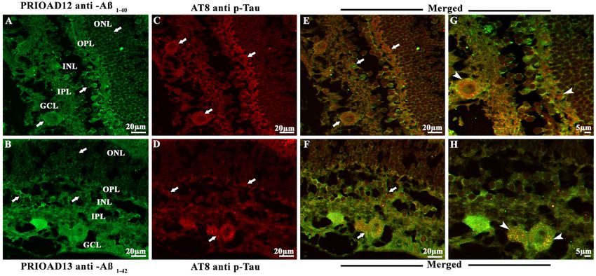
Page 2 of 21                             Habiba et al. Ageing Neur Dis 2022;2:7   https://dx.doi.org/10.20517/and.2022.06

Methods: This study used eye tissues from 30 dogs with a variety of ophthalmic or other orbital disorders, of which
7/30 were 1-2 years old. Tissues were subdivided into dogs of three different age groups: young (1-5 years old),
middle (6-10 years old), and old (≥ 11 years old).

Results: Following immunostaining of tissue sections with nanobodies against retinal Aβ1-40 and Aβ1-42 oligomers,
and antibodies against Aβ plaques (Aβp) and hyperphosphorylated Tau (p-Tau), our investigations revealed that
accumulation of Aβ1-40 and Aβ1-42 oligomers were widespread in the retina in all age groups. In contrast, Aβp were
detected in the middle and old age groups but not in the young age group. Furthermore, p-Tau staining was
observed in four old dogs only, while other dogs were p-Tau free. Interestingly, both Aβo and Aβp co-localized in
the middle and old age groups of dogs. Moreover, diffuse granular p-Tau co-localized with intracellular Aβo in the
old age group. Finally, we also observed co-localization of Aβo and Aβp in the retinal vasculature which might be
similar to cerebral amyloid angiopathy associated with AD.

Conclusion: As far as we know, the presence of amyloid and tau in the canine retina is hitherto unreported. If
similar, early-in-life subclinical retinal deposits occur in a human cohort perhaps identified by AD genetic risk
factors, following this group may offer the prospect of preclinical therapeutic intervention in imminent dementia, a
strategy recognized as likely necessary to impact this burgeoning disorder.

Keywords: Alzheimer’s disease, canine cognitive dysfunction, retina, early diagnosis, Aβo, Aβp, p-Tau

INTRODUCTION
The principal neuropathological lesions in Alzheimer’s disease (AD) brains include extracellular neuritic
and/or diffuse plaques containing amyloid-beta (Aβ), intracellular tau protein (p-Tau) in the form of
neurofibrillary tangles (NFTs) in addition to cerebral amyloid angiopathy (CAA), ubiquitin, severe synaptic
loss, neuronal death, and brain atrophy[1-4]. While the deposition of Aβ in the human brain has traditionally
been accepted as a major hallmark of AD[5], its accumulation is also observed in about 20% of cognitively
unimpaired, aged individuals[6,7]. For decades, the association of Aβ with AD has been demonstrated, but the
significance and impact of Aβ accumulation in healthy individuals are both poorly understood[8] and a
source of confusion. For example, previous studies did not establish a clear correlation with memory loss in
the aging brain[9-11]. Previous reports documented the progressive accumulation of both Aβ plaques (Aβp) in
the brain[12,13] and Aβ oligomers (Aβo) in the brain and periphery of cognitively unimpaired individuals[14,15].
A study by Lesne et al. measured the levels of three Aβo “species”, including Aβ trimers, Aβ*56 and Aβ
dimers, in brain tissues from 75 cognitively unimpaired individuals, including young children and
adolescents[14]. The authors showed that Aβ trimers were present in the central nervous system (CNS) of
children and adolescents, and their levels increased progressively with age, suggesting that this particular
Aβo could be used to track the potential progression into AD from a very young age. Another study
investigated the relationship between amyloid levels and memory performance[15]. This study, which
included 147 participants divided into three groups of adults 30-49, 50-69, and 70-89 years of age,
established a clear relationship between episodic memory performance and amyloid accumulation in the
youngest group[15]. A longitudinal study by Hanseeuw et al. demonstrated a correlation between Aβ/p-Tau
and declining cognition in 60 clinically normal individuals aged between 65-85 years[16]. The authors
concluded that there was a positive correlation between Aβ/p-Tau positron emission tomography (PET)
and cognition, where participants with high Aβ and tau were at higher risk of developing mild cognitive
impairment (MCI)[16]. These studies highlighted the importance of investigating and characterizing normal
aging in cognitively unimpaired younger individuals to identify those at risk of progressive increase of AD-
associated neuropathology changes and cognitive impairment and establish a time frame for early
diagnostic and therapeutic intervention. However, such studies are difficult to implement in human subjects
and will take decades to deliver any meaningful outcome[8,17].
Habiba et al. Ageing Neur Dis 2022;2:7   https://dx.doi.org/10.20517/and.2022.06                   Page 3 of 21

Currently available transgenic animal models of AD do not replicate the subtle clinical and pathological
features of the disease, as demonstrated by their lack of reproducible therapeutic outcomes[18,19]. As to the
dog’s age, there is an anatomically defined accumulation of Aβ peptides in the CNS, while less is known
about cerebral p-Tau deposition. Some animals will develop a progressive cognitive decline with loss of
learned behaviors and memory, a syndrome named canine cognitive dysfunction (CCD)[20-24], with CNS
changes similar to the neuropathological hallmarks characteristic of AD[25-28]. In those destined to develop
AD, amyloid precursor protein (APP) is sequentially cleaved by β and γ secretase leading to the production
of Aβ peptide fragments (36-43 amino acids), which then aggregate and deposit as plaques[4,5,29]. The canine
APP and Aβ peptides are 98% and 100% identical to their human counterparts[24]. In older dogs and others
affected with CCD, Aβ deposits as diffuse plaques, while the dense core of humans’ mature plaques is very
rare or absent[20,21,27,30].

There are two major isoforms of A β , Aβ1-40 (~80%-90%) and Aβ1-42 (~5%-10%) and three major
assemblies[31-34] recognized in man and animals/dogs, including monomeric Aβ, Aβo containing 12-24
monomers which become elongated to form protofibrils and finally insoluble fibrils[35]. Among these three
stages, Aβo is thought to be the most toxic to neurons and responsible for the structural and functional
pathologic changes associated with AD[36-39]. Likewise, a previous study reported that cognitive decline
occurs before the accumulation of Aβp in CCD, supporting the premise that earlier assembly states of Aβ
may be the toxic species[23]. In addition, CAA, ubiquitin, and severe synaptic loss have also been reported in
CCD[20,40-43]. Another cardinal pathological hallmark of AD is the accumulation of p-Tau[3,4]. Several
phosphorylation sites were identified on tau in aged dogs including Thr181[42], Ser422[43],
Ser202/Thr205[40,43], Ser396[30,40,43], Ser189, and Ser207[44]; however, demonstrating the presence of NFTs has
been infrequent compared to the consistent demonstration of amyloid oligomers and diffuse plaques. A
study by Schmidt and colleagues using anti-pT205, AT8, AT100, PHF-1, and anti-pT422 antibodies seeking
the presence of tau pathology in 24 dogs aged between two and nineteen years, showed that three 13-15-
year-old dogs displayed p-Tau and only one 15-year-old Pekingese dog displayed NFT-like appearance[43].

In AD, visual disturbances are one of the early complaints and include loss of color vision, impairment of
peripheral vision and object recognition, contrast sensitivity, and decreased visual memory and
perception[45-47]. A recent longitudinal study of 1349 older adults showed that poor visual acuity paralleled
the development of dementia[48], suggesting the possibility that ocular disturbances can be used as an early
predictor of dementia risk in the older population[48]. Moreover, post-mortem studies of AD and animal
models of AD demonstrated a strong association between retinal accumulation of Aβo[49,50]. Aβ plaques[51,52],
p-Tau[53,54] and brain depositions and cognitive decline, where retinal Aβo was shown to deposit earlier than
the brain and before deficits in cognition. Although, to our knowledge, age-dependent retinal deposition of
Aβo and/or Aβp has not been investigated in AD and cognitively unimpaired individuals, including children
and adolescents, our recent studies in AD mice models confirmed the conversion of cerebral and retinal Aβ
o to Aβp in an age-dependent manner, where retinal Aβo was detected as early as 3-month old APP/PS1
mice, before brain pathology and cognitive decline were observed[49,55].

In this study, we investigated the retinal accumulation of Aβo, Aβp, and p-Tau in three age groups of
genetically diverse and neurologically intact populations of dogs. Here, following immunohistochemistry
and immunofluorescence analysis, we confirmed the presence of retinal Aβ40 and Aβ42 oligomers, Aβp, and
p-Tau. Retinal Aβ40 and Aβ42 oligomers deposition was conspicuous and widespread and observed in all age
groups, including the young 1-5-year-old neurologically intact group. Moreover, retinal co-localization of

Page 4 of 21                           Habiba et al. Ageing Neur Dis 2022;2:7   https://dx.doi.org/10.20517/and.2022.06

Aβ40/Aβ42 oligomers and Aβp was observed in few middle-aged dogs and most dogs in the old neurologically
intact age group, while retinal co-localization of Aβ40/Aβ42 oligomers and p-Tau was only seen in the old
neurologically intact age group of dogs. Morphologically, extracellular Aβp deposits appeared as small, dot-
like rounded deposits, while intracellular p-Tau deposits adopted a diffuse appearance in the retinal
layers[54]. No NFTs or neuropil threads were observed in these dogs. Taken together, these results highlight
the importance of further investigations of AD-related pathology in the retina of presymptomatic children
and adolescents to gain insight into disease progression and potentially identify early at-risk individuals to
help implement speedy therapeutic interventions.

METHODS
Dog eye samples and animal ethics
Sections of eyes used in this study were prepared from archived cases in the Comparative Ocular Pathology
Laboratory of Wisconsin (COPLOW) at the Department of Pathobiological Sciences, School of Veterinary
Medicine, University of Wisconsin, Madison. Surgical enucleations had been submitted by practicing
veterinarians to COPLOW for routine pathological diagnosis. These canine eyes came from 30 random,
genetically diverse dogs (15 different breeds) ranging from 1 to 16 years of age with a broad variety of ocular
disorders [Tables 1 and 2]. Such tissues, historically submitted for disease investigation purposes, are not
subject to approval by institutional animal ethical regulations.
Tissue preparation
Enucleated eyes were submitted to the laboratory fixed in 10% neutral buffered formalin. On receipt, they
were examined for gross abnormalities before trimming, followed by processing overnight using a Leica
ASP300S tissue processor (Leica biosystem, Wetzlar, Germany). Tissues were then embedded in paraffin
blocks and sectioned at 4μm thickness using a Leica RM2235 microtome (Leica biosystem, Wetzlar,
Germany) and placed on charged slides. Sections were then stained with hematoxylin and eosin (H&E) and
Congo red (CR). Further sections were used for immunohistochemistry (IHC) and immunofluorescence
(IF) staining, and both upper and lower parts of the retina were assessed.
Hematoxylin and eosin staining
Paraffin sections were dewaxed by two changes of absolute xylene for 5 min each. Sections were rehydrated
using two changes of 100% ethanol for 2 min each, 95% ethanol for 3 min, 70% ethanol for 2 min, and
finally rinsed in deionized water for 2 min. H&E stain was then performed by adding Gill ll Haematoxylin
solution (Leica Biosystems, Wetzlar, Germany), followed by 1% acid alcohol and subsequently eosin stain
(Leica Biosystems, Wetzlar, Germany). Finally, the sections were dehydrated with increasing concentrations
of ethanol, from 70%, 95% to 100%, then dewaxed by two changes of xylene and finally mounted with
xylene-based mounting media (Sigma Aldrich, Missouri, United States)[56]. H&E staining was used to assess
retinal morphology, the neuronal population within the eye.
Congo red staining
Initially, the CR working solution was prepared by mixing 50 ml Congo red solution and 0.5 ml potassium
hydroxide solution supplied in the Congo red amyloid special stain kit (Leica Biosystems, Wetzlar,
Germany). Retinal sections were placed in the working solution for 20 min and then rinsed in 5-8 changes
of deionized water. This was followed by staining with Gill ll Haematoxylin (Leica Biosystems, Wetzlar,
Germany) for 1-3 min and rinsing in 3 changes of deionized water. Sections were then dehydrated in two
changes of 95% alcohol followed by three changes of absolute alcohol for one minute each. Finally, the
sections were cleared in two changes of xylene and mounted in a xylene miscible medium. Amyloid fibrils
appeared as dull to red brick under light microscopy (Olympus CX 43, Shinjuku, Tokyo, Japan) and apple
green birefringent under polarized light (Olympus CX 43, Shinjuku, Tokyo, Japan).

Habiba et al. Ageing Neur Dis 2022;2:7         https://dx.doi.org/10.20517/and.2022.06                                         Page 5 of 21

Table 1. Signalment for 30 dogs and their retinal Aβ oligomers and plaques IHC scores

                                                                                            Pathological Aβ IHC findings in the dog retina
Age groups                 Age (year) Sex Breed                                   Size
                                                                                          A11 - Aβ oligomers        4G8 - Aβ plaques
Young (1-5 years)          1              SF    Staffordshire terrier             Medium +                          -
                           1.4            M     Siberian husky                    Medium -                          -
                           1.8            SF    Mixed breed                       ND*     +++                       -
                           1.10           SF    Siberian husky                    Medium ++                         -
                           2              F     Chihuahua mix                     Toy     +++                       -
                           2              M     Bichon frise mix                  Small   +                         -
                           2              NM Shih Tzu                             Small   +                         -
                           3              NM German shepherd                      Large   +++                       -
                           3              F     Giant schnauzer                   Large  +                          -
                           3.6            M     Siberian husky                    Medium +                          -
Middle age (6-10 years)    7              NM Hound mixed                          Medium -                          -
                           8              SF    Bedlington terrier                Small   -                         -
                           8              SF    Cocker spaniel                    Medium -                          -
                           8              M     Mixed breed                       ND*     -                         -
                           8              M     Cocker spaniel                    Medium +                          -
                           8              F     German shepherd mix               Large   -                         -
                           8              SF    Great dane dog                    Giant   -                         -
                           9              M     Jack Russell terrier dog          Small   -                         -
                           9.5            SF    Boxer dog                         Large   -                         -
                           9.9            SF    Cocker spaniel                    Medium -                          -
Elderly (11-16 years)      11             NM Mixed breed                          ND*     -                         -
                           11             NM Beagle                               Small   -                         -
                           11.9           SF    Bouvier des flanders              Large   +                         -
                           12             SF    German shepherd                   Large   -                         ++
                           12             SF    German shepherd mix               Large   -                         -
                           12             NM Husky mix                            Medium -                          -
                           12.8           NM Shih-tzu                             Small   -                         ++
                           13             NM Border collie                        Medium -                          -
                           15             SF    Basset hound                      Medium -                          +
                           16             SF    Shih Tzu                          Small   -                         -

                                                                           [76]
Sizes were determined according to the American Kennel Club (AKC) . Size range between 34 - ≥ 54 kg is considered “giant”, 24-38 kg is
considered “large”, 15-29 kg is considered “medium”, 3-15 kg is considered “small”, and 0.9-4 kg is considered “toy”. Aβ oligomers and Aβ
plaques staining intensity were semi-quantitatively analyzed and scored across the retinal layers under the brightfield microscope (Olympus CX
43, Shinjuku, Tokyo, Japan). The total area was examined at 40 magnification and categorized into no immunostaining “-”; low immunoreactivity
found only in limited areas of the retinal layers “+”, moderate immunoreactivity where Aβ deposits were more apparent “++”; and finally strong
immunoreactivity with widespread A11 and 4G8 positive Aβ labeling were exhibited “+++”. ND*: Not determined; SF: spayed female; F: female;
NM: neutered male; M: male; IHC: immunohistochemical.

Immunohistochemical staining of amyloid-beta plaques and amyloid-beta oligomers
Eye sections that contained the retina were pre-treated using the 2100 antigen retriever (Aptum Biologics
Ltd, Southampton, United Kingdom) to expose the target epitopes. The sections were then treated with 90%
formic acid for 5 min at room temperature (RT) followed by cell membrane permeabilization, achieved by
using 0.1% Triton X for 1 min before the addition of 0.3% H2O2 for 15 min to inactivate endogenous
peroxidases. Sections were then blocked with protein block serum-free (Agilent, City, Country) for 15 min.
The sections were then stained for 1 h with the following primary antibodies in phosphate-buffered saline
(PBS): mouse purified 4G8 anti-Aβ17-24 (1:500; Bio legend, San Diego, CA, USA) or A11 rabbit anti-Aβo
(1:250; Merck Millipore, Burlington, MA, USA) antibodies. After washing with PBS, sections were
Page 6 of 21 Habiba et al. Ageing Neur Dis 2022;2:7 https://dx.doi.org/10.20517/and.2022.06
Table 2. Signalment for 30 dogs, their ophthalmological disorders, and retinal Aβ and p-Tau IF scores
Pathological IF findings in the dog
Age Ophthalmological disorders which retina
Age groups (year) Sex Breed Size
resulted in enucleation Aβ40 Aβ42 Aβ p-
oligomers oligomers Tau
Young (1-5 1 SF Staffordshire Medium Anterior segment dysgenesis + + - -
years) terrier
1.4 M Siberian husky Medium Anterior segment dysgenesis - - - -
1.8 SF Mixed breed ND* Anterior segment dysgenesis +++ +++ - -
1.10 SF Siberian husky Medium Anterior segment dysgenesis +++ +++ - -
2 F Chihuahua mix Toy Anterior segment dysgenesis +++ +++ - -
2 M Bichon frise mix Small Proptosis +++ +++ - -
2 NM Shih Tzu Small Melanoma limbal ++ ++ - -
3 NM German Large Rhabdomyosarcoma orbital +++ +++ - -
shepherd
3 F Giant schnauzer Large Phthisis bulbi ++ ++ - -
3.6 M Siberian husky Medium Anterior segment dysgenesis ++ ++ - -
Middle age 7 NM Hound mixed Medium Hemangiosarcoma - - - -
(6-10 years)
8 SF Bedlington Small Neoplasia - - - -
terrier
8 SF Cocker spaniel Medium Conjunctivitis +++ +++ + -
8 M Mixed breed ND* Conjunctival melanoma +++ +++ + -
8 M Cocker spaniel Medium Pre glaucoma +++ +++ +++ -
8 F German Large Mast cell tumor ++ ++ + -
shepherd mix
8 SF Great dane dog Giant Complex apocrine adenocarcinoma + + - -
9 M Jack Russell Small Sialoadenitis ++ ++ - -
terrier dog
9.5 SF Boxer dog Large Hemangioma + + - -
9.9 SF Cocker spaniel Medium Adenocarcinoma a orbital +++ +++ +++ -
Elderly (11-16 11 NM Mixed breed ND* Squamous cell carcinoma - - - -
years)
11 NM Beagle Small Melanoma conjunctival +++ +++ +++ -
11.9 SF Bouvier des Large Melanoma eyelid + + - -
flanders
12 SF German Large Melanoma eyelid +++ +++ +++ +++
shepherd
12 SF German Large Melanoma conjunctival +++ +++ +++ -
shepherd mix
12 NM Husky mix Medium Orbital carcinoma +++ +++ ++ +++
12.8 NM Shih-tzu Small Glaucoma +++ +++ ++ +++
13 NM Border collie Medium Melanoma skin +++ +++ +++ +++
15 SF Basset hound Medium Conjunctival hemangiosarcoma +++ +++ +++ -
16 SF Shih Tzu Small Squamous cell carcinoma + + - -
Existing ophthalmological conditions of the 30 neurologically intact unimpaired dogs were determined from the clinical report. Sizes were
determined according to the American Kennel Club (AKC). Size range between 34 - ≥ 54 kg is considered “giant”, 24-38 kg is considered “large”,
15-29 kg is considered “medium”, 3-15 kg is considered “small”, and 0.9-4 kg is considered “toy”. Aβ oligomers Aβ plaques and p-Tau staining
intensity were semi-quantitatively analyzed and scored across the retinal layers under the confocal microscope (LSM800, Zeiss, Oberkochen,
Germany). The total area was examined at 40 magnification and categorized into no immunostaining “-”; low immunoreactivity found only in
limited areas of the retinal layers “+”, moderate immunoreactivity where deposits were more apparent “++”; and finally strong immunoreactivity
with widespread Aβo, Aβp, and p-Tau labeling was exhibited “+++”. ND*: Not determined; SF: spayed female; F: Female; NM: neutered male; M:
male; IF: immunofluorescence.
incubated for 1 h at RT with the following secondary antibodies in PBS: HRP-conjugated anti-mouse IgG
(Sigma-Aldrich, Missouri, United States) or anti-rabbit IgG (Sigma-Aldrich, Missouri, United States). TheHabiba et al. Ageing Neur Dis 2022;2:7 https://dx.doi.org/10.20517/and.2022.06 Page 7 of 21 sections were then washed three times with PBS before the addition of the 3,3'-Diaminobenzidine substrate chromogen system and incubated for 5-10 min. The sections were then counterstained with hematoxylin for 1 min before mounting. Sections were finally imaged using Olympus CX 43 light microscopy (Shinjuku, Tokyo, Japan). The staining intensity was semi-quantitatively analyzed and scored across the retinal layers. Each bright field was examined at 40 × magnification and categorized into no immunostaining “-”; low immunoreactivity, found in limited areas of the retinal layers “+”; moderate immunoreactivity where Aβ deposits were numerous “++”; and finally strong immunoreactivity with widespread A11 and 4G8 positive Aβ labeling “+++” [Table 1]. Immunofluorescence detection and co-localization studies To investigate whether Aβo co-localized with Aβp or with p-Tau, we performed immunofluorescence double labeling using camelid-derived single domain anti-A40 (PRIOAD12) or anti-A42 (PRIOAD13) oligomer antibodies with 4G8 anti-Aβp antibody (Bio legend, San Diego, California, United States) or anti- phosphorylated tau AT8 antibody targeting amino acid residues Ser202/Thr205 (Thermo-fisher Scientific, Massachusetts, United States). Slides were processed as described above for IHC before adding the primary antibodies. The sections were double-stained with either PRIOAD 12 (1:500) or PRIOAD 13 (1:500)[57] and 4G8 antibody overnight (1:500) or AT8 (1:500) and either PRIOAD 12 (1:500) or PRIOAD 13 (1:500). Sections were then washed in Tris-buffered saline with 0.05% Tween 20 (TBST) and incubated with secondary antibodies at a dilution of 1:500, including goat anti-llama IgG conjugated to FITC (Bethyl Laboratories, Inc, Texas, USA) or donkey-anti-mouse IgG conjugated to Texas red (Sigma-Aldrich, Missouri, United States) for 2 hours at room temperature. Other sections were used as negative control and stained with secondary antibodies with the omission of the primary antibodies. Retinal sections derived from an APP/PS1 and TAU58/2 transgenic mice models[58,59] were used as positive control and stained with anti-Aβ and anti-P-Tau antibodies, respectively. The sections were covered, slipped with paramount aqueous mounting medium (Dako, Agilent, Santa Clara, California, United States), sealed, and dried overnight. Finally, the sections were visualized with LSM800 confocal microscope with a standard FITC/Texas Red double band-pass filter set (Zeiss, Oberkochen, Germany). RESULTS Histological assessment of the retina in 30 dogs We performed H&E staining of the canine eye sections and examined the retinal layers to assess their general morphological appearance and identify pathological changes such as vacuolation, neuronal death, and eosinophilic deposits[49,55]. No specific lesions were observed in the retinal layers of the young and middle age groups (data not shown). However, scattered eosinophilic deposits in the ganglion cell layer (GCL) and inner nuclear layer (INL) of the retina were noticeable in 5/10 dogs in the older age group (11-16 years old), including an 11-year-old Beagle, a 12-year-old German shepherd, a 12-year-old Husky mix, a 12.8-year-old Shih Tzu, and a 15-year-old Basset hound [Figure 1A and B]. Furthermore, CR staining used to detect any amyloid fibrils and CAA in eye sections of the dogs did not display any staining in the retina tissues from all age groups (data not shown). Immunohistochemical detection of Aβ oligomers and plaques in the retina of 30 dogs While demonstrated in the CNS of aged animals including cats and bears[24,60], dogs have been more comprehensively studied and shown to develop human type Aβ deposition at a very young age. Retinal tissues stained with A11 exhibited intracellular Aβo depositions in the outer nuclear layer (ONL), INL, and GCL [Figure 1C and D]. The intraneuronal Aβo intensity following A11 staining ranged from low (+), moderate (++) to strong (+++) in all dogs in the young age group except a 1.4 years old male Siberian husky
Page 8 of 21                                    Habiba et al. Ageing Neur Dis 2022;2:7       https://dx.doi.org/10.20517/and.2022.06

Figure 1. Photomicrographs of the microscopic lesions in the canine retina. The retina in a 12-year-old German shepherd dog.
Eosinophilic deposits (green arrows) were observed in the ganglion cell layer (GCL) and inner nuclear layer (INL). Representative of 10
cases of old-aged dogs. Hematoxylin and eosin (H&E) 40×. (B) Higher magnification of the deposits in image (A) (green arrowhead) in
the GCL. H&E 100×. (C) Retina of a 3-year-old German shepherd dog. Immunohistochemical staining with A11 anti-Aβo IgG antibody
exhibited cytoplasmic Aβo depositions in the GCL and INL (green arrows). Representative of 10 cases of young age group. IHC 40×.
(D) Higher magnification of deposits from image (C) in the GCL (green arrowheads). IHC 100×. (E) Retina of a 12-year-old German
shepherd mix dog. Immunohistochemical staining with 4G8 anti-Aβp IgG antibody exhibited extracellular Aβ aggregates in the GCL and
INL (green arrows). IHC 40×. (F) Higher magnification of deposit from image (E) in the GCL (green arrowhead). (G and H)
Representative retinal sections derived from dogs of all age groups stained with secondary antibodies with the omission of the primary
antibody did not show any depositions (40× and 100×, respectively). The photomicrographs were taken from the peripheral region of
the retina - away from the optic disc. Representative of all 30 dogs examined. IHC 100×. IHC: Immunohistochemistry.

that did not display any A11 positive stain [Supplementary Figure 1A]. Of note, retinal sections derived
from wild-type mice were used as negative control following immunofluorescence staining [Supplementary
Figure 1B and C].

A spayed female mixed breed aged 1.8 years, a female Chihuahua aged 2 years, and a neutered male German
shepherd aged 3 years showed strong (+++) accumulation of A11 positive Aβo [Figure 1C and D]. Two dogs
displayed a low amount (+) of A11 positive Aβo in the retinal layers in the middle and old age groups,
including an 8-year-old male Cocker spaniel and an 11.9-year-old spayed female Bouvier des Flanders
[Table 1]. In addition, 4G8-positive small rounded and dot-like extracellular Aβp deposits were observed in
the INL, inner plexiform layer (IPL), and GCL [Figure 1E and F] of the retina. They were structurally
different from the typical large diffuse plaques normally observed in dog brains with CCD[21,30,61]. The
extracellular Aβp intensity following 4G8 staining ranged from low (+) to moderate (++) in some dogs in
the old age group, including a 15-year-old spayed female Basset hound, a 12.8-year-old neutered male Shih
Tzu, and a 12-year-old spayed female German shepherd. Other dogs in this age group were all negative for
4G8 staining [Table 1, Figure 1E and F]. Representative retinal sections derived from dogs of all age groups
stained with secondary antibodies with the omission of the primary antibody did not show any depositions
[Figure 1G and H].
Immunofluorescence detection and co-localization of retinal Aβ40, Aβ42 oligomers, and Aβ plaques in
the retina of 30 dogs
To confirm the presence of retinal Aβ40 and Aβ42 oligomers and to determine whether Aβ40 and/or Aβ42
oligomers co-localized with Aβp in the retinas of different age groups and breeds of neurologically intact
dogs, we performed immunofluorescence double staining using PRIOAD12 (Aβ40 oligomers), PRIOAD13
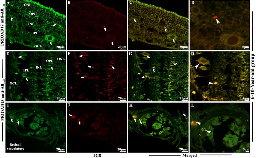
Habiba et al. Ageing Neur Dis 2022;2:7   https://dx.doi.org/10.20517/and.2022.06                 Page 9 of 21

(Aβ42 oligomers) camelid-derived single domain anti-A β oligomer and 4G8 anti-A β p antibodies.
Morphologically, Aβo appeared as globular and annular in shape[55,62] and deposited intracellularly in the
ONL, INL, and GCL [Figures 2-4]. 4G8 positive Aβ plaques appeared morphologically as dot-like and small
rounded extracellular deposits[50,54] in the outer plexiform layer (OPL), IPL, and GCL [Figures 2-4]. We
found that both Aβ40 and Aβ42 oligomers staining was widespread in the majority of the dogs in young
[Table 2, Figure 2A and B], middle [Table 2, Figure 3A and B], and old age groups [Table 2, Figure 4A and
B] except four dogs including a 1.4-years-old male Siberian husky, a 7-years-old neutered male Hound
mixed, an 8-years-old spayed female Bedlington terrier and an 11-years-old mixed breed. In comparison,
Aβp was absent in young dogs [Table 2, Figure 2C and D], but its presence in the middle age group was
moderate [Table 2, Figure 3D and E] and conspicuous in the old age group [Table 2, Figure 4D and E].
Therefore, no co-localization of Aβo and Aβp was noticed in the retinal layers of the young age group
[Figure 2E-H]. However, retinal Aβp was shown to co-localize with Aβ40 or Aβ42 in the GCL, IPL & INL
of the middle [Figure 3G, H, J and K] and old [Figure 4G, H, J and K] neurologically intact age groups.
Co-localization of Aβ oligomers and 4G8 positive Aβ plaques were also exhibited in the retinal vessel wall,
where young dogs didn’t exhibit any co-localization, the middle age group showed scarce co-localization
[Figure 3I and L], and the older age group exhibited conspicuous co-localization in the vessel wall [Figure
4I and L]. In the middle age group, two Cocker spaniels aged 8 years male and 9.9 years female respectively
exhibited widespread co-localization of Aβo and Aβp, and in the old age group, a neutered male Beagle
aged 11 years, two spayed female German shepherds aged 12 years, a neutered male Border collie aged 13
years, and a spayed female Basset hound aged 15 years displayed strong and widespread co-localization
of Aβo and Aβp. This co-localization study revealed an age-dependent distribution and co-
accumulation of Aβo and Aβp in the retina of the neurologically intact dogs. We found that the young
dogs exhibited widespread accumulation of Aβ40 and Aβ42 oligomers without any plaque deposits; in the
middle age group, Aβ40 and Aβ42 oligomers accumulation was less conspicuous and, in some cases, co-
localized with Aβp; and at the old age group, there was widespread and strong co-localization of Aβo
and Aβp strongly distributed in most dogs [Supplementary Figures 2 and 3]. Representative retinal
sections derived from dogs of all age groups stained with secondary antibodies with the omission of the
primary antibody did not show any co-localization [Supplementary Figure 4A-F]. Retinal sections
derived from an APP/PS1 mouse were used as positive control and confirmed the presence of Aβ40 and
Aβ42 oligomers [Supplementary Figure 5A and B] and Aβp [Supplementary Figure 5C][49,55].

Immunofluorescence co-localization of retinal Aβ oligomers and phosphorylated tau in the retina of
30 dogs
To confirm the presence of p-Tau and to investigate whether p-Tau co-localizes with Aβo, we performed
double fluorescence staining of Aβo and p-Tau using PRIOAD 12 (Aβ40) or PRIOAD 13 (Aβ42) anti-
oligomer[57] and AT8 anti-p-Tau antibody, targeting Ser202/Thr205[54]. Aβo appeared as globular and
annular in shape[55,62] [Figure 5A and B] and AT8 positive p-Tau appeared morphologically as diffuse and
granular intracellular deposits[54] in the OPL, INL, IPL, and GCL [Figure 5C and D]. Overall, Aβo and p-Tau
did not display consistent co-localization in all age groups, as P-tau was only detected in the old age group
and Aβ40 or Aβ42 oligomers were identified in all age groups. Among ten dogs in the old age group, only four
dogs have displayed the presence of p-Tau including a spayed female German shepherd and a neutered
male Husky aged 12 years, a neutered male Shih Tzu aged 12.8 years, and a neutered male Border collie aged
13 years, and they exhibited strong co-localization with Aβ40 or Aβ42 oligomers [Figure 5E-H].
Representative retinal sections derived from Tau 58/2 mouse were used as positive control and confirmed
the presence of AT8 positive p-Tau [Supplementary Figure 5D][58]. Representative retinal sections derived
from dogs of all age groups stained with secondary antibodies with the omission of the primary antibody
did not show any co-localization [Supplementary Figure 6A and B].
Page 10 of 21                                  Habiba et al. Ageing Neur Dis 2022;2:7       https://dx.doi.org/10.20517/and.2022.06

Figure 2. Immunofluorescence (IF) detection and co-localization of retinal amyloid-beta oligomers and amyloid-beta plaques in the
dogs of the 1-5-year-old group. Retinal co-staining of oligomers with anti-Aβ40 (PRIOAD12) or anti-Aβ42 (PRIOAD 13) camelid-derived
single domain antibodies (green) and plaques with anti-Aβ (4G8) antibody (red) of a 3-year-old German shepherd dog (A-H). A and B
show widespread accumulation of Aβ40 and Aβ42 oligomers in the GCL, INL, and ONL (white arrows) and retinal vasculature,
respectively. IF 40×. (C and D) No Aβp was detected in the retinal layers. IF 40×. (E and F) Co-localization of Aβo and Aβp was not
observed in the retinal layers of this animal (Aβo was present - white arrows). G and H are higher magnification 100× of images (A and
B) in the GCL and INL, ONL and retinal vasculature. Representative of 10 dogs in the younger age group (1-5 years). GCL: Ganglion cell
layer; INL: inner nuclear layer; ONL: outer nuclear layer.

Influence of demographic factors on the retinal deposition of Aβ oligomers, Aβ plaques, and
phosphorylated tau in 30 dogs
To investigate the influence of the demographic factors on retinal Aβ and p-Tau deposition, staining
intensity was compared with the age, breed, size, and sex of the neurologically intact dogs [Table 2]. Aβ40
and Aβ42 oligomers, A β p and p-Tau fluorescence intensity, were assessed at 40 × magnification.
Immunoreactivity throughout the retinal layers was semi-quantified and categorized into no
immunostaining “-”; low immunoreactivity, exhibited in limited areas of the retinal layers “+”; moderate
immunoreactivity where deposits were more apparent “++” and finally strong immunoreactivity with
widespread Aβo, Aβp and p-Tau labeling “+++” [Table 2]. After comparing the age of the dogs and
immunofluorescence staining scores, we found that both Aβ40 and Aβ42 oligomers staining was high in young
dogs, which slightly decreased in the middle age group, then finally, an upward trend was noticed in older
dogs [Table 2, Figure 6]. In comparison, Aβp was absent in young dogs, but its presence in the middle age
group was moderate and high in the old age group [Table 2, Figure 6]. Finally, p-Tau deposits were not
observed in the young and middle age groups, whereas old dogs exhibited widespread staining for p-Tau
[Table 2, Figure 6]. However, when the size of the dog was compared with the pathological outcome, we
found that six medium-sized dogs displayed strong Aβo staining and only two dogs exhibited strong p-Tau
staining [Table 2, Figure 7]. In addition, seven dogs revealed strong Aβp staining, of which four were
medium size [Table 2, Figure 7].
Influence of eye pathology on the retinal deposition of Aβ oligomers, Aβ plaques, and
phosphorylated tau in 30 dogs
Further, to understand whether the presence of pre-existing underlying eye pathology in the dogs may
influence retinal Aβ and p-Tau depositions, we compared their staining intensity with the reported
underlying clinical eye conditions [Table 2]. We found that four young dogs aged 1-5 years affected with
anterior segment dysgenesis, a spectrum of disorders that affect the development of the anterior segment,
including the cornea, iris, ciliary body, and lens[63-65], displayed "moderate" to "strong" intensity staining for
Aβ40 and Aβ42 oligomers [Table 2]. However, another two dogs in this age group and affected with anterior
segment dysgenesis showed little to no deposition of Aβ40 and Aβ42 oligomers [Table 2]. Eighteen out of
Habiba et al. Ageing Neur Dis 2022;2:7      https://dx.doi.org/10.20517/and.2022.06                                     Page 11 of 21

Figure 3. Immunofluorescence detection and co-localization of retinal amyloid-beta oligomers and amyloid-beta plaques in dogs of the
6-10-year-old group. Retinal co-staining with anti-Aβ40 (PRIOAD 12) and anti-Aβ42 (PRIOAD 13) camelid-derived single domain
antibody (green) and 4G8 antibody (red) of a 9-year-old Cocker spaniel dog (A-L). (A) A large number of Aβ40 and (B) Aβ42 oligomers
were found in the GCL, INL, and ONL (white arrows, 40×). (C) Detection of Aβ42 oligomers in the vasculature. 4G8 positive Aβ plaque-
like deposits were observed in the (D and E) GCL, IPL, INL, and OPL of the retina (white arrows, 40×). Widespread co-localization was
observed in the (G and H) retinal layers (white arrows, 40×). Co-localization of 4G8 positive Aβ plaque with (J) Aβ40 and (K) Aβ42
depositions (white arrowhead) showed with higher magnification (100×) in the GCL and INL of the same 9-year-old Cocker spaniel dog
retinal section. (C) Aβ oligomers and (F) 4G8 positive Aβ plaques were observed in the retinal vasculature (white arrows). (I and L) Co-
localization of Aβ oligomers and 4G8 positive Aβ plaques were exhibited with 40× and with higher 100× magnification in the retinal
vessel wall, respectively (white arrowhead). The photomicrograph was derived from the peripheral region of the retina - away from the
optic disc. Representative of 10 dogs examined from middle age group (6-10 years). GCL: Ganglion cell layer; IPL: inner plexiform layer;
INL: inner nuclear layer; OPL: outer plexiform layer; ONL: outer nuclear layer.

thirty dogs presented with eye neoplasms, including two in the young age group, seven in the middle age
group, and nine in the old age group [Table 2]. There was no clear relation between neoplasms and the
intensity of Aβ or p-Tau [Table 2]. Interestingly, eye inflammation (proptosis/ phthisis bulbi in young dogs
and conjunctivitis in a middle-aged dog) corresponded to Aβ40 and Aβ42 oligomers intensity that ranged from
"moderate" to "strong" [Table 2]. Finally, a case of pre-glaucoma in the middle age group and a case of
glaucoma in the old age group also matched well with Aβ40 and Aβ42 oligomers as well as Aβp intensity.
Overall, this analysis appears to indicate that no direct influence of pre-existing eye disorders on Aβ and
p-Tau intensity exists; however, the small size of the cohorts used in this study did not allow to reach a
substantive conclusion, and studies with much larger cohorts are needed.

DISCUSSION
Eye imaging can provide an opportunity to develop an easily accessible and point-of-care routine diagnostic
testing to help predict MCI/AD early[66,67]. A study by Ko and colleagues examined the retinal NFL thickness
and cognitive status of individuals aged 40 to 69 years over three years[68]. The authors found that
individuals with thinner NFL showed a higher incidence of reduced cognition.
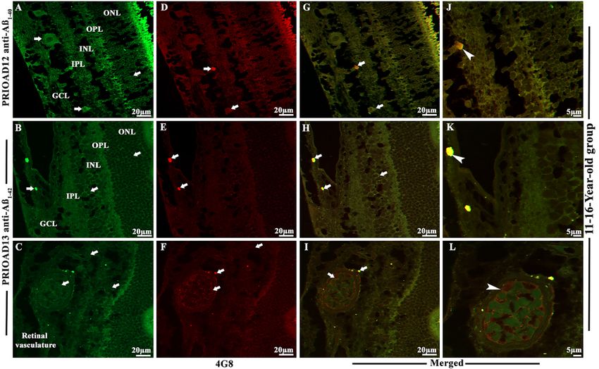
Page 12 of 21                                  Habiba et al. Ageing Neur Dis 2022;2:7       https://dx.doi.org/10.20517/and.2022.06

Figure 4. Immunofluorescence co-localization of retinal amyloid-beta oligomers and amyloid-beta plaques in dogs of 11-16-year-old
group. Retinal co-staining with anti-Aβ40 (PRIOAD 12) and anti-Aβ42 (PRIOAD 13) camelid-derived single domain antibody (green) and
4G8 antibody (red) of a 12-year-old German shepherd dog (A-L). A large number of (A) Aβ40 and (B) Aβ42 oligomers were found in the
GCL, INL, and ONL (white arrows, 40×). 4G8 positive Aβ plaque-like deposits were observed in the (D and E) GCL of the retina (white
arrows, 40×). Widespread co-localization was observed in the (G and H) retinal layers (white arrows, 40×). Co-localization of 4G8
positive Aβ plaque with (J) Aβ40 and (K) Aβ42 depositions (white arrowhead) showed with higher magnification (100×) in the GCL of
the same 12-year-old German shepherd dog retinal section. (C) Aβ oligomers and (F) 4G8 positive Aβ plaques were observed in the
retinal vasculature (white arrows). (I and L) Co-localization of Aβ oligomers and 4G8 positive Aβ plaques were exhibited with 40× and
also with higher 100× magnification in the retinal vessel wall, respectively (white arrowhead). The photomicrograph was derived from
the peripheral region of the retina - away from the optic disc. Representative of 10 dogs examined from elder age group (11-16 years).
GCL: ganglion cell layer; INL: inner nuclear layer; ONL: outer nuclear layer.

Despite the validation of the dog as a robust translational model for AD[24,30,69], only very limited studies
investigated AD-related changes in the young and neurologically intact dogs. One study by Stylianaki and
colleagues confirmed a similar pattern to that of humans; 61 dogs were subdivided into young (0-4 years
old), middle-aged (4-8 years old), aged cognitively normal (8-20 years old), and aged cognitively impaired
(8-17 years-old)[70]. The authors found that young dogs displayed the highest levels of total plasma Aβ42 and
Aβ42/Αβ40 ratio and the middle-aged dogs had the highest cerebrospinal fluid Aβ40 and Aβ42 when compared
to neurologically intact aged dogs. Our first step was to investigate the distribution and morphological
appearance of both Aβ40 and Aβ42 oligomers representing the canine lifetime, in the retinal layers of these
neurologically intact 30 dogs. Previous studies have shown that A11 anti-oligomer antibody binds to
different epitopes of the Aβo but also reacts with oligomeric aggregates of other proteins independent of
their primary sequences. Of note, A11 cannot differentiate between Aβ40 and Aβ42. In contrast, PRIOAD12
and PRIOAD13 nanobodies bind to Aβ40 and Aβ42, respectively. Previous studies reported an inverse
interrelationship between neurotoxicity and the size of Aβo, where the toxic effect of Aβo was shown to
decrease with increased size. The general molecular weight of Aβo was found to range between 10-100 kDa
in AD brain and included dimers to dodecamer[71]. A study by Lambert and colleagues first describes the
cytotoxic effect of the small diffusible Aβo on the hippocampal neurons[72]. The small Aβo referred to as Aβ-
derived diffusible ligands toxicity was tested in organotypic mouse brain slice cultures. The authors found
that 17 and 22 kDa small oligomers killed hippocampal neurons at nanomolar concentration[72]. In addition,
a study by Cizas and colleagues confirmed that oligomers larger than 30 KDa have a less toxic effect on the
Habiba et al. Ageing Neur Dis 2022;2:7      https://dx.doi.org/10.20517/and.2022.06                                    Page 13 of 21

Figure 5. Immunofluorescence co-localization of retinal amyloid-beta oligomers and hyperphosphorylated tau in dogs of 11-16-year-old
group. Retinal co-staining with anti-Aβ40 (PRIOAD 12) and anti-Aβ42 (PRIOAD 13) camelid-derived single domain antibody (green) and
AT8 antibody (red) of a 12-year-old German shepherd dog. A large number of (A) Aβ40 and (B) Aβ42 oligomers were found in the GCL,
INL, and ONL (white arrows, 40×). AT8 positive diffuse p-Tau-like deposits were observed in the (C and D) GCL, IPL, INL, and OPL of
the retina (white arrows, 40×). Widespread co-localization was observed in the (E and F) retinal layers (white arrows, 40×). Co-
localization of AT8 positive p-Tau with (G) Aβ40 and (H) Aβ42 depositions (white arrowhead) showed with higher magnification (100×)
in the GCL and OPL of the same 12-year-old German shepherd dog retinal section. The photomicrograph was derived from the
peripheral region of the retina - away from the optic disc. Representative of 10 dogs examined from elder age group (≥ 11 years). GCL:
Ganglion cell layer; IPL: inner plexiform layer; INL: inner nuclear layer; OPL: outer plexiform layer; ONL: outer nuclear layer.

Figure 6. Semiquantitative analysis of Aβ40 and Aβ42 oligomers, Aβ plaques, and p-Tau in neurologically intact young (1-5-years),
middle (6-10-years), and old (11-16-years) age groups of dogs. PRIOAD12 (Aβ40 oligomers), PRIOAD13 (Aβ42 oligomers), 4G8 (Aβp),
and AT8 (p-Tau) fluorescence immunoreactivity and intensities were examined and quantified at 40× magnification under the
fluorescence microscope. Semiquantitative analyses were compared with the three different age groups. A large amount of Aβ40 and
Aβ42 oligomers were found in young dogs, which decreased in middle age group, then finally, an upward trend was noticed in older
dogs. In comparison, Aβp were completely absent in young dogs, and then moderately found in middle, and large amounts in old age
groups. Finally, p-Tau deposits were not found in young and middle age groups of dogs, whereas old dogs exhibited a considerable
amount of p-Tau.
Page 14 of 21                                  Habiba et al. Ageing Neur Dis 2022;2:7      https://dx.doi.org/10.20517/and.2022.06

Figure 7. Influence of the size of dogs on the retinal deposition of amyloid-beta oligomers (Aβo), plaques (Aβp), and phosphorylated
tau (p-Tau) in cognitively unimpaired young (1-5-years), middle (6-10-years) and old (11-16-years) age groups of dogs. PRIOAD12
(Aβ40 oligomers), PRIOAD13 (Aβ42 oligomers), 4G8 (Aβp), and AT8 (p-Tau) fluorescence immunoreactivity and intensities were
examined and quantified at 40× magnification under the fluorescence microscope. In three different age groups, semiquantitative
analyses were compared with the size of the dogs. In young dogs, a large number of oligomers were displayed by the small and big size
dogs. The majority of medium-sized dogs displayed strong Aβo and Aβp staining intensity in the middle-aged dogs. Finally, among the
different sizes of old dogs, medium-sized breeds displayed the highest amount of Aβo, Aβp, and p-Tau staining intensity.

inhibition of long-term potentiation[73]. They also suggested the transition of sizes from small to large
correlate with the high to a low toxic effect of Aβo[73]. A11 specifically binds to the prefibrillar oligomers.
However, studies suggested that the specificity of A11 to AD-related oligomers might vary as it recognized
prefibrillar oligomers from various proteins that share a common structure including α-synuclein, islet
amyloid polypeptide, polyglutamine (PolyQ), lysozyme, and prion peptide[36]. A study by Glabe and
colleagues reported the ideal band size of prefibrillar oligomer-specific antibody A11 ranged from
approximately tetrameric up to ~75 kDa[35]. In our study, we confirmed the presence of an A11-specific
band at ~70KDa, confirming binding to prefibrillar oligomers in APP/PS1 mice. In comparison, camelid-
derived single-domain nanobodies bind to ~15KDa band representing the small oligomers. In agreement
with this current study, our previous report[55] also suggested that nanobodies were able to detect the toxic
small diffusible oligomers specific to AD, whereas A11 binds to the larger oligomers believed to be less toxic
to the neurons.

Studies suggested that in the canine brain, Aβo may be the toxic species responsible for cognitive decline
and can potentially be an early biomarker for the detection of CCD[23,74]. Naaman and colleagues recently
reported that Aβ42 oligomers cause extensive retinal neurotoxicity in rats when compared to fibrillary Aβ40
and Aβ42[75]. Our findings demonstrated extensive deposition of Aβ40 and Aβ42 oligomers in the retinal layers,
including GCL, INL, and ONL in 26/30 in the young, middle, and old age groups, except for a 1.4-year-old
male Siberian husky, a 7-year-old neutered male Hound mixed, an 8-year-old spayed female Bedlington
terrier, and an 11-year-old neutered male mixed breed. Moreover, we did not notice any difference in the
intensity of immunofluorescence (graded - to +++), the pattern of deposition, or morphology between Aβ40
and Aβ42 oligomers in all age groups. This presentation was unlikely influenced by the breed of the animals;
for instance, out of three Siberian Huskies, one dog failed to display Aβ40 and Aβ42 oligomers accumulation.
Remarkably, the younger age group, with an age range of 1-5 years, equivalent to humans aged 15-40 years
according to the American Kennel Club[76], has displayed extensive deposition of Aβ oligomers. Of interest,
a 2013 brain study investigated the presence of three different types of Aβ oligomers, including Aβ trimers,
Aβ*56, and Aβ dimers, in 75 cognitively unimpaired individuals aged 1 to 96 years[14]. Young children and
adolescents were positive for Aβ oligomers; the authors found an age-related accumulation of Aβ oligomers
where the level of the amyloid-β dimer was significantly higher in subjects in their 60s, and amyloid-β trimer
Habiba et al. Ageing Neur Dis 2022;2:7   https://dx.doi.org/10.20517/and.2022.06                   Page 15 of 21

in their 70s, whereas Aβ*56 level was significantly higher in individuals in their 40s. The investigators
proposed that Aβo, specifically Aβ*56, might trigger the pathological cascade in asymptomatic AD, a phase
that may be identifiable two decades before the clinical onset[14].

Having found that the eyes of 26/30 dogs contained Aβ40 and Aβ42 oligomers, our next step was to
demonstrate the presence/deposition of fibrillar Aβ in the canine retina of this same cohort. We show that
none of the young dogs displays 4G8-positive Aβ fibrils and plaques (Aβp). However, Aβp were observed in
some of the middle age group, where three dogs exhibited "low" staining intensity and two exhibited
"strong" staining intensity in the GCL, IPL & INL. Aβp were also observed in the old age group, where five
dogs showed "strong" signal intensity and two with "moderate" signal intensity in the GCL, IPL & INL. A
pattern, not surprising, emerges; Aβ40 and Aβ42 oligomers are deposited in the retina following birth and
subsequently, a fibrillar Aβ configuration, aggregated as plaques follow. Several studies have demonstrated
the presence of Aβp in the canine brain and suggested only a weak correlation with the severity of cognitive
dysfunction[77,78]. Schütt et al. investigated this question in further depth by studying dogs at differing ages,
specifically aged 9-15 years, including neurologically intact dogs and dogs with CCD, and compared their
amyloid burden with that of young dogs aged less than 6 years[69]. The authors reported that the levels of Aβ
deposition strongly correlated with the age of the dog but not to their cognitive capacity. Asking the same
questions regarding amyloid accretion in non-CNS neuronal populations, such as the retina, has not been
investigated in the canine, but some recent reports confirmed their presence in the retina of AD[50,54,79]. In
this study, we have demonstrated accumulation of retinal Aβp in the mature to an aged group of these 30
dogs which supports the hypothesis that Aβp might not influence the severity of cognitive deficits but can
be a predictor of AD development[11,17]. Overall, our study revealed that the Aβ deposition pattern in the
retina was such that Aβo was observed in all age groups, whereas Aβp accumulation was restricted to the
middle and more intensely the old age group of dogs, regardless of demographic criteria, breed and gender.
Interestingly, co-accumulation of both Aβo and Aβp were apparent in some middle and old dogs. This is in
agreement with our report this year, where 17-18 months old APP/PS1 mice showed high levels of
oligomers deposits in the retinal layers that co-localized with Aβp[55]. Previous neuropathological studies in
AD have shown that cerebral Aβo is usually present in early disease stages and is the most cytotoxic of Aβ
species, mostly responsible for neurotoxicity and synaptic dysfunction[34,80-83], whereas extracellular Aβp is
believed to accumulate at later ages and may act as a reservoir for Aβo[84]. Moreover, the involvement of Aβo
in retinal degeneration in AD has also been reported[85-87]. In the study reported herein, we noticed
widespread distribution of Aβo in the retinal layers of dogs of all age groups, indicating potential
involvement in retinal degeneration, perhaps leading to vision impairment in some dogs[88,89]. Of
importance, a previous study by Ozawa and colleagues that focused on web and paper-based surveys of dogs
aged ≥ 10 years to identify physical disturbances related to CCD showed that more than 90% of dogs
affected with CCD had vision impairment[90]. In addition to the neural retina, we observed Aβ deposition s
in the retinal microvasculature of young, middle, and old age groups, which might imitate CAA in AD[91-93].
Sharafi et al. suggested that retinal vasculature changes captured by hyperspectral imaging can differentiate
cerebral amyloid status between cognitively impaired and unimpaired individuals[94].
Central to AD pathogenesis is the intraneuronal deposition of hyperphosphorylated tau (p-Tau) in granular
form and eventual organization as NFTs, one of the cardinal neuropathological hallmarks[3,4]. In dogs with
CCD, p-Tau, unlike NFTs, has been identified in the brain, but until recently, only in some cases. We
speculate that, unlike human AD, p-Tau is only evident in pretangle granular form in dogs due to their
shorter lifespan (Tayebi & Habiba, unpublished observation). p-Tau neuropathology was shown to develop
about a decade before the formation of Aβp in AD brains and was hypothesized to trigger AD[95,96]. However,
Aβo accumulation was shown to precede and drive p-Tau accumulation and transneuronal spread across
synapses in the parietal cortex of AD[96].
You can also read