THIOL REDOX SWITCHES REGULATE THE OLIGOMERIC STATE OF CYANOBACTERIAL RRE1, RPAA AND RPAB RESPONSE REGULATORS
←
→
Page content transcription
If your browser does not render page correctly, please read the page content below
RESEARCH LETTER

Thiol redox switches regulate the oligomeric state of
cyanobacterial Rre1, RpaA and RpaB response regulators
Iskander M. Ibrahim1                 , Stephen J. L. Rowden2, William A. Cramer3, Christopher J. Howe2 and
Sujith Puthiyaveetil1
1 Department of Biochemistry and Center for Plant Biology, Purdue University, West Lafayette, IN, USA
2 Department of Biochemistry, University of Cambridge, UK
3 Department of Biological Sciences, Purdue University, West Lafayette, IN, USA

Correspondence                                       Cyanobacteria employ two-component sensor-response regulator systems to
I. M. Ibrahim, Department of Biochemistry            monitor and respond to environmental challenges. The response regulators
and Center for Plant Biology, Purdue
                                                     RpaA, RpaB, Rre1 and RppA are integral to circadian clock function and
University, West Lafayette, IN 47907, USA
                                                     abiotic stress acclimation in cyanobacteria. RpaA, RpaB and Rre1 are known
Tel: +1 765 494-8335
E-mail: Ibrahii@purdue.edu                           to interact with ferredoxin or thioredoxin, raising the possibility of their thiol
                                                     regulation. Here, we report that Synechocystis sp. PCC 6803 Rre1, RpaA
(Received 18 February 2022, revised 11               and RpaB exist as higher-order oligomers under oxidising conditions and that
March 2022, accepted 16 March 2022)                  reduced thioredoxin A converts them to monomers. We further show that
                                                     these response regulators contain redox-responsive cysteine residues with an
doi:10.1002/1873-3468.14340
                                                     Em7 around −300 mV. These findings suggest a direct thiol modulation of the
                                                     activity of these response regulators, independent of their cognate sensor
Edited by Peter Brzezinski
                                                     kinases.

                                                     Keywords: Hik2; RpaA; RpaB; Rre1; thiol regulation; TrxA

Two-component systems allow microorganisms to                                   1 (Rre1) and Regulator of Phycobilisome Association A
adapt rapidly to a wide range of environmental condi-                           and B (RpaA and RpaB) are conserved in all sequenced
tions through integrated regulation of gene expression.                         cyanobacteria. Homologues of Rre1 and RpaB are also
Consisting of a sensor histidine kinase and a response                          found in chloroplasts of certain non-green algae such as
regulator proteins, a two-component system employs                              Ycf29 and Ycf27, respectively [3,4].
transient phosphorylation and dephosphorylation cycles                             Most sensor kinases have just one cognate response
to convert an environmental stimulus into an appropri-                          regulator partner to which they transmit the environ-
ate physiological response. A typical cyanobacterial                            mental information through a phosphotransfer mecha-
genome encodes multiple two-component systems.                                  nism. Such two-component systems are usually found
These can vary from as few as five histidine kinases and                        as part of the same operon [5]. Some sensor kinases
six response regulators in the genome of Prochlorococ-                          and response regulators, on the other hand, are
cus MED4 to as many as 146 histidine kinases and 168                            encoded as orphan proteins. These two-component sig-
response regulators in the filamentous cyanobacterium                           nalling elements are frequently far more promiscuous
Nostoc punctiforme [1,2]. Of these, Response Regulator                          than their chromosomally paired counterparts [6].

Abbreviations
CSK, chloroplast sensor kinase; Em, midpoint potential; Em7, midpoint potential at pH 7; FTR, ferredoxin-thioredoxin reductase; H2O2, hydro-
gen peroxide; Hik, histidine kinase; hspA, heat shock protein A; LB, lysogeny broth; MBP, maltose-binding protein; NTR, NADPH-thioredoxin
reductase; PQ, plastoquinone; PSI, photosystem I; PSII, photosystem II; RpaA and RpaB, regulator of phycobilisome association A and B;
RppA, regulator of photosynthesis and photopigment-related gene expression A; Rre1, response regulator 1; SasA, Synechococcus adaptive
sensor A; Trx, thioredoxin.

FEBS Letters (2022) ª 2022 The Authors. FEBS Letters published by John Wiley & Sons Ltd on behalf of Federation of European Biochemical Societies   1
This is an open access article under the terms of the Creative Commons Attribution License, which permits use,
distribution and reproduction in any medium, provided the original work is properly cited.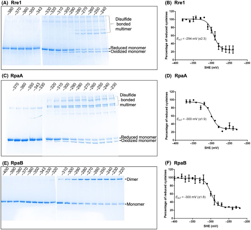
Thiol redox regulation of Rre1, RpaA, and RpaB                                                                                          I. Ibrahim et al.

Rre1 is an orphan response regulator and, as evidence                         The aspartate phosphorylation creates a high-energy
of its promiscuity, it has been found to work with two                        acyl phosphate in the response regulator receiver
sensor kinases – histidine kinases 2 and 34 (Hik2 and                         domain. This receiver domain modification results in
Hik34) [7,8]. Hik2, an orphan sensor kinase, likewise                         dimerisation or higher-order oligomerisation of the
interacts with, and competitively transfers phosphoryl                        response regulator, or, in some cases, it modifies the
groups to, not only Rre1 but also a second response                           interaction with the effector domain. This results in an
regulator called Regulator of Photosynthesis and                              appropriate output response – usually a change in gene
Photopigment-related gene expression A (RppA) [7,9].                          expression. A few response regulators have been found
Analysis of Synechocystis sp. PCC 6803 rre1 mutant                            to be regulated, independently of their cognate sensor
revealed that it is required for transcriptional control                      kinases, by modifications such as acetylation, serine/
of salt and heat tolerance genes [10,11]. Hik2 and its                        threonine phosphorylation or small molecule ligand
plant orthologue, Chloroplast Sensor Kinase (CSK),                            binding [26–28]. Indeed, Rre1, RpaA and RpaB have
were recently shown to sense the redox state of the                           been shown to interact with ferredoxin or thioredoxin
plastoquinone (PQ) pool via an iron-sulfur cluster [12].                      and in the case of RpaA and RpaB TrxA is able to
Furthermore, Arabidopsis CSK controls photosystem                             reduce them in vitro [23–25,29]. This raises the possi-
gene expression in a PQ redox-dependent manner [12–                           bility of direct thiol regulation of these response regu-
15]. Taken together, these observations support a role                        lators by the redox state of the photosynthetic electron
for Hik2-Rre1 in regulation of photosynthetic gene                            transport chain. In this study, we build on these obser-
expression. RppA also likely forms an additional phos-                        vations to understand the redox regulatory mechanism
photransfer pathway with the RppB sensor kinase                               of these response regulators in detail. We also included
found in the same operon. RppB is a nickel sensor his-                        RppA in our analysis given its interaction with Hik2
tidine kinase that regulates nickel responsive gene                           and its role in photosynthetic gene regulation.
expression in cyanobacteria [16]. In addition to its role
in nickel homeostasis, RppA has also been shown to                            Materials and methods
regulate photosynthetic gene transcription in response
to the redox state of the PQ pool [17], possibly via                          Construction of recombinant plasmids
Hik2 [7].
   RpaA and RpaB were first identified as specific reg-                       All clones used here are as described in [7]. Coding sequences
ulators of excitation energy transfer from the phycobil-                      for the full-length Rre1 (slr1783), RppA (sll0797), RpaA
isome antennae to reaction centres [3,18]. RpaA forms                         (sll0797), RpaB (slr0947), and TrxA (slr0623) were amplified
a two-component pair with the Synechococcus Adap-                             from Synechocystis sp. PCC 6803 genomic DNA using the
tive Sensor A (SasA) histidine kinase, which directly                         primer pairs listed in Table 1. PCR products were digested
interacts with the central circadian clock component                          with KpnI and XhoI endonucleases (New England BioLabs)
KaiC. RpaB forms a two-component pair with the his-                           and cloned into pETG-41A (EMBL) expression vector. The
                                                                              identities of the recombinant clones were confirmed by
tidine kinase 33 (Hik33, also known as NblS), and is
                                                                              sequencing (results not shown).
required for responses to multiple environmental con-
ditions including high light, oxidative stress and high
salinity. The circadian rhythm in cyanobacteria is gen-                       Expression and purification of recombinant
erated by a central oscillator composed of the KaiA,                          proteins
KaiB and KaiC proteins [19], which serves as an
                                                                              The recombinant plasmid constructs were introduced into
endogenous time-keeping system that allows cyanobac-
                                                                              BL21(DE3) chemically competent cells (Stratagene) by
teria to anticipate diurnal changes in environmental
                                                                              transformation. Colonies that grew on agar selection plates
conditions. Light entrains the circadian clock in
                                                                              were used to inoculate overnight starter cultures in 10 mL
cyanobacteria but not through photoreceptors [20,21].                         Lysogeny Broth (LB) supplemented with 100 µgmL−1
The light input is instead derived of the photosynthetic                      ampicillin. The overnight cultures were diluted to 1 : 100 in
electron transport chain [22]. Together, the SasA-                            l L LB medium and grown at 37 °C until an optical density
RpaA and Nbls-RpaB systems constitute the primary                             at 600 nm of 0.55 was reached. Recombinant protein over-
output components of the cyanobacterial circadian                             expression was initiated by adding IPTG at a final concen-
clock [23–25].                                                                tration of 0.5 mM. The cultures were subsequently grown at
   In a typical two-component system, the histidine                           16 °C for further 16 h. Cells were harvested by centrifuga-
kinase detects a signal and, upon activation and                              tion at 9000 × g for 10 min and the pellets were re-
autophosphorylation, transphosphorylates its cognate                          suspended in lysis buffer (20 mM Tris–HCl pH 8, 300 mM
response regulator on a conserved aspartate residue.                          NaCl, 25 mM imidazole and 1 mM PMSF) and lysed with an

2       FEBS Letters (2022) ª 2022 The Authors. FEBS Letters published by John Wiley & Sons Ltd on behalf of Federation of European Biochemical Societies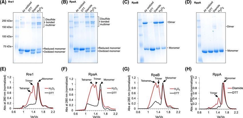
I. Ibrahim et al.                                                                                 Thiol redox regulation of Rre1, RpaA, and RpaB

Table 1. Primer pairs used for cloning Rre1, RpaA, RpaB, RppA
and TrxA. Sequences in lower case are restriction site overhangs                                                             ½DTTox
                                                                                                   Eh ¼ E0 þ 29:6 log
– Rre1F_MBP (cloned into pETG-41A)
                                                                                                                            ½DTTRed
  Forward: GCGCGCggtaccGTGGGCTTGAGTTTGCTG
                                                                                where [DTTox] and [DTTred] are molar concentrations of
  Reverse: GCGGCGctcgagCTAGACGATCGCCTCCAATTC
                                                                                oxidised and reduced DTT respectively and E0 is the stan-
– RppAF_MBP (cloned into pETG-41A)
                                                                                dard redox potential of DTT at pH 7.0 (−327 mV). The
  Forward: GCGCGCggtaccCGAATTTTGCTGGTGGAA
  Reverse: GCGGCGctcgagCTACAGTCTTGCTAATAGCTC                                    Em of the redox-responsive thiol group was calculated by
– RpaAF_MBP (cloned into pETG-41A)                                              fitting the data to the Nernst equation with the GRAPHPAD
  Forward GCGCggtaccATGCCTCGAATACTGATC                                          software.
  Reverse: GCGCGCctcgagCTACGTTGGACTACCGCC
– RpaBF_MBP (cloned into pETG-41A)
  Forward: GCGCGCggtaccGTGGTCGATGACGAGGCC                                       Size-exclusion chromatography
  Reverse: GCGGCGctcgagCTAGATTCTAGCTTCCAATTC
                                                                                To oxidize the recombinant proteins, 1.7 mg of Rre1,
– TrxA_MBP (cloned into pETG-41A)
                                                                                RpaA and RpaB were treated with 2 mM H2O2 and 1.7 mg
  Forward: GCGCGCggtaccAGTGCTACCCCTCAA
  Reverse: TGCGGCGctcgagAAGATATTTTTCTAGGGT
                                                                                of RppA with 2 mM diamide. The oxidant was removed by
                                                                                desalting with a MiniTrap G25 column. As previously
                                                                                stated, 2 mM DTT was used for reducing all target pro-
                                                                                teins. The oligomeric states of the oxidised and reduced
                                                                                proteins were determined with a Superdex S200 10/300GL
EmulsiFlex-C3 homogeniser (Avestin). The lysate was sepa-
                                                                                Increase (GE Healthcare Life Sciences) size exclusion chro-
rated by centrifugation at 39 000 × g for 20 min at 4 °C. The
                                                                                matography of 500 µL (0.85 mg) protein. The column was
supernatant was applied to a Ni2+ affinity column (Cytiva,
                                                                                equilibrated with a buffer containing 100 mM HEPES (pH
Uppsala, Sweden) and purified according to the manufac-
                                                                                8.0) and 100 mM NaCl for Rre1, RpaA and RppA or with
turer’s instructions. The purified proteins were buffer
                                                                                5% (v/v) glycerol, 300 mM NaCl and 100 mM HEPES (pH
exchanged with a PD-10 column into a buffer medium con-
                                                                                8.0) for RpaB. For reduced proteins, the column was equi-
taining 0.1 mM NaCl and 0.1 mM HEPES (pH 8.0).
                                                                                librated with the same buffer but with 2 mM DTT added.
                                                                                The molecular weight was determined by a calibration
Redox treatment                                                                 curve generated using the following standard proteins of
                                                                                known molecular weight: thyroglobulin (669 kDa), apofer-
Aliquots of 2.5 µM desalted full-length recombinant Rre1,                       ritin (443 kDa), β-amylase (200 kDa), bovine serum albu-
RpaA, RpaB and RppA proteins were incubated with a                              min (66 kDa) and carbonic anhydrase (29 kDa). Blue
final concentration of 2 mM cysteine-specific oxidant                           dextran (2000 kDa) was used to determine the void volume
diamide, H2O2 or with the reductant DTT for 30 min at                           (Vo).
room temperature. The reaction products were immediately
resolved by non-reducing sodium dodecyl sulfate–6 M urea–
8% (w/v) polyacrylamide gel electrophoresis.                                    Results

                                                                                Rre1, RpaA, RpaB and RppA contain conserved
Redox titration
                                                                                cysteine residues as putative redox regulation
The purified proteins were buffer exchanged with a PD-10                        target sites
column into a redox titration buffer (100 mM NaCl,
                                                                                To examine whether these response regulators contain
100 mM HEPES, pH 7.0). Aliquots of 2.5 µM Rre1, RpaA
or RpaB proteins were equilibrated for 2 h at redox poten-
                                                                                conserved cysteine residues that could function as thiol
tials ranging from −220 mV to −400 mV in a redox titra-                         modification sites, we aligned the amino acid sequence
tion buffer at 22 °C. The different redox potentials were                       of Rre1, RpaA, RpaB and RppA. Fig. 1 and Fig. S1-S4
achieved by mixing different ratios of oxidised and reduced                     show the presence of one or more conserved cysteine
DTT at a final concentration of 2 mM. The redox-titrated                        residues in all four response regulators. Cysteine resi-
proteins were resolved on a non-reducing SDS/urea/PAGE                          dues are found in the receiver domain, which carries
gel as before. The oxidised and reduced proteins migrated                       the aspartate phosphorylation site (D1 motif), as well
differently on the gel and their band intensities, as quanti-                   as in the DNA-binding domain. From the multiple
fied by the Image Lab software (Bio-Rad, Hercules, CA,                          sequence alignment of each protein orthologues (Fig.
USA), were used as a reporter for the redox titration. The                      S1-S4), it is apparent that the Synechocystis Rre1 con-
equilibrium redox potential (Eh) was calculated by the                          tains two cysteine residues within its receiver domain.
Nernst equation for a 2 electron redox reaction:                                Of these, cysteine 153 is highly conserved in other

FEBS Letters (2022) ª 2022 The Authors. FEBS Letters published by John Wiley & Sons Ltd on behalf of Federation of European Biochemical Societies   3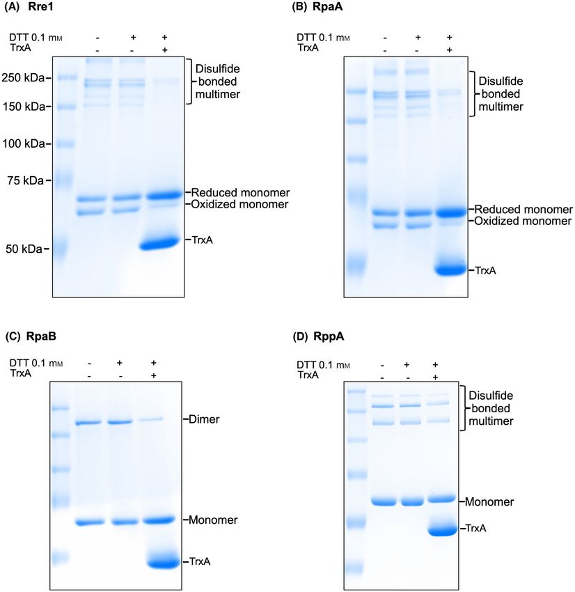
Thiol redox regulation of Rre1, RpaA, and RpaB I. Ibrahim et al.
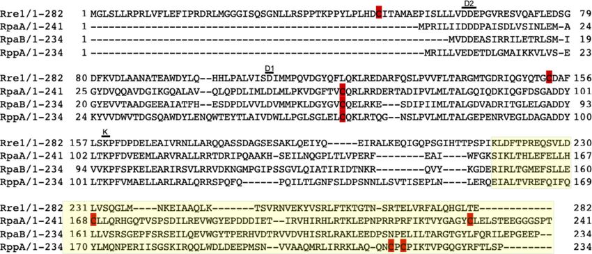
Fig. 1. Sequence alignment of full-length Synechocystis sp. PCC 6803 Rre1, RpaA, RpaB, and RppA response regulators. Putative redox-
active cysteines are shown in red. The divalent cation Mg2+-binding motif “D2”, phosphorylation site “D1”, and the lysine (K) motifs are
shown. The receiver domain is shaded in white, and the DNA binding domain in yellow.
cyanobacterial Rre1 sequences while the other cysteine arising from multiple intermolecular disulfide bonds
is poorly conserved (Fig. S1). Synechocystis RpaA, between three or four monomers (Fig. 2A,B). The
RpaB and RppA contain a cysteine residue within addition of 2 mM DTT to the air-oxidised samples
their receiver domains and RpaA and RppA contain resulted in monomerisation of all proteins (Fig. 2A,B).
two additional cysteines within their DNA-binding RpaB appears to dimerise via an intermolecular disul-
domains (Fig. 1). All three cysteines in RpaA are fide bond after being oxidised by the air or after treat-
almost completely conserved (Fig. S2), and the single ment with diamide or H2O2 (Fig. 2C), and the dimer
cysteine in RpaB is fully conserved (Fig. S3) as noted form can be converted to monomers after treatment of
earlier [29]. For RppA, only the receiver domain cys- the air-oxidised protein with 2 mM DTT (Fig. 2C). In
teine 65 is fully conserved (Fig. S4). contrast to these three response regulators, the air-
oxidised RppA protein migrated as a single monomer
band. However, after treatment with diamide, two
Redox conditions modulate the oligomeric state
additional bands, corresponding to higher-order oligo-
of Rre1, RpaA, RpaB and RppA
mers, were apparent (Fig. 2D). To check whether the
Having identified putative redox-responsive cysteine(s) effect of diamide could be reversed, the diamide-
in Rre1, RpaA, RpaB and RppA (Fig. 1), we exam- oxidised protein was first desalted to remove any resid-
ined whether redox treatments have any effect on the ual diamide present. The desalted protein was subse-
protein conformation or oligomeric state of these quently treated with 2 mM DTT. DTT treatment
response regulators. Full-length recombinant response indeed converts the higher-order oligomers into mono-
regulator proteins were incubated with 2 mM cysteine- mers, supporting the role of cysteines in the redox con-
specific oxidant diamide, or 2 mM H2O2, or 2 mM thiol trol of RppA oligomeric state (Fig. 2D).
reductant DTT and the redox-treated proteins were Size exclusion chromatography was used to probe
then resolved on an 8% non-reducing urea–SDS/ further the oligomeric states of these response regula-
PAGE gel. In the air-exposed untreated and diamide-, tors in their native state under oxidising (H2O2) and
or H2O2-treated Rre1 and RpaA samples, an oxidised reducing (DTT) conditions. Because the behaviour of
fast-migrating monomer protein band was present. proteins within the size exclusion gel matrix is more
This more compact form of the monomer presumably closely related to their hydrodynamic radius (Stokes
arose from an intramolecular disulfide bond and on radius, Rs) than their molecular weight, obtaining an
the gel it migrates slightly below the reduced monomer accurate molecular weight through this technique is
protein. Several less intense bands above 180 kDa were difficult. For example, proteins with elongated shape
also observed, with the higher-order oligomers likely might elute at a position that is significantly different
4 FEBS Letters (2022) ª 2022 The Authors. FEBS Letters published by John Wiley & Sons Ltd on behalf of Federation of European Biochemical SocietiesI. Ibrahim et al. Thiol redox regulation of Rre1, RpaA, and RpaB
Fig. 2. Effect of redox environment on the oligomeric state of Rre1, RpaA, RpaB, and RppA. (A–D) redox-treated proteins as resolved by
urea-SDS/PAGE. (E–H) oligomeric state of response regulators as determined by size exclusion chromatography. Panels (E–H) correspond to
samples oxidized with 2 mM H2O2 or reduced with 2 mM DTT, respectively.
from spherical proteins with the same molecular 70 kDa) eluted with apparent molecular weights of 84
weight. Size exclusion chromatography can neverthe- and 184 kDa, corresponding to monomer and trimer,
less provide an approximate molecular weight. To respectively (Fig. 2H). All four proteins eluted as a
that end, we used the standard proteins listed in the monomer in their reduced DTT-treated forms (Fig. 2E-
Materials and Methods and Fig. S5 to calculate the H). The results obtained with size exclusion chromatog-
approximate molecular weight of response regulators raphy thus corroborate the oligomerisation patterns
selected in this study. The oxidised form of Rre1 was seen in non-reducing urea–SDS/PAGE (Fig. 2A-D).
eluted with apparent molecular weights of 104, 210
and 297 kDa on a Superdex S200 column. The theo-
Midpoint potentials of redox-active cysteines in
retical molecular weight of a monomer of Rre1
Rre1, RpaA and RpaB suggest a regulatory role
tagged with MBP is 75 kDa. Thus, the eluted proteins
likely correspond to monomer, trimer and tetramer of Our results in Fig. 2 suggest that the response regula-
Rre1, respectively (Fig. 2E). Similarly, the oxidised tors studied here contain redox-active disulfide link-
RpaA (with a theoretical molecular weight of ages. Disulfide bonds might nevertheless have a
71 kDa) and RpaB (with a theoretical molecular structural rather than a regulatory role in these pro-
weight of 70 kDa) eluted as a monomer, trimer and teins. Disulfide bonds with structural roles, however,
tetramer with an apparent molecular weight of 103, typically have a very negative midpoint potential as
211 and 304 kDa for RpaA (Fig. 2F), and 109, 213 low as −470 mV [26] while cysteines with regulatory
and 306 kDa for RpaB (Fig. 2G). Interestingly, size roles have a higher midpoint potential, ranging from
exclusion chromatography does not show any RpaB −95 to −330 mV [27,28,30–32]. We therefore carried
dimers in contrast to the non-reducing gel (Fig. 2C). out a redox titration of the purified response regula-
It is likely that the disulfide bonded-dimers of RpaB, tors to determine their midpoint potentials. The results
resolved in the protein gel (Fig. 2C), form tetramers of redox titration for Rre1 (Fig. 3A,B), RpaA
(dimer of dimers) or other oligomers via noncysteine- (Fig. 3C,D) and RpaB (Fig. 3E,F) at pH 7 are shown.
mediated interactions. Such oligomers will only be Duplicate titrations gave an average Em value at pH 7
apparent in size exclusion chromatography but not in of −294 mV ( 2.5 mV) for Rre1, −300 mV ( 2 mV)
denaturing protein gel electrophoresis (Fig. 2C). The for RpaA, and −300 mV ( 1.8 mV) for RpaB. Since
RppA (with a theoretical molecular weight of all three Em7 values are higher than −330 mV, the
FEBS Letters (2022) ª 2022 The Authors. FEBS Letters published by John Wiley & Sons Ltd on behalf of Federation of European Biochemical Societies 5Thiol redox regulation of Rre1, RpaA, and RpaB I. Ibrahim et al.
Fig. 3. Redox titration of Rre1, RpaA, and RpaB. (A,C & E) Redox-titrated proteins separated by nonreducing urea-SDS/PAGE and stained
with Coomassie brilliant blue. (B,D & F) Plots of redox titration. From the intensity of the reduced and oxidized band species, the midpoint
potential Em was computed by fitting the data to the Nernst equation. SHE, standard hydrogen electrode potential. Error bars represent
SE from two replicate titrations.
lowest potential reported for a regulatory disulfide are consistent with regulatory thiols, we checked
[27,28,30–32], it is likely that the redox-active cysteines whether those cysteines are targets of the thioredoxin
of Rre1, RpaA and RpaB play a regulatory role. We did system. TrxA has been shown to interact with RpaA
not perform a titration for RppA because only a small and RpaB and reduce them in vitro [29]. Although
fraction of this protein could be oxidised with diamide. there is no direct evidence for Rre1 interaction with
thioredoxin, its redox potential (Fig. 3B) suggests that
it could also be a target of TrxA. Rre1 and RpaA pro-
TrxA-dependent reduction of Rre1, RpaA and
teins purified from E. coli mostly exist as a low molec-
RpaB
ular weight reduced and oxidised monomers with a
After establishing that Rre1, RpaA and RpaB contain small amount of oxidised higher-order oligomers
redox-active cysteines and have redox potentials that (Fig 4A,B). RpaB, as purified from bacteria, migrates
6 FEBS Letters (2022) ª 2022 The Authors. FEBS Letters published by John Wiley & Sons Ltd on behalf of Federation of European Biochemical SocietiesI. Ibrahim et al. Thiol redox regulation of Rre1, RpaA, and RpaB Fig. 4. TrxA-mediated reduction of the redox-active cysteines of Rre1, RpaA, RpaB, and RppA. TrxA-treated proteins as separated on a nonreducing urea-SDS/PAGE gel and stained with Coomassie. + or − sign indicates the presence or absence of 0.1 mM DTT or 5 mM TrxA, respectively. as a reduced monomer and an oxidised dimer (Fig. 4 RpaB, from dimer to monomer (Fig. 4A-C). These C). Treatment of all three recombinant proteins with results suggest that the redox-active cysteines of Rre1, 0.1 mM DTT had little effect on their oligomeric state. RpaA and RpaB could indeed be targets of TrxA reg- However, incubation of Rre1 and RpaA with 5 mM ulation in vivo. Treatment of RppA with 0.1 mM DTT TrxA in the presence of 0.1 mM DTT resulted in the and TrxA had only a small effect on its aggregation conversion of the oxidised higher-order oligomers or state (Fig. 4D), indicating that it is unlikely to be a oxidised monomers into a reduced monomer; for physiological TrxA substrate. FEBS Letters (2022) ª 2022 The Authors. FEBS Letters published by John Wiley & Sons Ltd on behalf of Federation of European Biochemical Societies 7
Thiol redox regulation of Rre1, RpaA, and RpaB                                                                                          I. Ibrahim et al.

Discussion                                                                    phosphorylation induces conformational changes in
                                                                              the α4-β5-α5 face of the receiver domain. This pro-
The Synechocystis genome contains six thioredoxin                             motes dimerisation of the response regulators via their
genes. These thioredoxin proteins are likely reduced by                       receiver domains. Phosphorylation-mediated dimerisa-
two separate enzymes: ferredoxin-thioredoxin reduc-                           tion thus enhances DNA-binding activity and ulti-
tase (FTR) and NADPH-thioredoxin reductase                                    mately the extent of transcriptional regulation by
(NTR), which receive reducing equivalents from ferre-                         response regulators [34]. An intriguing possibility is
doxin and NADPH, respectively. Through the thiol-                             that thiol modification represents an independent or
disulfide interconversion of target proteins, the thiore-                     synergistic (with phosphorylation) mechanism that
doxin system connects the redox state of photosyn-                            promotes dimerisation or oligomerisation of response
thetic electron transport with metabolic regulation of                        regulators. Our analyses of Rre1, RpaA, RpaB and
both light and dark reactions of photosynthesis. Light                        RppA by non-reducing gel electrophoresis and size
plays a critical role in fine-tuning gene expression in                       exclusion chromatography are consistent with this
cyanobacteria. High light treatment or moving the                             possibility. The DTT-reduced proteins eluted as mono-
cyanobacterial cells to darkness promotes the down-                           mers in size exclusion chromatography (Fig. 2E-H),
regulation of several photosystem genes, most notably                         which is consistent with results obtained by non-
genes encoding photosystem I (PSI) and phycobilisome                          reducing urea–SDS/PAGE (Fig. 2A-D). Under oxidis-
subunits [33]. Genes encoding photosystem II (PSII)                           ing condition of air, all proteins were converted into
components respond in the opposite direction as they                          higher-order oligomers. Expectedly, stronger oxidising
become upregulated in high light conditions. Interest-                        agents such as H2O2 or diamide enhanced oligomerisa-
ingly, the expression profiles of FTR, trxA and trxB                          tion (Fig. 2). It is possible that the disulfide-mediated
genes mimic that of PSI and phycobilisome genes.                              oligomerisation of these regulators is sufficient for
Notably, these three genes are further shown to be reg-                       enhancing their DNA-binding activity, or the thiol
ulated by the redox state of the PQ pool [33].                                modification likely acts synergistic with phosphoryla-
   Given previous reports of interaction between the                          tion. Whether the more compact internally disulfide-
ferredoxin-thioredoxin system and Rre1, RpaA and                              bonded Rre1 and RpaA monomers are functionally
RpaB, the current study sought to characterise                                relevant also remains to be determined (Fig. 2A,B).
whether these response regulators indeed contain                              Rre1 has recently been shown to be phosphorylated in
thioredoxin-targeted thiol-disulfide redox switches for                       vivo under reduced PQ pool condition during thermal
the regulation of their oligomeric state and activity.                        stress, possibly via its cognate sensor kinase Hik2.
All four response regulators contain at least one highly                      Phosphorylation of Rre1 leads to heat shock protein
conserved cysteine residue within their receiver domain                       A (hspA) gene transcript accumulation under the same
where phosphorylation takes place (Fig. 1, Fig. S1-S4).                       condition [11]. Interestingly, Bairagi et al. [11] also
This raises two interesting but not mutually exclusive                        find some hspA transcript accumulation even in the
possibilities: the redox-responsive cysteines might con-                      absence of Rre1 phosphorylation, suggesting a
trol the accessibility of the key aspartate phosphoryla-                      phosphorylation-independent regulation of the hspA
tion site for the sensor kinase and or the oligomeric                         gene. The oxidation-induced oligomerisation that we
state of the response regulators. Under oxidising con-                        report here might represent such a phosphorylation-
ditions, Rre1 and RpaA proteins formed an                                     independent pathway for regulation of Rre1 activity.
intramolecular disulfide-bonded monomer and inter-                               What is the nature of the in vivo redox signals that
molecular disulfide-bonded higher-order oligomers.                            likely act upon the thiol redox switches of Rre1, RpaA
RpaB under oxidising conditions formed a dimer via                            and RpaB? Redox regulation of transcription factors
an intermolecular disulfide bond as revealed by the                           is a widely recognised fundamental regulatory mecha-
non-reducing urea–SDS/PAGE (Fig. 2A-C). The oxi-                              nism for gene expression. The Fd-Trx system plays a
dised RppA had two high molecular weight bands,                               crucial role in this process. RpaA, RpaB and Rre1
corresponding to a trimer and probably another                                were shown to interact with TrxA [29,35] or Fd [36].
higher-order oligomer (Fig. 2D).                                              The RpaA–Fd interaction was found to be strongest
   Although response regulators usually have a single                         under reducing conditions, indicating that the interac-
DNA-binding domain, the active state of many                                  tion occurs in light and might serve as a diurnal input
response regulators is dimeric. This allows them to                           to downregulate RpaA transcriptional activity. The
recognise tandem or inverted DNA repeat elements for                          midpoint potentials of redox-active cysteines of Rre1,
transcriptional regulation. For FixJ, Spo0A, and                              RpaA and RpaB at pH 7 are −294, −300, and
OmpR/PhoB subfamily of response regulators,                                   −300 mV, respectively (Fig. 3). Cyanobacterial and

8       FEBS Letters (2022) ª 2022 The Authors. FEBS Letters published by John Wiley & Sons Ltd on behalf of Federation of European Biochemical Societies
I. Ibrahim et al. Thiol redox regulation of Rre1, RpaA, and RpaB
plant thioredoxins have an Em7 value of around References
−300 mV [37,38]. This redox potential of thioredoxin
1 Ashby MK, Houmard J. Cyanobacterial two-
is closer to the Em of Rre1, RpaA and RpaB (Fig. 3),
component proteins: structure, diversity, distribution,
making their TrxA-mediated reduction thermodynami-
and evolution. Microbiol Mol Biol Rev. 2006;70:
cally favourable. Indeed, in this study, and in [29]
472–509.
TrxA was able to reduce the oxidised cysteines of differ-
2 Mary I, Vaulot D. Two-component systems in
ent response regulators (Fig. 4). Thus, m-type thiore- Prochlorococcus MED4: genomic analysis and
doxin might thus downregulate the activity of these differential expression under stress. FEMS Microbiol
response regulators in vivo. An additional physiologi- Lett. 2003;226:135–44.
cally relevant activating redox signal is likely to be H2O2 3 Ashby MK, Mullineaux CW. Cyanobacterial ycf27 gene
as growth of cyanobacterial cells in ambient air at 40 °C products regulate energy transfer from phycobilisomes
induces oxidative stress in the form of H2O2 [39]. The to photosystems I and II. FEMS Microbiol Lett.
elevated H2O2 at high temperature could be an impor- 1999;181:253–60.
tant regulatory signal that mediates the oligomerisation 4 Puthiyaveetil S, Allen JF. Chloroplast two-component
of Rre1, RpaA and RpaB. Reducing conditions via the systems: evolution of the link between photosynthesis
Trx system might reverse the effect of H2O2. Taken and gene expression. Proc Biol Sci. 2009;276:2133–45.
together, our results suggest that thiol redox regulation 5 Laub MT, Goulian M. Specificity in two-component
is likely to be an important mechanism in the regulation signal transduction pathways. Annu Rev Genet.
of the activity of these cyanobacterial response regula- 2007;41:121–45.
tors independently of, or synergistic with, their cognate 6 Burger L, van Nimwegen E. Accurate prediction of
sensor kinase-mediated transphosphorylation. protein-protein interactions from sequence alignments
using a Bayesian method. Mol Syst Biol. 2008;4:165.
7 Ibrahim IM, Puthiyaveetil S, Allen JF. A two-
Acknowledgements component regulatory system in transcriptional control
of photosystem stoichiometry: redox-dependent and
The authors thank Gilbert E. Kayanja and Steven D.
sodium ion-dependent phosphoryl transfer from
McKenzie for discussions. SP thanks United States
cyanobacterial histidine kinase Hik2 to response
Department of Energy (DOE) for a research grant
regulators Rre1 and RppA. Front Plant Sci. 2016;7:137.
(DE-SC0020639). WAC thanks the U.S. Department
8 Paithoonrangsarid K, Shoumskaya MA, Kanesaki Y,
of Energy (DOE DE-SC0018238) and the Henry Kof-
Satoh S, Tabata S, Los DA, et al. Five histidine kinases
fler Professorship. SJLR and CJH thank the Biotech- perceive osmotic stress and regulate distinct sets of
nology and Biological Sciences Research Council genes in synechocystis. J Biol Chem. 2004;279:53078–86.
[grant numbers BB/L014130/1 and BB/T010525/1], the 9 Sato S, Shimoda Y, Muraki A, Kohara M, Nakamura
Engineering and Physical Sciences Research Council Y, Tabata S. A large-scale protein protein interaction
[grant number EP/F047940/1], and Spicer Consulting analysis in Synechocystis sp. PCC6803. DNA Res.
Ltd for financial support. 2007;14:207–16.
10 Kobayashi I, Watanabe S, Kanesaki Y, Shimada T,
Yoshikawa H, Tanaka K. Conserved two-component
Conflict of interest
Hik34-Rre1 module directly activates heat-stress
The authors declare no conflict of interest. inducible transcription of major chaperone and other
genes in Synechococcus elongatus PCC 7942. Mol
Microbiol. 2017;104:260–77.
Author contributions 11 Bairagi N, Watanabe S, Nimura-Matsune K, Tanaka K,
Tsurumaki T, Nakanishi S, et al. Conserved two-
IMI, SJLR, WAC, CJH and SP conceived, conceptu-
component Hik2-Rre1 signaling is activated under
alised and wrote the manuscript. IMI curated data
temperature upshift and plastoquinone-reducing
and devised methodology. All authors have read and
conditions in the cyanobacterium Synechococcus
agreed to the published version of the manuscript.
elongatus PCC 7942. Plant Cell Physiol. 2022;63:176–88.
12 Ibrahim IM, Wu H, Ezhov R, Kayanja GE, Zakharov
Data accessibility SD, Du Y, et al. An evolutionarily conserved iron-
sulfur cluster underlies redox sensory function of the
The dataset generated and analysed in the current Chloroplast Sensor Kinase. Commun Biol. 2020;3:13.
study is available as Supplementary Data S1. All other 13 Puthiyaveetil S, Kavanagh TA, Cain P, Sullivan JA,
data (if any) are available upon request. Newell CA, Gray JC, et al. The ancestral symbiont
FEBS Letters (2022) ª 2022 The Authors. FEBS Letters published by John Wiley & Sons Ltd on behalf of Federation of European Biochemical Societies 9Thiol redox regulation of Rre1, RpaA, and RpaB I. Ibrahim et al.
sensor kinase CSK links photosynthesis with gene 26 Gilbert HF. Molecular and cellular aspects of thiol-
expression in chloroplasts. Proc Natl Acad Sci USA. disulfide exchange. Adv Enzymol Relat Areas Mol Biol.
2008;105:10061–6. 1990;63:69–172.
14 Puthiyaveetil S, Ibrahim IM, Jelicic B, Tomasic A, 27 Wunderlich M, Jaenicke R, Glockshuber R. The redox
Fulgosi H, Allen JF. Transcriptional control of properties of protein disulfide isomerase (DsbA) of
photosynthesis genes: the evolutionarily conserved Escherichia coli result from a tense conformation of its
regulatory mechanism in plastid genome function. oxidized form. J Mol Biol. 1993;233:559–66.
Genome Biol Evol. 2010;2:888–96. 28 Lin TY, Kim PS. Urea dependence of thiol-disulfide
15 Puthiyaveetil S, Ibrahim IM, Allen JF. Evolutionary equilibria in thioredoxin: confirmation of the linkage
rewiring: a modified prokaryotic gene-regulatory relationship and a sensitive assay for structure.
pathway in chloroplasts. Philos Trans R Soc Lond B Biochemistry. 1989;28:5282–7.
Biol Sci. 2013;368:20120260. 29 Kadowaki T, Nishiyama Y, Hisabori T, Hihara Y.
16 López-Maury L, Garcı́a-Domı́nguez M, Florencio FJ, Identification of OmpR-family response regulators
Reyes JC. A two-component signal transduction system interacting with thioredoxin in the cyanobacterium
involved in nickel sensing in the cyanobacterium Synechocystis sp. PCC 6803. PLoS One. 2015;10:e0119107.
Synechocystis sp. PCC 6803. Mol Microbiol. 30 Krause G, Holmgren A. Substitution of the conserved
2002;43:247–56. tryptophan 31 in Escherichia coli thioredoxin by site-
17 Li H, Sherman LA. A redox-responsive regulator of directed mutagenesis and structure-function analysis. J
photosynthesis gene expression in the cyanobacterium Biol Chem. 1991;266:4056–66.
Synechocystis sp. Strain PCC 6803. J Bacteriol. 31 Huber-Wunderlich M, Glockshuber R. A single
2000;182:4268–77. dipeptide sequence modulates the redox properties of a
18 Ashby MK, Houmard J, Mullineaux CW. The ycf27 whole enzyme family. Fold Des. 1998;3:161–71.
genes from cyanobacteria and eukaryotic algae: 32 Wouters MA, Fan SW, Haworth NL. Disulfides as redox
distribution and implications for chloroplast evolution. switches: from molecular mechanisms to functional
FEMS Microbiol Lett. 2002;214:25–30. significance. Antioxid Redox Signal. 2010;12:53–91.
19 Ishiura M, Kutsuna S, Aoki S, Iwasaki H, Andersson 33 Perez-Perez ME, Martin-Figueroa E, Florencio FJ.
CR, Tanabe A, et al. Expression of a gene cluster Photosynthetic regulation of the cyanobacterium
kaiABC as a circadian feedback process in Synechocystis sp. PCC 6803 thioredoxin system and
cyanobacteria. Science. 1998;281:1519–23. functional analysis of TrxB (Trx x) and TrxQ (Trx y)
20 Rust MJ, Golden SS, O’Shea EK. Light-driven changes thioredoxins. Mol Plant. 2009;2:270–83.
in energy metabolism directly entrain the cyanobacterial 34 Toro-Roman A, Mack TR, Stock AM. Structural
circadian oscillator. Science. 2011;331:220–3. analysis and solution studies of the activated regulatory
21 Diamond S, Rubin BE, Shultzaberger RK, Chen Y, domain of the response regulator ArcA: a symmetric
Barber CD, Golden SS. Redox crisis underlies dimer mediated by the alpha4-beta5-alpha5 face. J Mol
conditional light-dark lethality in cyanobacterial Biol. 2005;349:11–26.
mutants that lack the circadian regulator, RpaA. Proc 35 Mata-Cabana A, Florencio FJ, Lindahl M. Membrane
Natl Acad Sci USA. 2017;114:E580–9. proteins from the cyanobacterium Synechocystis sp.
22 Mackey SR, Golden SS, Ditty JL. The itty-bitty time PCC 6803 interacting with thioredoxin. Proteomics.
machine genetics of the cyanobacterial circadian clock. 2007;7:3953–63.
Adv Genet. 2011;74:13–53. 36 Hanke GT, Satomi Y, Shinmura K, Takao T, Hase T.
23 Takai N, Nakajima M, Oyama T, Kito R, Sugita C, A screen for potential ferredoxin electron transfer
Sugita M, et al. A KaiC-associating SasA-RpaA two- partners uncovers new, redox dependent interactions.
component regulatory system as a major circadian Biochim Biophys Acta. 2011;1814:366–74.
timing mediator in cyanobacteria. Proc Natl Acad Sci 37 Mihara S, Sugiura K, Yoshida K, Hisabori T.
USA. 2006;103:12109–14. Thioredoxin targets are regulated in heterocysts of
24 Iwasaki H, Williams SB, Kitayama Y, Ishiura M, cyanobacterium Anabaena sp. PCC 7120 in a light-
Golden SS, Kondo T. A kaiC-interacting sensory independent manner. J Exp Bot. 2020;71:2018–27.
histidine kinase, SasA, necessary to sustain robust 38 Yoshida K, Hara S, Hisabori T. Thioredoxin selectivity
circadian oscillation in cyanobacteria. Cell. for thiol-based redox regulation of target proteins in
2000;101:223–33. chloroplasts. J Biol Chem. 2015;290:19540.
25 Espinosa J, Boyd JS, Cantos R, Salinas P, Golden SS, 39 Hasegawa H, Tsurumaki T, Imamura S, Sonoike K,
Contreras A. Cross-talk and regulatory interactions Tanaka K. The circadian rhythm regulator RpaA
between the essential response regulator RpaB and modulates photosynthetic electron transport and alters
cyanobacterial circadian clock output. Proc Natl Acad the preferable temperature range for growth in a
Sci USA. 2015;112:2198–203. cyanobacterium. FEBS Lett. 2021;595:1480–92.
10 FEBS Letters (2022) ª 2022 The Authors. FEBS Letters published by John Wiley & Sons Ltd on behalf of Federation of European Biochemical SocietiesI. Ibrahim et al. Thiol redox regulation of Rre1, RpaA, and RpaB
Supporting information Fig. S2. Conserved cysteines of RpaA.
Fig. S3. Conserved cysteine of RpaB.
Additional supporting information may be found Fig. S4. Conserved cysteines of RppA.
online in the Supporting Information section at the end Fig. S5. Protein standard curve.
of the article. Data S1. Datasets used to plot Fig. 2E,F and Fig. 3B,
Fig. S1. Conserved cysteine of Rre1. D,F.
FEBS Letters (2022) ª 2022 The Authors. FEBS Letters published by John Wiley & Sons Ltd on behalf of Federation of European Biochemical Societies 11You can also read