Therapeutic Ultrasound in Cardiovascular Medicine - the ...
←
→
Page content transcription
If your browser does not render page correctly, please read the page content below
REVIEW ARTICLE

Therapeutic Ultrasound in
Cardiovascular Medicine
Olivia C. Coiado, PhD , Jacques Lowe, BS, William D. O’Brien Jr, PhD

                                                   An advantage of therapeutic ultrasound (US) is the ability to cause controlled
                                                   biological effects noninvasively. Depending on the magnitude and frequency of
                                                   exposure parameters, US can interact in different ways with a variety of biologi-
                                                   cal tissues. The development and clinical utility of therapeutic US techniques are
                                                   now rapidly growing, especially with regard to the application of US pulses for
                                                   cardiac pacing and the potential treatment of cardiovascular diseases. This review
                                                   outlines the basic principles of US-based therapy in cardiology, including the
                                                   acoustic properties of the cardiovascular tissue, and the use of US in therapeutic
                                                   cardiovascular medicine.
                                                   Key Words—animal studies; bioeffects; cardiology; cardiovascular diseases;
                                                   human studies; therapeutic ultrasound

                                                          P      rogress in the application of ultrasound (US)-based medical
Received February 20, 2020, from the
Department of Biomedical and Translational                       technologies has grown out of advances in US biophysics,
Sciences (O.C.C.); Carle Illinois College of
Medicine (O.C.C., J.L.); and Bioacoustics
                                                                 which consider the mechanisms by which US and biological
Research Laboratory, Department of Electri-               tissues interact with one another.1,2 Ultrasound-induced bioeffect
cal and Computer Engineering (W.D.O);                     studies have focused on US’s effects on biomaterials and tissues.
University of Illinois at Urbana-Champaign,               Conversely, biological materials’ effects on US waves are the basis
Urbana, Illinois, USA. Manuscript accepted
for publication August 8, 2020.
                                                          of US imaging. The scientific basis of risk assessment (bioeffects)
      We thank Rachel Rigg, PhD, and Owen
                                                          and image production (imaging) are provided with the under-
McCarty, PhD, for helpful feedback. This                  standing of the interaction of US with tissue, as shown in Figure 1.
work was supported by the National Institutes                  The amplitude of the US wave decreases with the penetration
of Health (grant R37 EB002641).                           distance whenever US energy is propagated into the tissue. The
      Address correspondence to Olivia                    resultant attenuation is due to both scattering and absorption.
C. Coiado, PhD, Department of Bioengineer-
ing, Carle Illinois College of Medicine, Univer-          Absorption represents the part of the US wave that is converted
sity of Illinois at Urbana-Champaign,                     to a temperature increase, whereas scattering is the part of the
1406 W Green St, Urbana, IL 61801, USA.                   wave that changes direction. Since the medium can absorb acous-
      E-mail: coiado@illinois.edu                         tic energy that is converted into heat energy, termed a thermal
Abbreviations
                                                          mechanism, heating may occur if the heat production rate is
ASD, atrial septal defect; AVD, aortic                    greater than the heat removal rate. Acoustically generated cavita-
valve disease; AVN, atrioventricular node;                tion is a nonthermal mechanism that has received the most atten-
CW, continuous wave; DF, duty factor;
ECWT, extracorporeal shock wave ther-
                                                          tion, in which US energy is concentrated by cavitation bubbles.
apy; FDA, Food and Drug Administra-                       Acoustic cavitation broadly refers to bubble activity induced by
tion; HIFU, high-intensity focused                        US in a biological material that contains preexisting gaseous
ultrasound; M, maximum temporal inten-                    spaces. Mechanisms related to cavitation may include radiation
sity.; MR, mitral regurgitation; PD, pulse
duration; PRF, pulse repetition frequency;                force, microstreaming, shock waves, free radicals, microjets, and
PRPA, peak rarefactional pressure ampli-                  strain. Cardiac arrhythmia is a reported bioeffect of myocardial
tude; SATA, spatial-average temporal-                     contrast echocardiography, which occurs at relatively high peak
average; SPPA, spatial-peak pulse-aver-
age; SVT, supraventricular tachycardia;
                                                          rarefactional pressure amplitudes (PRPAs), which are also associ-
US, ultrasound                                            ated with the destabilization and destruction of the gas body from
                                                          the US pulses. In particular, US-induced radiation force has
doi:10.1002/jum.15493                                     received increased attention because of the challenge of

      © 2020 American Institute of Ultrasound in Medicine | J Ultrasound Med 2021; 40:1061–1076 | 0278-4297 | www.aium.org
Coiado et al—Therapeutic Ultrasound in Cardiovascular Medicine
understanding tissues’ mechanical effects, which do caused a rhythmic contraction in the ventricular mus-
not have gas bodies. The described mechanisms of cles of frogs. During the 1980s and 1990s, therapeutic
US biophysics, as well as applications, will be dis- US began to evolve, and by the 2000s, there were a
cussed in the context of therapeutic US in cardiovas- few tentative uses of US to treat cardiovascular condi-
cular medicine. tions in humans. The techniques and technology
Clinical applications of US in diagnostics are developed during this period are indicated in Tables 1
widespread and include obstetrics, gynecology, neu- and 2 and discussed below.
rology, cardiology, radiology, oncology, ophthalmol-
ogy, emergency medicine, and surgery. Diagnostic US
is known for providing images of organs and systems Historical Perspective of Ultrasound in
to help diagnose medical issues, whereas therapeutic Cardiology
US is used not to produce images but to treat and
promote healing. Therapeutic US applications include Before 2000: in vitro and in vivo Studies
physical therapy,3,4 thrombolysis,5 hyperthermia,6 The mechanism of therapeutic US on the cardiovas-
lithotripsy,7 histotripsy,8 ablation therapy,9 and pro- cular system was first identified by the increase and
motion of hemostasis.10 The adoption of US-based decrease of zones of pressure in the cardiac tissue,
therapies in the clinic has benefited from being nonin- causing a rhythmic contraction in the ventricular mus-
vasive and safe compared with alternative modalities cles13 and possible changes in the diastolic force.15–17
and techniques.11 Additionally, US has a potential car- Studies also suggested that US irradiation might
diovascular application as an alternative energy source accelerate calcium release or accumulation in the car-
for electrical defibrillators, based on the ability of US diac tissue.18–20
to produce desired noninvasive biological effects.12 One of the first in vitro studies13 used frog’s
Herein, we will review the use of therapeutic US hearts stimulated by 2 oscillator tubes, a bank of oil
in medicine, outlining the basic biophysical US princi- condensers, and quartz plates to generate “high-fre-
ples and highlighting studies that suggest US can be quency” (continuous wave [CW] 340-kilocycle)
used as part of a therapeutic strategy to treat select sound waves on the heart muscle of frogs and turtles
cardiovascular conditions. An effort has been made to to investigate possible bioeffects. These US waves
discuss in vitro and in vivo studies in the past caused a rhythmic contraction in the ventricular mus-
92 years to include a historical overview of therapeu- cles; it was suggested that the effect was not due to
tic US within cardiovascular medicine. temperature changes. It was also considered unlikely
The interaction between US and cardiovascular that the waves were passing through the thoracic wall
tissue has been studied since the 1920s13,14, when it because the author speculated that membranes
was discovered that high-frequency sound waves absorbed the waves. Other hypotheses were that the
agitation of the medium might be responsible for the
stimulation because the heart was sensitive to
Figure 1. Diagram of US biophysics (modified from O0 Brien2).
stretching, which causes contractions. Another expla-
nation provided was the effect of the electrical field,
because high-frequency waves were more likely to
produce zones of condensations and rarefactions of
the medium and locally increase and decrease pres-
sure, thus causing the rhythmic contraction in the
heart.13
In a more recent study,15 papillary muscles of rats
had exposure to CW 2.3-MHz US in the spatial-
average temporal-average (SATA) intensity range of
1.1 to 3.3 W/cm2. The experimental setup consisted
of a muscle bath, US transducer, and radiofrequency
power (Figure 2A). Electrical-stimuli strain gauge
1062 J Ultrasound Med 2021; 40:1061–1076Coiado et al—Therapeutic Ultrasound in Cardiovascular Medicine
measures of the papillary muscles were used to assess properties such as the diastolic force or resting force
the US effect on the contractility of the muscles. The before contraction, the developed force or the maxi-
strain gauge measures yielded contractile muscle mum systolic force achieved, and the time to peak
Table 1. Time Line of Pertinent Developments Relating to Therapeutic Ultrasound in Cardiology
Development Study Type US Type Study Year
High-frequency sound waves caused In vivo (frog) US parameters Harvey 1929
rhythmic contraction in ventricular muscles
Decrease of the diastolic force in isolated In vitro (rat) Continuous waves, 1.1–3.3 W/cm2 at Mortimer et al 1980
papillary muscle using a frequency of 2.3 MHz
2.3 MHz
Cardiac defibrillation caused by US with a In vitro (dog) Continuous waves, 4 W/cm2 at 500 kHz Smailys et al 1981
frequency of 500 kHz and an intensity of
10 W/cm2
Application of 1 MHz in isolated papillary In vitro (rat) Continuous waves, 1 W/cm2 at 1 MHz Forester et al 1982
muscle caused a decrease of the diastolic
force
Potentiation of developed force (953 kHz) In vitro (rat) Pulsed waves, 1 W/cm2 SATA at 963 kHz Forester et al 1984
Decrease of the diastolic force, increase of In vitro Continuous waves, 1.5 W/cm2 SATA at Mortimer et al 1984
the action potential amplitude 953 kHz
Inotropic effect of CW US related to intensity In vitro (rat) Continuous waves, 0.25–2 W/cm2 SATA at Forester et al 1985
963 kHz
US treatment increased calcium uptake with In vitro Pulsed waves, 0.5 W/cm2 peak, 1 Hz, 2-ms Mortimer and 1988
increasing exposure time PD Dyson
Increase in the intracellular concentration of In vitro and in Continuous waves, 60–480 W/cm2 at Dinno et al 1989
calcium ions vivo 1 MHz
Arrhythmias In vitro (rats) Continuous waves, 3 W/cm2 at 543 kHz Zakharov et al 1991
Premature ventricular contraction In vivo (frog) Continuous waves, 5–10 MPa at 0.7–6 MHz Dalecki et al 1991
Changes in cardiac rhythm and aortic In vivo (frog) Pulsed waves, 390–2.400 W/cm2 peak, Dalecki et al 1993
pressure 0.5–2 Hz, 5–ms PD
Premature contraction of the myocardium In vivo Pulsed waves, 25–800 W/cm2 peak, Macrobbie 1997
(mouse) 0.5–2 Hz, 5-ms PD et al
No significant changes in the contraction and In vitro (rats) Continuous waves, 0.3 MPa at 2.25 MHz Salz et al 1997
a significant effect of US on the stimulation
threshold in myocardial cells
Arrhythmias In vivo (rats) Pulsed waves, 15.9 MPa, 1700 Hz PRF, Zachary et al 2002
1.3–μs PD
Frequency-dependent arrhythmogenic effect In vitro (rat) Continuous waves, 0.3 W/cm2 at Petrishchev 2003
45–298 kHz et al
Positive chronotropic effect In vivo Continuous waves, 2.9 W/cm2 SATA at Kuma et al 2006
(guinea 1 MHz
pig)
Cardiac pacing Humans Pulsed waves, 16.4 W/cm2 at 320-330 KHz Echt et al 2006
Cardiac pacing In vivo (pig) Pulsed waves, 2 kW/cm2 peak, 1.4–2-Hz Towe and Rho 2006
PRF, 5-ms PD
Cardiac pacing Humans Pulsed waves, 350 kHz, 0.5-ms PD Lee et al 2007
Irreversible cardiomyocyte injury In vivo (rats) Continuous waves, 1 MPa at 1.7 MHz Miller et al 2009
Negative chronotropic effect In vivo (rats) Pulsed waves, 300 W/cm2 peak, 1 Hz, 2-ms Buiochi et al 2012
PD
Negative chronotropic effect-dependent DF In vivo (rats) Pulsed waves, 190 W/cm2 peak, 4–6 Hz, Coiado et al 2014
2-ms PD
Negative chronotropic effect and the vagus In vivo (rats) Pulsed waves, 190 W/cm2 peak, 4–6 Hz, Coiado et al 2015
nerve role 167–250-ms PD
Negative chronotropic effect in different In vivo (rats) Pulsed waves, 190 W/cm2 peak, 4–6 Hz, Coiado et al 2017
sexes and ages 167–250-ms PD
J Ultrasound Med 2021; 40:1061–1076 1063Coiado et al—Therapeutic Ultrasound in Cardiovascular Medicine
Table 2. Summary of Selected Experimental Setups, Including In Vitro, in vivo and Human Studies
Experimental Setup Study Year
Frog’s heart was stimulated using 2 1-kW oscillator tubes designed for Harvey 1929
an induction furnace, a bank of oil condensers, and coaxial coils.
Quartz plates varying in thickness from 7–14 mm generated waves
from 10,000–700,000 cycles or an electric field (50-kV maximum).
The muscle bath assembly for suspending and maintaining the Mortimer et al 1980, 1984
papillary muscle is shown in Figure 2A. US transducer consisted of a Forester et al 1982, 1984, 1985
piezoelectric disk of 13 mm in diameter; the electrical generating
system consisted of a signal generator amplified by a radiofrequency
power amplifier.
The experimental arrangement for exposure of cell suspensions to US Mortimer and Dyson 1988
is shown in Figure 2B. US was generated by a commercial US
therapy unit. The exposure chamber was made from thin-wall
stainless steel tubing. The exposure vessel was positioned so that
the center of the chamber was 100 mm from the face of the
transducer. A US-absorbing material was placed at the end of the
exposure tank.
Pulsed focused US with 543 kHz was applied on rats’ hearts isolated by Zakharov et al 1991
the Langendorff method.
0.3-MHz piezoceramic crystals were clamped on the back of a 50-cm- Dalecki et al 1991
diameter planoconcave lens with a focal length of 54 cm. The crystal
bank was charged to 5.5 kV, causing the crystals to expand. To fire
the lithotripter, the crystals were shortened to ground. The shorting
switch was triggered by a circuit, which sensed the R-wave of the
frog’s electrocardiogram (Figure 2C).
The aorta of frogs was catheterized and coupled to a pressure Dalecki et al Macrobbie et al 1993
transducer; high-intensity pulsed US at 1.2 MHz stimulated the 1997
myocardial tissue. Signals from the electrocardiogram, pressure
transducer, and power to the US were input to a chart recorder and
digital oscilloscope for data display and recording.
The exposure system consisted of 2 vessels: a larger thermostatted Salz et al 1997
tank containing the US transducer, the exposure cell (isolated
myocardial cell of adult rats), and a sound absorber. The axis of the
US field and the laser beam formed an angle of about 45 to allow
the undisturbed measurement of light intensity by a charge-coupled
device camera. The exposure cell could be moved in the x-y
direction to position the myocyte of interest in the laser beam.
The low-power pulse-echo capability of the exposure system displayed Zachary et al 2002
on a digital oscilloscope was used to adjust the calibrated
transducer’s focal region center 6 mm posterior to the skin surface
echo. Fine tuning of the transducer’s position was then done until 3
distinct echoes were seen within the focal region. The US beam axis
was approximately perpendicular to the heart at the position of the
black dot, with the beam’s focal region within the heart. The
oscilloscope’s echo signals were also used to visually determine
whether the US field interacted with the contrast agent within the
circulatory system during exposure.
Langendorff perfusion setup and US generator. Petrishchev et al 2003
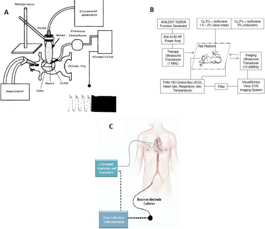
US generator system (Figure 3A). Kuma et al 2006
A steerable bipolar electrophysiology catheter incorporating a receiver Echt et al 2006
electrode into the tip and circuitry to convert US energy to electrical Lee et al 2007
energy was inserted transvenously into the heart. A US-transmitting
transducer was placed on the chest wall with US gel. The output
waveform of the receiver electrode was monitored while the
transmitter was moved on the chest wall to target the receiver. US-
(Continues)
1064 J Ultrasound Med 2021; 40:1061–1076Coiado et al—Therapeutic Ultrasound in Cardiovascular Medicine
Table 2. Continued
Experimental Setup Study Year
mediated pacing with minimum voltage but consistent capture was
obtained for 12 s (Figure 3C).
The transducer element was driven by a custom-built field effect Towe and Rho 2006
transistor power amplifier using a ferrite core step-up transformer that
provided up to 4 kV of excitation corresponding to a peak electrical
power.
The electrocardiographic signal was amplified and sent to an Miller et al 2009
oscilloscope and to a digitizer. The digitized electrocardiogram was
analyzed with the aid of software, which provided automated
collection of data on the heart rate and the numbers of normal
complexes. The software also partially automated detection of
premature complexes.
A 1–3.5-MHz US transducer was driven by a function generator Buiochi et al 2012
connected to a radiofrequency power amplifier. The rat’s heart was Coiado et al 2014, 2015, 2017
exposed to pulsed US, and the cardiac parameters were monitored
with imaging US (Figure 3B).
force or the period between the stimulation of the recurrent US effect.16,17 With CW 1-MHz, 1-W/cm2
muscle and the maximum rate of rising systolic force SATA intensity US, a decrease of diastolic force was
(dF/dt). After mechanical parameters and tempera- observed in rat hearts without simultaneous modifica-
ture measurements were obtained, a proportional tion of the developed force. These changes were
increase of the bath’s ambient temperature was per- attributed to nonthermal effects because temperature
formed. The contractile properties of cardiac muscle changes were not observed. It was also speculated
were affected by temperature. The comparison that US accelerated a release or accumulation of cal-
between the US responses versus the thermal equiva- cium.16 The effect of US exposure (4 seconds,
lent response showed that both equivalent changes in 1.0 W/cm2 SATA at 963 kHz) on the post-tetanic
temperature and US lead to a significant decrease of potentiation of isolated isometrically contracting pap-
both the time to peak force and developed force. A illary muscle of rats caused a potentiation of the
decrease of diastolic tension was observed compared developed force. This showed an important connec-
with the equivalent thermal intervention.15 An tion between contracting cardiac muscles and short-
in vitro study19 using a US therapy unit exposed term CW US exposure. Another study22 showed that
fibroblasts cells to pulsed waves at 1 Hz, and the US the release and transport of calcium were highly likely
treatment increased calcium uptake with increasing to be involved in the potentiation of the developed
exposure time (Figure 2B). force. Ultrasound (CW 1 MHz, 1.5 W/cm2 SATA)
In a subsequent study,21 CW 500-kHz, 10-W/cm2 caused simultaneous alterations in isolated cardiac
SATA intensity US yielded a cardiac defibrillation and muscle.17 In a previous study,15 a decrease of diastolic
antiarrhythmic effect in the heart of dogs. The anti- tension had been observed, whereas in this later
arrhythmic effect of US can be generated with a lower study, an increase of the action potential amplitude
intensity for the ventricular myocardium than that was reported in addition to the decrease of the dia-
required for the heart as a whole. The effect can be stolic force. Ultrasound (CW 1 MHz, 0.25–2 W/cm2
explained by the hypothesis that the US waves are SATA) was applied to the rat isolated papillary mus-
partially absorbed and partially dispersed by the large cle, and after US stimulation, a positive inotropic
mass of the myocardium, resulting in an inhibition of effect was observed to be linearly related to the US
the electrical activity and a decrease of its refractory intensity.18 Therapeutic-level CW US16,22 was reported
period of the cells of the myocardium. to increase the force and enhance the effect of graded
In vitro and in vivo studies in subsequent years intensities on rat isolated papillary muscle contractile
found that the decrease of the diastolic force is a performance. Based on studies by Mortimer et al15,17
J Ultrasound Med 2021; 40:1061–1076 1065Coiado et al—Therapeutic Ultrasound in Cardiovascular Medicine
and Forester et al,16,18,22 the US stimulation might shortly after exposure ceased. Studies in isolated per-
accelerate a release or accumulation of calcium in the fused hearts of rats with physiologic saline showed
cardiac tissue, and the effect mechanism was viewed that acoustic cavitation was followed by a decrease of
to be with nonthermal effects. the developed pressure, and no US effects below the
More recent reports demonstrate that US appli- same acoustic cavitation intensity were found.26,27
cation can reduce the threshold for cardiac electrical Vykhodteseza et al28 performed an investigation
excitation23 and produce positive inotropic effects in into the effects of high-intensity pulsed US (1.2 MHz,
isolated myocardial preparations by increasing the up to 2000 W/cm2 SPPA) for the treatment of brain
influx of calcium into cardiac cells.12,24 In an in vivo disorders. The multiple pulsed experiments in rabbit
study in frogs25 (Figure 2C), high-intensity US pulses brain demonstrated that the histologic effects varied
(1.2 MHz, up to 2000 W/cm2 spatial-peak pulse- from zero visual damage of tissue to local hemor-
average [SPPA]) caused changes in the heart rate but rhage. As the pulse duration (PD), number of pulses,
did not demonstrate effects because the rhythm of and repetition frequency increased, the severity of the
the heart and aortic pressure returned to normal tissue damage increased. Another in vivo study29
Figure 2. In vitro experimental setups. A, Papillary muscle exposed at 953-kHz continuous waves (modified from Mortimer et al17). B, Cells
exposed at 1-Hz pulsed waves (modified from Mortimer and Dyson19). C, Frogs’ heart exposed at 0.5- to 2-Hz pulsed waves (modified from
Dalecki et al49).
1066 J Ultrasound Med 2021; 40:1061–1076Coiado et al—Therapeutic Ultrasound in Cardiovascular Medicine
showed that free gas bubbles were induced in living heart of rats likely resulted from parasympathetic
mammalian tissue by 0.75-MHz US irradiation at stimulation or direct mechanical US stimulation of
680 mW/cm2; however, the study did not show his- aortic baroreceptors with consequent stimulation of
tologic results. There were no effects of US below the the baroreceptor reflex.30,31 In a porcine model, the
cavitation intensity found. The importance of this combination of a radiation force mechanism and tis-
work is to explain the bioeffects involved in cardiac sue vibration was suggested as a possible cause of the
pacing during US exposure and find a therapeutic US cardiac pacing.32
application for cardiac conditions. Arrhythmia occurred coincidentally with diagnos-
tic US exposures and ceased after US exposures
After 2000: in vitro and in vivo Studies stopped.33 The heart abnormalities were induced
Studies after 2000 suggested that the decrease of the principally when the contrast agent interacted with
heart rate effect caused by US application on the US during application (pulsed 3.1-MHz frequency,
Figure 3. In vivo and human experimental setups. A, Positive chronotropic effect observed in guinea pigs (modified from Kuma et al51). B,
Negative chronotropic effect observed in rats (Modified form Buiochi et al30). C, Cardiac pacing in humans (modified from Echt et al42).
J Ultrasound Med 2021; 40:1061–1076 1067Coiado et al—Therapeutic Ultrasound in Cardiovascular Medicine
1.3-millisecond PD, 1700-Hz pulse repetition fre- markedly arrhythmogenic effect and a negative chrono-
quency [PRF], and 15.9-MPa PRPA), and the study tropic efffect.35 In the future, the rat model will likely be
suggested that US pulses may have the potential to translated to large animals such as pigs or dogs to evalu-
cause arrhythmias via their biomechanical interactions ate the feasibility and safety of therapeutic US. The
with contrast agents.34 major contribution of this research will be to improve
Transthoracic cardiac US stimulation (pulsed treatment with pacemakers.
1 MHz, 3-MPa PRPA) at an approximately 1% duty In vivo pulsed experiments (1 MHz, 2–3-MPa
factor (DF) induced a negative chronotropic effect in PRPA [≈133–300-W/cm2 SPPA], ≈1.0% DF,
rat hearts without damage to the hemodynamic sys- 2-millisecond PD, 4–6-Hz PRF) also have demon-
tem.30 A likely mechanism to explain the negative strated that US applied to the chest has the potential
chronotropic response to pulsed US exposure would to cause a negative chronotropic effect without dam-
be reflex vagal activation and sympathetic inhibition age to the cardiac tissue.30,40,41 To study this effect, a
involving the baroreceptor reflex (eg, by direct bilateral vagotomy was performed, with a small verti-
mechanical stimulation of aortic baroreceptors by cal midline incision 1 cm superior to the sternum of
US) or the Bezold-Jarisch reflex. This reflex can be the rat, to minimize the time the carotid artery was
followed by apnea, bradycardia, and hypotension, cannulated for the measurement of arterial pressure.40
depends on intact vagi, and is mediated through cra- It was hypothesized that the negative chronotropic
nial nervous medullary centers controlling respiration, effect was a direct mechanism caused by US pulses,
heart rate, and vasomotor tone. and the parasympathetic nervous system did not play
Another in vivo study31 showed a negative chro- a role.
notropic effect in rat hearts (pulsed 3.5 MHz, 2-MPa
PRPA [≈133-W/cm2 SPPA], 0.25%–1.0% DF, Translation to Human Studies
2-millisecond PD, 4–5-Hz PRF) showed that the DF In medicine, US has been used in both diagnosis and
more likely influenced cardiac pacing than the pulsed disease treatment. Ongoing medical research on US
pressure amplitudes. Another bioeffect of myocardial has stimulated the improvement of existing tech-
contrast echocardiography is cardiac arrhythmia, niques and development of new applications.30,40–43
which occurs at high PRPAs that are correlated with Echt et al42 investigated the use of US as an alterna-
destruction and gas body destabilization caused by tive source of energy for pacing without leads
the US pulses. Experiments in perfused rat hearts (Figure 3C). One potential research application is sex
demonstrated that US (1 MHz, 3-MPa PRPA differences not only in cardiology but also in other
[300-W/cm2 SPPA], 1% DF, 5-millisecond PD, 2-Hz specialties.44 In the United States, deaths due to car-
PRF) exerted a markedly arrhythmogenic effect on diovascular disease in women exceed those in men,
the heart, which, in addition to a mild negative chro- and cardiovascular disease remains the primary cause
notropic effect, might cause a deleterious influence of death worldwide. Physiologic differences between
on blood pumping.35 women and men and cultural factors can contribute
The study of cardiac pacing using US (313–- to the development of cardiovascular disease.33 Of
385 kHz, 22.7-W/cm2 mean PRPA [0.74–112-W/cm2 note, the incidence and the increased rate of cardio-
SPTA], mechanical indexCoiado et al—Therapeutic Ultrasound in Cardiovascular Medicine
sex and/or the sex hormone estrogen may contribute known by mathematical modeling techniques53–62 and
to the sexual dimorphism in the heart and to a better has been estimated for various exposure conditions.63,64
outcome of cardiac diseases in women is supported The US wave propagation transports and dissi-
by cardiovascular disease animal models. In a recent pates energy; the average energy density is
study, the role of age and sex in the decrease of the
ðT
heart rate was investigated with exposure of the rat 1 ρ 2
heart to 3.5-MHz pulsed US.48 The study showed a hEi = Eðx,t Þt = U op + U 2on : ð1Þ
T 0 2
negative chronotropic effect caused by pulsed US,
and ovarian hormones were responsible for different The instantaneous intensity is defined as the dot
US-induced cardiac bioeffects. product of the US pressure and particle velocity, but
Over the years, various US exposure models have because these 2 quantities are in phase, the dot prod-
been developed. Pulsed US has been shown to inter- uct is pu. Its temporal average representation is
fere in the cardiac activity of the turtle,13 dog,21 frog,49 given by
mouse,50 pig,32 guinea pig,51 and rat 30,34,35,40,41. Kuma
ðT
et al51 showed a positive chronotropic effect in hearts 1 ρc 2
of guinea pigs (Figure 3A), whereas Coiado et al31 I= pudt = U op − U 2on , ð2Þ
observed a negative chronotropic effect in hearts of T 0 2
rats (Figure 3B). Animal models have been developed
for different kinds of research, specifically models to where Uop and Uon are the particle velocity amplitudes
identify sex-related differences, including brain injury, for the positive and the negative directed components.
atherosclerosis, toxicology, autoimmune diseases, hor- For a progressive US plane wave propagating in only
mones, and stress/alcohol consumption.52 Coiado and the positive x direction, U 2on = 0 , for standing waves
O0 Brien41 investigated whether sex differences could U 2op = U 2on , then U 2op = U 2o :
affect the outcomes in cardiac US therapy. The study
showed the feasibility and biophysics of a new technol- ρ 1 2
hEi = U 20 = p ð3Þ
ogy that uses US pulses to achieve cardiac leadless pac- 2 2ρc2 0
ing without causing undesirable side effects. To
overcome the limitation of pacemaker leads, a new and
technology that used pulsed US at a mean frequency
of 350 kHz and an 0.5-millisecond PD to achieve car- ρ 1 2 p0 U 0
I = U 20 = p = ð4Þ
diac pacing was considered the first human demonstra- c 2ρc 0 2
tion of cardiac stimulation.36 The safety and feasibility
of cardiac stimulation using an alternative energy In tissue, at the site (spatial peak) where the US
source proved to be feasible and safe.36,42 spatial-peak temporal-average intensity is ITA, the rate
of heat generation per unit volume is given by the
expression55,57
Thermal and Nonthermal Mechanisms
*
app
Thermal Q_ = 2αI TA = , ð5Þ
ρc
The amplitude of the US wave decreases with distance
whenever it propagates into tissue or any attenuating where
material; this attenuation is due to absorption and scat-
tering. Scattering can be defined as a portion of the pp*
wave that changes direction, and absorption is a mecha- I TA = , ð6Þ
nism that represents that portion of the wave energy 2ρc
that is converted into an increase of temperature (heat).
where α is the US amplitude absorption coefficient,
The thermal mechanism is relatively well studied.
which increases with increasing frequency; p and p* are
The increase of temperature produced by US is well
J Ultrasound Med 2021; 40:1061–1076 1069Coiado et al—Therapeutic Ultrasound in Cardiovascular Medicine
the instantaneous US pressure and its complex conju- that the latter one includes a cooling duration of 2–
gate, respectively; ρ is density; and cc is sound speed. 0.2 seconds = 1.8 seconds.
The product of p and p*is equal to the US pressure
amplitude squared, p20 , at the specific location in the Nonthermal
medium where Q_ is determined and can be thought Both first- and second-order US quantities have been
of as a spatial-peak temporal-average quantity: involved in nonthermally produced biological
effects.66 Acoustically generated cavitation is the non-
thermal mechanism that has received the most atten-
αp 2
Q_ = 0 : ð7Þ tion, principally from US contrast agent microbubbles.
ρc Outstanding literature reviews of cavitation have been
published.54,63,64,66–78
For a given ITA, the maximum temperature Concerns have been addressed regarding the
increase, ΔTmax, under the assumption that no heat is interaction of US with contrast agents in humans and
lost by convection, conduction, or any other processes potential bioeffects of inertial cavitation.66,76 Some
to remove heat, is described approximately below65: studies raised these concerns by documenting the
hemolysis of erythrocytes in cell suspensions in
Q_ Δt human and in mice that contained contrast agents
ΔT max = , ð8Þ
Cv that were exposed to pulsed US.79–85 Hemorrhage in
the vascular beds of the intestine and skin86,87 plus
where Δt is the time duration (or also the PD of a damage to cells in the heart88 were also studied in
single pulse) of exposure, and Cv is the tissue’s heat dogs and mice, respectively, after exposure to pulsed
capacity per unit volume. Equation 8 is valid exclu- US and intravenous injection of a contrast agent. in vivo
sively for short exposure times (Coiado et al—Therapeutic Ultrasound in Cardiovascular Medicine
Whether there are other biophysical effects such as of US energy during pacing attempts. The authors
permanent or temporary risk-related tissue responses mentioned in this review that more studies are neces-
has yet to be studied extensively. sary to elucidate the mechanisms of muscle contractil-
However, US-induced temporal-average force has ity, including beat-beat variability and the negative
been a mechanism implicated in the association with chronotropic effect.
the tactile response103–107 and cardiac changes in Diagnostic US operates on the hypothesized
frogs25,108 and pigs.32 The radiation force in biological premise that it is noninvasive, low cost, and safe
tissues is estimated to range from 0.1% to 1% of the compared with other diagnostic imaging modalities.
instantaneous US pressure amplitude. Considering a In the mid-1970s the safety of US and the regulatory
radiation pressure of 1% of a 3-MPa peak pressure parameters were discussed. In the early 1990s, the
amplitude wave, a transient pressure of 30 kPa United States Food and Drug Administration (FDA)
(0.3 atm) would be created on the heart. This tran- implemented the output display standard.2 The FDA’s
sient increase of pressure is similar to the occurrence stipulated regulatory upper limits for cardiac applications
described during a precordial thump, a single blow of 430 mW/cm2 for the derated (0.3-dB/cm/MHz)
that has the potential to promote defibrillation.109 spatial peak and either 1.9 for the mechanical index
or 190 mW/cm2 (Table 3). The US-induced tissue
damage seen in the earlier years of its application
Discussion and Conclusions shows that lung tissue can be damaged at diagnostic
levels. However, through careful and detailed experi-
In cardiovascular medicine, the pulsed wave velocity mental and theoretical studies, it has been shown that
and a pressure myograph are commonly used to mea- the severity of damage is not clinically significant.113
sure the elastic properties of blood vessels.111 Scan- In the 2000s, the FDA approved therapeutic US
ning acoustic microscopy has also been used as a for cardiovascular use to treat cardiac arrhythmias and
method for mapping mechanical properties of iso- ischemic heart disease with the use of high-intensity
lated cells and tissues.110 Some studies that used these focused ultrasound (HIFU) and extracorporeal shock
techniques showed increased stiffness of car- wave therapy (ECWT; Table 4). Although diagnostic
diomyocyte cells with age.111,112 However, these tech- US leads to low or negligible increases in tissue tem-
niques were not used in the reviewed studies to perature, the use of therapeutic US in cardiovascular
explore the possible mechanical properties of the car- applications can cause thermal (HIFU) and nonther-
diac tissue after US application. mal (ECWT) bioeffects.114
Coiado and O0 Brien41 observed a negative chro- The delivery of nanoparticle carriers for drug and
notropic effect in young female rats; one of the gene therapy using microbubbles and US has
hypotheses is that the US effect is weight dependent. expanded in recent years. This technology was origi-
It may be possible that US field interacts with more nally approved for use in echocardiography; the use
cardiac structures in smaller animals than in larger of a microbubble contrast agents improved the quality
animals. However, it is not clear what biomechanism of US images due to the difference in acoustic imped-
is involved in the negative chronotropic effect.41 The ance between their gaseous core and the surrounding
authors hypothesized that the decrease of the heart medium and their nonlinear oscillation in an acoustic
rate could be a direct or indirect US (mechanical) field.115 More recently, the use of US in combination
stimulation of aortic baroreceptors that can cause bra- with nanoparticles has been shown to enhance the
dycardia. It is well known that the velocity of sound
in bone is different from that in other tissues. In some
cardiac therapeutic studies,30–31,36,40–43 the US was Table 3. The FDA’s Preamendment Levels of Diagnostic US
Devices (Modified From O0 Brien2)
used as an external and alternative source of energy
to pace the heart. Lee et al36 showed a refractive Derated Intensity Values
effect from the ribs, with attenuation and absorption Application ISPTA, mW/cm2 ISPPA, W/cm2 IM, W/cm2
during US stimulation. To minimize these mechanical
Cardiac 430 190 310
effects, the heart was exposed to different amplitudes
J Ultrasound Med 2021; 40:1061–1076 1071Coiado et al—Therapeutic Ultrasound in Cardiovascular Medicine
efficacy of drug delivery and reduce side effects of and (2) high-risk devices undergo premarket approval,
drugs, including treatment of Alzheimer disease, car- the most stringent type of device application required
diovascular disease, and cancer.116 Although the by the FDA. For the low- to moderate-risk devices, fed-
potential use of high-intensity US for drug delivery eral law requires new device manufacturers to register
can also cause heat, for drug delivery applications, the with the FDA and notify the agency at least 90 days
intensity range of 0.3 to 3 W/cm2 is used. The FDA before they start selling their devices. This premarket
has recommended that an intensity that causes notification must prove the device is as safe and effective
heating of tissues of less than 1 C,116 to avoid heat and substantially equivalent to a similar, legally marketed
high intensities, can be applied when the pulse length device. No evidence from clinical studies is needed. For
(pulse cycles/US frequency) and PRF (pulses per a high-risk or class III device, to gain FDA approval,
second) are reduced.117 Although there are many there must be enough scientific evidence to prove the
benefits of using US, US energies higher than cavita- device is safe and effective for its intended use.119
tion can affect the cell integrity and can thermally and Studies using pulsed US are clinically important
sonochemically induce permanent damage to lipid for the identification of an alternative and leadless
membranes and cause denaturation of proteins and source of energy for cardiac pacing. Other studies
DNA.118 over the years have shown a potential US application
The first HIFU device was approved by the FDA for cardiac pacing. Thus, the feasibility and safety of
in October 2004 and, more recently, the use of US therapeutic US demonstrates its potential for the
with microbubbles for diagnostic applications.116 treatment of cardiovascular diseases.
Therapeutic US for cardiac applications has advanced, The exploration of US as an alternative therapy
especially as an approach to catheter-based ablation for cardiac pacing in vitro and in vivo up to human
of arrhythmias and for treatment of ischemic heart studies has shown its potential as a therapeutic
disease.114 The FDA approval process for new medi- technology. This research motivates the develop-
cal devices, including therapeutic cardiovascular treat- ment of new therapeutic US applications and
ment using US, can be long and tedious. The FDA advancements. The future of US in the medical
classifies the devices by risk: (1) low- to moderate- field will ultimately depend on collaboration and
risk devices are typically subjected to what is called integration between engineering concepts and car-
premarket notification, also called PMN or 510(k); diovascular studies.
Table 4. Classification of US Waves and Applications in the Medical Field
Classification US Parameter FDA-Approved Clinical Applications Potential Cardiovascular Applications
2
US intensity Low-intensity US (Coiado et al—Therapeutic Ultrasound in Cardiovascular Medicine
References 19. Mortimer AJ, Dyson M. The effect of therapeutic ultrasound on cal-
cium uptake in fibroblasts. Ultrasound Med Biol 1988; 14:499–506.
1. O’Brien WD Jr. Assessing the risks for modern diagnostic ultra- 20. Dinno MA, Dyson M, Young SR, Mortimer AJ, Hart J,
sound imaging. Jpn J Appl Phys 1998; 37:2781–2788. Crum LA. The significance of membrane changes in the safe and
2. O’Brien WD Jr. Ultrasound: biophysics mechanisms. Prog effective use of therapeutic and diagnostic ultrasound. Phys Med
Biophys Mol Biol 2007; 93:212–255. Biol 1989; 34:1543–1552.
3. Lehmann JF. The biophysical mode of action of biologic and 21. Smailys AD, Dulevičius Z, Muckus K, Daukša K. Investigation of
therapeutic ultrasonic reactions. J Acoust Soc Am 1953; 25:17–25. the possibilities of cardiac defibrillation by ultrasound. Resuscitation
4. Lehmann JF, Guy AW. Ultrasound therapy. In: Reid JM, 1981; 9:233–242.
Sikov MR (eds) Interaction of Ultrasound with Biological Tissues. 22. Forester GV, Mortimer AJ, Roy OZ, Bateson D, Keon WJ. Effect
Washington, DC: Department of Health Education and Welfare, of brief ultrasound exposure on post-tetanic potentiation in car-
1973:141–152. DHEW publication 73–8008, BRH/DBE 73–1. diac muscle. Pflügers Arch 1984; 400:208–210.
5. Singh MR, Rosenschein U, Ho KK. Treatment of saphenous 23. Salz HR, Rosenfeld EH, Wussling M. Effect of ultrasound on the
vein bypass grafts with ultrasound thrombolysis: a randomized contraction of isolated myocardial cells of adult rats. Ultrasound
study. Circulation 2003; 107:2331–2336. Med Biol 1997; 23:143–149.
6. Dalecki D. Mechanical bioeffects of ultrasound. Annu Rev Biomed 24. Petrishchev NN, Vlasov TD, Galagudza MM, Makov YN. Effect
Eng 2004; 6:229–248. of low-frequency low-intensity ultrasound on contractile function
7. Leighton TG, Cleveland RO. Lithotripsy. Proc Inst Mech Eng H of isolated heart. Bull Exp Biol Med 2002; 4:327–329.
2010; 224:317–242. 25. Dalecki DK, Keller BB, Raeman CH, Carstensen EL. Effects of
8. Xu T, Hall LZ, Fowlkes JB, Cain CA. Effects of acoustic parame- pulsed ultrasound on the frog heart, I: thresholds for changes in
ters on bubble cloud dynamics in ultrasound tissue erosion (his- cardiac rhythm and aortic pressure. Ultrasound Med Biol 1993;
totripsy). J Acoust Sci Am 2007; 122:229–236. 19:385–390.
9. Voogt MJ, Trillaud H, Kim YS, et al. Volumetric feedback ablation 26. Zakharov SI, Bogdanov KY, Rozenshtraukh LV. Arrhtymogenic
of uterine fibroids using magnetic resonance-guided high intensity action of acoustic cavitation on the isolated rat heart perfused
focused ultrasound therapy. Eur Radiol 2012; 22:411–417. with physiological saline. Bull Exp Biol Med 1991; 111:575–578.
10. Vaezy S, Noble ML, Keshavarzi A, et al. Liver hemostasis with 27. Zakharov SI, Bogdanov KY, Rosenshtraukh LV, Gavrilov LR,
high-intensity ultrasound. J Ultrasound Med 2004; 23:217–225. Yushin VP. The effect of acoustic cavitation on the contraction
11. Fagenholz PJ, Murray AF, Noble VE, Baggish AL, Harris NS. Ultra- force and membrane potential of rat papillary muscle. Ultrasound
sound for high altitude research. Ultrasound Med Biol 2012; 38:1–12. Med Biol 1989; 15:561–565.
12. Petrishchev NN, Vlasov TD, Galagudza MM, Makov YN, 28. Vykhodtseza NH, Hynynen K, Damianou C. Histologic effects of
Minasyan CM. Frequency-dependent effects of low-intensity high intensity pulsed ultrasound exposure with subharmonic emis-
ultrasound on activity of isolated heart. Bull Exp Biol Med 2003; sion in rabbit brain in vivo. Ultrasound Med Biol 1995; 21:969–979.
3:239–241. 29. ter Haar G, Daniels S, Eastaugh KC, Hill CR. Ultrasonically
13. Harvey EN. The effect of high frequency sound waves on heart induced cavitation in vivo. Br J Cancer 1982; 45:151–155.
muscle and other irritable tissues. Am J Physiol 1929; 91:284–290. 30. Buiochi EB, Miller RJ, Hartman E, et al. Transthoracic cardiac
14. Harvey EN. Biological aspects of ultrasonic waves: a general sur- ultrasonic stimulation induces a negative chronotropic effect.
vey. Biol Bull 1930; 59:306–325. IEEE Trans Ultrason Ferroelectr Freq Control 2012; 59:2655–2661.
15. Mortimer AJ, Roy OZ, Trollope BJ, et al. A relationship between 31. Coiado OC, O’Brien WD Jr. The role of the duty factor in
ultrasonic intensity and changes in myocardial mechanics. Can J ultrasound-mediated cardiac stimulation. J Acoust Soc Am 2014;
Physiol Pharmacol 1980; 58:67–73. 136:231–235.
16. Forester GV, Roy OZ, Mortimer AJ. Enhancement of contractil- 32. Towe BC, Rho R. Ultrasonic cardiac pacing in the porcine
ity in rat isolated papillary muscle with therapeutic ultrasound. model. IEEE Trans Biomed Eng 2006; 53:1446–1448.
J Mol Cell Cardiol 1982; 14:475–477. 33. Miller DL, Dou C, Lucchesi BR. Cardiac arrhythmia and injury
17. Mortimer AJ, Bresden B, Forester GV, Roy OZ. System for the induced in rats by burst and pulsed mode ultrasound with gas
measurement of the effects of ultrasound on membrane electrical body contrast agent. J Ultrasound Med 2009; 28:1519–1526.
and mechanical properties of the myocardium. Med Biol Eng 34. Zachary JF, Hartleben SA, Frizzell LA, O’Brien WD Jr. Arrhythmias
Comput 1984; 22:24–27. in rat hearts exposed to pulsed ultrasound after intravenous injec-
18. Forester GV, Roy OZ, Mortimer AJ. Ultrasound intensity and tion of a contrast agent. J Ultrasound Med 2002; 21:1347–1356.
contractile characteristics of rat isolated papillary muscle. Ultra- 35. Coiado OC, Costa ET, Bassani R. Arrhythmogenic effect of
sound Med Biol 1985; 11:591–598. power ultrasound in perfused rat hearts. Paper presented at:
J Ultrasound Med 2021; 40:1061–1076 1073Coiado et al—Therapeutic Ultrasound in Cardiovascular Medicine
World Congress on Medical Physics and Biomedical Engineer- 51. Kuma FU, Ueda N, Ito H, et al. Effects of ultrasound energy
ing; May 26–31, 2012; Beijing, China. application on cardiac performance in open-chest guinea pigs: an
36. Lee KL, Lau CP, Tse HF, et al. First human demonstration of in vivo pilot study. Circ J 2006; 70:1356–1361.
cardiac stimulation with transcutaneous ultrasound energy deliv- 52. Curry BB III. Animal models used in identifying gender-related
ery: implications for wireless pacing with implantable devices. differences. Int J Toxicol 2001; 20:153–160.
J Am Coll Cardiol 2007; 50:877–883. 53. Robinson TC, Lele PP. An analysis of lesion development in the
37. Lawton JS, Moon MR, Curci JA, et al. Management of arterial brain and in plastics by high intensity focused ultrasound at low
injuries caused by laser extraction of indwelling venous pacemaker megahertz frequencies. J Acoust Soc Am 1972; 51:1333–1351.
and defibrillator leads. Pacing Clin Electrophysiol 2006; 29:917–920. 54. Nyborg W. Intermediate Biophysical Mechanics. Menlo Park, CA: Cum-
38. Venkataraman G, Hayes DL, Strickberger SA. Does the risk-benefit mings Publishing Co; 1975.
analysis favor the extraction of failed, sterile pacemaker and defibril- 55. Nyborg W. Heat generation by ultrasound in a relaxing medium.
lator leads? J Cardiovasc Electrophysiol 2009; 20:1413–1415. J Acoust Soc Am 1981; 70:310–312.
39. Hamid S, Arujuna A, Ginks M, et al. Pacemaker and defibrillator 56. Lerner RM, Carstensen EL, Dunn F. Frequency dependence of
lead extraction: predictors of mortality during follow-up. Pacing thresholds for ultrasonic production of thresholds for ultrasonic
Clin Electrophysiol 2010; 33:209–216. production of thermal lesions in tissue. J Acoust Soc Am 1973; 54:
40. Coiado OC, Buiochi EB, O’Brien WD Jr. Ultrasound-induced 504–506.
heart rate decrease: role of the vagus nerve. IEEE Trans Ultrason 57. Cavicchi TJ, O’Brien WD Jr. Heat generated by ultrasound in an
Ferroelectr Freq Control 2015; 62:329–336. absorbing medium. J Acoust Soc Am 1984; 70:1244–1245.
41. Coiado OC, O’Brien WD Jr. The negative chronotropic effect in 58. Cavicchi TJ, O’Brien WD Jr. Heating distribution color graphics
rat heart stimulated by ultrasonic pulses: the role of sex and age. for homogeneous lossy spheres irradiated with plane wave ultra-
J Ultrasound Med 2017; 36:799–808. sound. IEEE Trans Sonics Ultrason 1985; 32:17–25.
42. Echt DS, Cowan MW, Riley RE, Brisken AF. Feasibility and 59. Nyborg WL, Steele SR. Temperature elevation in a beam of
safety of a novel technology for pacing without leads. Heart ultrasound. Ultrasound Med Biol 1983; 9:611–620.
Rhythm 2006; 3:1202–1206. 60. Nyborg WL, O’Brien WD Jr. An alternative simple formula for
43. Lee KL, Tse HF, Echt DS, Lau CP. Temporary leadless pacing temperature estimate. J Ultrasound Med 1989; 8:653–654.
in heart failure patients with ultrasound-mediated stimulation 61. Curley M. Soft tissue temperature rise caused by scanned, diag-
energy and effects on the acoustic window. Heart Rhythm 2009; nostic ultrasound. IEEE Trans Ultrason Ferroelectr Freq Control
6:742–748. 1993; 40:59–66.
44. Nowak B, Misselwitz B, Expert Committee “Pacemaker,” Institute 62. Lubbers JH. Time to threshold (TT), a safety parameter for heating
of Quality Assurance Hessen, et al. Do gender differences exist in by diagnostic ultrasound. Ultrasound Med Biol 2003; 29:755–764.
pacemaker implantation? Results of an obligatory external quality 63. National Council on Radiation Protection and Measurements.
control program. Europace 2010; 12:210–215. Biological Effects of Ultrasound: Mechanisms and Clinical Implications.
45. Reckelhoff JF. Gender difference in the regulation of blood pres- Bethesda, MD: National Council on Radiation Protection and
sure. Hypertension 2001; 37:1199–1208. Measurements; 1983. Report 74.
46. Patten RD. Models of gender differences in cardiovascular dis- 64. National Council on Radiation Protection and Measurements.
ease. Drug Discov Today Dis Models 2007; 4:227–232. Exposure Criteria for Medical Diagnostic Ultrasound, I: Criteria Based
47. Maric C. Sex differences in cardiovascular disease and hyperten- on Thermal Mechanisms. Bethesda, MD: National Council on
sion: involvement of the renin-angiotensin system. Hypertension Radiation Protection and Measurements; 1992. Report 113.
2005; 46:475–476. 65. Fry WJ, Fry RB. Temperature changes produced in tissue during
48. Mahmoodzadeh S, Fliegner D, Dworatzek E. Sex and gender dif- ultrasonic irradiation. J Acoust Soc 1953; 25:6–11.
ferences in pharmacology. In. Handbook of Experimental Pharma- 66. National Council on Radiation Protection and Measurements.
cology. Berlin, Germany: Springer-Verlag; 2012:23–48. Exposure Criteria for Medical Diagnostic Ultrasound, II: Criteria Based
49. Dalecki D, Keller BB, Carstensen EL, Neel DS, Palladino JL, on all Known Mechanisms. Bethesda, MD: National Council on
Noordergraaf A. Thresholds for premature ventricular contrac- Radiation Protection and Measurements; 2002. Report 140.
tions in frog hearts exposed to lithotripter fields. Ultrasound Med 67. Flynn H. Physics of acoustic cavitation in liquids. In: Mason WP
Biol 1991; 17:341–346. (ed). Physical Acoustics: Principles and Methods. Vol lB. New York,
50. Macrobbie AG, Raeman CH, Child SZ, Dalecki D. Thresholds NY: Academic Press; 1964:1B,57.
for premature contractions in murine hearts exposed to pulsed 68. Flynn H. Cavitation dynamics, I: a mathematical formulation.
ultrasound. Ultrasound Med Biol 1997; 23:761–765. J Acoust Soc Am 1975; 57:1379–1396.
1074 J Ultrasound Med 2021; 40:1061–1076Coiado et al—Therapeutic Ultrasound in Cardiovascular Medicine
69. Flynn H. Cavitation dynamics, II: free pulsations and models for 86. Miller DL, Quddus J. Diagnostic ultrasound activation of contrast
cavitation bubbles. J Acoust Soc Am 1975; 58:1160–1170. agent gas bodies induces capillary rupture in mice. Proc Natl Acad
70. Flynn H. Generation of transient cavities in liquids by microsec- Sci USA 2000; 97:10179–10184.
ond pulses of ultrasound. J Acoust Soc Am 1982; 72:1926–1932. 87. Miller DL, Quddus J. Sonoporation of monolayer cells by diag-
71. Nyborg W. Acoustic streaming. In: Mason WP (ed). Physical nostic ultrasound activation of contrast agent. Ultrasound Med Biol
Acoustics: Principles and Methods. Vol 2B. New York, NY: Aca- 2000; 26:661–667.
demic Press; 1964. 2B 88. Skyba DP, Price RJ, Linka AZ, Skalak TC, Kaul S. Direct in vivo
72. Coakley W, Nyborg W. Cavitation; dynamics of gas bubbles; visualization of intravascular destruction of microbubbles by ultra-
applications. In: Fry FJ (ed). Ultrasound: Its Applications in Medi- sound and its local effects on tissue. Circulation 1998; 98:290–293.
cine and Biology. New York, NY: Elsevier; 1978:77–160. 89. Killam AG, Greener Y, McFerran BA, et al. Lack of bioeffects of ultra-
73. Apfel R. Acoustic cavitation. In: Edmonds PD (ed). Ultrasonics: sound energy after intravenous administration of FS069 (Optison) in
Methods of Experimental Physics. Ser 356. New York, NY: Aca- the anesthetized rabbit. J Ultrasound Med 1998; 17:349–356.
demic Press; 1981;355–411. 90. Wible JH Jr, Galen KP, Wojdyla JK, et al. Microbubbles induce
74. Leighton TG. The Acoustic Bubble. New York, NY: Academic renal hemorrhage when exposed to diagnostic ultrasound in anes-
Press; 1994. thetized rats. Ultrasound Med Biol 2002; 28:1535–1546.
75. ter Haar G. The new British Medical Ultrasound Society Guide- 91. Hwang JB, Brayman AA, Reidy MA, et al. Vascular effects induced
lines for the safe use of diagnostic ultrasound equipment. Ultrasound. by combined 1-MHz ultrasound and microbubble contrast agent
2010;18(2):50–51. http://dx.doi.org/10.1258/ult.2010.100007. treatments in vivo. Ultrasound Med Biol 2005; 31:553–564.
76. American Institute of Ultrasound in Medicine. Mechanical 92. Miller DL, Gies RA. The influence of ultrasound frequency and
bioeffects from diagnostic ultrasound: AIUM consensus state- gas-body composition on the contrast agent-medicated enhance-
ments. J Ultrasound Med 2000; 19:68–168. ment of vascular bioeffects in mouse intestine. Ultrasound Med
77. Suslick K. Sonochemistry and sonoluminescence. In Encyclopedia Biol 2000; 26:307–313.
of Physical Science and Technology. Vol 17. 3rd ed. San Diego, CA: 93. Kobayashi NY, Yasu T, Yamada S, et al. Endothelial cell injury in
Academic Press; 2001:363–376. venule and capillary induced by contrast ultrasonography. Ultra-
78. Hoff L. Acoustic Characterization of Contrast Agents for Medical sound Med Biol 2002; 28:949–956.
Ultrasound Imaging. Dordrecht, the Netherlands: Kluwer Aca- 94. Kobayashi NY, Yasu T, Yamada S, et al. Influence of contrast
demic Publishers; 2001. ultrasonography with perflutren lipid microspheres on micro-
79. Williams AR, Kubowicz G, Cramer E, Schlief R. The effects of vessel injury. Circ J 2003; 67:630–636.
the microbubble suspension SH U 454 (Echovist) on 95. Schlachetzki FH, Hölscher T, Koch HJ, et al. Observation on the
ultrasound-induced cell lysis in a rotating tube exposure system. integrity of the blood-brain barrier after microbubble destruction
Echocardiography 1991; 8:423–433. by diagnostic transcranial color-coded sonography. J Ultrasound
80. Dalecki D, Raeman CH, Child SZ, et al. Hemolysis in vivo from expo- Med 2002; 21:419–429.
sure to pulsed ultrasound. Ultrasound Med Biol 1997; 23:307–313. 96. Hynynen KM, McDannold M, Martin H, Jolesz FA,
81. Miller DL, Gies RA, Chrisler WB. Ultrasonically induced hemoly- Vykhodtseva N. The threshold for brain damage in rabbits
sis at high cell and gas body concentrations in a thin-disc expo- induced by bursts of ultrasound in the presence of an ultrasound
sure chamber. Ultrasound Med Biol 1997; 23:625–633. contrast agent (Optison). Ultrasound Med Biol 2003; 29:
82. Miller DL, Gies RA. Enhancement of ultrasonically-induced 473–481.
hemolysis by perfluorocarbon-based compared to air-based echo- 97. Van der Wouw PA, Brauns AC, Bailey SE, Powers JE,
contrast agents. Ultrasound Med Biol 1998; 24:285–292. Wilde AA. Premature ventricular contractions during triggered
83. Miller DL, Gies RA. Gas-body–based contrast agent enhances imaging with ultrasound contrast. J Am Soc Echocardiogr 2000;
vascular bioeffects of 1.09 MHz ultrasound on mouse intestine. 13:288–294.
Ultrasound Med Biol 1998; 24:1201–1208. 98. Dalecki D, Rota C, Raeman CH, Child SZ. Premature cardiac
84. Poliachik SC, Chandler WL, Mourad PD, et al. Effect of high- contractions produced by ultrasound and microbubble contrast
intensity focused ultrasound on whole blood with and without agents in mice. Acoust Res Lett Online 2005; 6:221–226.
microbubble contrast agent. Ultrasound Med Biol 1999; 25: 99. Nightingale KK, Kornguth PJ, Walker WF, McDermott BA,
991–998. Trahey GE. A novel ultrasonic technique for differentiating cysts
85. Brayman AM, Miller MW. Sonolysis of Albunex-supplemented, from solid lesions: preliminary results in the breast. Ultrasound
40% hematocrit human erythrocytes by pulsed 1-MHz ultra- Med Biol 1995; 21:745–751.
sound: pulse number, pulse duration and exposure vessel rotation 100. Fatemi M, Greenleaf JF. Ultrasound-stimulated vibro-acoustic
dependence. Ultrasound Med Biol 1999; 25:307–314. spectrography. Science 1998; 280:82–85.
J Ultrasound Med 2021; 40:1061–1076 1075You can also read