Therapeutic afucosylated monoclonal antibody and bispecific T- cell engagers for T- cell acute lymphoblastic leukemia
←
→
Page content transcription
If your browser does not render page correctly, please read the page content below
Open access Original research
Therapeutic afucosylated monoclonal
J Immunother Cancer: first published as 10.1136/jitc-2020-002026 on 17 February 2021. Downloaded from http://jitc.bmj.com/ on July 3, 2022 by guest. Protected by copyright.
antibody and bispecific T-cell engagers
for T-cell acute lymphoblastic leukemia
Daniele Caracciolo ,1 Caterina Riillo,1 Andrea Ballerini,2 Giuseppe Gaipa,3
Ludovic Lhermitte,4,5 Marco Rossi,1 Cirino Botta,6 Eugénie Duroyon,4,5
Katia Grillone,1 Maria Eugenia Gallo Cantafio,1 Chiara Buracchi,3 Greta Alampi,3
Alessandro Gulino,7 Beatrice Belmonte,7 Francesco Conforti,8 Gaetanina Golino,1
Giada Juli,1 Emanuela Altomare,1 Nicoletta Polerà,1 Francesca Scionti,1
Mariamena Arbitrio,9 Michelangelo Iannone,9 Massimo Martino,10
Pierpaolo Correale,11 Gabriella Talarico,12 Andrea Ghelli Luserna di Rorà,13
Anna Ferrari,13 Daniela Concolino,14 Simona Sestito,14 Licia Pensabene,14
Antonio Giordano,15 Markus Hildinger,16 Maria Teresa Di Martino,1
Giovanni Martinelli,13 Claudio Tripodo,7 Vahid Asnafi,4,5 Andrea Biondi,3
Pierosandro Tagliaferri,1 Pierfrancesco Tassone1,15
To cite: Caracciolo D, Riillo C, ABSTRACT Both UMG1-BTCEs demonstrated highly effective killing
Ballerini A, et al. Therapeutic Background T-cell acute lymphoblastic leukemia activity against T-ALL cells in vitro. We demonstrated that
afucosylated monoclonal (T-ALL) is an aggressive disease with a poor cure this effect was specifically exerted by engaged activated
antibody and bispecific T-cell T cells. Moreover, UMG1-BTCEs effectively antagonized
rate for relapsed/resistant patients. Due to the lack of
engagers for T-cell acute
T-cell restricted targetable antigens, effective immune- tumor growth at concentrations >2 log lower as compared
lymphoblastic leukemia. Journal
for ImmunoTherapy of Cancer therapeutics are not presently available and the treatment with ahuUMG1, with significant mice survival advantage in
2021;9:e002026. doi:10.1136/ of chemo-refractory T-ALL is still an unmet clinical different T-ALL models in vivo.
jitc-2020-002026 need. To develop novel immune-therapy for T-ALL, we Conclusion Altogether our findings, including the safe
generated an afucosylated monoclonal antibody (mAb) UMG1-epitope expression profile, provide a framework
►► Prepublication history and (ahuUMG1) and two different bispecific T-cell engagers for the clinical development of these innovative immune-
additional material is published (BTCEs) against UMG1, a unique CD43-epitope highly and therapeutics for this still orphan disease.
online only. To view please visit selectively expressed by T-ALL cells from pediatric and
the journal online (http://dx.doi. adult patients.
org/10.1136/jitc-2020-002026). Methods UMG1 expression was assessed by BACKGROUND
immunohistochemistry (IHC) on a wide panel of normal T-cell acute lymphoblastic leukemia (T-ALL)
DC and CR contributed equally.
tissue microarrays (TMAs), and by flow cytometry on is an aggressive hematological malignancy
Accepted 17 January 2021 healthy peripheral blood/bone marrow-derived cells, on derived from the abnormal proliferation
10 different T-ALL cell lines, and on 110 T-ALL primary
of aberrant intra- thymic T- cell progeni-
patient-derived cells. CD43-UMG1 binding site was defined
tors.1 2 Although T-ALL was historically asso-
through a peptide microarray scanning. ahuUMG1 was
generated by Genetic Glyco-Engineering technology from ciated with a substantially worse outcome as
a novel humanized mAb directed against UMG1 (huUMG1). compared with B- cell ALL (B- ALL), inten-
BTCEs were generated as IgG1-(scFv)2 constructs with sive chemotherapy regimens have recently
bivalent (2+2) or monovalent (2+1) CD3ε arms. Antibody improved the prognosis of T-ALL patients.3–6
dependent cellular cytotoxicity (ADCC), antibody dependent However, approximately 20% of pediatric
cellular phagocytosis (ADCP) and redirected T-cell and 50% of adult patients experience disease
cytotoxicity assays were analysed by flow cytometry. relapse/progression after first- line chemo-
© Author(s) (or their In vivo antitumor activity of ahUMG1 and UMG1-BTCEs therapy with a dismal outcome.7 8 In fact, in
employer(s)) 2021. Re-use was investigated in NSG mice against subcutaneous and these patients, the only approved agent is
permitted under CC BY-NC. No orthotopic xenografts of human T-ALL.
commercial re-use. See rights nelarabine, which can provide temporary
Results Among 110 T-ALL patient-derived samples, 53
and permissions. Published by benefit in a minority of cases only (30%),9
(48.1%) stained positive (24% of TI/TII, 82% of TIII and
BMJ. while few eligible patients can benefit from
42.8% of TIV). Importantly, no expression of UMG1-epitope
For numbered affiliations see
was found in normal tissues/cells, excluding cortical allogeneic hematopoietic cell transplantation
end of article.
thymocytes and a minority (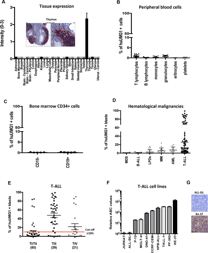
Open access
B-cell antigens, such as CD19, CD20 and CD22, via chain and the variable domain of the light chain of the
J Immunother Cancer: first published as 10.1136/jitc-2020-002026 on 17 February 2021. Downloaded from http://jitc.bmj.com/ on July 3, 2022 by guest. Protected by copyright.
chimeric antigen receptors (CAR-T) or bispecific T-cell murine antibody to the corresponding human IgG1
engagers (BTCEs), and have dramatically empowered constant domains. Humanized H(1-4)/L(1-4) variants
the treatment of relapsed/refractory B- ALL patients, were generated by identifying murine complementary
the treatment landscape of relapsed/refractory T- ALL determinant regions that were grafted onto a human
is still completely orphan and lacks immunotherapeutic antibody framework. The IgG1 isotype was used for all
options. Therefore, the development of innovative immu- humanized variants. Sixteen humanized antibody vari-
notherapeutics is urgently awaited. ants were constructed by replacing selected residues in
We present here a promising experimental therapeutic the closest human germ line sequence of the framework
approach based on the targeting of a unique epitope regions, with the aim to preserve potentially structurally
of CD43 (UMG1), which is highly expressed in cortical- important residues of the murine counterpart. Addi-
derived T-ALL cells. We developed an afucosylated form tionally, eight hybrid CHL(1-4) and H(1-4) CL variants
of the humanized mAb UMG1 (ahuUMG1) and two were generated. Recombinant genes were placed into
different BTCEs that, respectively, simultaneously bind the Evitria vector plasmid and transfected (with eviFect,
UMG1- epitope on T-ALL cells and CD3ε (by bivalent Evitria) into CHO K1 cells. Cells were grown after trans-
or monovalent arm) to induce cell-mediated killing of fection in animal-component free, serum-free medium
epitope- expressing leukemic cells. We performed an (eviMake2, Evitria). Supernatant was harvested by centrif-
extensive analysis of the epitope expression on normal ugation and subsequently sterile filtered (0.2 µm filter).
tissue/cells, and we investigated the in vitro and in vivo The antibodies were purified using MabSelect SuReresin.
activity of these agents in different models of human Each of the humanized antibody has been screened for
T-ALL. The final aim of our study was the translational its affinity to the target (estimated by Mean Fluorescence
development of UMG1- based immune- therapeutics in Intensity, MFI) on two different cell lines (HPB-ALL and
the poor therapeutic landscape of T-ALL. H9) as compared with chimeric and hybrid mAbs by flow
cytometry. Each screening has been performed twice, for
a total of four replicates. All tests have been performed
MATERIAL AND METHODS under the same conditions: all mAbs were used at a final
For a more detailed description of the methods used, see concentration of 1 µg/mL; rituximab (Roche) has been
online supplemental data. used as IgG1 negative control; FITC Mouse Anti-Human
IgG (BD Biosciences) has been used as secondary mAb.
Cell lines
Ke-37, PF-382, TALL-1, HPB- ALL, DND-41, MOLT-4, Generation of UMG1-BTCEs
JURKAT, p12-ichikawa and ALL-SIL were purchased by Evitria cloned the UMG1 cDNAs into Evitria’s vector
DSMZ. CCRF-CEM cell lines was obtained by ATCC. system using conventional (non- PCR based) cloning
Ke-37, PF-382, TALL-1, DND-41, ALL-SIL, CCRF-CEM, techniques. The Evitria vector plasmids were gene synthe-
MOLT-4, JURKAT, p12-ichikawa were cultured in RPMI- sized. Plasmid DNA was prepared under low-endotoxin
1640 (Gibco, Life Technologies, Carlsbad, California, conditions based on anion exchange chromatography.
USA), supplemented with 10% fetal bovine serum (Lonza DNA concentration was determined by measuring the
Group, Basel, Switzerland), 100 U/mL penicillin and 100 absorption at a wavelength of 260 nm. Correctness of the
mg/mL streptomycin (Gibco, Life Technologies), and sequences was verified with Sanger sequencing (with up to
maintained at 37°C in a 5% CO2 atmosphere. two sequencing reactions per plasmid depending on the
HPB-ALL cell line was cultured in RPMI-1640 supple- size of the cDNA). Evitria used suspension-adapted CHO
mented with 20% fetal bovine serum. K1 cells (originally received from ATCC and adapted to
serum-free growth in suspension culture at Evitria) for
Patient samples production. The seed was grown in eviGrow medium, a
BM o PB leukemia cells were collected from children chemically defined, animal-component free, serum-free
with T-ALL enrolled in the AIEOP ALL 2009 protocol at medium. Cells were transfected with eviFect, and cells
the Pediatric Clinic of University of Milano Bicocca, San were grown after transfection in eviMake2. Supernatant
Gerardo Hospital and Hôpital Necker Enfants-Malades was harvested by centrifugation and subsequent filtration
of Paris. Adult T-ALL samples were collected from IRST (0.2 µm filter). The antibody was purified using MabSe-
IRCCS of Meldola, in compliance with bioethical stan- lect SuRe. Monovalent CD3 binding BTCE was generated
dards. Mononuclear cells were collected by Ficoll-Paque using “Knobs-into-holes” technology.
Plus (GE Healthcare) centrifugation and washed twice
in culture medium (RPMI-1640 supplemented with 10% UMG1 expression analyses
Fetal Bovine Serum, FBS). UMG1 expression on bone marrow, peripheral blood and
T-ALL cell lines was evaluated by flow cytometry. Expres-
Antibody humanization and chimerization sion on normal tissue microarrays (TMAs) was evaluated
Chain Heavy Chain Light (CHCL) chimeric antibody by IHC. Detailed information can be found in online
was generated by fusing the variable domain of the heavy supplemental methods section.
2 Caracciolo D, et al. J Immunother Cancer 2021;9:e002026. doi:10.1136/jitc-2020-002026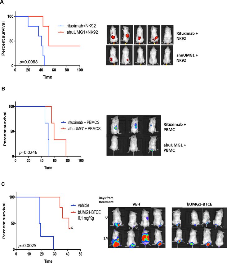
Open access
In vitro antibody dependent cellular cytotoxicity assays Sciences) after 15 min from tail vein injection of Redi-
J Immunother Cancer: first published as 10.1136/jitc-2020-002026 on 17 February 2021. Downloaded from http://jitc.bmj.com/ on July 3, 2022 by guest. Protected by copyright.
Target cells were incubated with Peripheral Blood Ject 2-DeoxyGlucosone (2-DG) (Perkin Elmer). In the
Mononuclear Cells (PBMCs) at effector to target cell disseminated model, CCRF-CEM-Luc+ were intravenous
ratios (E:T) 25:1 with rituximab (50 µg/mL), ahuUMG1 injected in NSG mice. Seven days after tumor cell injec-
(25–100 µg/mL) antibodies or medium alone, o/n at tion, NSG mice were intravenous injected with human
37°C in RPMI/10% fetal calf serum. Residual T lympho- PBMC (20×106 cells) and then randomized to receive
cytes CD8+ (CD45+, CD3+ and CD8+), T lymphocytes rituximab (IgG1, control group) or ahuUMG1, at the
CD4+ (CD45+, CD3+ and CD4+), B lymphocytes (CD45+, dose of 15 mg/kg weekly. Intraperitoneally injection of
CD3− and CD19+) and T-ALL cells (CD45dim, CD3 dim/− mAbs was started at day 3 after PBMC engraftment. In
and CD5+) were analyzed by flow cytometry using a BD vivo evaluation of tumor volume was performed using
FACS CANTO II. The proportion of cells remaining was IVIS LUMINA II Imaging System.
expressed as a percentage of control cultures incubated For UMG1-BTCEs in vivo evaluation, CCRF-CEM-Luc+
without the antibody. The following monoclonal anti- were intravenous injected in NSG mice. After tumor
bodies were used for antigen detection: anti-CD3 PerCP engrafment, PBMC from healthy donors, as source of
Cy5.5 (Biolegend), anti-CD4 PE (Biolegend), anti-CD8 T cells, were engrafted. After 7 days, mice were weekly
APC- H7 (Biolegend), anti- CD45 PO (Thermo Fisher treated with intraperitoneally injection of UMG1-BTCE
Scientific), anti-CD19 PC7 (Beckman Coulter) and anti- (0.1 mg/kg) or vehicle. In vivo evaluation of tumor
CD5 FITC (BD). volume was performed using IVIS LUMINA II Imaging
System.
Redirected T cell cytotoxicity assay
T-ALL cell lines were labeled with Far Red (Invitrogen) Statistical analysis
viable marker, according to manufacturer instructions. Each experiment was performed at least three times and
Labeled cells were cocultured with PBMCs at different values are reported as means±SD. Comparisons between
E:T ratio, in the presence of increasing concentrations of groups were made with Student’s t-test, while statistical
UMG1-BTCE or relative controls for 24–48 hours at 37°C significance of differences among multiple groups was
and 5% CO2, and then stained with 7ADD (BD). Cyto- determined by GraphPad software ( www.graphpad.
toxicity was detected by flow cytometry (Attune NxT Flow com). Graphs were obtained using Graphpad Prism
cytometer, Thermo Fisher Scientific) as the percentage V.6.0. P value of less than 0.05 was accepted as statistically
of 7AAD+/Far Red + cells. In the citotoxicity experiment significant.
with T cell depletion, immunomagnetic cell sorting using
CD4 and CD8 microbeads (MACS Miltenyi Biotec) were
used. RESULTS
Generation and characterization of humanized UMG1 mAb
In vivo studies We generated a novel subclone, named UMG1 (ICLC
Four to six weeks-old female NSG (NOD.Cg-PrkdcscidIl- Biobank, Genova, Italy, 2016) from the original
2rgtm1Wjl/SzJ) mice were purchased from Charles River hybridoma12 by long- term subcloning procedures for
Laboratories (Wilmington, Massachusetts, USA). During clone selection and induction of activation-induced cyti-
experiments, animals were regularly monitored and euth- dine deaminase which may produce slight diversity in
anized when tumors were > 2 cm3 or when signs of disease- mAb reactivity and changes in scFv sequences among
related symptoms or graft-versus-host disease developed. the original hybridoma and subclones.13 14 The mAb
We performed two different experiments for in vivo produced was differentially reactive on cells/tissues, as
activity evaluation of ahuUMG1 in T-ALL mouse models. compared with original mAb. On the sequence of murine
In the limited disease model, 10×106 HPB-ALL cells UMG1, we developed 1 chimeric, 8 chimeric/humanized
were subcutaneously injected. At day 7 mice were and 16 different humanized mAb variants, which under-
randomized into two groups (cohorts of five animals) went evaluation by flow cytometry for affinity on T-ALL
of treatment1: rituximab+NK92+interleukin (IL)-22; cells. A clone based on H3 and L4 chains combination
ahuUMG1+NK92+IL-2. The day after leukemic cells (data not shown) was selected for affinity and named
injection, antibodies were intraperitoneally injected at a humanized UMG1 mAb (huUMG1).
dose of 15 mg/kg once a week, 15×106 NK92 cells were
intravenous injected and 1800 UI/mL of IL-2 (Proleukin; huUMG1 recognizes a unique epitope on CD43 extracellular
Novartis Pharma, Nürnberg, Germany) was intraperi- domain
toneally administered. Treatment started the day after Although CD43 has been reported to be essential for
leukemic cells injection. Tumors were measured with cali- the original parental clone murine mAb binding,15 16 the
pers two times a week and tumor volume was calculated specific binding site of huUMG1 was not defined so far.
according to the formula (width2×length)/2=mm3, where To this aim, a linear epitope mapping was performed
width was the shorter of the two measurements. Further- through a peptide microarray scan. This assay revealed
more, in vivo evaluation of tumor volume was performed that the amino- acid sequence INEGSPLW, included
using IVIS LUMINA II Imaging System (Caliper Life from aa71 to aa78 of the extracellular domain of CD43
Caracciolo D, et al. J Immunother Cancer 2021;9:e002026. doi:10.1136/jitc-2020-002026 3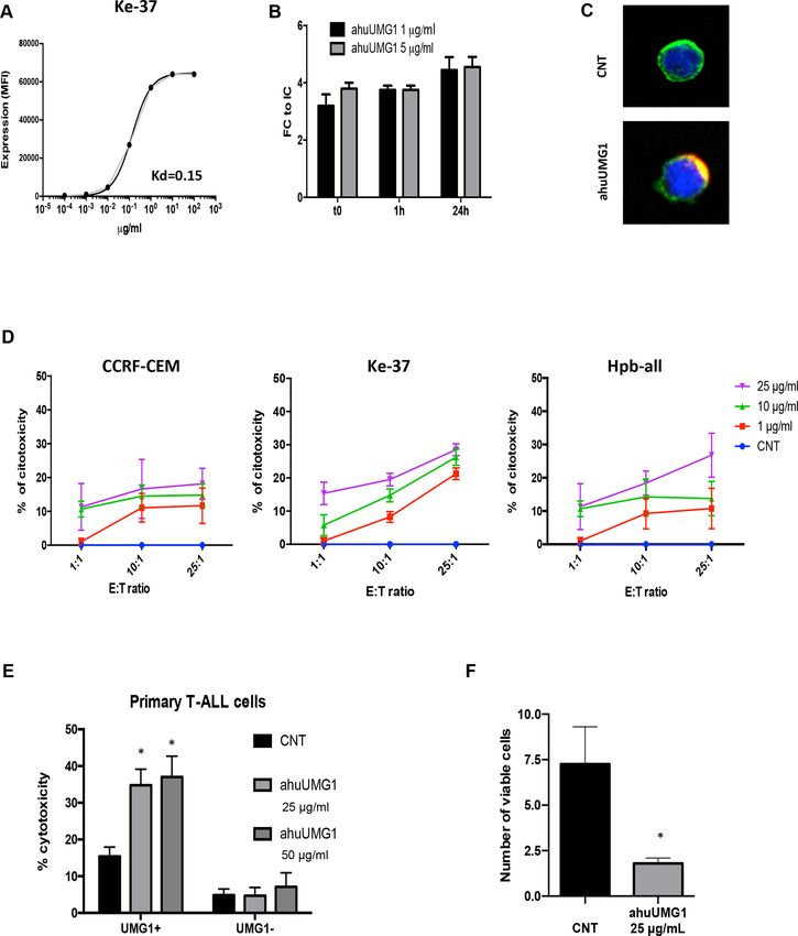
Open access
is the binding peptide of huUMG1 (online supplemental subgroup), 33.3% was huUMG1 positive, thus suggesting
J Immunother Cancer: first published as 10.1136/jitc-2020-002026 on 17 February 2021. Downloaded from http://jitc.bmj.com/ on July 3, 2022 by guest. Protected by copyright.
figure 1A). To confirm this finding, the full- length a potential role of UMG1 in the most aggressive type of
coding sequence (CD43 #1) and a cDNA that lacks the T-ALL (online supplemental table).
sequence encoding for the huUMG1 binding site (CD43 Consistently with data on pediatric/adult primary
#2) have been cloned into expression vectors and sepa- leukemic cells, strong reactivity towards T-ALLcell lines
rately transfected into HEK293 cells, that do not express was observed. Indeed, among T- ALL cell lines, 7/10
CD43. A strong binding of huUMG1 to the CD43 #1 posi- clearly expressed the target, while one showed minor
tive control was found, while no binding was observed expression and two lines no expression. In particular,
for CD43 variant #2 (online supplemental figure 1B). UMG1-epitope was expressed in a range between 125,400
Moreover, to confirm that huUMG1 recognizes a unique and 1858 antigen molecules on Ke-37 and p-12 Ichikawa
epitope, a competitive binding assay among huUMG1 and cells, respectively (figure 1F,G).
commercially available CD43 antibodies (1G10, MEM-59, Taken together these findings indicate that the UMG1-
L10 clones) was performed. None of the tested anti-CD43 epitope is a promising immune- therapeutic target for
clones competes with huUMG1 in binding CCRF-CEM T-ALL patients. At this aim, we developed two species
and HPB-ALL cells (online supplemental figure 1C). of innovative tools: (i) an afucosylated humanized mAb
Taken together, these findings indicate that huUMG1 UMG1 (ahuUMG1) and (ii) two different BTCEs, in the
recognizes a previously undescribed unique epitope of aim to empower the therapeutic targeting of UMG1-
CD43. epitope on leukemic cells.
Reactivity of huUMG1 Afucosylated huUMG1 binding activity on T-ALL cells
To investigate the translational relevance of UMG1- It is well known that afucosylation of IgG1 mAbs signifi-
epitope, the pattern of huUMG1 reactivity on a wide cantly improves Antibody Dependent Cellular Cyto-
panel of normal TMA was first evaluated according to toxicity (ADCC) if compared with the fucosylated form
FDA standards. A highly restricted pattern of reactivity of the same mAb. The afucosylated form of huUMG1
was found. The only strong positive staining was detected (ahuUMG1) was generated by the Genetic Glyco-
on cortical lymphocyte progenitors within the thymus. Engineering/ADCC (GlymaxX) technology.
No other normal tissues, including vital organs, were Dose titration experiments were performed to
reactive. This pattern of expression of the UMG1-epitope define the apparent constant of dissociation (Kd) with
is convincingly predictive of safety for clinical translation ahuUMG1 on Ke-37 cell line. Average apparent Kd was
of immune-targeting agents (figure 1A). estimated at 0.15 µg/mL, while the saturation of binding
In peripheral blood, huUMG1 reacted only with a was reached at concentrations of about 1.00 µg/mL
very small subset of peripheral blood T lymphocytes (figure 2A). Taking into account that downregulation of
(55% of huUMG1 positive blasts. Furthermore, among ahuUMG1 was investigated in vitro. HPB-ALL, CCRF-CEM
tested early T- lineage progenitor leukemias (EGIL- TI and Ke-37 cell lines were exposed to ahuUMG1 25–50
4 Caracciolo D, et al. J Immunother Cancer 2021;9:e002026. doi:10.1136/jitc-2020-002026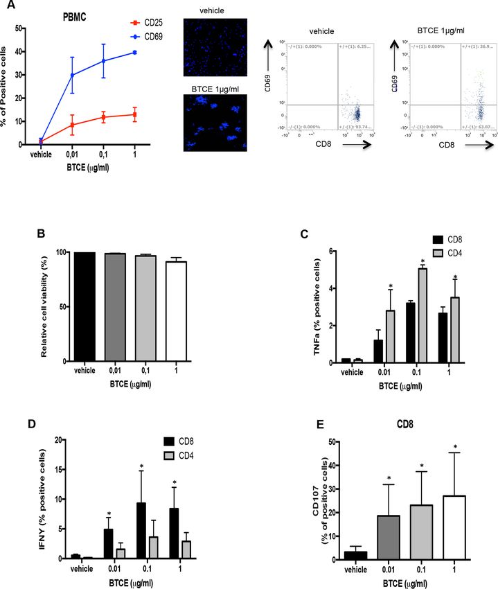
Open access
J Immunother Cancer: first published as 10.1136/jitc-2020-002026 on 17 February 2021. Downloaded from http://jitc.bmj.com/ on July 3, 2022 by guest. Protected by copyright.
Figure 1 UMG1 expression on human healthy and leukemic cells. (A) Immunohistochemical staining intensity (score 0–3)
of huUMG1 on normal tissues. Strong immunohistochemical staining of huUMG1 on human thymic cortical is shown. (B)
UMG1 expression on human healthy donor peripheral blood cells. (C) UMG1 expression on CD19+/− CD34+ human healthy
bone marrow cells. (D) UMG1 expression on a panel of hematological malignancies. (E) Focus of UMG1 expression on T-ALL
blasts from 110 patients according to EGIL classification: TI/TII (50 cases), TIII (39 cases), TIV (21).21 (F) Relative fluorescence
quantitation of UMG1 as evaluated by Fluorescence-activated cell sorting (FACS) analysis. (G) Immunohistochemistry analysis
of UMG1 in a UMG1-positive cell line (Ke-37) as compared with a UMG1-negative one (ALL-SIL). EGIL, European Group for
the Immunological Classification of Leukemias; FACS, Fluorescence-activated cell sorting; huUMG1, humanized monoclonal
antibody directed against UMG1; T-ALL, T-cell acute lymphoblastic leukemia.
µg/mL and cell survival and proliferation were evaluated. increasing concentrations of ahuUMG1 for 24 hours.
Cell viability was not affected after 72 hours of treatment Viable cells were detected by flow cytometry as 7AAD−/
(online supplemental figure 3A) indicating that targeting Far Red+ cells. Importantly, ahuUMG1 demonstrated
of UMG1- epitope does not trigger direct cytotoxicity. in vitro ADCC against UMG1-positive cells (HPB-ALL,
Additionally, ahuUMG1 does not induce complement CCRF-CEM and Ke-37) (figure 2D). Moreover, the anti-
dependent cytotoxicity (data not shown). leukemic activity of ahuUMG1 was evaluated on primary
Based on these data, ADCC against T-ALL cells was cells by coculturing healthy donor’s derived PBMCs with
evaluated after ahuUMG1 exposure. UMG1- positive patient-derived malignant cells exposed to escalating
T-ALL cell lines HPB-ALL, CCRF-CEM and Ke-37 were doses of ahuUMG1. A significant induction of ADCC
cocultured with human healthy donors-derived PBMCs was observed in UMG1+ blasts only (figure 2E). Impor-
at different effector/target ratios in the presence of tantly, in the same primary samples, cytotoxic effects
Caracciolo D, et al. J Immunother Cancer 2021;9:e002026. doi:10.1136/jitc-2020-002026 5Open access
J Immunother Cancer: first published as 10.1136/jitc-2020-002026 on 17 February 2021. Downloaded from http://jitc.bmj.com/ on July 3, 2022 by guest. Protected by copyright.
Figure 2 ahuUMG1 in vitro activity characterization. (A) Average apparent Kd evaluated by flow cytometry on Ke-37 cell line.
(B) UMG1epitope shedding evaluation on HPB-ALL cell line treated with 1 µg/mL and 5 µg/mL of Dy634-labeled huUMG1.
MFI fold change (FC) for each time point (t0, 1 hour, 24 hours) as compared with isotype control (IC) as evaluated by FACS
analysis, is shown. (C) Immunofluorescence analysis of epitope internalization was evaluated on Ke-37 cells. The images
show membrane colocalization of huUMG1 (red) and wheat germ agglutinin (green) 48 hours after treatment. (D) Percentage of
cytotoxicity in T-ALL huUMG1 positive CCRF-CEM, HPB-ALL and Ke-37, cocultured in the presence of PBMCs and ahuUMG1
25 µg/mL or IgG1 25 µg/mL at 25:1 E:T ratio for 12 hours. (E) Percentage of cytotoxicity in UMG1-positive (nine cases) as
compared with UMG1-negative (six cases) T-ALL primary blasts treated with ahuUMG1 25 and 50 µg/mL or IgG1 50 µg/mL and
cocultured with PBMCs at 25:1 E:T ratio for 12 hours. (F) Percentage of viable cells (HPB-ALL) treated for 7 days with ahuUMG1
25 µg/mL and cocultured with PBMCs (E:T=500:1) as compared with control. *pOpen access
J Immunother Cancer: first published as 10.1136/jitc-2020-002026 on 17 February 2021. Downloaded from http://jitc.bmj.com/ on July 3, 2022 by guest. Protected by copyright.
A B
CNT
25000 ahuUMG1 1 µg/ml 50
CD107 (% of positive cells)
ahuUMG1 10 µg/ml
20000 40
ahuUMG1 25 µg/ml * * *
MFI CD1615000 * 30
* *
10000 * 20
* * * * *
5000 10
0 0
l
l
l
T
37
EM
L
/m
m
m
N
AL
/
/
ke
C
µg
µg
µg
-C
B-
RF
10
25
5
HP
CC
ahuUMG1
C D
100 100
IFNY (% of positive cells)
80
% of phagocytosis
80
*
60 * * 60 *
40 40
20 20
0 0
l
l
T
l
l
l
T
/m
m
/m
/m
/m
CN
CN
/
µg
µg
µg
µg
µg
0
50
10
1
5
10
ahuUMG1 ahuUMG1
I E
8 vehicle
CD107 (% of positive cells)
Doxo 0.2 nM
*
6 MTX 0.2 nM
Doxo + MTX 0.2nM
4
2
0
l
T
/m
N
C
µg
1
ahuUMG1
Figure 3 Mechanism of antitumor action of ahuUMG1. CD16 MFI downregulation (A), interferon (IFN)-γ production (B) and
% of CD107a positivity (C) in effector cells (PBMCs) cocultured with CCRF-CEM, after ahuUMG1 treatment as compared to
CNT. (D) ahuUMG1 induction of antibody dependent cellular phagocytosis on CCRF-CEM cell line cocultured with human
healthy macrophages in 5:1 E:T ratio for 4 hours. (E) Cytotoxic effects induced by ahuUMG1 and cytotoxic drug combination
on HPB-ALL cells. *pOpen access
J Immunother Cancer: first published as 10.1136/jitc-2020-002026 on 17 February 2021. Downloaded from http://jitc.bmj.com/ on July 3, 2022 by guest. Protected by copyright.
A UMG1-BTCE B
An3-UMG1
T cell (an3-CD3 FITC) 30000
Expression (MFI)
20000
10000
Kd=0.57
T-ALL cell (an3-UMG1 APC/DAPI
0
An3-CD3 10-4 10-3 10-2 10-1 100 101 102 103
µg/ml
C
CCRF-CEM Ke-37 HPB-ALL
100 100 100 1 µg/ml
1 µg/ml
1 µg/ml
0,1 0,1 µg/ml
80 80µg/ml
0,1 80µg/ml
% of citotoxicity
% of citotoxicity
% of citotoxicity
0,01 µg/ml 0,01 µg/ml
0,01 µg/ml
60 60 vehicle
60 vehicle
vehicle
40 40 40
20 20 20
0 0 0
.1
1
1
1
.1
:1
1
1
.1
:1
1
1
:
1:
5:
1:
5:
1:
5:
20
20
10
10
20
10
E:T ratio E:T ratio E:T ratio
D E
100 Primary T-ALL cells 100
Primary T-ALL cells
100 * vehicle
**
80
vehicle 80 1 µg/ml
vehicle
of citotoxicity
80
1 µg/ml
% cytotoxicity
1 µg/ml
% cytotoxicity
60 60 *
60
40 4040 *
%
20 2020
0 0 0
UMG1+ UMG1- PBMC
UMG1+ CD8 UMG1- CD4
total depleted depleted
Figure 4 bUMG1-BTCE redirects T lymphocytes to kill T-ALL cells in vitro (A) left panel: schematic representation of
the bUMG1-BTCE structure. Right panel: coupling of a CD3+ T cell on Ke-37 cells by bUMG1 BTCE as assessed by
immunofluorescence microscopy. T cells are stained with anti-CD3 (FITC), CCRF-CEM cells with anti-UMG1-APC and DAPI
counterstaining; (B) equilibrium dissociation constants (KD) for bUMG1 BTCE binding to UMG1 on Ke-37 cells; (C) redirected
T-cell-mediated lysis monitored by viable target cell Far Red labeled T-ALL cell lines (CCRF-CEM, Ke-37, HPB-ALL) cocultured
with PBMCs at different E:T ratio and treated with increasing bUMG1 BTCE concentrations for 48 hours. (D) Redirected T-cell-
mediated lysis of UMG1-positive as compared with UMG1-T-ALL primary blasts cocultured for 48 hours with PBMCs at 10:1
E:T ratio and treated with 1 µg/mL of bUMG1 BTCE. (E) CCRF-CEM cells incubated with full or CD8 or CD4-cell-depleted
PBMCs as effector cells and treated with 1 µg/mL at 10:1 E:T ratio. *pOpen access
Importantly, bUMG1-BTCE demonstrated in vitro cyto- in prolonged survival of treated mice of almost 1 month
J Immunother Cancer: first published as 10.1136/jitc-2020-002026 on 17 February 2021. Downloaded from http://jitc.bmj.com/ on July 3, 2022 by guest. Protected by copyright.
toxicity in T-ALL UMG1-positive cells with an EC50 of (figure 6B).
0.01 µg/mL in CCRF-CEM and HPB-ALL and 0.1 µg/mL Taken together these findings demonstrate a signifi-
in Ke-37 cells at E:T ratio of 10:1 (figure 4C). Conversely, cant antitumor activity of ahuUMG1 against T-ALL xeno-
as formal proof of target specificity, no cytotoxic effects grafts in vivo.
were observed after BTCE treatment of ALL-SIL (online Finally, to investigate in vivo bUMG1- BTCE activity,
supplemental figure 4A). Moreover, T-ALL cell viability CCRF- CEM- Luc+ were engrafted in NSG mice. After
was not affected after 72 hours treatment in the absence tumor development, PBMCs from healthy donors, as
of effector cells (Supplemental figure 4B), indicating that source of T cells, were engrafted. After 7 days, mice were
bUMG1-BTCE does not trigger direct cytotoxic effects. weekly intraperitoneally treated with bUMG1-BTCE (0.1
Activity of bUMG1-BTCE toward primary T-ALL primary mg/kg) or vehicle. Of note, significantly reduced tumor
cells was next tested. bUMG1-BTCE induced cytotoxic growth and prolonged survival were observed in bUMG1-
activity only in UMG1+ samples, wherein blasts were BTCE treated mice, as compared with control mice
90% lysed (figure 4D). In particular, to demonstrate T (figure 6C).
cell-mediated bUMG1-BTCE cytotoxicity, T-ALL cell lines Taken together, these results indicate that bUMG1-
were cocultured with total human PBMCs or immunomag- BTCE is a highly effective and promising tool to be inves-
netic T-CD8 or T-CD4 cell-depleted PBMCs. Importantly, tigated in non- human primates for setting the better
while minimal cytotoxic activity was observed in T-CD8 conditions for a First-in-Human study in patients with
depleted samples compared with total PBMCs, a residual T-ALL.
T-ALL cell lysis was found in T-CD4 depleted samples,
thus demonstrating that bUMG1-BTCE cytotoxicity on Monovalent CD3ε binding empowers UMG1-BTCE activity
T-ALL cells is mainly mediated by cytotoxic CD8 T-lym- Several studies have shown that the valency for CD3ε
phocytes (figure 4E). Finally, functional effects induced binding may affect the efficacy of the BTCE molecules.
by bUMG1-BTCE treatment on PBMCs cocultured with Indeed, by limiting the crosslinking of CD3 molecules
T-ALL cell lines were evaluated. Importantly, upregu- on the surface of T cells, CD3ε monovalent binding may
lation of early and late T-cell activation markers, such reduce T-cell unspecific activation and exhaustion. On
as CD25 and CD69, was observed (figure 5A), without these premises, a UMG1-BTCE with monovalent CD3ε
affecting viability of healthy donors’ PBMCs (figure 5B). arm (mUMG1-BTCE) was generated and the binding of
Additionally, bUMG1- BTCE induced release of Tumor this new construct to UMG1-epitope was assessed (online
Necrosis Factor-α (TNF-α) and Interferon-γ (IFN-γ) and supplemental figure 5A). To characterize this novel agent
(figure 5C,D). Consistent with their prominent cytotoxic format (2+1), effector cells were cocultured with T-ALL
function, T-CD8 lymphocytes were positive to CD107a cell lines (CCRF-CEM and Ke-37) at 10:1 E:T ratio in the
activation marker (figure 5E). presence of CD3ε bivalent or monovalent UMG1-BTCEs
(0.1 µg/mL) for 24 hours. Consistently, as compared with
Antitumor activity of ahuUMG1 and bUMG1-BTCE in vivo bivalent BTCE, the monovalent construct resulted in
Based on in vitro findings, we aimed to validate the ther- lower PD1, TIM3 and TIGIT expression thus confirming
apeutic effectiveness of ahuUMG1 and bUMG1 BTCE in a reduction of T cell exhaustion (figure 7A). Interest-
different murine models of human T-ALL. ingly, a stronger redirected cytotoxicity on T-ALL cells
In the first model, HPB-ALL cells were injected subcu- (figure 7B, online supplemental figure 5B) and higher
taneously in NSG mice and animals were then random- proliferation of effector cells (online supplemental figure
ized to receive intraperitoneal rituximab (IgG1, control C) were observed after treatment with the CD3ε monova-
group) or ahuUMG1, both treated at a dose of 15 mg/kg lent construct, as compared with the bivalent one. Impor-
weekly. We found that ahuUMG1 strongly reduced tumor tantly, as for bUMG1-BTCE, in the absence of effector
growth as compared with control mice (figure 6A). Inter- cells, T-ALL cell viability was not affected by mUMG1-
estingly, 60 days after cell engraftment, IHC analysis of BTCE (data not shown).
tumors retrieved from treated animals showed high NK To investigate the translational relevance of our in vitro
cell infiltration in ahuUMG1 treated mice, as compared findings, in vivo activity of mUMG1-BTCE on the dissemi-
with control group (not shown). nated mouse model of T-ALL was evaluated. Importantly,
In the second model, mice were systemically (intrave- a significant survival advantage and reduction of tumor
nous) injected with CCRF-CEM-Luc+ cells to recapitu- growth (figure 7C,D), together with a lower expression
late the disseminated feature of the disease. Seven days of T cell exhaustion markers (online supplemental figure
after tumor cell injection, NSG mice were intravenous 5D), were observed in mUMG1-BTCE treated mice, as
injected with human PBMCs and then randomized to compared with control mice.
receive rituximab (IgG1, control group) or ahuUMG1, at Overall, these findings suggest that a 2+1 format of
the dose of 15 mg/kg weekly 3 days after PBMCs engraft- UMG1-BTCE must be preferred to a 2+2 construct, in
ment. As compared with rituximab, ahuUMG1 induced a clinical translation aimed at an immune-therapeutic
a significant inhibition of tumor growth that translated approach to T-ALL patients.
Caracciolo D, et al. J Immunother Cancer 2021;9:e002026. doi:10.1136/jitc-2020-002026 9Open access
J Immunother Cancer: first published as 10.1136/jitc-2020-002026 on 17 February 2021. Downloaded from http://jitc.bmj.com/ on July 3, 2022 by guest. Protected by copyright.
Figure 5 bUMG1-BTCE activates T lymphocytes against T-ALL cells. (A) CCRF-CEM cells were cocultured for 24 hours
with PBMC (E:T=10:1) in the presence of bUMG1 BTCE or vehicle. Left: percentages of CD69+ and CD25+ PBMCs. Middle:
morphological phenotype (rosetting) of DAPI-stained lymphocytes after BTCE-mediated activation. Right: representatives
FACS traces showing percentage of CD69 positivity on effector CD8 T cells after treatment with 1 µg/mL of UMG1-BTCE. (B)
PBMCs were cocultured with Far Red labeled CCRF-CEM at E:T ratio of 10:1 and then treated with increasing bUMG1-BTCE
concentrations for 48 hours. Cell viability of effector cells was evaluated as the percentage of Far Red negative cells not stained
by 7-AAD. (C–D) Percentages of TNF-α (C) and interferon (IFN)-γ (D) CD4+ and CD8+ cells and percentage of CD107a. (E) CD8+
cells, after 24 hours of incubation of CCRF-CEM cells cocultured with PBMC (E:T=10:1) in presence of bUMG1 BTCE or vehicle.
*pOpen access
J Immunother Cancer: first published as 10.1136/jitc-2020-002026 on 17 February 2021. Downloaded from http://jitc.bmj.com/ on July 3, 2022 by guest. Protected by copyright.
Figure 6 ahuUMG1 and UMG1-BTCE exert antileukemic activity in in vivo models of T-ALL. (A, B) In vivo activity of ahuUMG1
(15 mg/kg) after once a week intraperitoneal injection compared with rituximab at equimolar dose. (A) 5×10⁶ HPB-ALL cells were
injected subcutaneously in NSG mice. The day after leukemic cells injection, antibodies were intraperitoneal injected at a dose
of 15 mg/kg once a week, 15×106 NK92 cells were intravenous injected and 1800 UI/mL of interleukin 2 was intraperitoneal
administered. The treatment started the day after leukemic cells injection. Survival curves (Kaplan-Meier) (left) and IVIS imaging
(right) in a subcutaneous model of disease are shown. (B) NSG mice were systemically (intravenous) injected with 1×10⁶ CCRF-
CEM-Luc+. Seven days after tumor cell injection, animals were intravenous injected with human PBMCs (20×106 cells). Three
days after PBMCs engraftment, mice were randomized to receive intraperitoneal rituximab (IgG1, control group) or ahuUMG1
at the dose of 15 mg/kg weekly. Survival curves (Kaplan-Meier) (left) and IVIS imaging (right) are shown. (C) CCRF-CEM-Luc+
were intravenous injected in NSG mice. After tumor engraftment, PBMCs from healthy donors were engrafted. After 7 days,
mice were weekly treated with intraperitoneal injection of bUMG1-BTCE (0.1 mg/kg) or vehicle. Left: survival curves (Kaplan-
Meier) of each group (log-rank test, pOpen access
J Immunother Cancer: first published as 10.1136/jitc-2020-002026 on 17 February 2021. Downloaded from http://jitc.bmj.com/ on July 3, 2022 by guest. Protected by copyright.
Figure 7 CD3ε monovalent binding reduces T cell exhaustion empowering antileukemic activity of UMG1-BTCE. (A–C) CCRF-
CEM cells were cocultured for 24 hours with PBMC (E:T=10:1) in the presence of monovalent or bivalent UMG1 BTCE (0.1 µg/
mL): (A) FACS analysis (tSNE) of exhaustion markers expression on effector cells. (B) Redirected cytotoxicity assay on CCRF-
CEM cells. (C) In vivo activity of monovalent UMG1-BTCE (0.1 mg/kg) after weekly intraperitoneal injection. Survival curves
(Kaplan-Meier) of each group (log-rank test, pOpen access
leukemia. However, these effects occurred together with a such as CARs or bispecific NK engagers,35 therefore
J Immunother Cancer: first published as 10.1136/jitc-2020-002026 on 17 February 2021. Downloaded from http://jitc.bmj.com/ on July 3, 2022 by guest. Protected by copyright.
dose-dependent and profound CD4+ lymphocytopenia and expanding the framework for a novel immunothera-
consequent immunosuppression.22 Although expressed by peutic strategy for this still incurable orphan disease.
majority of T-ALL, CD7 is also expressed by bone-marrow
stem cells, progenitor cells committed to lymphocytic differ- Author affiliations
1
entiation at prethymic stages, by cortical and medullary Department of Experimental and Clinical Medicine, Magna Græcia University of
Catanzaro, Catanzaro, Italy
thymocytes, and up to mature stage by circulating T cells 2
BiovelocITA srl, Milano, Italy
and NK cells. Targeting CD7 could therefore result in heavy 3
Centro Ricerca M. Tettamanti, Clinica Pediatrica Università Milano-Bicocca,
and prolonged cell-mediated immunosuppression. Similarly, Ospedale San Gerardo, Monza, Italy
4
the wide expression of CD5 in hematopoietic compartments Université de Paris, Institut Necker-Enfants Malades, Institut National de Recherche
suggest that its targeting in T- ALL might also affect cell- Médicale U1151, Paris, France
5
Laboratory of Onco-Hematology, Assistance Publique-Hôpitaux de Paris, Hôpital
mediated immune-surveillance. Necker Enfants-Malades, Paris, France
On these premises, the discovery of novel T-ALL selec- 6
Hematology Unit, Annunziata Hospital, Cosenza, Italy
tive antigens is an unmet need for the treatment of this 7
Tumor Immunology Unit, Department of Health Sciences, Human Pathology
aggressive disease. Anti-CD96 mAb (TH-111) showed only Section, University of Palermo, Palermo, Italy
8
limited reactivity with blood and bone marrow nucleated Pathology Unit, Annunziata Hospital, Cosenza, Italy
9
IRIB-CNR, Catanzaro, Italy
cells but stained a major (78.3%) subset of T-ALL (ALL), 10
Stem Cell Transplant Program, Clinical Section, Department of Hemato-Oncology
representing a potential tool for clinical translation but and Radiotherapy, Grande Ospedale Metropolitano Bianchi-Melacrino-Morelli,
still awaiting for translational development.23 Reggio Calabria, Italy
11
In this scenario, the advantage of here presented Medical Oncology Unit, “Bianchi-Melacrino-Morelli” Grand Metropolitan Hospital,
UMG1-epitope is the lack of expression by hematopoietic Reggio Calabria, Italy
12
Immunotransfusion Service Unit, Pugliese-Ciaccio Hospital, Catanzaro, Italy
stem cells as well as by normal tissues, excluding cortical 13
Istituto Scientifico Romagnolo per lo Studio e la Cura dei Tumori (IRST) IRCCS,
thymocytes and a minority of T lymphocytes. We there- Meldola, Italy
fore do not expect immunodeficiency associated with 14
Department of Medical and Surgical Sciences, Pediatric Unit, University "Magna
the targeting of UMG1- epitope expressing thymocytes Graecia" of Catanzaro, Catanzaro, Italy
15
for the residual role in adult life, while in young patients, Sbarro Institute for Cancer Research and Molecular Medicine, Center for
Biotechnology, College of Science and Technology, Temple University, Philadelphia,
emigrating T cells are generated very early in the embry-
Pennsylvania, USA
onic life and persist for decades, thus making unlike 16
Evitria, Zurich, Switzerland
impairment of T-cell repertoire in adult life.24
The heavily glycosylated protein CD43 exists in many Twitter Emanuela Altomare @ea
variants25 that are differentially expressed by T-cell during Acknowledgements We thank Dr Ivana Criniti for her helpful support in study
ontogenesis and activation, or with aberrant expression coordination and assistance.
in cancer cells.26 27 This explains why, even if the UMG1- Contributors DC, CR, ABa, CB, KG, MEGC, CBu, GG, GJ, EA, NP and FS, performed
epitope is expressed on CD43 core protein structure, experiments and/or analyzed the data. GG, LL, MR, ED, GA, MA, AGLDR, AF, MM,
its pattern of expression significantly differs from other MTDM, PC and GT provided biological samples and analyzed the data. PT developed
the mAb. MH developed bioconstructs generation technology. AG, BB, FC and MI
epitopes recognized by available anti- CD43 mAbs.28–30 performed IHC analysis. ABa, GG, LL, MR, CB, PC, DCo, SS, LP, AG, MH, MTDM,
Recently, other anti-CD43 antibodies have been described GM, CT, VA and AB provided critical evaluation of experimental data and of the
for potential use in the treatment of acute myeloid manuscript. DC, CR, ABa, PTagliaferri and PTassone conceived the study and wrote
leukemia (AML).31 32 For instance, a novel mAb and a the manuscript. PTagliaferri and PTassone supervised the study.
BTCE against a different glycoform of CD43 expressed on Funding This work has been supported by BiovelocITA and partially by the Italian
AML have been recently investigated.33 34 However, due to Association for Cancer Research (AIRC)/CARICAL Multi Unit Regional No.16695,
2015/18 (PI: PT); AIRC IG 2017 and AIRC 5x1000, No. 21147 (PI: AB); Fondazione
its wide expression on cells of the hematopoietic system Alessandro Maria Zancan ONLUS “GrandeAle ONLUS”; Fondazione M. Tettamanti De
and other tissues, CD43 cannot provide the best target Marchi; TRANSCAN-2 Fondazione Regionale per la Ricerca Biomedica (to AB).
option for immunotherapeutic approaches, whereas the Competing interests None declared.
CD43-expressing UMG1-epitope is of major value for its
Patient consent for publication Not required.
highly restricted expression.
Ethics approval All in vivo experiments were approved by the Institutional Ethical
To the best of our knowledge, our findings describe the Committee of the Magna Graecia University and all the procedures were performed
first BTCE targeting T-ALL with a potential very low risk according to the Institutional Animal Care-approved protocols and guidelines.
of T-cell immunosuppression. Importantly, BTCEs do not Provenance and peer review Not commissioned; externally peer reviewed.
require ex vivo manipulation and are off-the-shelf thera-
Data availability statement All data relevant to the study are included in the
peutics, with easier use in daily practice over CAR-T. article or uploaded as supplementary information.
In conclusion, we demonstrated that ahuUMG1 and Supplemental material This content has been supplied by the author(s). It
UMG1-BTCEs could be safe and effective T-ALL specific has not been vetted by BMJ Publishing Group Limited (BMJ) and may not have
therapeutics to be explored in a First-in-Human clinical been peer-reviewed. Any opinions or recommendations discussed are solely
trial and to be developed in the front-line as well as main- those of the author(s) and are not endorsed by BMJ. BMJ disclaims all liability
and responsibility arising from any reliance placed on the content. Where the
tenance treatment, such as blinatumomab for the control content includes any translated material, BMJ does not warrant the accuracy and
of MRD. Our preclinical findings allow also the design of reliability of the translations (including but not limited to local regulations, clinical
alternative immune-UMG1-based therapeutic strategies, guidelines, terminology, drug names and drug dosages), and is not responsible
Caracciolo D, et al. J Immunother Cancer 2021;9:e002026. doi:10.1136/jitc-2020-002026 13Open access
for any error and/or omissions arising from translation and adaptation or 15 Cecco L, Bond HM, Bonelli P, et al. Purification and characterization
J Immunother Cancer: first published as 10.1136/jitc-2020-002026 on 17 February 2021. Downloaded from http://jitc.bmj.com/ on July 3, 2022 by guest. Protected by copyright.
otherwise. of a human sialoglycoprotein antigen expressed in immature
thymocytes and fetal tissues. Tissue Antigens 1998;51:528–35.
Open access This is an open access article distributed in accordance with the 16 de Laurentiis A, Gaspari M, Palmieri C, et al. Mass spectrometry-
Creative Commons Attribution Non Commercial (CC BY-NC 4.0) license, which based identification of the tumor antigen UN1 as the
permits others to distribute, remix, adapt, build upon this work non-commercially, transmembrane CD43 sialoglycoprotein. Mol Cell Proteomics
and license their derivative works on different terms, provided the original work is 2011;10:M111.007898–7898.
properly cited, appropriate credit is given, any changes made indicated, and the use 17 Bene MC, Castoldi G, Knapp W, et al. Proposals for the
is non-commercial. See http://c reativecommons.org/licenses/by-nc/4.0 /. immunological classification of acute leukemias. European group for
the immunological characterization of leukemias (EGIL). Leukemia
1995;9:1783–6.
ORCID iD
18 Manches O, Lui G, Chaperot L, et al. In vitro mechanisms of
Daniele Caracciolo http://orcid.org/0000-0001-7870-7565 action of rituximab on primary non-Hodgkin lymphomas. Blood
2003;101:949–54.
19 Gökbuget N, Dombret H, Bonifacio M, et al. Blinatumomab for
minimal residual disease in adults with B-cell precursor acute
lymphoblastic leukemia. Blood 2018;131:1522–31.
20 Zhang Z, Zhang M, Goldman CK, et al. Effective therapy for a
REFERENCES murine model of adult T-cell leukemia with the humanized anti-CD52
1 Ballesteros-Arias L, Silva JG, Paiva RA, et al. T cell acute monoclonal antibody, Campath-1H. Cancer Res 2003;63:6453–7.
lymphoblastic leukemia as a consequence of thymus autonomy. J 21 Alcantara M, Tesio M, June CH, et al. Car T-cells for T-cell
Immunol 2019;202:1137–44. malignancies: challenges in distinguishing between therapeutic,
2 Van Vlierberghe P, Ferrando A. The molecular basis of T cell acute normal, and neoplastic T-cells. Leukemia 2018;32:2307–15.
lymphoblastic leukemia. J Clin Invest 2012;122:3398–406. 22 Kim YH, Duvic M, Obitz E, et al. Clinical efficacy of zanolimumab
3 Winter SS, Dunsmore KP, Devidas M, et al. Improved survival for (HuMax-CD4): two phase 2 studies in refractory cutaneous T-cell
children and young adults with T-lineage acute lymphoblastic lymphoma. Blood 2007;109:4655–62.
leukemia: results from the children's Oncology Group AALL0434 23 Gramatzki M, Ludwig WD, Burger R, et al. Antibodies TC-12
methotrexate randomization. J Clin Oncol 2018;36:2926–34. ("unique") and TH-111 (CD96) characterize T-cell acute lymphoblastic
4 Schrappe M, Valsecchi MG, Bartram CR, et al. Late MRD response leukemia and a subgroup of acute myeloid leukemia. Exp Hematol
determines relapse risk overall and in subsets of childhood T-cell all: 1998;26:1209–14.
results of the AIEOP-BFM-ALL 2000 study. Blood 2011;118:2077–84. 24 Haynes BF, Hale LP, Weinhold KJ, et al. Analysis of the adult thymus
5 Annino L, Vegna ML, Camera A, et al. Treatment of adult acute in reconstitution of T lymphocytes in HIV-1 infection. J Clin Invest
lymphoblastic leukemia (all): long-term follow-up of the GIMEMA all 1999;103:921.
0288 randomized study. Blood 2002;99:863–71. 25 Tuccillo FM, de Laurentiis A, Palmieri C, et al. Aberrant glycosylation
6 Hoelzer D, Gökbuget N. T-Cell lymphoblastic lymphoma and T-cell as biomarker for cancer: focus on CD43. Biomed Res Int
acute lymphoblastic leukemia: a separate entity? Clin Lymphoma 2014;2014:1–13.
Myeloma 2009;9 Suppl 3:S214–21. 26 Perkey E, Maurice De Sousa D, Carrington L, et al. GCNT1-Mediated
7 Peirs S, Frismantas V, Matthijssens F, et al. Targeting BET proteins O-Glycosylation of the Sialomucin CD43 Is a Sensitive Indicator of
improves the therapeutic efficacy of Bcl-2 inhibition in T-cell acute Notch Signaling in Activated T Cells. J Immunol 2020;204:1674–88.
lymphoblastic leukemia. Leukemia 2017;31:2037–47. 27 Modak M, Majdic O, Cejka P, et al. Engagement of distinct epitopes
8 Cheng Z, Yi Y, Xie S, et al. The effect of the JAK2 inhibitor on CD43 induces different co-stimulatory pathways in human T cells.
TG101209 against T cell acute lymphoblastic leukemia (T-ALL) is Immunology 2016;149:280–96.
mediated by inhibition of JAK-STAT signaling and activation of the 28 Alvarado M, Klassen C, Cerny J, et al. MEM-59 monoclonal antibody
crosstalk between apoptosis and autophagy signaling. Oncotarget detects a CD43 epitope involved in lymphocyte activation. Eur J
2017;8:106753–63. Immunol 1995;25:1051–5.
9 DeAngelo DJ, Yu D, Johnson JL, et al. Nelarabine induces complete 29 Nong YH, Remold-O'Donnell E, LeBien TW, et al. A monoclonal
remissions in adults with relapsed or refractory T-lineage acute antibody to sialophorin (CD43) induces homotypic adhesion and
lymphoblastic leukemia or lymphoblastic lymphoma: cancer and activation of human monocytes. J Exp Med 1989;170:259–67.
leukemia group B study 19801. Blood 2007;109:5136–42. 30 Mambole A, Baruch D, Nusbaum P, et al. The cleavage of neutrophil
10 Bakr M, Rasheed W, Mohamed SY, et al. Allogeneic hematopoietic leukosialin (CD43) by cathepsin G releases its extracellular domain
stem cell transplantation in adolescent and adult patients with and triggers its intramembrane proteolysis by presenilin/gamma-
high-risk T cell acute lymphoblastic leukemia. Biol Blood Marrow secretase. J Biol Chem 2008;283:23627–35.
Transplant 2012;18:1897–904. 31 Kim S, Hong JW, Cho W-D, et al. Characterization of two novel
11 Brammer JE, Saliba RM, Jorgensen JL, et al. Multi-Center analysis mAbs recognizing different epitopes on CD43. Immune Netw
of the effect of T-cell acute lymphoblastic leukemia subtype and 2014;14:164–70.
minimal residual disease on allogeneic stem cell transplantation 32 Jeon YK, Min HS, Lee YJ, et al. Targeting of a developmentally
outcomes. Bone Marrow Transplant 2017;52:20–7. regulated epitope of CD43 for the treatment of acute leukemia.
12 Tassone P, Bond H, Bonelli P, et al. UN1, a murine monoclonal Cancer Immunol Immunother 2011;60:1697–706.
antibody recognizing a novel human thymic antigen. Tissue Antigens 33 Gillissen MA, de Jong G, Kedde M, et al. Patient-Derived antibody
1994;44:73–82. recognizes a unique CD43 epitope expressed on all AML and has
13 Xin H, Cutler JE. Hybridoma passage in vitro may result in reduced antileukemia activity in mice. Blood Adv 2017;1:1551–64.
ability of antimannan antibody to protect against disseminated 34 Bartels L, de Jong G, Gillissen MA, et al. A Chemo-enzymatically
candidiasis. Infect Immun 2006;74:4310–21. linked bispecific antibody Retargets T cells to a sialylated epitope on
14 Bradbury ARM, Trinklein ND, Thie H, et al. When monoclonal CD43 in acute myeloid leukemia. Cancer Res 2019;79:3372–82.
antibodies are not monospecific: hybridomas frequently express 35 Flemming A. Trifunctional antibodies unleash NK cells. Nat Rev
additional functional variable regions. MAbs 2018;10:539–46. Cancer 2019;19:369.
14 Caracciolo D, et al. J Immunother Cancer 2021;9:e002026. doi:10.1136/jitc-2020-002026You can also read