Effects of Annurca Flesh Apple Polyphenols in Human Thyroid Cancer Cell Lines
←
→
Page content transcription
If your browser does not render page correctly, please read the page content below
Hindawi
Oxidative Medicine and Cellular Longevity
Volume 2022, Article ID 6268755, 14 pages
https://doi.org/10.1155/2022/6268755

Research Article
Effects of Annurca Flesh Apple Polyphenols in Human Thyroid
Cancer Cell Lines

 Francesca Maria Orlandella ,1 Peppino Mirabelli ,1 Anna Elisa De Stefano,2,3
 Paola Lucia Chiara Iervolino,3,4 Neila Luciano ,2,3 Stefania D’Angelo ,2
 and Giuliana Salvatore 1,2,3
 1
 IRCCS SYNLAB SDN, Naples, Italy
 2
 Dipartimento di Scienze Motorie e del Benessere, Università di Napoli Parthenope, Naples, Italy
 3
 CEINGE-Biotecnologie Avanzate S.c.a.r.l., Naples, Italy
 4
 Dipartimento di Scienze Biomediche Avanzate, Università di Napoli Federico II, Naples, Italy

 Correspondence should be addressed to Stefania D’Angelo; stefania.dangelo@uniparthenope.it
 and Giuliana Salvatore; giuliana.salvatore@uniparthenope.it

 Received 20 September 2021; Revised 14 January 2022; Accepted 25 January 2022; Published 17 February 2022

 Academic Editor: Ferdinando Chiaradonna

 Copyright © 2022 Francesca Maria Orlandella et al. This is an open access article distributed under the Creative Commons
 Attribution License, which permits unrestricted use, distribution, and reproduction in any medium, provided the original work
 is properly cited.

 Among natural macromolecules, the polyphenol extract from Annurca flesh (AFPE) apple could play a potential therapeutic
 role for a large spectrum of human cancer also by exerting antioxidant properties. Thyroid cancer is a common neoplasia in
 women, and it is in general responsive to treatments although patients may relapse and metastasize or therapy-related side
 effects could occur. In this study, we explored the effects of AFPE on papillary (TPC-1) and anaplastic (CAL62) thyroid
 cancer cell line proliferation and viability. We found that AFPE exposure induced a reduction of cell proliferation and cell
 viability in dose-dependent manner. The effect was associated with the reduction of phosphorylation of Rb protein. To
 study the mechanisms underlying the biological effects of AFPE treatment in thyroid cancer cells, we investigated the
 modulation of miRNA (miR) expression. We found that AFPE treatment increased the expression of the miR-141, miR-145,
 miR-200a-5p, miR-425, and miR-551b-5p. Additionally, since natural polyphenols could exert their beneficial effects through
 the antioxidant properties, we investigated this aspect, and we found that AFPE treatment reduced the production of reactive
 oxygen species (ROS) in CAL62 cells. Moreover, AFPE pretreatment protects against hydrogen peroxide-induced oxidative
 stress in thyroid cancer cell lines. Taken together, our findings suggest that AFPE, by acting at micromolar concentration in
 thyroid cancer cell lines, may be considered a promising adjuvant natural agent for thyroid cancer treatment approach.

1. Introduction Therefore, it is essential to find alternative therapeutic strat-
 egies to treat unresponsive form.
Thyroid cancer, the most common endocrine tumor, is tradi- Among plant-derived compounds, phytochemicals exert
tionally classified into different subtypes: papillary, follicular, multiple physiological functions and have been shown to be
Hürthle cells, medullary, and undifferentiated or anaplastic helpful in the protection against DNA damage and genomic
carcinoma [1]. The most frequent form is papillary thyroid instability [6, 7]. Several phytochemicals (as resveratrol or
carcinoma (PTC), for which the standard treatments are taxol analogues) isolated from medicinal plants are also
often successful, although a fraction of patients could develop commonly used for therapy against various types of cancer
drug resistance, recurrence, and metastasis [2, 3]. The most [8–10]; additionally, other phytochemicals (as curcumin)
aggressive subtype is undifferentiated or anaplastic thyroid can decrease side effects of chemotherapy and improve qual-
cancer (ATC) characterized by poor overall survival [4, 5]. ity of life in cancer patients [6, 11].
2 Oxidative Medicine and Cellular Longevity

 Polyphenols, the most abundant natural macromolecules 2.2. AFPE Polyphenol Extraction and Characterization of
among phytochemicals, are chemical substances enclosed Polyphenol Content. AFPE extraction from Annurca apple
within the plants that show several anticancer properties was carried out as previously indicated by D’Angelo et al.
through the inhibition of cell proliferation and motility [44]. Briefly, 40 grams of Annurca apple flesh were homoge-
and the induction of apoptosis; in addition, they exert anti- nized, using a Tefal Rondo 500 homogenizer, in 40 ml solution
oxidant and antiangiogenesis activity with few side effects of 80% methanol and 20% water plus 180 mM HCl, for 10
[12–20]. Polyphenols are also able to regulate the immunity minutes. After centrifugation (18.000 x g for 30 minutes),
system by reducing the expression of cytokines, by inactivat- the slurry was dried under vacuum by Univapor Concentrator
ing nuclear factor kappa-light-chain-enhancer of activated B Centrifuge (model Univapo 100 H-Uni Equip, Munich, Ger-
cells (NF-κB) and by inhibiting the mitogen-activated pro- many). The dried sample was mixed in 10 ml of phosphate-
tein kinase (MAPK) and phosphatidylinositide 3-kinases/ buffered saline (PBS) and frozen at -80°C [44, 45].
protein kinase B (PI3K/AKT), mammalian target of rapamy- The total polyphenolic content in the extract obtained
cin complex 1 (mTORC1), and Janus chinasi (JAK)/signal was assessed by the Folin-Ciocalteu colorimetric method
transducer and activator of transcription (STAT) [13]. [43]. Briefly, 100 μl of the extracts were mixed with the
Finally, evidences show that polyphenol compounds are Folin–Ciocalteu phenol reagent (500 μl), deionized water
able to modulate the epithelial-to-mesenchymal transition (900 μl), and Na2CO3 (7.5% w/v, 4 ml). The sample was
(EMT) by upregulating the expression of epithelial markers incubated 1 hour at room temperature. Then, the absor-
such as E-cadherin and by inhibiting the expression of mes- bance at 765 nm was measured using a Cary ultraviolet–vis-
enchymal markers [21, 22]. Based on these data, polyphenol ible spectrophotometer (Varian) (Agilent Technologies,
consumption represents one of the strategies proposed by Santa Clara, USA) and compared to a standard curve of
clinical chemoprevention in the context of the predictive, catechin solutions [43, 44]. To provide AFPE an arbitrary
preventive, personalized medicine (3PM) [7]. molar concentration, since it is a mixture of several phenolic
 In this frame, in vitro and in vivo studies have been compounds with different molecular weights, its polyphenol
performed in different types of thyroid cancer, and several concentration was expressed as milligrams of catechin
reviews well summarize the beneficial effects that the various equivalents (EqC)/100 g of Annurca flesh fresh weight.
phytochemicals exert in thyroid cancer cells [14, 23–28]. The chemical characterization of AFPE was performed
 Apple fruits contain high levels of polyphenols and other as reported by Vuoso et al. [43, 45]. Separation of polyphe-
phytochemical compounds. The biological effects resulting nols was done by HPLC using reversed-phase chromatogra-
by the consumption of apple polyphenols have been investi- phy on a 5 μm column Kromasil C18 column (150 × 4:6 mm),
gated in numerous studies showing its antioxidant and using a Beckman Apparatus (Gold-126) with a UV detector
antiangiogenic ability [29]. Apple fruit displays also cardio- fixed at 278 nm. On the basis of the retention time of standard
protective effects, antiarteriosclerosis, and antihypertensive references, the main o-diphenols (+)-catechin, (-)-epicatechin,
activity by reducing low-density lipoprotein oxidation and and chlorogenic acid were identified [43].
also by decreasing glucose levels and lipid uptake [30–32].
Furthermore, among the benefits of apple fruit, there is also 2.3. Cell Cultures. The human anaplastic (CAL62) and
the capability to contribute to prevent tumor formation in papillary (TPC-1) thyroid cancer cell lines were grown in
different types of human cancer [29, 33–35]. Dulbecco’s modified Eagle’s medium (DMEM) (Thermo
 Malus pumila Miller cv. Annurca is an apple variety with Fisher Scientific, Waltham, MA, USA) containing 10% fetal
a “Protected Geographical Indication” of the Campania bovine serum (FBS), 100 U/ml penicillin, 100 mg/ml of
region [36] accounting for approximately 5% of Italian apple streptomycin, and 200 mM of L-glutamine (Thermo Fisher
production. Compared to other varieties, it is richer in cate- Scientific) at 37°C in a humidified incubator containing 5%
chin, epicatechin, and chlorogenic acid endowing a stronger CO2 and 95% humidity. All experiments were performed
antioxidant activity [37]. We have previously shown the in medium containing 10% FBS.
antiproliferative effect of flesh polyphenol extract from
Annurca flesh apple in human HaCaT keratinocytes [38] 2.4. Cell Counting. Cell counts were evaluated by direct cell
and in human breast carcinoma cells [39–43]. counting in trypan blue reagent. In detail, TPC-1 and
 Within this frame, in this paper, we have investigated the CAL62 cells were plated in 6-well plates at a density of
role of polyphenol extract activities from Annurca flesh 5 × 10⁴ and 3 × 10⁴ cells, respectively, and kept in DMEM
polyphenolic extract (AFPE) on the malignant phenotype supplemented with 10% of FBS. The day after plating, the
of human thyroid cancer cells. medium was replaced by fresh complete medium with dif-
 ferent concentrations (50, 100, 250, 500, 750, and 1000 μM
 EqC, i.e., 14.5, 29, 70, 145, 210, and 290 μg EqC/ml) of
2. Materials and Methods AFPE while the untreated cells were used as a control.
 To determine the number of live and dead cells, 24 hours
2.1. Apple Samples. Annurca apple (Malus pumila cv. after treatments, CAL62 and TPC-1 cells were collected by
Annurca) fruits (weight ~100 g) were collected in Giugliano trypsinization, resuspended in PBS, and stained with 0.4%
(Naples, Italy) in October, immediately after the fruit (green trypan-blue (Bio-Rad, Richmond, VA, USA) according to
peel) had been harvested. Fruits were reddened in the “melai” manufacturer’s instructions and counted with a TC10™
according to peculiar proceedings and then used [35, 36]. Automated Cell Counter (Bio-Rad). Then, the percentage
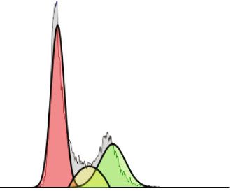
Oxidative Medicine and Cellular Longevity 3

of viability (viability %) was obtained dividing the number of CAL62 cells were seeded in 96-well plate at the density of
viable cells by the number of total cells (viable + dead cells). 5 × 10³ cells/well in FBS-supplemented medium and allowed
 to attach overnight. Then, cells were incubated with AFPE
2.5. Cell Cycle Analysis. Cell cycle progression was deter- (250 or 500 μM EqC) for 24 hours or with H2O2 (0.05, 0.5,
mined by flow cytometry using the Cytoflex instrument or 1 mM) for 4 hours, alone or in combination. Cells were
purchased from Beckman Coulter (Milan, Italy). To this then washed once with PBS, labeled with the fluorogenic
aim, cells were stained using Coulter DNA PREP Reagents
 dye 2 ′ ,7 ′ –dichlorofluorescin diacetate (DCFDA) for 45
Kit, (Beckman Coulter) according to manufacturer’s instruc-
 minutes protected from light, washed with Buffer 1X (pro-
tions. Briefly, TPC-1 and CAL62 (5 × 10⁴ cells) were treated vided in the kit), and analyzed on a microplate reader
with AFPE for 24 hours at the doses of 250 and 500 μM at (EnSpire Multimode plate reader by PerkinElmer, Milan,
37°C with 5% CO2. After cellular detachment, cells were Italy) with the excitation at 485 nm and the emission at
incubated with DNA PREP LPR reagent for 15 minutes 535 nm according to manufacturer’s instructions. ROS
and then with DNA PREP STAIN, containing 50 μg/ml of production was measured every 30 minutes for 4 hours.
propidium iodide, for 1 hour. TPC-1 and CAL62 stained The unlabeled CAL62 cells were analyzed and used as nega-
cells were acquired using the Cytoflex flow cytometer tive controls.
(Beckman Coulter). Cell cycle progression was analyzed
using Kaluza Analysis Software 2.1 (Beckman Coulter) 2.9. Cell Death Analysis. For the discrimination of live/dead
applying the Michael Fox algorithm. cells, we used 7-AAD staining. Specifically, thyroid cancer
2.6. Western Blot. Proteins were lysed in ice using JS Buffer cells (TPC-1 and CAL62) were pretreated with 500 μM of
supplemented with protease inhibitors and quantified using AFPE for 20 hours; then, the peroxide hydrogen (H2O2) at
a Bradford assay (Bio-Rad) according to standard protocols. concentration of 1 mM was added to the medium for other
 Protein lysates were separated in a sodium dodecyl 4 hours. At the end of the treatments, cells were detached
sulphate–polyacrylamide gel electrophoresis (SDS-PAGE); by trypsin, collected by centrifugation, and resuspended in
then, the nitrocellulose membranes were hybridized with 100 μl DPBS supplemented with 1% FBS containing 5 μl of
the following antibodies: rabbit polyclonal phospho-Rb a 10 μg/ml 7-AAD solution for 10 minutes in the dark before
(Ser795) diluted 1 : 1000 (#9301, Cell Signaling, Danvers, analysis. For the discrimination of early, late apoptosis and
USA) and mouse monoclonal anti-α-Tubulin diluted of necrosis, CAL62 cells untreated or treated were also
1 : 10000 (T-9026, Sigma-Aldrich, St. Louis, USA). Second- stained using Annexin V (Beckman Coulter) according to
ary anti-mouse and anti-rabbit antibodies coupled to horse- the manufacturer and analyzed.
radish peroxidase were diluted 1 : 3000 and purchased from
Bio-Rad. The bands were detected by an enhanced chemilu- 2.10. Statistical Analysis. The statistical analysis was per-
minescence detection kit (ECL, Thermo Fisher Scientific) formed with the GraphPad Prism 9 software (La Jolla, CA,
and analyzed by the Image Lab™ Software (Bio-Rad). USA), and the data are shown as the mean ± standard error
 of the mean (SEM). Differences were regarded as significant
2.7. RNA Extraction and q-RT-PCR. Total RNA containing when p < 0:05.
miRNAs was extracted from CAL62 cells untreated or For the differences between untreated and AFPE treat-
treated with AFPE 500 μM EqC for 24 hours using TRIzol ment group, Student’s t tests were used, while for the multi-
reagent (Thermo Fisher Scientific) in accordance with stan- ple comparisons between the four experimental groups
dard procedures and quantified using a NanoDrop spectro- (untreated, AFPE, H2O2, and AFPE/H2O2), ANOVA was
photometer (Thermo Fisher Scientific). calculated followed by a post hoc test (Bonferroni).
 cDNA was reverted using a miScript II RT Kit (Cat.
Number 218161) following manufacturer’s instructions. 3. Results
 The mature miRNA expression was determined by the
real-time PCR (q-RT-PCR) using SYBR Green PCR Master 3.1. Determination of Polyphenolic Content in AFPE. The
Mix (Cat. Number 218073). The following miRNA-specific total polyphenol amount in AFPE was quantified by Folin-
miScript primers (Cat. Number 218300) were used: miR-141 Ciocalteu assay and resulted to be approximately 126 mg of
(ID MS00003507), miR-145 (ID MS00003528), miR-200a-5p catechin per 100 g of apple flesh; this concentration was
(ID MS00009009), miR-551b-5p (ID MS00010157), and comparable to that measured in other studies [40, 42, 44].
miR-425 (ID MS00009695). All reagents were purchased from Then, we analyzed the polyphenols profile by HPLC in
Qiagen (Hilden, Germany). which the main o-diphenols (+)-catechin, (-)-epicatechin,
 The Ct-value of miRNAs was normalized with snU6 (ID and chlorogenic acid were detected on the basis of the reten-
MS00029204) used as endogenous control, and the fold tion time of standard references [43]. The data agree with
changes were calculated using the formula 2−ΔΔCt . Three the literature [35, 36, 43, 44].
independent experiments were performed in triplicate.
 3.2. AFPE Inhibits the Viability and Cell Cycle Progression of
2.8. Measurements of ROS Production. To quantitatively Anaplastic Thyroid Cancer Cell Line. Anaplastic thyroid
assess the reactive oxygen species (ROS) production, the cancer cell line, CAL62, was treated with increasing doses
DCFDA/H2DCFDA- Cellular ROS Assay Kit (ab113851) of AFPE (from 50 to 1000 μM EqC) for 24 hours, and the
purchased from Abcam (Cambridge, UK) was used. Briefly, number of live and dead cells and cell viability were
4 Oxidative Medicine and Cellular Longevity

 CAL62

 90 120 300 Algorithm : Michael H.Fox
 80
 ⁎⁎ 100 %Diploid: 100
 Cell number (x1000)

 70 %G1:55,23 at 162,43
 200

 Untreated
 %S:19,28
 80

 Counts
 60

 Viability (%)
 ⁎⁎⁎ %G2:25,5 at 309,11
 %CV: 9,54
 50 ⁎⁎⁎ G2/G1 : 1,9
 40 ⁎⁎⁎ 60 ⁎⁎ 100
 30
 ⁎⁎⁎ 40
 ⁎ All Events : 17010

 20 ⁎⁎ 0
 20
 10 0 100 200 300 400 500 600 700 800 900 1000
 0 0 PI F
 T 50 0 0 0 00
 T

 50

 0

 0

 0

 00
 25 50 75
 25

 50

 75
 N

 N 10
 10 Algorithm : Michael H.Fox

 AFPE ( M) AFPE ( M) 200 %Diploid: 100
 %G1:59,86 at 163
 150 %S: 9,7

 250 μM
 Counts
 Dead cells %G2:30,44 at 312
 %CV: 11,04
 Live cells 100 G2/G1 : 1,91

 All Events : 14500
 (a) (b) 50
 0
 0 100 200 300 400 500 600 700 800 900 1000
 PI F
 PE ⁎⁎
 NT AF 120
 Relative expression of

 100 Algorithm:Michael H.Fox
 150
 phospho-Rb

 110 kDa Phospho-Rb 80 %Diploid: 100
 %G1:62,89 at 169,44
 (Ser795) 60 %S: 9,51

 500 μM
 Counts
 100 %G2:27,59 at 305,36
 40 %CV: 12,41
 G2/G1 : 1,8
 55 MIN α-Tubulin 20 50 All Events : 12075
 0
 NT AFPE
 0
 (d) 0 100 200 300 400 500 600 700 800 900 1000
 PI F

 (c)

 G1
 S
 G2

Figure 1: Effects of AFPE treatment on proliferation and viability of anaplastic thyroid cancer cell line. (a) Cell counting was performed by
trypan blue reagent after treatment with increasing doses of AFPE for 24 hours. Black bars represent the viable cells (assessed by trypan blue
exclusion), and dashed bars represent the dead cells. (b) The percentage of cell viability (viable cells/total cells) was calculated in CAL62 cell
line treated with AFPE and counted in trypan blue dye. (c) Cell cycle analysis after propidium iodide (PI) staining of CAL62 untreated (NT)
or treated with 250 or 500 μM of AFPE EqC for 24 hours. Representative fluorescence-activated cell sorting (FACS) plots are shown applying
the Michael Fox algorithm. (d) Protein extracts from CAL62 cells treated with 500 μM of AFPE EqC for 24 hours and untreated were
analyzed by Western blot using antibodies for phospho-Rb (Ser795) and for α-tubulin. Representative figures from at least four
independent experiments are shown (left panel). Relative expression level of phospho-protein was calculated using α-tubulin as an
endogenous control (right panel). Values are expressed as mean of at least three different experiments ± standard error of the mean
(SEM). ∗ p < 0:05; ∗∗ p < 0:01; ∗∗∗ p < 0:001 versus control (NT).

simultaneously evaluated by counting cells after trypan blue 3.3. AFPE Reduced the Phosphorylation of Rb. To better
staining. investigate the molecular mechanisms underlying the effects
 As shown in Figure 1(a), the treatment with AFPE on cell cycle induced by AFPE treatments, we analyzed the
significantly reduced cell proliferation in a dose-dependent phosphorylated levels of the cell cycle regulator Rb by
manner; appreciable changes in cell number were observed Western blot. Consistent with the results obtained by flow
already at 50 μM of AFPE EqC. cytometry, treatment with 500 μM AFPE EqC for 24 hours
 As reported in Figure 1(b), viability percentages in reduces the phosphorylation of Rb protein in CAL62 cells
CAL62 were approximately 46, 28, and 13% after treatments (Figure 1(d)) compared to control. This result confirms that
with 500, 750, and 1000 μM EqC of AFPE, respectively, in AFPE treatment acts on G1/S phase of the cell cycle through
comparison to untreated (NT) cells that showed approxi- Rb signaling pathway.
mately 100% of viability.
 Additionally, 24 hours after treatments with 250 and 3.4. Anticancer Effects of AFPE are Mediated by Deregulation
500 μM of AFPE, cell cycle progression was analyzed by flow of miRNA Expression. MicroRNAs (miRNAs) are small mol-
cytometry with propidium iodide staining. Figure 1(c) shows ecules able to drive different biological process through
that the dose of 500 μM EqC of AFPE induces a slight accu- downregulation of specific target genes. Deregulation of
mulation of CAL62 cells at G1 phase compared to untreated miRNA expression drives a significant contribution in sev-
cells. eral human neoplasia, including thyroid cancer development
Oxidative Medicine and Cellular Longevity 5

 CAL62
 8 ⁎⁎⁎ 20 60 ⁎⁎
 ⁎⁎

 (relative to control)
 (relative to control)

 (relative to control)
 6 15

 Fold change
 40
 Fold change

 Fold change
 4 10

 20
 2 5

 0 0 0
 NT AFPE NT AFPE NT AFPE
 miR-141 miR-145 miR-200a-5p
 ⁎⁎
 8 250
 ⁎⁎

 200
 (relative to control)

 6 (relative to control)
 Fold change

 Fold change

 150
 4
 100
 2
 50

 0 0
 NT AFPE NT AFPE
 miR-425 miR-551b-5p

Figure 2: Effects of AFPE treatment on miRNA expression levels. The relative expression level of the indicated miRNAs was measured in
CAL62 cells untreated (named NT) or treated with 500 μM of AFPE EqC for 24 hours by q-RT-PCR. Fold change was calculated using the
formula 2−ΔΔCt in which the Ct value of miRNA was normalized with an internal control (snU6). Bar represents the mean ± SEM of three
independent experiments performed in triplicate. ∗∗ p < 0:01; ∗∗∗ p < 0:001.

and progression, since they can act as onco-miR or tumor dead cells with trypan blue reagent after 24 hours from
suppressor miRNAs [46, 47]. Notably, in literature, it is also AFPE treatments.
reported that polyphenols exert their anticancer properties The results obtained evidence that up to 250 μM AFPE
by targeting different miRNAs [48, 49]. Thus, it is possible EqC, no appreciable changes in cell number and cell viability
that the anticancer activity exerted by AFPE is in part due were observed. The number of live cells was decreased after
to the modulation of miRNA expression also in our cell treatments with higher doses of AFPE, and in parallel, the
model system. number of dead cells was increased (Figures 3(a) and 3(b)).
 To verify if AFPE can modulate miRNA expression Accordingly, as reported in Figure 3(b), when we treated
levels, CAL62 cells were treated with 500 μM of AFPE EqC TPC-1 cell line with increasing doses of AFPE for 24 hours,
for 24 hours, and q-RT-PCR was performed to analyze the the reduction of viability occurred: from approximately
expression level of several miRNAs that are known to be 100% in untreated (NT) cells to less than 50% in TPC-1
involved in thyroid carcinogenesis. Figure 2 shows that treated with 750 and 1000 μM EqC of AFPE.
CAL62 cells treated with 500 μM of AFPE EqC presented The effects of AFPE treatments were also evaluated by
an increased expression level of the members of miR-200 flow cytometry analysis of the cell cycle distribution. As
family (miR-200a-5p and miR-141) and of miR-145, miR- reported in Figure 3(c), in agreement with the results in
425, and miR-551-5p. ATC cells, AFPE induced an accumulation of TPC-1 popu-
 These data suggest that miRNA targeting is one of the lation in G1 phase compared to untreated cells.
mechanisms modulated by AFPE to exert its anticancer All together, these data unveiled that the proliferation
activity in thyroid carcinoma cell lines. and the viability were reduced in human papillary and
 anaplastic thyroid cancer cells exposed to Annurca apple
 extracts in a dose-dependent manner.
3.5. AFPE Inhibits the Viability and Cell Cycle Progression of
Papillary Thyroid Cancer Cell Line. To further confirm the
results obtained, we also tested the viability of a papillary 3.6. Antioxidant Effect of AFPE on Human Thyroid Cancer
thyroid cancer cell line, TPC-1, after treatment with increas- Cells. Since the involvement of polyphenol extract from
ing doses of AFPE (ranging from 50 to 1000 μM EqC). Annurca apple fruits in the regulation of reactive oxygen
 The effects obtained on cell proliferation and on cell via- species (ROS) generation is reported [41], we investigated
bility were simultaneously evaluated by counting viable and the effects of AFPE on oxidative stress.
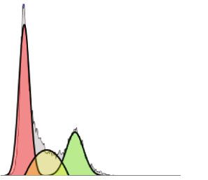
6 Oxidative Medicine and Cellular Longevity

 TPC-1
 350 Algorithm : Michael H.Fox
 %Diploid: 100
 300 %G1:55,47 at 108,6
 300 %S: 12,11
 Cell number (x1000)

 %G2:32,41 at 213,82

 Untreated
 250

 Counts
 %CV: 14,42
 200 G2/G1 : 1,91

 200 All Events : 15918
 ⁎⁎⁎ 100
 150 ⁎⁎⁎
 ⁎⁎⁎
 100 0
 0 100 200 300 400 500 600 700 800 900 1000
 50
 PI F
 0
 300 Algorithm : Michael H.Fox %Diploid: 100
 50
 T

 0

 0

 0

 00
 25

 50

 75
 N

 %G1:60,12 at 109,2

 10
 %S: 12,15
 %G2:27,73 at 212,25
 200

 250 μM
 AFPE ( M) %CV: 14,53

 Counts
 G2/G1 : 1,93
 Dead cells
 All Events : 13935
 Live cells 100

 (a)
 0
 120 0 100 200 300 400 500 600 700 800 900 1000
 PI F
 100
 400 Algorithm : Michael H.Fox
 %Diploid: 100
 80
 Viability (%)

 %G1:67,07 at 108,01
 %S: 17,21
 300
 60 ⁎⁎ ⁎⁎ %G2:25,67 at 210,18

 500 μM
 Counts

 %CV: 14,11
 G2/G1 : 1,95
 200
 40
 All Events : 14894
 100
 20

 0 0
 0 100 200 300 400 500 600 700 800 900 1000
 50
 T

 0

 0

 0

 00
 25

 50

 75
 N

 PI F
 10

 AFPE ( M) (c)
 (b) G1
 S
 G2

Figure 3: Effects of AFPE treatment on the proliferation and viability in papillary thyroid cancer cell line. (a) TPC-1 cells were treated with
the indicated doses of AFPE, and after 24 hours, cells were collected by trypsinization, stained for 5 minutes with trypan blue, and counted;
black bars represent the viable cells, and dashed bars represent the dead cells. (b) After counting cells with trypan blue reagent, the
percentage of cell viability (viable cells/total cells) was calculated. Values are expressed as mean of two different experiments ± standard
error of the mean (SEM). ∗∗ p < 0:01; ∗∗∗ p < 0:001. (c) Cell cycle analysis after propidium iodide staining was analyzed by flow cytometry
in TPC-1 cell line treated with different doses of AFPE for 24 hours. Representative fluorescence-activated cell sorting (FACS) plots are
shown applying the Michael Fox algorithm.

 Thus, we initially verified if AFPE exerts antioxidant or the exposure to hydrogen peroxidase (H2O2). To this aim,
prooxidant effects in our cell model system. To this aim, first, we treated CAL62 cells with different doses of H2O2
CAL62 cell line was treated with 250 and 500 μM of AFPE (0.05, 0.5, and 1 mM) and measured ROS production every
EqC for 24 hours, and the production of reactive oxygen 30 minutes for 4 hours using the DCFDA/H2DCFDA-
species (ROS) within the live cells was quantitatively Cellular ROS assay kit. As shown in Figure 4(b), we observed
assessed by the DCFDA/H2DCFDA-Cellular ROS assay a significant increased level of ROS production starting from
kit. As shown in Figure 4(a), treatment with 250 μM of 1 hour of treatment with the lower dose of H2O2 (0.05 mM)
AFPE EqC decreased the production of ROS of ~3.6-fold, and continuing progressively in time-dependent manner.
while 500 μM of AFPE EqC treatment reduces the produc- CAL62 cells treated with the highest concentrations of
tion of ROS of ~5-fold compared to untreated (NT) cells. H2O2 (0.5 and 1 mM) presented a significantly increased
These data suggest that AFPE exerts antioxidant effects in level of ROS starting 30 minutes with the maximum peak
thyroid cancer cell line in dose-dependent manner. after 1 hour and 30 minutes (Figure 4(b)).
 Next, we verified if AFPE treatment is also able to protect Based on this experiment, we started using the lower
thyroid cancer cells from extracellular stress signal such as dose of H2O2 (0.05 mM) to investigate the protective effect
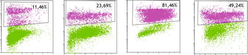
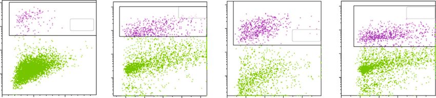
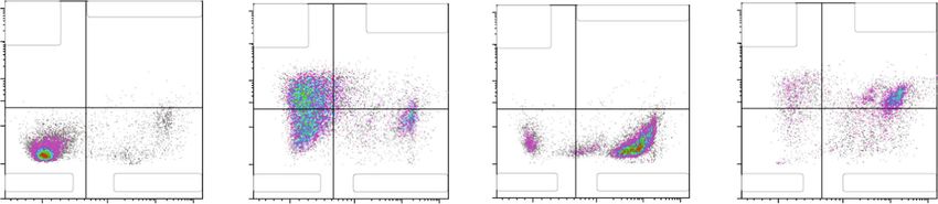
Oxidative Medicine and Cellular Longevity 7

 CAL62
 350000
 ⁎⁎⁎b, c ⁎⁎⁎ b, c ⁎⁎⁎b, c ⁎⁎⁎
 ⁎⁎⁎ b, c ⁎⁎⁎ b, c
 300000 b, c ⁎⁎⁎
 ⁎⁎⁎ b, c ⁎⁎⁎
 250000
 a ⁎⁎⁎

 Fluorescence activity
 40000 ⁎⁎⁎
 a ⁎⁎⁎ a
 a ⁎⁎⁎
 Fluorescence activity

 200000
 30000 ⁎⁎ a ⁎⁎⁎
 b, c ⁎⁎⁎
 150000
 a ⁎⁎⁎
 20000
 100000
 a ⁎⁎⁎
 10000 50000

 0 0
 NT AFPE [250 μM] AFPE [500 μM] 0 0:30 1 1:30 2:00 2:30 3:00 3:30 4:00
 Time (hours)

 Untreated H2O2 [0.5 mM]
 H2O2 [0.05 mM] H2O2 [1 mM]

 (a) (b)

 ⁎⁎⁎
 ⁎⁎⁎
 ⁎⁎⁎ ⁎⁎⁎
 400000
 ⁎⁎⁎ ⁎
 Fluorescence activity

 300000 ⁎⁎⁎ ⁎⁎
 ⁎⁎⁎

 200000

 100000 ⁎⁎

 0
 AFPE – + – + – +
 H2O2 (0.05 mM) – – + + – –
 H2O2 (1 mM) – – – – + +
 (c)

Figure 4: Effects of AFPE treatment on the production of reactive oxygen reagents (ROS) in anaplastic thyroid cancer cell line. (a) CAL62
cells untreated or treated for 24 hours with AFPE (250 or 500 μM EqC) were stained with the fluorogenic dye DCFDA (2 ′ ,7 ′ -
dichlorofluorescein diacetate) for ROS detection. Fluorescence activity was read with the excitation at 485 nm and the emission at
535 nm. Significant differences were analyzed using paired t-test. (b) ROS quantification within the CAL62 cells treated with different
doses of H2O2 (0.05, 0.5, and 1 mM) was determined using DCFDA. Fluorescence activity was measured every 30 minutes for 4 hours.
Significant difference compared to the untreated cells was analyzed using T-test. Letter “a” indicates the significant difference between
untreated and H2O2 (0.05 mM), letter “b” significant difference between untreated and H2O2 (0.5 mM), and letter “c” significant
difference between untreated and H2O2 (1 mM). (c) ROS production in CAL62 cells untreated, treated with AFPE (500 μM EqC) for
24 hours, treated with H2O2 (0.05 or 1 mM) for 4 hours, or pretreated with AFPE (500 μM EqC) for 20 hours and then treated
with H2O2 (0.05 or 1 mM) for 4 hours. Statistical analysis was performed using ANOVA followed by a Bonferroni post hoc test. All
the data are shown as mean ± SEM of at least three independent experiments in triplicate. ∗ p < 0:05; ∗∗ p < 0:01; ∗∗∗ p < 0:001.

exerted by APFE in our model system. Thus, we induced the Overall, these results provided evidence that AFPE exerts
oxidative stress with H2O2 (0.05 mM) treatment in CAL62 antioxidant activity by inhibiting ROS production in thyroid
cells pretreated for 20 hours with AFPE (500 μM EqC). As cancer cell lines.
shown in Figure 4(c), CAL62 treated for 4 hours with
H2O2 presented a high production of ROS, while CAL62 3.7. Characterization of the Protective Effect of AFPE on
pretreated with AFPE presented a reduction in the genera- Oxidative Stress. Next, we also further characterize the pro-
tion of ROS compared to cells treated with H2O2 alone. tective role of AFPE in oxidative stress in thyroid cancer cell
Additionally, we also found that the pretreatment with line by evaluating early, late apoptosis or necrosis. To this
AFPE protects cells from the ROS production induced by aim, CAL62 cells were seeded in medium containing 10%
highest doses of H2O2 (1 mM) (Figure 4(c)). FBS in 6-well plates at the density of 1 × 10⁵ cells/well and
8 Oxidative Medicine and Cellular Longevity

 CAL62
 NT AFPE H2O2 AFPE/ H2O2
 106 106
 106 106

 105 105
 105 105
 7-AAD – FL3

 104 104
 104 104

 103 103
 103 103

 102 102
 200 400 600 800 1000 0 200 400 600 800 200 400 600 800 1000 0 200 400 600 800
 (× 103) (× 103) (× 103)
 (× 103)

 SSC–A

 (a)

 ⁎⁎

 ⁎⁎

 ⁎⁎⁎

 100 ⁎⁎⁎ ⁎⁎
 90
 80
 70
 Cell death (%)

 60
 50
 40
 30
 20
 10
 0
 NT AFPE H2O2 AFPE/H2O2

 (b)

Figure 5: Effects of AFPE treatment against H2O2-induced oxidative stress in anaplastic thyroid cell line. CAL62 cell line was pretreated
with AFPE, and then, the oxidative stress was induced by H2O2 treatment. After 24 hours, the percentage of dead cells (%) was
determined by flow cytometry. (a) Representative fluorescence-activated cell sorting (FACS) plots are shown from one of two
independent experiments. (b) Bar plot of the experimental data, values are expressed as mean of two different experiments ± standard
error of the mean (SEM). ∗∗ p < 0:05; ∗∗∗ p < 0:001. NT: untreated cells; H2O2: cells treated with hydrogen peroxide; AFPE: cells treated
with AFPE alone; AFPE/H2O2: cells pretreated with AFPE and stressed using hydrogen peroxide. For the multiple comparisons,
statistical analysis between the four experimental groups (NT, AFPE, H2O2, and AFPE/H2O2) and ANOVA followed by a post hoc test
(Bonferroni) were performed.

the day after treated with AFPE (500 μM EqC) for 24 hours tive), and 14.87% of necrosis (Annexin V negative/7-AAD
alone or pretreated with AFPE (500 μM EqC) for 20 hours positive). Treatment with H2O2 induced mainly cellular
and then treated with H2O2 (1 mM) for 4 hours or only necrosis (89.45%) and not apoptosis. AFPE pretreatment
treated with H2O2 (1 mM) for 4 hours. The percentage of could counteract H2O2 oxidative damage; indeed, we found
cell death was examined by flow cytometry analysis with that cells treated with AFPE for 20 hours and then with
7-AAD staining. H2O2 for 4 hours show 14.5% and 52.15% of, respectively,
 As shown in Figure 5, the treatment with AFPE (500 μM early and late apoptosis. Furthermore, AFPE pretreatment
EqC) alone induced an increase in cell death percentage that reduced the percentage of necrotic cells to 25.97% in com-
raised from 11.3% to 20.2% in CAL62 cells; H2O2 alone parison to 89.45% detected in H2O2 treated cells (Figure 6).
caused a significant increase in the percentage of cell death Then, we analyzed the protective effect of AFPE on oxi-
resulting to be approximately 79% in CAL62, while in the dative stress in TPC-1 cells. As shown in Figure 7, the treat-
cells pretreated with AFPE, cell death percentage was ment of TPC-1 cells with AFPE (500 μM EqC) alone induced
approximately 46%. an increase in cell death that raised from 2.9% to 17.9%.
 Then, to better characterize the effects on cell death, we H2O2 alone caused a significant increase in the percentage
stained CAL62 cells treated with AFPE (500 μM EqC) or of cell death resulting to be approximately 38%, while in
H2O2 (1 mM) or the combination with both Annexin V the TPC-1 cells pretreated with AFPE, cell death percentage
and 7-AAD and analyzed the results by flow cytometry. was approximately 27% (Figure 7).
CAL62 cells treated with only AFPE disclosed: 40.85% of These results provided evidence that, in human thyroid
early apoptosis (Annexin V positive/7-AAD negative), cancer cells, death induced by oxidative stress was reduced
10.32% of late apoptosis (Annexin V positive/7-AAD posi- by the pretreatment with AFPE.
Oxidative Medicine and Cellular Longevity 9

 CAL62

 Untreated AFPE H2O2 AFPE+H2O2
 107 Early Late apoptosis: 0,42% 107 Early Late apoptosis: 107 Early Late apoptosis: 0,58% 107 Early Late apoptosis:
 apoptosis: apoptosis: 10,32% apoptosis: apoptosis: 52,15%
 106 0,01% 106 40,85% 106 0,14% 106 14,50%
 Annexin V FITC-A

 105 105 105 105
 40000 40000 40000 40000
 20000 20000 20000 20000

 0 0 0 0
 Live: 91,99% Necrosis: 7,58% Live: 33,96% Necrosis: 14,87% Live: 9,83% Necrosis: 89,45% Live: 7,39% Necrosis: 25,97%
 –20000 –20000 –20000 –20000
 0 104 105 106 0 104 105 106 0 104 105 106 0 104 105 106

 7-AAD - FL3

Figure 6: Effects of AFPE on apoptosis and necrosis in CAL62 cells. CAL62 cells were treated with AFPE (500 μM EqC), H2O2 (1 mM), or
the combination as described above. Early and late apoptosis and necrosis were evaluated by staining cells with Annexin V and 7-AAD and
by analyzing with flow cytometry. Shown are early (Annexin V positive/7-AAD negative) and late (Annexin V positive/7-AAD positive)
apoptotic and necrotic (Annexin V negative/7-AAD positive) cells.

 TPC-1
 NT AFPE H2O2 AFPE/H2O2
 107 107
 107 107
 18,82% 25,04%

 106 2,80%
 106
 106 106
 35,10%
 7-AAD – FL3

 105 105
 105 105

 104 104
 104 104

 0 500 1000 200 400 600 800 1000 0 200 400 600 800 0 200 400 600 800
 (× 103) (× 103) (× 103) (× 103)
 SSC –A

 (a)
 ⁎⁎
 ⁎⁎
 50 ⁎⁎

 40
 Cell death (%)

 30
 20
 10
 0
 NT AFPE H2O2 AFPE/H2O2

 (b)

Figure 7: Effects of AFPE treatment against H2O2-induced oxidative stress in papillary thyroid cell line. TPC-1 cell line was pretreated
with AFPE, and then, the oxidative stress was induced by H2O2 treatment. After 24 hours, dead cells (%) were determined by flow
cytometry. (a) Representative plots of cell death analysis obtained by fluorescence-activated cell sorting (FACS). One of two
independent experiments is shown. (b) Bar plot of the experimental data, values of percentage of dead cells are expressed as mean
of two different experiments ± standard error of the mean (SEM). ∗∗ p < 0:01. NT: untreated cells; H2O2: cells treated with hydrogen
peroxide; AFPE: cells treated with AFPE alone; AFPE/H2O2: cells pretreated with AFPE and stressed using hydrogen peroxide. For the
multiple comparisons, statistical analysis between the four experimental groups (NT, AFPE, H2O2, and AFPE/H2O2) and ANOVA
followed by a post hoc test (Bonferroni) were performed.

4. Discussion apple fruit could have an important role in preventing
 cancer by exerting antioxidant activity [7, 29]. Indeed, the
Phytochemicals are compounds available in plant-including beneficial effects of these compounds are attributable, at least
foods characterized by antioxidant and antitumor abilities in part, to their ability to scavenge the ROS [44, 50, 51].
[6, 18]. Among these natural substances, polyphenols in However, in other context, polyphenols act also as
10 Oxidative Medicine and Cellular Longevity

prooxidant agents since they are able to generate ROS invasion, and metastasis [62–64]. It is also reported that
and induce cellular oxidative stress and, consequently, cell the restoration of miR-141 and of miR-200a expression
death [39, 41, 52, 53]. induced an epithelial morphology in ATC cells [65].
 In addition to these abilities, several in vitro studies Concerning miR-425, upregulated by AFPE in CAL62
showed that in human cancer cell lines, apple polyphenols cells, in literature is described to be decreased in PTC tissues;
inhibit cell proliferation and invasion [35, 43, 54]. furthermore, the transfection of miR-425 mimic decreases
 For all these reasons, the growing scientific interest is the motility phenotype of PTC cells [66]. Interestingly, the
focused on identifying the biological mechanisms and the aberrant expression of miR-425 is also correlated to the oxi-
signal transduction pathways underpinning the chemo- dative stress in breast cancer cell lines where it regulates the
preventive activities of these compounds. apoptosis by PTEN axis [67, 68]. Indeed, it has been recently
 Pathological conditions of the thyroid are often associ- highlighted that miRNA plays an important role in the
ated with oxidative stress that results mainly from excessive homeostasis of ROS during carcinogenesis [67, 69, 70].
production of ROS, including H2O2 possibly concurring to Finally, previous studies revealed that the polyphenols
cell transformation and genomic instability [55]. Since the exert their health benefits in mouse model by regulating
thyroid follicular cells require H2O2 to synthesize thyroid the expression of several miRNAs including miR-551b-5p
hormones (T3 and T4), in thyroid gland, it is very difficult that we also found to be upregulated in CAL62 following
to regulate the balance between ROS production and scav- the AFPE exposure [71, 72].
enging [55]. Indeed, despite the rate of ROS production is Concerning other molecular mechanisms deregulated by
efficiently controlled and removed by natural repair mecha- AFPE in thyroid cancer cells, there are evidences that AFPE
nisms, it can occur that the endogenous antioxidant systems inhibits the expression and activity of several oncoproteins
are not sufficed. These mechanisms may be supported by related to cell survival and proliferation such as AKT, NF-
exogenous substances with genoprotective and antioxidative κB, and β-CATENIN [41]. These oncogenic pathways are
activity that can be provided to the organism from a diet rich also altered in thyroid cancer, suggesting a possible negative
in food of plant origin [56, 57]. Interestingly, it was observed regulation of them by AFPE exposure [73–76].
that polyphenols such as genistein, resveratrol, and querce- Here, we have also investigated the effects of AFPE treat-
tin might be effective for thyroid cancer therapy, including ment against H2O2-induced oxidative stress in papillary and
for anaplastic thyroid cancer [14, 26, 58]. anaplastic thyroid cell lines. Our in vitro results show that, in
 In this context, within this study, we aimed to deepen the human thyroid cancer cell lines, cell death induced by oxida-
knowledge on the effects of polyphenol extracts obtained tive stress was reduced by the pretreatment with AFPE.
from Annurca apple flesh (AFPE). We have observed that The knowledge regarding the polyphenols as modulator
when AFPE was administered for 24 hours, the proliferation of oxidative stress based on their differential redox status is
of thyroid cancer cell lines (CAL62 and TPC-1) was inhib- already documented in human cancer [77].
ited in a dose-dependent manner. By flow cytometry, we also This could offer a window of opportunities in thyroid
found that AFPE treatment induced cell death in both cancer since AFPE can lead to apoptosis and cell cycle arrest
CAL62 and TPC-1 cell lines. Coherently, with our results, of malignant cells, but it is also able to protect human cells
in breast cancer, AFPE inhibits cell growth, causes cell cycle from the DNA breakage induced by reactive oxygen species.
arrest, and induces apoptosis in MDA-MB-231 cell line [41]. Thus, based on these findings, this study suggest that
 Here, we also investigated the molecular mechanisms development of phytochemical could be important for
underlying the effects observed following AFPE treatments thyroid cancer treatment.
in CAL62 cell line, and we found that the anticancer activity A limitation of this work is the high concentration of
induced by AFPE exposure is associated with deregulated AFPE used in the thyroid cancer cell lines that are above
expression of several miRNAs which are known to be those achievable in vivo. It should be also pointed out that
involved in thyroid cancer and in mediating the effects of all the experiments described here have been performed in
polyphenol. Indeed, we found that the expression level of medium containing 10% serum.
miR-141, miR-145, miR-200a-5p, miR-425, and miR-551b- Polyphenols are low bioavailability compounds with
5p resulted upregulated in CAL62 treated with AFPE with poor solubility in water. The absorption in the colon is
respect to untreated cells. different for the diverse groups of polyphenols; indeed,
 In literature, several papers reported that miR-145 is the flavan-3-ol derivates demonstrate higher absorption
downregulated in thyroid cancer where it acts as a tumor rate compared to quercetin [78]. Polyphenols are absorbed
suppressor by impairing cancer cell growth, cell motility, by the gut as aglycones and then released into the blood
and metastasis through targeting AKT3 [59] and RAB5C after chemical change that modifies their effects [79]. Dur-
[60] genes or by regulating the NF-κB pathway [61]. ing gastrointestinal digestion, several modifications occur
 Interestingly, in our cell model system, we found that that could affect their bioavailability, stability, and bioac-
miR-141 and miR-200a-5p are increased after AFPE expo- tivity [80].
sure. It is well known that the members of miR-200 family Importantly, the bioavailability of polyphenols is limited
(miR-200a, miR-200b, miR-200c, miR-141, and miR-429) by their extensive metabolism [81, 82]. Additionally, several
play an important role in thyroid cancer. In thyroid cancer, factors influence the bioavailability of polyphenols: environ-
it is reported that the loss of miR-141 and of miR-200a mental factors, food processing, interaction with other com-
expression is associated with thyroid cancer proliferation, pounds, genetic factors, the composition of microflora, and
Oxidative Medicine and Cellular Longevity 11

the length of the microbiome [83, 84]. Indeed, Wruss Ministero dell’Istruzione, dell’Università e della Ricerca,
et al. studied the pharmacokinetic of polyphenols in apple Progetti di Ricerca di Rilevante Interesse Nazionale (PRIN)-
juice, in healthy subjects, and discovered numerous differ- Bando 2017 (grant 2017MHJJ55).
ences in apple polyphenol pharmacokinetics [85]. Also,
Tenore and colleagues evaluated the bioaccessibility and
bioavailability of AFPE, by simulating an in vitro gastro- References
intestinal digestion [86]. Moreover, novel biotechnologies
for improving bioavailability of polyphenols have been [1] J. A. Fagin and S. A. Wells Jr., “Biologic and clinical perspec-
developed based on different approaches. For example, tives on thyroid cancer,” The New England journal of medi-
the encapsulation of polyphenols could contribute to the cine., vol. 375, no. 11, pp. 1054–1067, 2016.
intensification in their shelf life and could avoid their loss [2] M. R. Haroon Al Rasheed and B. Xu, “Molecular alterations in
in activity, thus improving the bioavailability [87]. Not- thyroid carcinoma,” Surg Pathol Clin., vol. 12, no. 4, pp. 921–
 930, 2019.
withstanding, further studies are necessary on the bio-
availability and bioaccessibility of polyphenols with the [3] M. Schlumberger and S. Leboulleux, “Current practice in
 patients with differentiated thyroid cancer,” Nature Reviews
aim of developing novel strategies for the production of
 Endocrinology, vol. 18, 2021.
functional foods.
 [4] N. Pozdeyev, M. M. Rose, D. W. Bowles, and R. E. Schweppe,
 “Molecular therapeutics for anaplastic thyroid cancer,” Semi-
5. Conclusion nars in Cancer Biology, vol. 61, pp. 23–29, 2020.
In conclusion, our findings suggest that AFPE could be a [5] V. Tiedje and J. A. Fagin, “Therapeutic breakthroughs for
 metastatic thyroid cancer,” Nature Reviews. Endocrinology,
promising tool for thyroid cancer treatment. Indeed, we
 vol. 16, no. 2, pp. 77-78, 2020.
found that AFPE acts on cell proliferation and viability. On
 [6] A. Hosseini and A. Ghorbani, “Cancer therapy with phyto-
the other hand, AFPE also acts as an antioxidant modulator
 chemicals: evidence from clinical studies,” Avicenna J Phy-
protecting cells from oxidative stress in the presence of oxi- tomed., vol. 5, no. 2, pp. 84–97, 2015.
dant agent such as peroxide hydrogen. Here, we have also
 [7] L. Koklesova, A. Liskova, M. Samec et al., “Genoprotective
clarified some of the biological mechanisms involved in the activities of plant natural substances in cancer and chemopre-
effects of this polyphenol in thyroid cancer, as deregulation ventive strategies in the context of 3P medicine,” The EPMA
of several cancer-related miRNAs and decreased phosphory- Journal, vol. 11, no. 2, pp. 261–287, 2020.
lation of Rb protein. Nevertheless, other studies are needed [8] S. J. Hosseinimehr and S. A. Hosseini, “Radiosensitive effect of
to better dissect the molecular mechanisms underlying the curcumin on thyroid cancer cell death induced by radioiodine-
effects of AFPE on the thyroid cancer cells. 131,” Interdisciplinary Toxicology, vol. 7, no. 2, pp. 85–88,
 2014.
Abbreviations [9] A. Rauf, M. Imran, M. S. Butt, M. Nadeem, D. G. Peters, and
 M. S. Mubarak, “Resveratrol as an anti-cancer agent: a review,”
7-AAD: 7-Amino-actinomycin D (7-AAD) Critical Reviews in Food Science and Nutrition, vol. 58, no. 9,
ATC: Anaplastic thyroid cancer pp. 1428–1447, 2018.
AFPE: Annurca flesh polyphenolic extract [10] L. Zhao, X. Yuan, J. Wang et al., “A review on flavones target-
CatEq: Catechin equivalent ing serine/threonine protein kinases for potential anticancer
DMEM: Dulbecco’s modified Eagle’s medium drugs,” Bioorganic & Medicinal Chemistry, vol. 27, no. 5,
FACS: Fluorescence-activated cell sorting p. 677, 2019.
FBS: Fetal bovine serum [11] L. Shu, K. L. Cheung, T. O. Khor, C. Chen, and A. N. Kong,
PI: Propidium iodide “Phytochemicals: cancer chemoprevention and suppression
PBS: Phosphate-buffered saline of tumor onset and metastasis,” Cancer Metastasis Reviews,
PTC: Papillary thyroid cancer. vol. 29, no. 3, pp. 483–502, 2010.
 [12] M. Dvorakova and P. Landa, “Anti-inflammatory activity of
Data Availability natural stilbenoids: a review,” Pharmacological Research,
 vol. 124, pp. 126–145, 2017.
The data underlying this study are included in the article; [13] N. Yahfoufi, N. Alsadi, M. Jambi, and C. Matar, “The immuno-
further inquiries can be directed to the corresponding modulatory and anti-inflammatory role of polyphenols,”
authors. Nutrients, vol. 10, no. 11, p. 1618, 2018.
 [14] H. J. Shin, K. A. Hwang, and K. C. Choi, “Antitumor effect of
Conflicts of Interest various phytochemicals on diverse types of thyroid cancers,”
 Nutrients, vol. 11, no. 1, p. 125, 2019.
The authors declare no conflict of interest. [15] M. Boccellino and S. D'Angelo, “Anti-obesity effects of poly-
 phenol intake: current status and future possibilities,” Interna-
Acknowledgments tional Journal of Molecular Sciences, vol. 21, no. 16, p. 5642,
 2020.
We thank R.M. Mariniello for contribution with initial cell [16] S. D'Angelo, “Current evidence on the effect of dietary poly-
viability experiments. This work was in part supported by phenols intake on brain health,” Current Nutrition & Food
the Ricerca Corrente, Italian Ministry of Health, and by Science, vol. 16, no. 8, pp. 1170–1182, 2020.
12 Oxidative Medicine and Cellular Longevity

[17] S. D'Angelo, “Polyphenols: potential beneficial effects of these [34] R. R. Watson, V. R. Preedy, and S. Zibadi, Polyphenols in
 phytochemicals in athletes,” Current Sports Medicine Reports, Human Health and Disease, Academic Press, 2013.
 vol. 19, no. 7, pp. 260–265, 2020. [35] S. T. Lin, S. H. Tu, P. S. Yang et al., “Apple polyphenol phlor-
[18] A. Ullah, S. Munir, S. L. Badshah et al., “Important flavonoids etin inhibits colorectal cancer cell growth via inhibition of the
 and their role as a therapeutic agent,” Molecules, vol. 25, no. 22, type 2 glucose transporter and activation of p53-mediated sig-
 p. 5243, 2020. naling,” Journal of Agricultural and Food Chemistry, vol. 64,
[19] B. Mariarosaria, L. Quagliuolo, and D. A. Stefania, “Annurca no. 36, pp. 6826–6837, 2016.
 apple biophenols’ effects in combination with cisplatin on [36] R. Lo Scalzo, A. Testoni, and A. Genna, “'Annurca' apple fruit,
 A549 cells,” Current Nutrition & Food Science, vol. 17, no. 1, a southern Italy apple cultivar: textural properties and aroma
 pp. 111–120, 2021. composition,” Food Chemistry, vol. 73, no. 3, pp. 333–343,
[20] R. Meccariello and S. D'Angelo, “Impact of polyphenolic-food 2001.
 on longevity: an elixir of life. An overview,” Antioxidants [37] A. Napolitano, A. Cascone, G. Graziani et al., “Influence of
 (Basel)., vol. 10, no. 4, p. 507, 2021. variety and storage on the polyphenol composition of apple
[21] H. Amawi, C. R. Ashby, T. Samuel, R. Peraman, and A. K. flesh,” Journal of Agricultural and Food Chemistry, vol. 52,
 Tiwari, “Polyphenolic nutrients in cancer chemoprevention no. 21, pp. 6526–6531, 2004.
 and metastasis: role of the epithelial-to-mesenchymal (EMT) [38] S. D'Angelo, R. La Porta, M. Napolitano, P. Galletti,
 pathway,” Nutrients, vol. 9, no. 8, p. 911, 2017. L. Quagliuolo, and M. Boccellino, “Effect of Annurca apple
[22] A. Bahrami, M. Majeed, and A. Sahebkar, “Curcumin: a potent polyphenols on human HaCaT keratinocytes proliferation,”
 agent to reverse epithelial-to-mesenchymal transition,” Cellu- Journal of Medicinal Food, vol. 15, no. 11, pp. 1024–1031,
 lar Oncology (Dordrecht), vol. 42, no. 4, pp. 405–421, 2019. 2012.
[23] L. Dal Maso, C. Bosetti, C. La Vecchia, and S. Franceschi, “Risk [39] S. D'Angelo, E. Martino, C. P. Ilisso, M. L. Bagarolo,
 factors for thyroid cancer: an epidemiological review focused M. Porcelli, and G. Cacciapuoti, “Pro-oxidant and pro-
 on nutritional factors,” Cancer Causes & Control, vol. 20, apoptotic activity of polyphenol extract from Annurca apple
 no. 1, pp. 75–86, 2009. and its underlying mechanisms in human breast cancer cells,”
[24] W. J. Choi and J. Kim, “Dietary factors and the risk of thyroid International Journal of Oncology, vol. 51, no. 3, pp. 939–948,
 cancer: a review,” Clin Nutr Res., vol. 3, no. 2, pp. 75–88, 2014. 2017.
[25] L. Allegri, F. Rosignolo, C. Mio, S. Filetti, F. Baldan, and [40] S. D'Angelo, E. Martino, and G. Cacciapuoti, “Effects of
 G. Damante, “Effects of nutraceuticals on anaplastic thyroid Annurca apple (Malus pumila cv Annurca) polyphenols on
 cancer cells,” Journal of Cancer Research and Clinical Oncol- breast cancer cells,” Current Nutrition & Food Science,
 ogy, vol. 144, no. 2, pp. 285–294, 2018. vol. 15, no. 7, pp. 745–751, 2019.
[26] S. A. Ozturk, E. Alp, A. S. Yar Saglam, E. Konac, and E. S. [41] E. Martino, D. C. Vuoso, S. D’Angelo et al., “Annurca_ apple
 Menevse, “The effects of thymoquinone and genistein treat- polyphenol extract selectively kills MDA-MB-231 cells
 ment on telomerase activity, apoptosis, angiogenesis, and sur- through ROS generation, sustained JNK activation and cell
 vival in thyroid cancer cell lines,” Journal of Cancer Research growth and survival inhibition,” Scientific Reports, vol. 9,
 and Therapeutics, vol. 14, no. 2, pp. 328–334, 2018. no. 1, p. 13045, 2019.
[27] X. Zheng, B. Jia, X. Song et al., “Preventive potential of [42] D. C. Vuoso, S. D’Angelo, R. Ferraro et al., “Annurca apple
 resveratrol in carcinogen-induced rat thyroid tumorigenesis,” polyphenol extract promotes mesenchymal-to-epithelial tran-
 Nutrients, vol. 10, no. 3, p. 279, 2018. sition and inhibits migration in triple-negative breast cancer
[28] J. Sharifi-Rad, S. Rajabi, M. Martorell et al., “Plant natural cells through ROS/JNK signaling,” Scientific Reports, vol. 10,
 products with anti-thyroid cancer activity,” Fitoterapia, no. 1, p. 15921, 2020.
 vol. 146, article 104640, 2020. [43] D. C. Vuoso, M. Porcelli, G. Cacciapuoti, and S. D’Angelo,
[29] S. H. Tu, L. C. Chen, and Y. S. Ho, “An apple a day to prevent “Biological activity of MelAnnurca flesh apple biophenols,”
 cancer formation: reducing cancer risk with flavonoids,” Jour- Current Nutrition & Food Science, vol. 16, no. 8, pp. 1149–
 nal of Food and Drug Analysis, vol. 25, no. 1, pp. 119–124, 1162, 2020.
 2017. [44] S. D'Angelo, A. Cimmino, M. Raimo, A. Salvatore, V. Zappia,
[30] N. Balasuriya and H. P. Rupasinghe, “Antihypertensive prop- and P. Galletti, “Effect of reddening-ripening on the antioxi-
 erties of flavonoid-rich apple peel extract,” Food Chemistry, dant activity of polyphenol extracts from cv. 'Annurca' apple
 vol. 135, no. 4, pp. 2320–2325, 2012. fruits,” Journal of Agricultural and Food Chemistry, vol. 55,
[31] G. C. Tenore, D. Caruso, G. Buonomo et al., “Annurca (Malus no. 24, pp. 9977–9985, 2007.
 pumila Miller cv. Annurca) apple as a functional food for the [45] R. Nasso, V. Pagliara, S. D'Angelo, R. Rullo, M. Masullo,
 contribution to a healthy balance of plasma cholesterol levels: and R. Arcone, “Annurca apple polyphenol extract affects
 results of a randomized clinical trial,” Journal of the Science acetyl-cholinesterase and mono-amine oxidase in vitro
 of Food and Agriculture, vol. 97, no. 7, pp. 2107–2115, 2017. enzyme activity,” Pharmaceuticals (Basel), vol. 14, no. 1,
[32] B. A. Gayer, E. E. Avendano, E. Edelson, N. Nirmala, E. J. p. 62, 2021.
 Johnson, and G. Raman, “Effects of intake of apples, pears, [46] S. Ghafouri-Fard, Z. Shirvani-Farsani, and M. Taheri, “The
 or their products on cardiometabolic risk factors and clinical role of microRNAs in the pathogenesis of thyroid cancer,”
 outcomes: a systematic review and meta-analysis,” Curr Dev Noncoding RNA Res., vol. 5, no. 3, pp. 88–98, 2020.
 Nutr, vol. 3, no. 10, p. nzz109, 2019. [47] D. Misiak, M. Bauer, J. Lange et al., “MiRNA deregulation
[33] D. A. Hyson, “A comprehensive review of apples and apple distinguishes anaplastic thyroid carcinoma (ATC) and sup-
 components and their relationship to human health,” Advances ports upregulation of oncogene expression,” Cancers (Basel),
 in Nutrition, vol. 2, no. 5, pp. 408–420, 2011. vol. 13, no. 23, p. 5913, 2021.
Oxidative Medicine and Cellular Longevity 13

[48] D. Milenkovic, B. Jude, and C. Morand, “miRNA as molecular by targeting insulin receptor substrate 2,” American Journal
 target of polyphenols underlying their biological effects,” Free of Translational Research, vol. 8, no. 3, pp. 1471–1481, 2016.
 Radical Biology & Medicine, vol. 64, pp. 40–51, 2013. [64] Y. Xu and J. Wang, “Long noncoding RNA XIST
[49] M. Majidinia, A. Karimian, F. Alemi, B. Yousefi, and A. Safa, promotes proliferation and invasion by targeting miR-
 “Targeting miRNAs by polyphenols: novel therapeutic strat- 141 in papillary thyroid carcinoma,” Oncotargets and Therapy,
 egy for aging,” Biochemical Pharmacology, vol. 173, no. 173, vol. Volume 11, pp. 5035–5043, 2018.
 article 113688, 2020. [65] J. Braun, C. Hoang-Vu, H. Dralle, and S. Hüttelmaier, “Down-
[50] K. Wolfe, X. Wu, and R. H. Liu, “Antioxidant activity of apple regulation of microRNAs directs the EMT and invasive poten-
 peels,” Journal of Agricultural and Food Chemistry, vol. 51, tial of anaplastic thyroid carcinomas,” Oncogene, vol. 29,
 no. 3, pp. 609–614, 2003. no. 29, pp. 4237–4244, 2010.
[51] S. D’Angelo and D. Sammartino, “Protective effect of Annurca [66] L. Shao, W. Sun, H. Zhang et al., “Long non-coding RNA
 apple extract against oxidative damage in human erythro- AGAP2-AS1 increases the invasiveness of papillary thyroid
 cytes,” Current Nutrition & Food Science, vol. 11, no. 4, cancer,” Aging (Albany NY), vol. 12, no. 18, pp. 18019–
 pp. 248–256, 2015. 18032, 2020.
[52] A. J. León-González, C. Auger, and V. B. Schini-Kerth, “Pro- [67] C. Lu, D. Zhou, Q. Wang et al., “Crosstalk of microRNAs and
 oxidant activity of polyphenols and its implication on cancer oxidative stress in the pathogenesis of cancer,” Oxidative Med-
 chemoprevention and chemotherapy,” Biochemical Pharma- icine and Cellular Longevity, vol. 2020, 2415313 pages, 2020.
 cology, vol. 98, no. 3, pp. 371–380, 2015. [68] M. Perdoncin, A. Konrad, J. R. Wyner et al., “A review of miR-
[53] A. V. A. Pirozzi, P. Imbimbo, A. D’Agostino et al., “Antioxi- NAs as biomarkers and effect of dietary modulation in obesity
 dant and hypolipidemic activity of açai fruit makes it a valu- associated cognitive decline and neurodegenerative disorders,”
 able functional food,” Antioxidants (Basel)., vol. 10, no. 1, Frontiers in Molecular Neuroscience, vol. 14, article 756499,
 p. 40, 2020. 2021.
[54] Y. Zhou, J. Zheng, Y. Li et al., “Natural polyphenols for preven- [69] J. He and B. H. Jiang, “Interplay between reactive oxygen spe-
 tion and treatment of cancer,” Nutrients, vol. 8, no. 8, p. 515, cies and microRNAs in cancer,” Curr Pharmacol Rep., vol. 2,
 2016. no. 2, pp. 82–90, 2016.
[55] R. Ameziane El Hassani, C. Buffet, S. Leboulleux, and [70] S. Ciesielska, I. Slezak-Prochazka, P. Bil, and J. Rzeszowska-
 C. Dupuy, “Oxidative stress in thyroid carcinomas: biological Wolny, “Micro RNAs in regulation of cellular redox homeo-
 and clinical significance,” Endocrine-Related Cancer, vol. 26, stasis,” International Journal of Molecular Sciences, vol. 22,
 no. 3, pp. R131–R143, 2019. no. 11, p. 6022, 2021, Jun 02;22(11).
[56] M. Carocho and I. C. Ferreira, “A review on antioxidants, [71] D. Milenkovic, C. Deval, E. Gouranton et al., “Modulation of
 prooxidants and related controversy: natural and synthetic miRNA expression by dietary polyphenols in apoE deficient
 compounds, screening and analysis methodologies and future mice: a new mechanism of the action of polyphenols,” PLoS
 perspectives,” Food and Chemical Toxicology, vol. 51, pp. 15– One, vol. 7, no. 1, article e29837, 2012.
 25, 2013. [72] R. R. Bansode, J. R. Khatiwada, J. N. Losso, and L. L. Williams,
[57] M. J. Vallejo, L. Salazar, and M. Grijalva, “Oxidative stress “Targeting microRNA in cancer using plant-based proantho-
 modulation and ROS-mediated toxicity in cancer: a review cyanidins,” Diseases., vol. 4, no. 4, p. 21, 2016, Apr 28;4(2).
 on in vitro models for plant-derived compounds,” Oxidative [73] M. Shinohara, Y. J. Chung, M. Saji, and M. D. Ringel, “AKT
 Medicine and Cellular Longevity, vol. 2017, 4586069 pages, in thyroid tumorigenesis and progression,” Endocrinology,
 2017. vol. 148, no. 3, pp. 942–947, 2007.
[58] H. J. Kang, Y. K. Youn, M. K. Hong, and L. S. Kim, “Antipro- [74] M. Xing, “Genetic alterations in the phosphatidylinositol-3
 liferation and redifferentiation in thyroid cancer cell lines by kinase/Akt pathway in thyroid cancer,” Thyroid, vol. 20,
 polyphenol phytochemicals,” Journal of Korean Medical Sci- no. 7, pp. 697–706, 2010.
 ence, vol. 26, no. 7, pp. 893–899, 2011. [75] T. Dong, Z. Zhang, W. Zhou et al., “WNT10A/β-catenin path-
[59] M. Boufraqech, L. Zhang, M. Jain et al., “miR-145 suppresses way in tumorigenesis of papillary thyroid carcinoma,” Oncol-
 thyroid cancer growth and metastasis and targets AKT3,” ogy Reports, vol. 38, no. 2, pp. 1287–1294, 2017.
 Endocrine-Related Cancer, vol. 21, no. 4, pp. 517–531, 2014. [76] C. Giuliani, I. Bucci, and G. Napolitano, “The role of the tran-
[60] W. Zhang, W. Ji, T. Li, T. Liu, and X. Zhao, “MiR-145 scription factor nuclear factor-kappa B in thyroid autoimmu-
 functions as a tumor suppressor in papillary thyroid cancer nity and cancer,” Front Endocrinol (Lausanne)., vol. 9, p. 471,
 by inhibiting RAB5C,” International Journal of Medical Sci- 2018.
 ences, vol. 17, no. 13, pp. 1992–2001, 2020. [77] A. M. Mileo and S. Miccadei, “Polyphenols as modulator of
[61] G. Chen, Y. Gao, G. Wang, G. Dai, and L. Tong, “MiR-145 oxidative stress in cancer disease: new therapeutic strategies,”
 inhibits the migration and invasion of papillary thyroid carci- Oxidative Medicine and Cellular Longevity, vol. 2016, Article
 noma cells through NF-κB pathway regulation,” Journal of ID 6475624, 2016.
 Cellular Biochemistry, vol. 121, no. 5-6, pp. 3325–3332, 2020. [78] S. Hagl, H. Deusser, B. Soyalan et al., “Colonic availability of
[62] Z. Zhang, Z. B. Liu, W. M. Ren, X. G. Ye, and Y. Y. Zhang, “The polyphenols and D-(−)-quinic acid after apple smoothie con-
 miR-200 family regulates the epithelial-mesenchymal transi- sumption,” Molecular Nutrition & Food Research, vol. 55,
 tion induced by EGF/EGFR in anaplastic thyroid cancer cells,” no. 3, pp. 368–377, 2011.
 International Journal of Molecular Medicine, vol. 30, no. 4, [79] C. Manach, A. Scalbert, C. Morand, C. Rémésy, and
 pp. 856–862, 2012. L. Jiménez, “Polyphenols: food sources and bioavailability,”
[63] S. Dong, X. Meng, S. Xue, Z. Yan, P. Ren, and J. Liu, “Micro- The American journal of clinical nutrition., vol. 79, no. 5,
 RNA-141 inhibits thyroid cancer cell growth and metastasis pp. 727–747, 2004.
You can also read